Nature Medicine doi: /nm.2548
|
|
|
- Elinor Heather Jones
- 5 years ago
- Views:
Transcription
1
2
3
4
5 Supplementary Table 1: Genotypes of offspring and embryos from matings of Pmm2 WT/F118L mice with Pmm2 WT/R137H mice total events Pmm2 WT/WT Pmm2 WT/R137H Pmm2 WT/F118L Pmm2 R137H/F118L offspring 117 (100%) 60 (51.3%) 31 (26.5%) 26 (22.2%) 0 (0%) events at dpc 44 (100%) 10 (22.7%) 13 (29.5%) 21 (47.7%) 0 (0%) events at 9.5-dpc 105 (100%) 22 (20.9%) 24 (22.9%) 28 (26.7%) 31 (29.5%)
6 Supplementary Table 2: Genotypes of offspring and embryos from matings of mannose supplemented Pmm2 WT/118L mice with Pmm2 WT/R137H mice total Pmm2 WT/WT Pmm2 WT/R137H Pmm2 WT/118L Pmm2 R137H/F118L living offspring (100%) (30%) (26.67%) (21.67%) (21.67%) events at dpc (100%) (27.45%) (21.56%) (37.25%) (13.72%)
7
8 Methods Gene targeting and generation of chimera Molecular biological procedures were performed by following standard procedures with materials of ThermoFisher Scientific, unless otherwise stated. Two Pmm2 gene targeting constructs were generated. A 1.3 kbp floxed neomycin-cassette with two loxp-sites in the same orientation was inserted as a SalI fragment into intron 2 of the 9.6 kbp Pmm2 fragment, leading to a fragment size of 10.9 kbp. Mutations F118L and R137H were inserted into exon 5 of the 4.1 kbp BamHI fragment by site-directed mutagenesis. The 10.9 kbp and 4.1 kbp BamHI fragments were were re-ligated, subcloned and digested with restriction enzyme BglI to generate the respective gene targeting vectors. 25 μg of each construct was used to transfect embryonic stem (ES) cells by electroporation (240 V cm 1, 500 μf). Cultivation of mouse embryonic fibroblasts and ES cells were performed as described 7. ES cell clones were analysed by Southern blot with an external probe. Insertion of mutations F118L or R137H into exon 5 was analysed by dye-determined sequencing. To prevent potential artefacts by the inserted neomycin cassette, the selection marker was deleted from the ES cell genome by Cre recombinase in a second round of electroporation with 25 µg of a Cre recombinase carrying plasmid. Analysis of the ES cells was performed as described above. Correctly modified Pmm2 WT/F118L and Pmm2 WT/R137H ES-clones without neomycin cassette were microinjected into mouse blastocysts. Resultant chimeras were used to generate Pmm2 WT/R137H and Pmm2 WT/F118L F1-offspring. Used symbols: X: homologous recombination; black box: exon; grey triangle: loxp-site; white box framed by two loxp-sites: floxed neomycin cassette; star: mutation F118L or R137H, grey box: external probe. Sequences of used primers are available on request. Mouse maintenance Mice were housed in a specific pathogen-free barrier facility and maintained on a 12- hour light/ 12-hour dark cycle at the University Heidelberg. All studies were carried out with littermates maintained on a C57BL/6J background. Animal procedures were
9 approved by the appropriate regional council in Karlsruhe (Germany) and the University of Heidelberg (Germany). Genotyping For genotyping exon 5 of the Pmm2 gene was amplified and sequenced. Template DNA was extracted from mouse tails, embryo heads, amnion and MEF by standard procedures or from paraffin embedded embryo slices by using QIAamp DNA Mini Kit (QIAGEN). PCR and sequencing was carried out by standard procedures. Cell lines and cell culture MEFs were harvested from littermates of embryonic day 9.5. Primary fibroblasts were harvested from ear skin biopsies. Cells were maintained at 37 C under 5% CO 2 in `Dulbecco s Modified Eagle s medium High Glucose (PAA). The medium was supplemented with 10% fetal calf serum (PAN Biotech GmbH) and 1x Penicillin/ Streptomycin (100 U ml 1 Penicillin, PAA; 0.2 mg ml 1 Streptomycin, PAA). Measurement of Pmm activity Pmm activity measurement in tissue or mouse fibroblasts was performed as described 13. Isoelectric focussing of serum transferrin Isoelectric focussing of serum transferrin was carried out as described 14 with 1.2µl of a 1:25 dilution of serum and antibody `rabbit-anti-mouse transferrin (1:3 in physiological salt solution, Sigma-Aldrich). Histology and lectin histochemistry
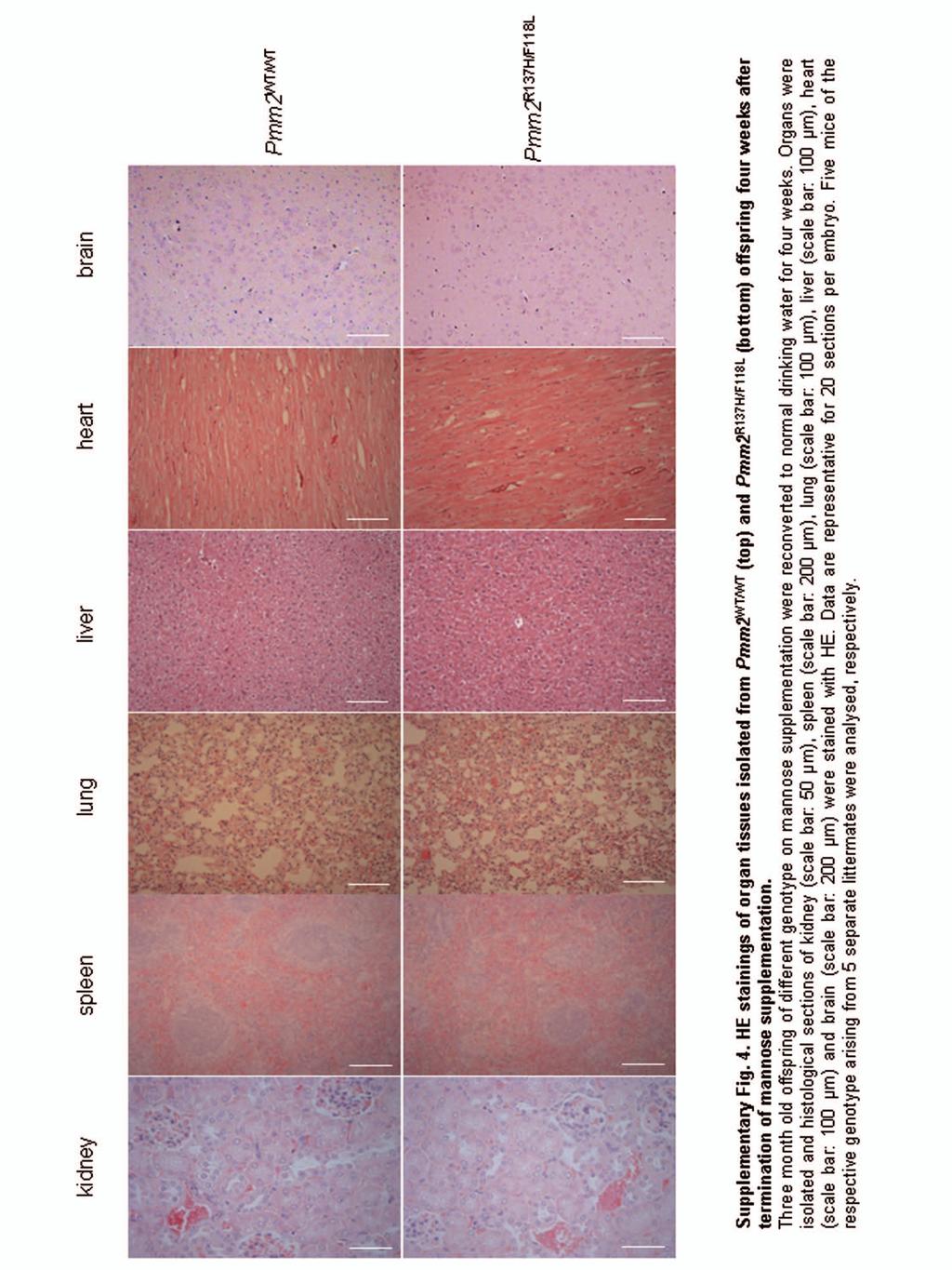
10 Embryos or mice were harvested at different days of development. Whole embryos or organs were fixed in 4% paraformaldehyde, embedded in paraffin and cut in 3 µm serial sections. After mounting on glass slides, staining with hematoxylin and eosin (HE; Sigma-Aldrich) was performed. For lectin histochemistry adjacent sections were used. Deparaffinised embryo sections were incubated with biotinylated WGA (Vector Laboratories) in a humidity chamber for 1 hour at room temperature. After washing three times in 10 mm HEPES (ph 7.5) for 5 min the slices were incubated with Steptavidin-FITC (Vector Laboratories) for 1 h in a humidity chamber in the absence of light and washed three times again. Slices were covered with Vectashield HardSet Mounting Medium including Diamino-2-phenylindol (Vector Laboratories) for nuclei staining. After drying overnight in darkness, fluorescence analysis was carried out by visualisation of WGA-binding with a fluorescence microscope (Leica). Determination of mannose concentration in sera Measurement of mannose concentration in sera of mice was carried out as described 15. Mannose supplementation Female mice were supplemented with D(+)-mannose (Sigma-Aldrich) enriched water (9 mg mannose ml drinking water 1 ) seven days before mating with heterozygous male of the corresponding other genotype. Oral mannose supplementation of mice with different genotypes was stopped as indicated in the text. Statistics For statistical analyses, Student s t test was performed to compare Pmm2 R137H/F118L to littermate values of Pmm2 WT/WT for all quantitative assays. Error bars are s.e.m.
11 References 13. Körner, C., Lehle, L. & von Figura, K. Glycobiology 8, (1998). 14. Niehues, R. et al. J. Clin. Invest. 101, (1998). 15. Etchison, J.R. & Freeze, H.H. Clin. Chem. 43, (1997).