Pan Stanford Series on Biomedical Nanotechnology
|
|
|
- Benedict Horton
- 5 years ago
- Views:
Transcription

1
2
3 Pan Stanford Series on Biomedical Nanotechnology Series Editors Vladimir Torchilin and Mansoor Amiji Titles in the Series Vol. 1 Handbook of Materials for Nanomedicine Vladimir Torchilin and Mansoor Amiji, eds (Hardcover) (ebook) Vol. 2 Nanoimaging Beth A. Goins and William T. Phillips, eds (Hardcover) (ebook) Vol. 3 Biomedical Nanosensors Joseph Irudayaraj, ed (Hardcover) (ebook) Vol. 5 Nanotechnology for Cancer Julia Ljubimova, ed Vol. 6 Nanotechnology for Delivery of Therapeutic Nucleic Acids Dan Peer, ed Vol. 7 Nanotechnology for Delivery of DNA and Related Materials Bengt Fadeel, ed Vol. 8 Translation Industrial Nanotechnology Thomas Redelmeier, ed Vol. 4 Inorganic Nanomedicine Bhupinder Singh Sekhon, ed. 2012
4
5 Published by Pan Stanford Publishing Pte. Ltd. Penthouse Level, Suntec Tower 3 8 Temasek Boulevard Singapore editorial@panstanford.com Web: British Library Cataloguing-in-Publication Data A catalogue record for this book is available from the British Library. Nanoimaging Copyright 2011 by Pan Stanford Publishing Pte. Ltd. All rights reserved. This book, or parts thereof, may not be reproduced in any form or by any means, electronic or mechanical, including photocopying, recording or any information storage and retrieval system now known or to be invented, without written permission from the publisher. For photocopying of material in this volume, please pay a copying fee through the Copyright Clearance Center, Inc., 222 Rosewood Drive, Danvers, MA 01923, USA. In this case permission to photocopy is not required from the publisher. ISBN (Hardcover) ISBN (ebook) Printed in the USA
6 Contents Preface Chapter 1 Chapter 2 Chapter 3 Chapter 4 Chapter 5 Chapter 6 Chapter 7 Chapter 8 Chapter 9 Chapter 10 Chapter 11 Chapter 12 Chapter 13 Combined Contrast and Therapeutic Nanocarriers for Oncologic MRI Yoshinori Kato and Arvind P. Pathak Nano-Size Superparamagnetic Magnetic Resonance Contrast Agents Alexei A. Bogdanov, Jr. In vivo Imaging of Immunotherapy Using Nanoparticles Christopher M. Long and Jeff W. M. Bulte Radiolabeled Liposomes as Theranostic Agents William T. Phillips, Beth Goins, Keitaro Sou, and Ande Bao Antibody-Targeted Liposomes and Micelles for Imaging Applications Tamer Elbayoumi and Vladimir Torchilin Quantum Dot-Based Multimodality Imaging Agents Weibo Cai and Xiaoyuan Chen Multimodal Imaging of Dendrimers Michelle R. Longmire, Peter L. Choyke, and Hisataka Kobayashi Multifunctional, Multimodality Cancer Imaging with Water-Soluble Synthetic Polymer Nanoparticles Marites P. Melancon, Xiaoxia Wen, Guodong Zhang, and Chun Li PLGA-Based Optical Imaging in Breast Cancer Hareesh B. Naira and Rajeshwar R. Tekmal Radiolabeled Gold Nanoshells for in vivo Imaging: Example of Methodology for Initial Evaluation of Biodistribution of a Novel Nanoparticle Huan Xie, Zheng Jim Wang, Ande Bao, Beth Goins, and William T. Phillips Combined Photoacoustic and Ultrasound Imaging of Metal Nanoparticles in vivo Kimberly Homan, Srivalleesha Mallidi, Erika Cooley, and Stanislav Emelianov Imaging Carbon Nanotubes in vivo: A Vignette of Imaging Modalities at the Nanoscale Khuloud T. Al-Jamal and Kostas Kostarelos Ultrasound Contrast Microbubbles: In vivo Imaging and Potential Therapeutic Applications Amanda Caissie, Raffi Karshafian, Kullervo Hynynen, and Gregory J. Czarnota vii Index 293
7
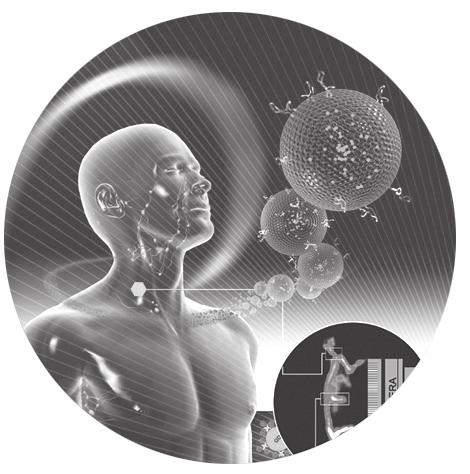
8 Preface The last decade has seen significant advances in the fabrication of new nanostructures, including nanoparticles. In parallel, considerable progress in the design of new imaging equipment and development of contrast agents has occurred. The translation of these nanostructures into clinical nanomedicine and diagnostic imaging applications is emerging rapidly with a burgeoning growth of different nanoparticles being tested. This volume of the Pan Stanford Series on Biomedical Nanotechnology, edited by Vladimir Torchilin and Mansoor Amiji, is devoted especially to reviewing the current status of the use of nanoparticles in in vivo imaging applications. The book begins with a review of nanoparticles for oncologic imaging, a major clinical application (Chapter 1). Chapters 2 5 focus on in vivo imaging of iron oxide particles and liposomes, some of the first nanoparticles to be used as diagnostic imaging agents clinically. Chapters 6 12 highlight the use of new nanoparticles that are currently at the preclinical stage of testing, such as quantum dots, dendrimers, polymer-based nanoparticles, gold nanoshells, carbon nanotubes, and metal nanorods. Finally, although not technically nanoparticles, no comprehenisive review on imaging contrast agents would be complete without the inclusion of microbubbles for ultrasound contrast (Chapter 13). This volume has attempted to represent the major clinical imaging modalities of magnetic resonance imaging (MRI), computed tomography (CT), positron emission tomography (PET), single photon emission computed tomography (SPECT), and ultrasound as well as optical methods. The overall thrust of this volume highlights the specific advantages of nanoparticles for drug delivery and imaging applications. Nanoparticles are able to target the delivery of therapeutic and imaging agents and retain these agents at local sites using different approaches not possible with small (free) molecules. In conjunction, noninvasive imaging can monitor the biodistribution of the nanoparticles as drug carriers and contrast agents and more importantly determine how successfully the intended target was reached. Moreover, the versatile nature of nanoparticles allows for their use as multimodality imaging agents as well as theranostics for carrying both diagnostic and therapeutic agents. Thus, readers should find that the principles learned from the in vivo imaging of one type of nanoparticle are useful for investigations of other kinds of nanoparticles. The editors are especially grateful to the numerous authors who agreed to devote their time and effort to share their research on the current use of nanoparticles for imaging applications. We would also like to acknowledge Julie Barker for her invaluable assistance with the organization and formatting of the book chapters and Jonathan Sumner for his help in formatting and labeling many of the images in this book. Beth A. Goins William T. Phillips
9