Description of supplementary material file
|
|
|
- Edwina Pope
- 5 years ago
- Views:
Transcription
1 Description of supplementary material file In the supplementary results we show that the VHL-fibronectin interaction is indirect, mediated by fibronectin binding to COL4A2. This provides additional information confirming specificity of VHL to COL4A2 but is not necessary for overall understanding of the manuscript. Other sections include detailed description of Materials and Methods, references, and acknowledgements.
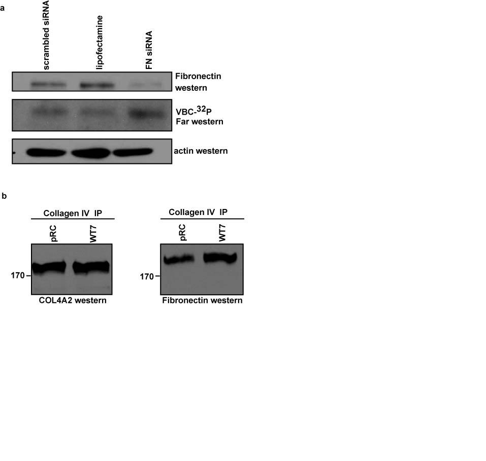
2 Kurban et al., Supplementary Material: Supplementary Results Fibronectin binds collagen IV and interacts indirectly with VHL It was previously shown that VHL interacts with fibronectin (Ohh et al., 1998); however our data in Figure 2d suggest that this interaction is not direct. To further clarify these results, WT7 cells were transfected with fibronectin sirna followed by western blot analysis to determine efficiency of fibronectin knockdown. Fibronectin was downregulated in cells transfected with fibronectin sirna whereas no effect was observed with control scrambled sirna or lipofectamine-only transfected cells (Supplementary 1a upper panel). Far-western analysis revealed that fibronectin knockdown did not decrease the intensity of the band interacting with the VBC- 32 P complex (Supplementary 1a middle panel) indicating that the band directly interacting with VHL is not fibronectin. To determine whether the association of fibronectin with VHL was through collagen IV binding, collagen IV immunoprecipitation from total extracts of cells lacking VHL (prc) or cells expressing WT-VHL (WT7) was performed followed by blotting for COL4A2 and fibronectin. Our results showed that fibronectin co-immunoprecipitated with COL4A2 (Supplementary 1b) as was observed upon VHL immunoprecipitation from WT7 and prc cells followed by western blot analysis for fibronectin (Figure 2d). These data suggest that VHL co-immunoprecipitates with COL4A2 which is bound to COL4A1, and the collagen IV complex then binds to fibronectin. The interaction between fibronectin and collagen IV has been previously reported (Matsuoka et al., 2004). Therefore, our results show that the interaction of VHL with fibronectin is indirect and mediated by binding of fibronectin to collagen IV.
3 Supplementary Figure 1.
4 Supplementary Materials and Methods Antibodies The primary antibodies used were: rabbit anti-human collagen type IV (Abcam), mouse anticollagen IV alpha 2 (MAB1910) (Chemicon), mouse anti-human fibronectin (BD Pharmingen), rabbit anti-hif-2α (Novus), mouse anti-ha (12CA5) (Roche), mouse anti-vhl (BD Pharmingen). Rat monoclonal collagen IV alpha 2 (H22) as well as rat monoclonal collagen IV alpha 1 (H11) were as described previously (Ninomiya et al., 1995). Collagen 18 antibody was obtained from Dr. Jose Teodoro (McGill University). Anti-calnexin antibodies were kindly provided by Dr. David Thomas (McGill University). Secondary antibodies used were: FITCconjugated anti-mouse and rhodamine-conjugated goat anti-mouse (Jackson Laboratories), Alexafluor 488-conjugated anti-rat (Molecular Probes, Invitrogen), HRP-conjugated anti-mouse (Jackson Laboratories) as well as HRP-conjugated anti-rabbit (Amersham). Mass Spectrometry Proteins were resolved by SDS-PAGE, and excised gel fragments digested with trypsin. Peptides were analysed by LC-MS/MS at the Centre Protéomique de L Est du Québec, Laval University, Québec. MS spectra were matched to the SwissProt database using Mascot algorithm (Matrix Science). Immunoprecipitation and Western blot Cells were lysed using 25 mm Tris HCl, ph 7.5, 100 mm NaCl, 1% NP40, 5 mm iodoacetamide and protease inhibitors (Roche). Lysates were incubated overnight with the corresponding antibodies. A 50:50 mix of protein G and protein A agarose (Upstate) was added for another 3 hours. The samples were then washed five times with 50 mm Tris-HCl, ph 7.5, 150 mm NaCl,
5 0.1% NP40, 10% glycerol. Laemmli buffer 2x containing DTT was added to the samples followed by 5 min boiling and resolving on SDS-PAGE gels. MG132, DFO and CoCl 2 treatments Cells were treated with either 100μM MG132 (Sigma) for 4 hrs, 100 μm desferrioxamine (DFO) (Sigma) for 6 hrs or 100μM CoCl 2 (Sigma) for 24 hrs. Far Western Far-Western analysis using VBC- 32 P was performed as described (Iwai et al., 1999). Immunostaining of xenograft tumors The mouse xenograft tumor assay was performed as previously described (Kurban et al., 2006). Tumors were embedded in optimal cutting temperature freezing medium (Tissue Tek, Sakura Finetek USA, Inc.) and stored at 80 C. Frozen sections of the various tumors were fixed in icecold acetone and blocked with 1% (w/v) normal bovine serum albumin (BSA). The sections were incubated with anti-col4a1 (H11) followed by secondary Alexa 488-conjugated goat anti-rat before mounting in Faramount fluorescent mounting medium (DAKO Canada Inc.). Acknowledgements This work was supported by operating grants from Kidney Foundation of Canada and Cancer Research Society. We thank Dr. W.G. Kaelin for providing cell lines, S. Welbourn, Dr. M-C Gingras, Dr. N. Beauchemin, Dr. J. Teodoro and Dr. M. Bouchard for help, discussion, and critical reading of the manuscript. G.K., N.R., E.D. were recipients of CIHR Cancer Consortium Training Grants, E.D. was a recipient a Peter Quinlan Fellowship and a fellowship from the Simone and Cino del Duca Foundation. V.H. is a recipient of the FRSQ studentship. A.P. holds a Canada Research Chair in Molecular Oncology.
6 Supplementary titles and legends to figures Supplementary Figure 1. Fibronectin binds collagen IV and interacts indirectly with VHL. (a) WT7 cells were transfected with either scrambled sirna (control), lipofectamine only (control), or fibronectin sirna followed by western blot analysis using anti-fibronectin antibody (upper panel) and far western analysis using VBC- 32 P (middle panel). Actin was used as loading control (lower panel). (b) Collagen IV immunoprecipitation from total lysates of prc and WT7 cell lysates followed by western blot analysis using anti-col4a2 (H22) (left panel) and antifibronectin antibodies (right panel). Supplemental References Matsuoka Y, Kubota H, Adachi E, Nagai N, Marutani T, Hosokawa N et al (2004). Insufficient folding of type IV collagen and formation of abnormal basement membranelike structure in embryoid bodies derived from Hsp47-null embryonic stem cells. Mol Biol Cell 15: Ninomiya Y, Kagawa M, Iyama K, Naito I, Kishiro Y, Seyer JM et al (1995). Differential expression of two basement membrane collagen genes, COL4A6 and COL4A5, demonstrated by immunofluorescence staining using peptide-specific monoclonal antibodies. J Cell Biol 130: