Fundamentals of Biochemistry
|
|
|
- Cecil O’Neal’
- 5 years ago
- Views:
Transcription
1 Donald Voet Judith G. Voet Charlotte W. Pratt Fundamentals of Biochemistry Second Edition Chapter 6: Proteins: Three-Dimensional Structure Copyright 2006 by John Wiley & Sons, Inc.

2 1958, John Kendrew Any regularity?

3 The atomic structure of myoglobin as a stick model A sequence of amino acids to a stable functional 3D form

4 Levels of protein structure
5 Secondary structure The peptide group: a rigid planar structure trans conformation

6 ~40% double bond character: due to resonance interactions: C-N bond is 0.13 Å shorter than its single bond C=O bond is 0.02 Å longer than that of aldehydes and ketones Trans conformation: more stable than cis conformation

7 Extended conformation of a polypeptide main chain: backbone The conformation of the backbone by rotation angles around the Cα-N bond and the Cα-C bond
 180º The angles increase when rotated clockwise as viewed from Cα")
8 Torsion angles of the polypeptide backbone the Cα-N bond (Φ) 180º the Cα-C bond (Ψ) 180º The angles increase when rotated clockwise as viewed from Cα

9 Steric interaction between adjacent peptide groups Conformational freedom is restricted
10 The Ramachandran diagram Calculation of sterically allowed angles/observed angles in the proteins Blue: sterically allowed angles for all residues except Gly and Pro Green: extreme limits for unfavorable atomic contacts Orange circles: conformational angles of secondary structures
11 Gly
 diameter: 12 Å intra chain H bonds C=O of the nth residues N-H of the (n+4)th residue side chains project")
12 Regular secondary structure α helix β sheet turns The α helix right handed helical 3.6 residues/turn 1.5 Å /residue 5.4 Å /turn (pitch) diameter: 12 Å intra chain H bonds C=O of the nth residues N-H of the (n+4)th residue side chains project outward

13 Space-filling model of α helix

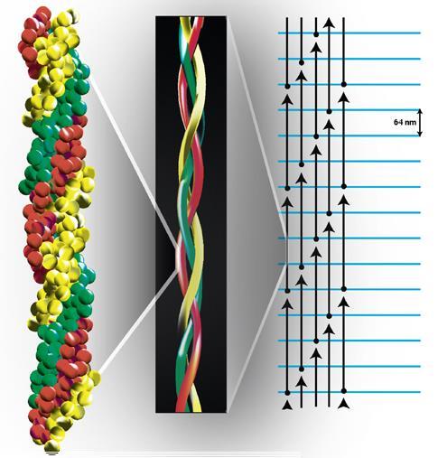
14 β sheets extended structure: pleated sheets inter chain H bonds H-bond difference Antiparallel is more stable than parallel

15 Pleated appearance (not fully extended)

16 Space filling model average of 6 strands


17 Diagram of a β sheet in bovine carboxypeptidase A Mixed: parallel and antiparallel Right-handed twist Complex topology (connectivity)

18 Connections between adjacent strands in β sheets
19 C. Fibrous proteins fibrous proteins: stiff, elongated, fibrous, insoluble globular proteins: compact, highly folded, globular, soluble some proteins have both regions Fibrous proteins: keratin, collagen keratin: structural proteins of hair, horn, nails, feathers α keratins in mammals (α-helical) β keratins in birds and reptiles (β-pleated) α keratins in mammals: over 30 variants X-ray diffraction pattern resembles that of α helix 5.1 Å spacing coiled coil structure: left-handed


20 Coiled coil structure 7-residue pseudorepeat Nonpolar at a and d 3.5 residue repeat per turn Inclined ~18 relative to one another Interdigitated side chains


21 High order α keratin structure Dimeric coiled coil Head to tail association: protofilament Dimerized protofilaments: microfibril Rich in Cys disulfide bond depending on the content: hard and soft Permanent waves reduction & oxidation


22 The chemistry of perms. Reference: J. Soc. Cosmet. Chem. 1996, 47,
 >33 genetically distinct chains")

23 Collagen: a triple helix Left-handed polypeptides twisted into right-handed superhelical structure Most abundant vertebrate protein Major connective tissue protein (muscle, tendons, ligaments, skin) >33 genetically distinct chains assembled into >20 collagen varieties in different tissues Type I collagen two α1(i) + one α2(i) chains molecular mass ~285 kd a width of ~14 Å a length of ~3000 Å epimysium, perimysium, endomysium: collagens muscle fiber: consists of myofibrils (actin, myosin)


24 Tendon and ligament Skin collagen
25 Collagen polypeptide composition ~1/3: Gly 15-30%: Pro The others: 4-OH Pro, 3-OH Pro, 5-OH Lys Typical polypeptide: Gly - x - y (over ~1000 residues)

26 Hydroxylation after protein synthesis prolyl hydroxylase ascorbic acid as a cofactor
 The bulky and relatively inflexible Pro and Hyp confer rigidity on the entire assembly viewed from N-terminal H-bonds in white color (between Gly N & Pro")
27 Typical polypeptide: Gly - x - y (x: Pro, y: Hyp) Pro prevents forming α helix structure (lack of backbone N-H) Why Gly is so frequent every 3 rd residues in the crowded center N-H of each Gly generates H-bond with C-O of X (Pro) The bulky and relatively inflexible Pro and Hyp confer rigidity on the entire assembly viewed from N-terminal H-bonds in white color (between Gly N & Pro O)
28
29 Cross-linking side chains in collagen Devoid of Cys Occur near the N- and C- termini (Lys & His) Lysyl oxidase Increased cross-linking with age Collagen diseases Lathyrism Osteogenesis imperfecta Ehlers-Danlos syndrome

30 Collagen fibril formation

31 As of 2003, there were more than 28 different types of collagen identified
 Variations in standard secondary structure β bulge Turn and")
32 Nonrepetitive protein structure in globular proteins Irregular structures: coil less ordered than α helix & β sheet different from random coil (disordered in denatured state) Variations in standard secondary structure β bulge Turn and loops

33 Propensity (P) of a residue to occur in regular secondary structure Useful for predicting the secondary str of proteins with known a.a.
34 Reverse turns (β bends) in polypeptide chains Occur at protein surfaces Gly 180º flip Pro

35 Ω loops: extended loops of 6-16 residues