Supplementary Information
|
|
|
- Cameron Wilson
- 5 years ago
- Views:
Transcription
 The cell pellet of VVD-III (VVD 36 C108A:M135I:M165I) is green compared to VVD-I (wild type VVD 36) due to the accumulation of the neutral semiquinone radical.")
 Absorption spectrum of purified VVD-III, shown in (b), reveals a mixture of oxidized (450 nm) and neutral semiquinone (577 nm, 621 nm) states of FAD.")
1 Supplementary Information Supplemental Figure 1. VVD-III purifies in a reduced state. (a) The cell pellet of VVD-III (VVD 36 C108A:M135I:M165I) is green compared to VVD-I (wild type VVD 36) due to the accumulation of the neutral semiquinone radical. (b) Purified VVD-I is yellow, VVD-II (VVD 36 M135I:M165I) is pale green, while VVD-III is dark green. (c) Absorption spectrum of purified VVD-III, shown in (b), reveals a mixture of oxidized (450 nm) and neutral semiquinone (577 nm, 621 nm) states of FAD. (d) Size Exclusion Chromatography (SEC) profile of VVD-III shows that the different redox forms of VVD separate by size; fractions corresponding to the peaks are shown above. (e) Absorption spectra of fraction #14 (d), which elutes earlier than #18, contains a neutral semiquinone state of VVD-III, whereas #18 contains the oxidized form.
 Time course for photoreduction of VVD-III to a neutral semiquinone with a 30 mw 448-nm laser, as monitored by")
 Reoxidation of VVD-III under ambient aerobic conditions.")
2 Supplementary Figure 2. Photoreduction of VVD-III. (a) Time course for photoreduction of VVD-III to a neutral semiquinone with a 30 mw 448-nm laser, as monitored by absorption spectroscopy (note the two isosbestic points at 347 nm and 497 nm). (b) Reoxidation of VVD-III under ambient aerobic conditions. Precipitation of some protein during the experiments causes light scattering and greater absorption at wavelengths <500 nm compared to the dark-adapted sample.
3 Supplementary Figure 3. Photoreduction of YF1 C62A. (a) Photoreduction of YF1 C62A in the presence of 10 mm TCEP was followed by absorption spectroscopy. The sample was continuously illuminated with 455-nm light (50 mw cm -2 ), and absorption spectra were recorded using a diode-array spectrophotometer. To avoid excitation of the sample by probe light, spectra were recorded with the tungsten lamp only, with the deuterium lamp turned off; hence spectra below ~370 nm suffer from poor signal-to-noise ratio. From top to bottom, spectra were recorded 1, 62, 122, 182, 242, 302, 362, 422, 482, 542, 602, and 662 s after onset of illumination. (b) Following saturating photoreduction to the NSQ state, the recovery was monitored in the dark under otherwise identical experimental settings. From top to bottom, spectra were recorded 0, 120, 240, 390, 570, 750, 930, 1440, 1800, 2160, 2520, 2880, 3240, 3600, 3960, 4320, 4750, 6190, and 7630 s after blue-light illumination ceased.
4 a
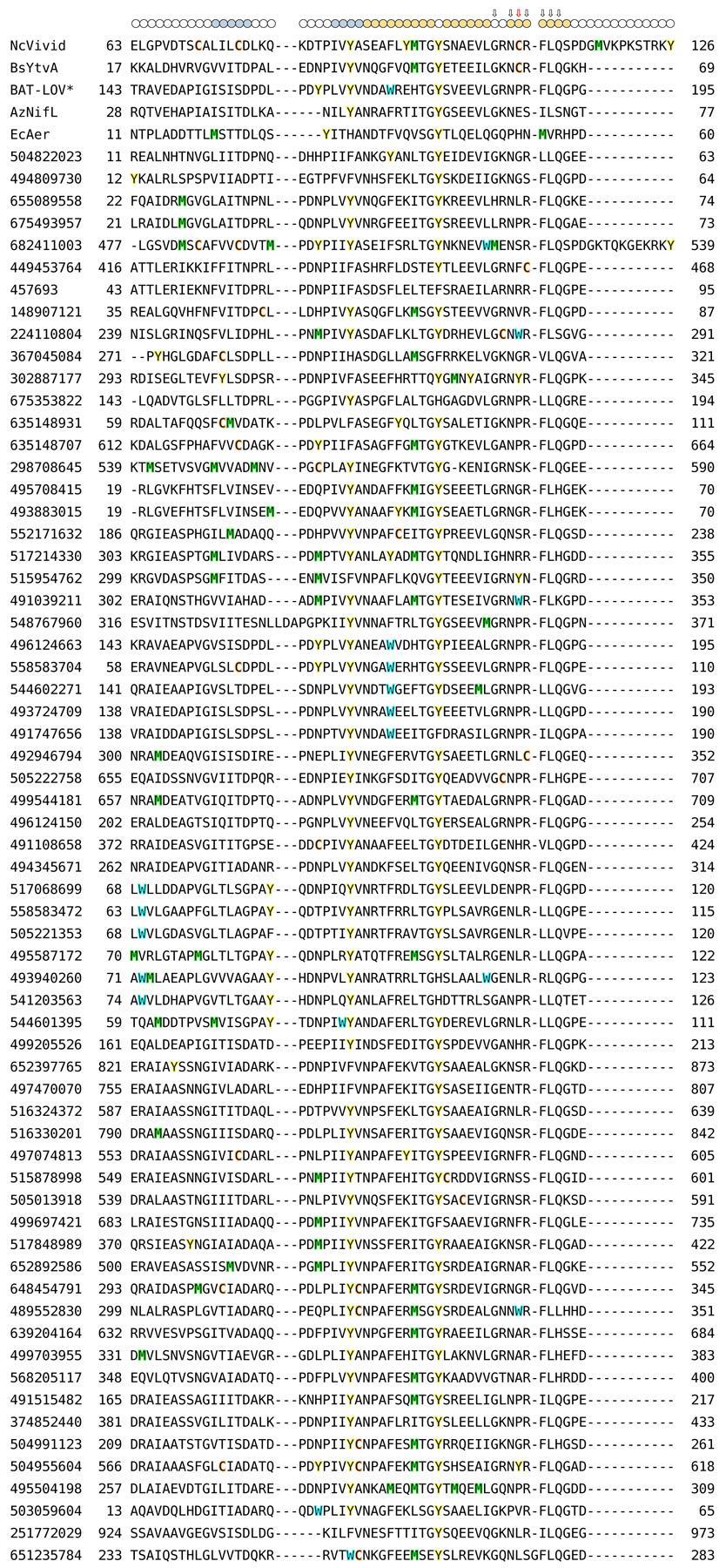
5
6 b
7 Supplementary Figure 4. Extended alignment of natural Cys-less LOV* domains. (a) Sequence alignment of VVD, BAT-LOV*, B. subtilis YtvA, A. vinelandii NifL, E. coli Aer, and LOV-like proteins lacking the adduct-forming cysteine. Tyr residues (yellow), Trp (blue), Cys (orange), and Met (green) are highlighted; circles above the alignment indicate the secondary structure in VVD with β sheets in blue and α helices in orange; arrows denote LOV-conserved residues, with the adduct-forming Cys in VVD and B. subtilis YtvA marked by a red arrow. (b) Dendrogram of the alignment shown in (a).
 Reverse-phase HPLC trace of flavin moieties released from BAT-LOV* produced through recombinant expression in E. coli.")
, and from these elution profiles, we estimate that 85-90% of BAT-LOV* can be reconstituted with either FAD or FMN.")
8 C Supplementary Figure 5. Flavin binding and reconstitution of recombinantly expressed BAT- LOV*. (a) Reverse-phase HPLC trace of flavin moieties released from BAT-LOV* produced through recombinant expression in E. coli. BAT-LOV* primarily copurifies with riboflavin, although small amounts of FMN and FAD are also bound. (b) Absorbance spectra of WT BAT- LOV* before (grey) and after (black) reconstitution with FAD. Apoprotein separates from the flavin-bound protein on SEC (see Fig. 5d), and from these elution profiles, we estimate that 85-90% of BAT-LOV* can be reconstituted with either FAD or FMN. These estimates also match those based on flavin spectral quantification relative to protein absorption. (c) SEC traces of BAT-LOV* reconstituted with FMN in the dark and after broad-spectrum light exposure, as for the FAD-bound protein shown in Fig. 5d.
9 Supplementary Figure 6. Relative photoreduction yields of VVD-III and BAT-LOV*. (a) VVD-III reduces to the NSQ by orders of magnitude more rapidly than BAT-LOV* when exposed to 448- nm light. However, photoreduction yields of BAT-LOV* can be increased to rival those of VVD-III by either replacing the three flavin-proximal aromatic residues (BAT-III) with Phe or substituting the Pro residue that occupies the position which normally forms an adduct with a Cys residue (P188C). Addition of a Cys residue on Iβ ~13 Å from the flavin ring has only a small effect (N252C), as does addition of exogenous reductants. Reconstitution with FMN instead of FAD has an insignificant effect. (b) In the presence of air, there is a small lag phase for WT BAT-LOV* photoreduction that is not present under anaerobic conditions. (c) Time courses for photoreduction of VVD and BAT variants. (d) Recovery of photoreduced BAT-III to the oxidized state in aerobic solution.
10 Supplementary Figure 7. Chemical reduction and recovery of BAT-LOV*. Treatment of BAT- LOV* with the low potential reductant Cr:EDTA generates a fully reduced flavin (blue line, broad absorption at 550 nm represents oxidation products of the Cr:EDTA reagent; purple line, with Cr:EDTA subtraction). Reoxidation after 80 min (shown with (dark grey) and without (light grey) Cr:EDTA subtraction) recovers the oxidized flavin, which is more evident after removal of the reductant by buffer exchange (red). A substantial amount of flavin remains bound to the protein after reoxidation.
11 a b Supplementary Figure 8. Multi-angle light scattering of BAT-LOV* and BAT-III. (a) Molar mass measurements from MALS are shown across the SEC elution profiles for dark-adapted BAT- LOV*-FAD (black) and BAT-LOV* exposed to a 100 W broad-spectrum visible light source for 1.5 h prior to chromatography (blue). (b) MALS-SEC data of BAT-III in the dark-adapted and photoreduced state. The shift in BAT elution profile does not accompany a substantial change in molecular weight. Elution times are standardized to WT BAT-LOV*.
phen 3. Over time, most of the reoxidized flavin dissociates from the protein (green trace).")
 3 ](ClO 4 ) 3 solution was immediately added.")
12 Supplementary Figure 9. Reoxidation of the BAT-P188C photoproduct. The spectrum of the light-excited protein strongly resembles that of a reduced HQ state. Furthermore, the light state can be rapidly converted to the oxidized flavin with the oxidant Co(III)phen 3. Over time, most of the reoxidized flavin dissociates from the protein (green trace). Some flavin is also unrecovered and presumably irreversibly photobleached. Unlike C4a adducts, the reduced state is also insensitive to bases, such as imidazole. In a 0.2-cm path length cuvette, 10 μl of 1 mm protein was photoreduced for 20 minutes under 448-nm laser light, after which, 1 μl of 1.2 mm [Co(phen) 3 ](ClO 4 ) 3 solution was immediately added. Spectra for the reoxidized species were collected within one minute (dark grey trace).