Supplemental Figure Legends:
|
|
|
- Sherman Carter
- 5 years ago
- Views:
Transcription
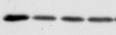
1 Supplemental Figure Legends: Fig S1. GFP-ABRO1 localization. U2OS cells were infected with retrovirus expressing GFP- ABRO1. The cells were fixed with 3.6% formaldehyde and stained with antibodies against GFP. Fig S2. is required to maintain protein levels of, ABRO1, and in the human normal fibroblast BJ cells and the human mammary epithelial MCF 10A cells. (Left) Protein levels of, ABRO1, and were decreased in human normal fibroblast BJ cells. Three different sirnas targeting were used for transfection. Forty-eight hours after transfection, cell lysates were collected and analyzed using indicated antibodies. (Right) Protein levels of, ABRO1,, and were decreased in the -shrna treated mammary epithelial MCF10A cells. Retroviral expression vector containing sh#1, as well as a control retroviral shrna construct against the firefly gene (FF), were introduced into MCF 10A cells and selected for stable cell lines with knockdown of the protein. Cell lysates were then analyzed with western blot using indicated antibodies. Fig S3. Semi-quantitative RT-PCR analysis for mrna levels of genes encoding various protein components in -shrna treated cells. Total RNA extracted from stably transfected U2OS cells containing either control vector containing an shrna hairpin against firefly gene (FF), or shrnas against (sh #1 and #2) was subjected to multiplexed RT-PCR using the 18s rrna gene as an internal loading control. The number of thermo cycles ranged from cycles for each target gene tested. Fig S4. Protein stability in the presence of a proteasomal inhibitor MG132. (A) Stably transfected U2OS cells containing either a control vector (FF), or targeting shrna vector (sh#1) were subjected to 5uM MG132 treatment and lysed at indicated times. Whole cell protein extracts were analyzed by western blot analysis for levels of target proteins. Levels of p53 and cyclin B1 were shown as positive controls. Pro-apoptotic effects of MG132 were observed at 18hr incubation timepoint, including changes in cell morphology and increased cell death. (B) Doxycycline inducible knockdown U2OS stable cell line was generated with lentiviruses carrying shrna#1 hairpin against regulated by a doxycycline response promoter as shown in the Fig.2C in the manuscript. Cells were cultured in the absence of doxycycline until induction. Cells were treated with or without 2µg/ml doxycycline for 4 days. Cells were then treated or not treated with 10 um MG132 for 8 hours. Cell lysates were analyzed by western blot using indicated antibodies. P53 protein level was shown as a positive control in response to MG132 treatment. Fig S5. Protein stability for components of the complex and ABRO1 complex. shrna-treated cells and rescued cells with expression of the wild type HA- and Flagtagged, as well as control cells treated with an shrna hairpin to the fruit fly gene (FF) lines, were treated with (0.1mg/ml) cycloheximide and analyzed at the indicated times. Cell lysates were analyzed by western blot. Fig S6. binding to components of the /BRCA1 A complex is phosphorylationindependent. 293T cells were transiently transfected with constructs carrying HA- and Flagtagged wildtype (WT) or mutants as indicated. Cell lysates were treated or not treated with a lambda protein phosphatase (NEB) at 30 C for 30 minutes before being used for immunoprecipitations with anti-flag antibodies. Immunoprecipitates were then analyzed by western blot with antibodies to,, and. Different forms of were indicated. 1
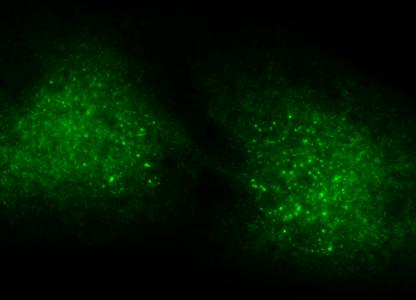
2 Fig S7. An alignment of UEV domains and generation of YSP mutant of. Fig S8. GFP-tagged PR mutant lacking the PXXR motif failed to form IR induced foci (IRIF). U2OS cells were infected with retrovirus containing GFP-tagged wild type (WT), PR mutant or R305A mutant. Cells were treated with 10Gy IR, incubated at 37 C for 2 hour, and fixed with 3.6% formaldehyde. Immunofluorescence was carried out with antibodies against BRCA1, GFP and appropriate secondary antibodies. Fig S9. GFP-tagged YSP mutant is defective in forming IRIF. U2OS cells were infected with retrovirus containing GFP-tagged wild type (WT) or YSP mutant. Cells were treated with 10Gy IR, incubated at 37 C for 2 hour. Cells were then treated with 0.5% Triton X-100 at room temperature for 5 min for extraction, then fixed with 3.6% formaldehyde. Immunofluorescence was carried out with antibodies against BRCA1, GFP and appropriate secondary antibodies. Fig S10. BRCA1 protein level is not changed in the cells with knockdown of or. (A) Retroviral expression vector containing two different shrna hairpins against, as well as a control retroviral shrna construct against the firefly gene (FF), were introduced into U2OS cells and selected for stable cell lines with knockdown of the protein. Cell lysates were then analyzed by western blot analysis using indicated antibodies. (B) U2OS cells were transfected with three different sirnas against. Forty-eight hours after transfection, cell lysates were analyzed by western blot using indicated antibodies. Fig S11. s DUB activity is not required for its interaction with components of the /BRCA1 A complex and the ABRO1/BRISC complex. 293T cells were transiently transfected with constructs carrying HA- and Flag-tagged wild type (WT) or DUB mutant (MT) as indicated. DUB mutant is generated with two active site Zn 2+ -binding histidine residues (H 122 H 124 ) replaced with glutamines (Q 122 Q 124 ). Immunopricipitations were carried out with anti-flag antibodies from total cell lysates. Immunoprecipitates were then analyzed by Western blot with antibodies to,,, ABRO1, or HA. Fig S12. A diagram showing two distinct -containing complexes, the /BRCA1 A complex and the ABRO1/BRISC complex. 2

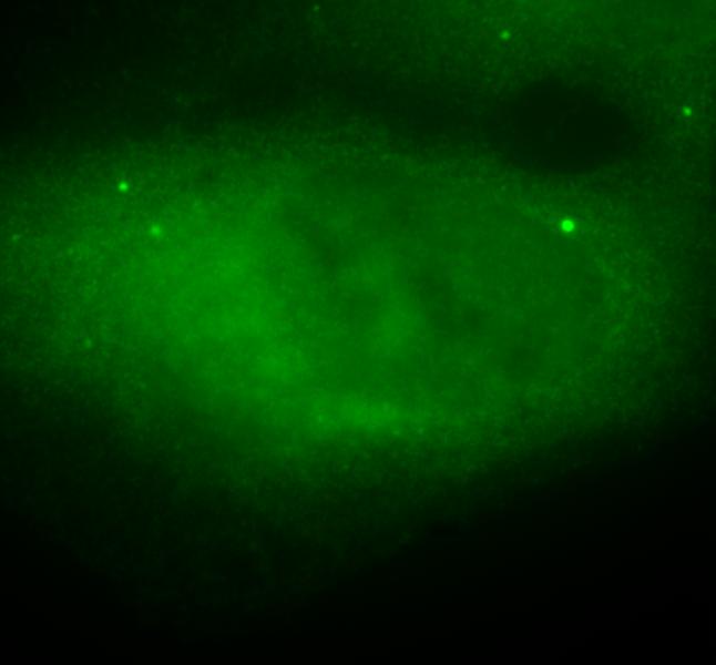
3 Fig. S1 GFP-ABRO1 DAPI


4 Fig. S2 sirnas Mock FF sh#1 Abro1 BJ cells MCF10A cells


5 Fig. S3 253 bp 116 bp 253 bp 123 bp sh FF #1 #2 18s 18s Abro1 299 bp 253 bp 253 bp 157 bp 253 bp 142 bp 18s 18s Abro1 18s

 MG132")

6 Fig. S4 A (MG132 5uM) FF - 4hr 18hr - sh#1 4hr 18hr B Dox (2ug/ml, 4day) MG132 (10uM, 8h) ABRO1 ABRO1 p53 Cyclin-B1 P53
7 Fig. S5 CHX (0.1 mg/ml) MSCV/FF 0h 1/2 MSCV/FF 0h MSCV/ MSCV/ HF-/ FF sh#1 sh# (hrs) HF--p HF- endo- ABRO1

8 Fig. S6 Lambda PP IB IB (Input) IP: HF- WT Δ1-19 Δ40-56 Input HF--p HF- Endo-
9 Fig. S7 h_1 DATNCLRITDLKSGCTSLTPGPNCDRFKLHIP...YAGETLKWDIIFNAQYPELPPDFIFGEDAE _ 2 IAAFLSHFGTGVVEYDAE.GFTKLTLLLM...WKDFCFLVHIDLPLFFPRDQPTLTFQSVYH hrad6a KRLQEDPPAGVSGAPSEN.NIMVWNAVIFGPEGTPFEDGTFKLTIEFTEEYPNKPPTVRFVSKMF hubch5 SDLQRDPPAHCSAGPVGD.DLFHWQATIMGPPDSAYQGGVFFLTVHFPTDYPFKPPKIAFTTKIY h_1 FLP...DPSALQNLASWNPSNPECLLLV.VKELVQQYHQFQCSRLRESSRLMFEYQTLLE _ 2 FTNSG..QLYSQAQKNYPYSPRWDGNEMAKRAKGCQGSRDACSPWEQVLAFAVAKTGCKLLQ hrad6a HPNVYADGSICLDILQNRWSPTYDVSSILTSIQSLLDEPNPNSPANSQAA.QLYQENKREYE hubch5 HPNINSNGSICLDILRSQWSPALTVSKVLLSICSLLCDPNPDDPLVPDIA.QIYKSDKEKYN *** YSP AAA


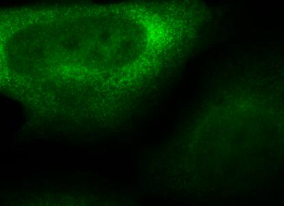
10 Fig. S8 BRCA1 WT PR mt R305A GFP-NBA MERGE DAPI


11 Fig. S9 BRCA1 WT YSP mt GFP- Merge DAPI
12 Fig. S10 A BRCA1 FF sh #1 #2 B BARD1 Chk1 ABRO1 BRCA1 sirnas Mock #1 #2 #3


")

13 Fig. S11 IP: αflag mock WT MT Input (5%) mock WT MT Abro1 HA- HA- (long exposure)

14 Fig. S12 Double Strand Break (DSB) Ub Ub Ub Ub P BRCA1 ABRO1 / BRCA1 A complex ABRO1 / BRISC complex Nucleus Cytoplasm