Spirochaete flagella hook proteins self-catalyse a lysinoalanine covalent crosslink for motility
|
|
|
- Jean Hampton
- 5 years ago
- Views:
Transcription
1 SUPPLEMENTARY INFORMATION ARTICLE NUMBER: DOI: /NMICROBIOL Spirochaete flagella hook proteins self-catalyse a lysinoalanine covalent crosslink for motility Authors: Michael R. Miller, Kelly A. Miller, Jiang Bian, Milinda E. James, Sheng Zhang, Michael Lynch, Patrick S. Callery, Justin M. Hettick, Andrew Cockburn, Jun Liu, Chunhao Li, Brian R. Crane, and Nyles W. Charon* *Corresponding author. ncharon@hsc.wvu.edu This file includes: Supplementary Figures Supplementary Tables 1-6. Captions for supplementary videos 1-4 NATURE MICROBIOLOGY 1
 PFs and PHs were reacted with antibodies directed to FlaB.")
2 Supplementary Fig 1. Western blot and Imperial staining analysis of T. denticola periplasmic flagella (PFs), polyhooks (PHs), and in vitro synthesized high molecular weight complex (HMWC). (a) PFs and PHs were reacted with antibodies directed to FlaB. PFs (b) and PHs (c) were treated with various agents or boiled and analyzed by western blot with antibodies directed to FlgE: Formic acid (FA), β-mercaptoethanol (BME), dithiothreitol (DTT). Although some breakdown of the HMWC occurred with some treatments, no monomeric rflge was detected. (d) In vitro synthesized HMWC s were reacted with BME or DTT and analyzed by SDS-PAGE and Imperial stained. (e) In vitro synthesized HMWC s of four mutant proteins after one week incubation under cross-linking conditions. Note that K165A, N179A, and C178A fail to form HMWCs. Blots and gels were repeated at least 3 times, and the data shown are representative. Figure e was only done once. 2
3 Supplementary Fig 2. LC-MS analysis of T. denticola rflge tryptic peptides (a) Comparison of UV elution profile taken from the monomer and multimer samples of T. denticola rflge. Differences in peptide absorbance are boxed. (b) MS total ion count profile of tryptic digests in (a). Regions of significant difference are boxed and were analyzed further for peptide identification. A major peptide of MW = Da was identified only in the multimer sample. Note that there is 11 min offset between the UV and MS traces that was calibrated as described in the Methods Section. 3
 Representative extracted ion chromatograms (XICs) of the tryptic peptide derived from in vitro formed HMWC multimeric species reveal the expected molecular mass for the lysinoalanine crosslink")
4 Supplementary Fig 3. MS/MS characterization of T. denticola rflge N175A substitution. (a) Representative extracted ion chromatograms (XICs) of the tryptic peptide derived from in vitro formed HMWC multimeric species reveal the expected molecular mass for the lysinoalanine crosslink between K165 and dehydroalanine 178 (d) in the derivative inter-peptide, thereby confirming the C-terminal sequence. The N175A mutation is identified in yellow. XIC s for both the cross-linked, and noncrosslinked carbamidomethyl (cab)-modified peptides are shown. (b) Representative MS/MS spectrum matching to a b 3-b 10 ion series. 4

5 30 Supplementary Fig. 4. Test of 3 models of cross-linking. MS data obtained from in vitro formed T. denticola rflge HMWC was examined to determine the accuracy of the Protea instrument and to test 3 models whereby T13-T14 is linked to T15, with the loss of 34 Da. The ppm errors of m/z values (M ion) for 1000 peptides automatically identified as FlgE by the software were plotted (blue line). The software was set to allow an error of up to 50 ppm, but it is clear from the graph that the instrument is accurate to within ~5 ppm. It also appears that it was biased to give a slight negative ppm error. In this run the observed mass unique to the HMWC was This was compared to the predicted masses from the three models to determine the ppm error for each model [loss of one SH 2 (red), two NH 3 s (yellow) or one H 2O 2 (green)], The loss of one SH 2 ( ) provided an excellent fit; loss of one H 2O 2 ( ) could not be eliminated, but did not fit as well; loss of two NH 3 s was eliminated as a possibility, based on this analysis. 5
, ~150 kda, and >250 kda HMWCs were excised, and the protein electroeluted.")
6 Intensity (mv) a kda B Supplementary Fig 5. Lysinoalanine (LAL) analysis of FlgE monomer and HMWC. In vitro crosslinked T. denticola rflge was electrophoresed in agarose, regions corresponding to monomer (50 kda), ~150 kda, and >250 kda HMWCs were excised, and the protein electroeluted. (a) Electroeluted proteins were electrophoresed and Imperial stained (outlined lanes): 1 = dual color protein markers ; 2 = electroeluted 50 kda monomer; 3 = electroeluted ~150 kda HMWC; 4 = electroeluted >250 kda HMWC. Proteins were submitted for amino acid analysis: (b) Partial amino acid chromatogram of >250 kda HMWC; arrow indicates the position of LAL, well resolved from other amino acids. (c) *The # of Gly, Ala,Tyr in one FlgE. Ť The expected ratio of LAL to Gly, Ala and Tyr in a HMWC of 10 FlgEs joined by 9 LALs. Ratios of LAL to Gly, Ala and Tyr determined in the 3 electroeluted protein fractions from one Td rflge preparation. The levels of LAL in the HMWCs were similar to that in a complex of 10 FlgE monomers joined by 9 LALs. LAL levels were much lower in the monomer, likely arising from dehydroalannine in the monomer (Supplementary Fig. 7) forming intra-molecular LAL cross-links. In four Td rflge preparations, the level of LAL was 3-8 times higher (mean of 5) in the >250 kda HMWC than in the monomer. 6
7 Supplementary Fig. 6. MS/MS spectrum of isotopically mixed T. denticola cross-linked peptide. Representative MS/MS spectrum of the peptide representing a 15 N (red) - 14 N (blue) cross-link as indicated in the inset. Mass corresponds to that expected for the LAL cross-link, with a b-ion series extending to N-terminal residue 12. Mass peaks at low and high m/z ratios are shown at 10x amplitude. 7
 MS analysis: Above: XIC of tryptic digests from momeric rflge treated with BME to inhibit cross-linking.")
8 Supplementary Fig. 7. BME blocks cross-linking by adding to dehydroalaine. (a) Inhibition of T. denticola rflge cross-link formation by addition of BME as shown in a representative gel. (b) MS analysis: Above: XIC of tryptic digests from momeric rflge treated with BME to inhibit cross-linking. ~10% of the detected peptides contain a BME adduct with dehydroalanine (Cα-CH 2-S-(CH 2) 2-OH). The remaining peptides retain Cys at position 178 and thus undergo modification by iodoacetamide to form a carbamidomethyl modification. Below: MS/MS spectrum of m/z = identifying the target peptide with an adduct between BME and dehydroalanine in the form of a thio-ether linkage 8
9 Supplementary Fig. 8. The cross-linked peptide is highly represented in flagellar PHs extracted from Td. a) Representative Extracted Ion Chromatograms (XICs) of the cross-linked peptide in Td PHs. XICs are shown for the [M+nH] n+ n=3,4,5 tryptic peptides from the PHs. Top trace shows the total ion counts for all chromatographed peptides vs. LC retention time (min). b) MS/MS spectrum of cross-linked peptide of Td PH FlgE. Mass peaks at low and high m/z ratios are shown at 5x or 10x amplitude. 9
 Phylogenetic tree analysis was carried out across 176 unique bacterial FlgE sequences based on alignment of Td peptide: IINTSGQTEDLNIPIGQKLDAKATTSVNYACNLDKRLPELPE.")
, Lys165 is invariant, and position 178 is either Cys, or sometimes Ser, two residues that are both known to participate in")
10 a IINTSGQTE DLNIPIGQKLDAKATTSVNYACNLDK RLPELPE b ,179 Supplementary Fig. 9. Conserved residues in the spirochete cross link region. (a) Phylogenetic tree analysis was carried out across 176 unique bacterial FlgE sequences based on alignment of Td peptide: IINTSGQTEDLNIPIGQKLDAKATTSVNYACNLDKRLPELPE. Conservation of the cross-linking peptide distinguishes two families whose sequence conservation via WebLogo is shown. In spirochetes and the closely related Synergistetes (92 sequences), Lys165 is invariant, and position 178 is either Cys, or sometimes Ser, two residues that are both known to participate in lysinoalanine formation. Asn179 and Leu180 are also invariant in this family. The Td amino acid sequence is shown in italics, with those residues essential for cross-linking underlined and numbered below. (b) Other types of bacteria (84 sequences) do not conserve the cross-linking residues in their FlgE sequences, but do conserve Asn179 and Leu
 that fail to cross-link FlgE and have altered motility, and examined by cryo-em. These structures were compared to that of the WT (Fig. 1a), and representative Cryo-EM images are shown.")
11 Supplementary Fig.10. Cryo-EM of TdK165A and TdN179A. PFs were purified from the above two mutants and TdC178A (Fig. 4c) that fail to cross-link FlgE and have altered motility, and examined by cryo-em. These structures were compared to that of the WT (Fig. 1a), and representative Cryo-EM images are shown. No discernable differences in the flagellar hook and filament structure were detected among the mutants compared to the WT. 11

12 Supplementary Figure 11. Cryo-EM reconstructions of wild-type and mutants. Top panels show tomographic slides from the cell tips of WT, TdK165A, TdC178A, TdN179A mutants, respectively. The bottom panels show the zoom-in views from the area highlighted in the corresponding top panel. Both flagella and hooks remain intact in periplasmic space in all strains. Representative Cryo-EM reconstructions are presented. 12
13 13
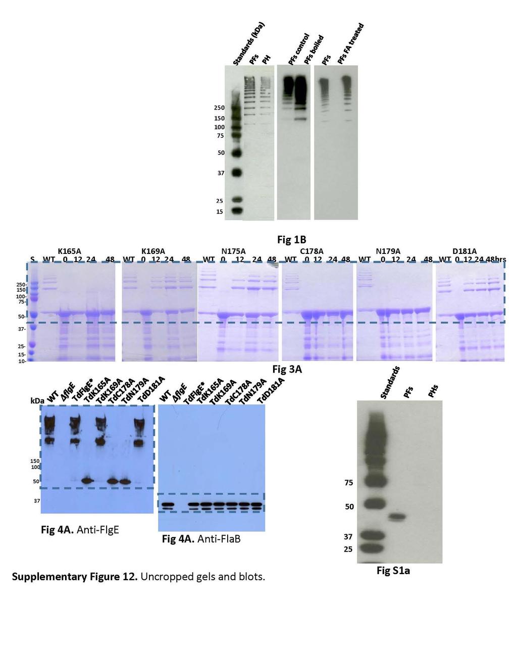
.")
14 Supplementary Fig 12 (continued). Uncropped gels and blots. 14
15 Supplementary Table 1. T13, T13-T14 and T15 Peptide abundances. Tryptic peptides involved in lysinoalainine formation were quantified by MS extracted ion chromatograms (XICs) in their uncrosslinked forms for monomeric FlgE and the HMWC. The uncrosslinked peptides in their various forms were standardized against two peptides that do not participate in cross-linking (red and blue sequences). See Supplementary Table 2 for summary. Peptide Residu es Peptide Sequence Modification Monomer Peak Area HMWC Peak Area ATTSVNYAcNLDK C9 - Carbamidomethyl 7.92E E ATTSVNYAcnLDK C9- Carbamidomethyl; N10 - Deamidated 1.13E E ATTSVNYAcNLDKR C9 - Carbamidomethyl 5.12E E+08 Σ 8.55E E IINTSGQTEDLNIPIGQK 4.02E E E E IInTSGQTEDLNIPIGQK N3 - Deamidated 3.94E E IINTSGQTEDLNIPIGQKL DAK IINTSGQTEDLNIPIGQKL DAKATTSVNYAcNLDK 1.38E E E E+07 C31 - Carbamidomethyl 3.08E E+07 Σ 3.49E E (std1) AQILESTWSTEFK 7.90E E E E+08 Σ 8.00E E (std2) VYDSFGEAHELQIDFAR 9.32E E E E+09 Σ 1.10E E+10 Supplementary Table 2. Non-crosslinked peptide ratios in monomer compared to HMWC. Summary of peptide abundance data presented in Supplementary Table 1. a standardized to std1 (red), b standardized to std2 (blue) Peptides Monomer a HMW a Monomer/ HMW a Monomer b HMW b Monomer/ HMW b Average of Std1 and Std
16 16
17 17
18 18
19 19
20 Supplementary Table 6. Oligonucleotide primers used in this study. Prim ers Sequences a Note b P1 CACCATGATGAGATCATTATTTTCGGG Over-expression of TDE2768; F P2 CTATCGTTTCAAGTTCAAGAC Over-expression of TDE2768; R P3 ATGATGAGATCATTATTTTCGGG Amplification of TDE2768; F P4 CCTATAGGTCAAGCACTTGATGCAAAGG Site-mutagenesis of TDE2768(165K-A); F P5 CCTTTGCATCAAGTGCTTGACCTATAGG Site-mutagenesis of TDE2768(165K-A); R P6 GGTCAAAAACTTGATGCAGCGGCAACCAC Site-mutagenesis of TDE2768(169K-A); F P7 GTGGTTGCCGCTGCATCAAGTTTTTGACC Site-mutagenesis of TDE2768(169K-A); R P8 CCACAAGTGTAGCCTATGCTTGTAACCTTG Site-mutagenesis of TDE2768(175N-A); F P9 CAAGGTTACAAGCATAGGCTACACTTGTGG Site-mutagenesis of TDE2768(175N-A); R P10 GTGTAAACTATGCTGCTAACCTTGATAAGAGGCTG Site-mutagenesis of TDE2768(178C-A); F P11 CAGCCTCTTATCAAGGTTAGCAGCATAGTTTACAC Site-mutagenesis of TDE2768(178C-A); R P12 CTATGCTTGTGCCCTTGATAAGAGGCTGCC Site-mutagenesis of TDE2768(179N-A); R P13 GGCAGCCTCTTATCAAGGGCACAAGCATAG Site-mutagenesis of TDE2768(179N-A); F P14 GCTTGTAACCTTGCTAAGAGGCTGCCTG Site-mutagenesis of TDE2768(181D-A); R P15 CAGGCAGCCTCTTAGCAAGGTTACAAGC Site-mutagenesis of TDE2768(181D-A); F P16 GGGATGCTTCAGCAGAC 5 -flank region of TDE2768; F P17 GCTGCTGCGTAACATAATTATTGCCTCCTAATTG 5 -flank region of TDE2768- aacc; R P18 ATGTTACGCAGCAGCAACGATG aacc cassette; F P19 TTAGGTGGCGGTACTTGGGTC aacc cassette; R P20 GTACCGCCACCTAGGTATGGTATAATATAGGG 3 -flank region of TDE2768- aacc; F P21 CGGCTTGAATTCCAAGTAC 3 -flank region of TDE2768; R P22 ATGAACAAAAATATAAAATATTCTC ermb cassette; F P23 TTATTTCCTCCCGTTAAATAATAG ermb cassette; R P24 CCTAGGAGGCAATAATTATGATGAGATCATTATTTTC TdFlgE* formation, TDE2768; F P25 CCTAGGTTCGTTTCAAGTTCAAGACTG TdFlgE* formation, TDE2768; R P26 GAGAATATTTTATATTTTTGTTCATAATTATTGCCT 5 -flank region of TDE2768-ermB; R CCTAATTG P27 CTATTATTTAACGGGAGGAAATAACCTAGGTATGG TATAATATAGGG 3 -flank region of TDE2768-ermB; F a Underlined portions show the engineered overlapping base pairs and italic show the AvrII restriction enzyme cutting-site; b Primer orientation: F, forward; R, reverse. 20
21 Videos: All videos were taken at 400X by dark-field microscopy. Supplementary video 1. Cells WT T. denticola in 1% methylcelluose. Note cells had notable translational motility. Cells of replacement mutant TdFlgE* had identical swim behavior. Supplementary video 2. Cells of mutant flge in 1% methylcellulose. Note cells deleted of flge were completely non-motile. Supplementary video 3. Cells of substitution mutant TdC178A in 1% methylcellulose. Note cells of TdC178A generated motion but lacked translational motility. Cells of substitution mutants TdK165A and TdN179A had identical swim behavior. Supplementary video 4. Cells of substitution mutant TdK169A in 1% methylcellulose. Note cells of TdK169A had notable translational motility similar to that of the WT and TdFlgE*. Cells of substitution mutant TdD181A had identical swim behavior. 21