Fluorescent Antibody technique Can identify microorganisms in clinical specimens and can detect the presences of a specific antibody in
|
|
|
- Sheila Foster
- 5 years ago
- Views:
Transcription
1 Lecture:7 Practical immunit Fluorescent Antibody technique Can identify microorganisms in clinical specimens and can detect the presences of a specific antibody in serum. These techniques combine fluorescent dyes such as fluorescein isothiocyanate (F I T C) with Ab to make them fluoresce when exposed to ultraviolet light these procedures are quick, sensitive, and very specific, the FA test for rabies can be performed in a few hours and has an accuracy rate close to 100% Fluorescent Antibody techniques are of two types direct & indirect. Direct FA tests are usually used to identify a microorganism in a clinical specimen, during this procedure, the specimen containing the Ag to be identified fixed on to a slide (fluorescein labeled Ab) are then added, and the slide is incubated briefly next the slide is washed to remove any Ab not bound to Ag and is then examined under the fluorescence microscope for yellow green fluorescence. Indirect FA tests are used to detect the presence of a specific Ab in serum following exposure to a microorganism, they are often more sensitive then direct tests during this procedure, known Ag is fixed on to a slide the test serum is then added, and if antibody that microbe is present it reacts with the Ag to form a bound complex in order for the Ag Ab complex to be seen, fluorescein labeled Antihuman immune serum globulin(anti-hisg), An antibody that reacts 1
, it is examined under a fluorescence microscope, if the known Ag fixed to the slid appears fluorescent, the Ab specific to the")
2 specifically with any human Ab, is added to the slide Anti-HISG will be present only if the specific Ab has reacted with its Ag and is therefore present as well.after the slide has been incubated and washed (to remove unbound Ab ), it is examined under a fluorescence microscope, if the known Ag fixed to the slid appears fluorescent, the Ab specific to the test Ag is present. Enzyme-linked immunosorbent assay (ELISA) Enzyme-linked immunosorbent assay (ELISA) the most widely used of a group of tests known as enzyme immunoassay (EIA) there are two basic methods 2
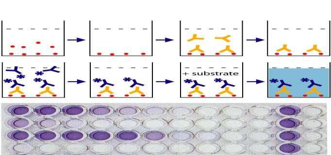
3 The direct ELISA detects Ag and the indirect ELISA detects Ab Amicrotiter plat with numerous shallow wells is used in both procedure. Variations of the test exist for example, the reagents can be bound to tiny latex particles rather than to the surfaces the microtiter plates ELISA procedures are popular primarily because they require little interpretive skill to read the results tend be clearly positive of clearly negative, ELISA test are also used to screen blood for Ab to HIV. (Direct ELISA) A "sandwich" ELISA, is used to detect sample antigen. The steps are : 1- In the first step the Ab specific to Ag to be detected is adsorbed to the surface of the wells of the microtiter plate. 2- A patients sample containing unidentified Ag is then added to each well, if the Ag specifically with the Ab adsorbent to the well, the Ag (drug in a urine test, for example ) will be retained there when the well is washed free of unbound Ag 3-A specific antibody is added, and binds to antigen ( hence the 'sandwich': the Ag is stuck between two antibodies), this risible only because the second added Ab is linked to an enzyme such as horseradish peroxidase or alkaline phosphatase, unbound enzyme linked Ab is washed from the well. 4-The plate is washed to remove the unbound antibody-enzyme conjugates. 5-the enzymes substrate(tmb*tetramethylbenzidin*) is added to it enzymatic activity is indicated by a color change that can be visually 3

4 detected the test will be positive if the Ag has reacted with adsorbed Ab in the first step. If the Ag was not specific for the Ab adsorbed to the wall of the well, the test will be negative because the unbound Ag will have been washed 4
5 A common use of the direct ELISA test to detect the presence of drug in urine the availability of monoclonal Ab has been essential to the widespread use of the type of test. Indirect ELISA 1. Micro-well plates are incubated with antigens, washed up and blocked with BSA 2. Samples with antibodies are added, incubate and washed. 3.Enzyme linked secondary antibody are added, incubate and washed. 4. A substrate(tmb*tetramethylbenzidin*) is added, and enzymes on the antibody elicit a chromogenic or fluorescent signal. The addition of an enzyme substrate - chromogen reagent causes color to develop. This color is directly proportional to the amount of bound sample antibody. The more antibody present in the sample, the stronger the color development in the test wells. This format of indirect ELISA is suitable for determining total antibody level in samples ( Newcastle disease virus, B. abortus, etc.). 5
6 6