Supporting Information
|
|
|
- Giles Davidson
- 5 years ago
- Views:
Transcription
1 Title Structure of bacterial cellulose synthase subunit D Hu, Song-Qing; Gao, Yong-Gui; Tajima, Kenji; Sunagaw Author(s) Yoda, Takanori; Shimura, Daisuke; Satoh, Yasuharu; M CitationProceedings of the National Academy of Sciences of t Issue Date Doc URL Type article (author version) Additional There Information are other files related to this item in HUSCAP File Information Supporting_Information.pdf (Supporting Information) Instructions for use Hokkaido University Collection of Scholarly and Aca
2 Supporting Information Text Bacterial strains and growth conditions Escherichia coli (E. coli) JM109 and BL21(pREP-4) were grown in Luria-Bertani (LB) broth at 37 C. Isopropyl-β-D(-)-thiogalactopyranoside (IPTG) was used as an inducer of genes. When needed, the antibiotics ampicillin (Ap) and kanamycin (Km) were added to the medium at final concentrations of 100 and 25 µg/ml, respectively. The above media with 1.5% (w/v) agar were used for cell growth on plates. Preparation of His 6 -tagged AxCeSD The expression and purification of selenomethionine (SeMet)-substituted AxCeSD with a His 6 -tag at the N-terminus (N_AxCeSD) were carried out as described previously (1). Using primer pairs corresponding to the specified 5' and 3' termini (Tab. S2), the PCR products were digested with NcoI/HindIII, and then cloned into pet-28b (Novagen, San Diego, CA, USA) to construct AxCeSD with a His 6 -tag at the C-terminus. AxCeSD with a His 6 -tag at the N-terminus (N_AxCeSD) or C-terminus (C_AxCeSD) was prepared by the His 6 -tag system as described previously (1). Crystallization and data collection The crystallization and data collection of N_AxCeSD were performed as described 1
3 previously (1). Single crystals of C_AxCeSD were grown at 20 ºC for 1-2 weeks by the hanging drop vapor diffusion method. Each drop was prepared by mixing 2.5 µl of protein solution (10 mg/ml) and the same volume of reservoir solution containing 0.2 M Li 2 SO 4, 0.1 M phosphate citrate (ph 5.4), and 10% (v/v) isopropanol. The diffraction data were collected on the in-house X-ray diffraction equipment of R-AXIS 4 ++ (Rigaku, Tokyo, Japan) at -173 ºC after crystals were soaked into reservoir solution buffer containing 20 % glycerol. Crystals of C_AxCeSD-CPT complex were obtained by soaking native C_AxCeSD crystals in the reservoir solution containing 3 mm CPT for 140 min then moved into cryoprotectant buffer (reservoir solution containing 3 mm CPT and 20 % glycerol) before data collection on beamline BL41XU (SPring-8, Japan). All diffraction data were indexed, integrated, scaled, and merged with the program HKL2000 (2). Crystallographic parameters and data collection statistics are shown in Table S1.. Small angle X-ray scattering Small-angle X-ray scattering (SAXS) measurements were carried out at SPring-8 beamline 40B2 of Japan (3). A wavelength of 1.0 Å was used, and the specimen-to-detector distance was 2 m. The condition of data collection was determined to use 1.75 mg/ml of N_AxCesD with an exposure time of 60 s at room 2
4 temperature. The SAXS data were normalized to the intensity of the incident beam and processed for background subtraction using the standard procedures with the program package PRIMUS (4). The Rg volume and the discrepancies between the calculated and experimental scattering curves were calculated and minimized using the program CRYSOL (5) as described previously (6, 7). Preparation of axcesd gene deletion mutant strain (DBCD) An axcesd gene deletion mutant strain of A. xylinum ATCC was prepared by homologous recombination with the ampicillin resistance gene used as a marker gene. Preparation of a plasmid to delete the axcesd gene was performed according to the procedure reported by Saxena et al. (8). Deletion of the axcesd gene was confirmed by PCR using SP(bcsD) and AP(bcsD) as a set of specific primers (Tab. S2) and Western-blotting analysis. A band of an amplicon with larger molecular weight than that of native axcesd gene (Fig. S5a, lane 2) was observed when a genomic DNA from a candidate of axcesd gene deletion mutant strain was used as a template of the PCR (Fig. S5a, lane 3), suggesting that an antibiotic-registant gene was inserted into the genomic DNA of the candidate. A protein band corresponding to AxCeSD was not observed in the sample prepared from the candidate of axcesd gene deletion mutant strain (Fig. S5b, lane 3). From these results, we concluded that the axcesd gene was 3
5 deleted in the candidate strain, which was designated as DBCD. 4
6 References 1. Hu S, et al. (2008) Purification, crystallization and preliminary X-ray studies of AxCesD required for efficient cellulose biosynthesis in Acetobacter xylinum. Protein Pept Lett 15: Otwinowski Z, Minor W (1997) Processing of X-ray diffraction data collected in oscillation mode. Methods Enzymol276: Fujisawa T, et al. (2000) Small-angle X-ray scattering station at the SPring-8 RIKEN beamline. J Appl Crystallogr 33: Konarev PV, Volkov VV, Sokolova AV, Koch MHJ, Svergun DI (2003) PRIMUS: a Windows PC-based system for small-angle scattering data analysis. J ApplCrystallogr 36: Svergun DI, Barberato C, Koch MHJ (1995) CRYSOL - a program to evaluate X-ray solution scattering of biological macromolecules from atomic coordinates. J Appl Crystallogr 28: Leulliot N, et al. (2008) Structure of the yeast trna m7g methylation complex. Structure 16: Boczkowska M, et al. (2008) X-ray scattering study of activated Arp2/3 complex with bound actin-wca. Structure 16: Saxena IM, et al. (1994) Characterization of genes in the cellulose-synthesizing 5
7 operon (acs operon) of Acetobacter xylinum: implications for cellulose crystallization. J Bacteriol 176,
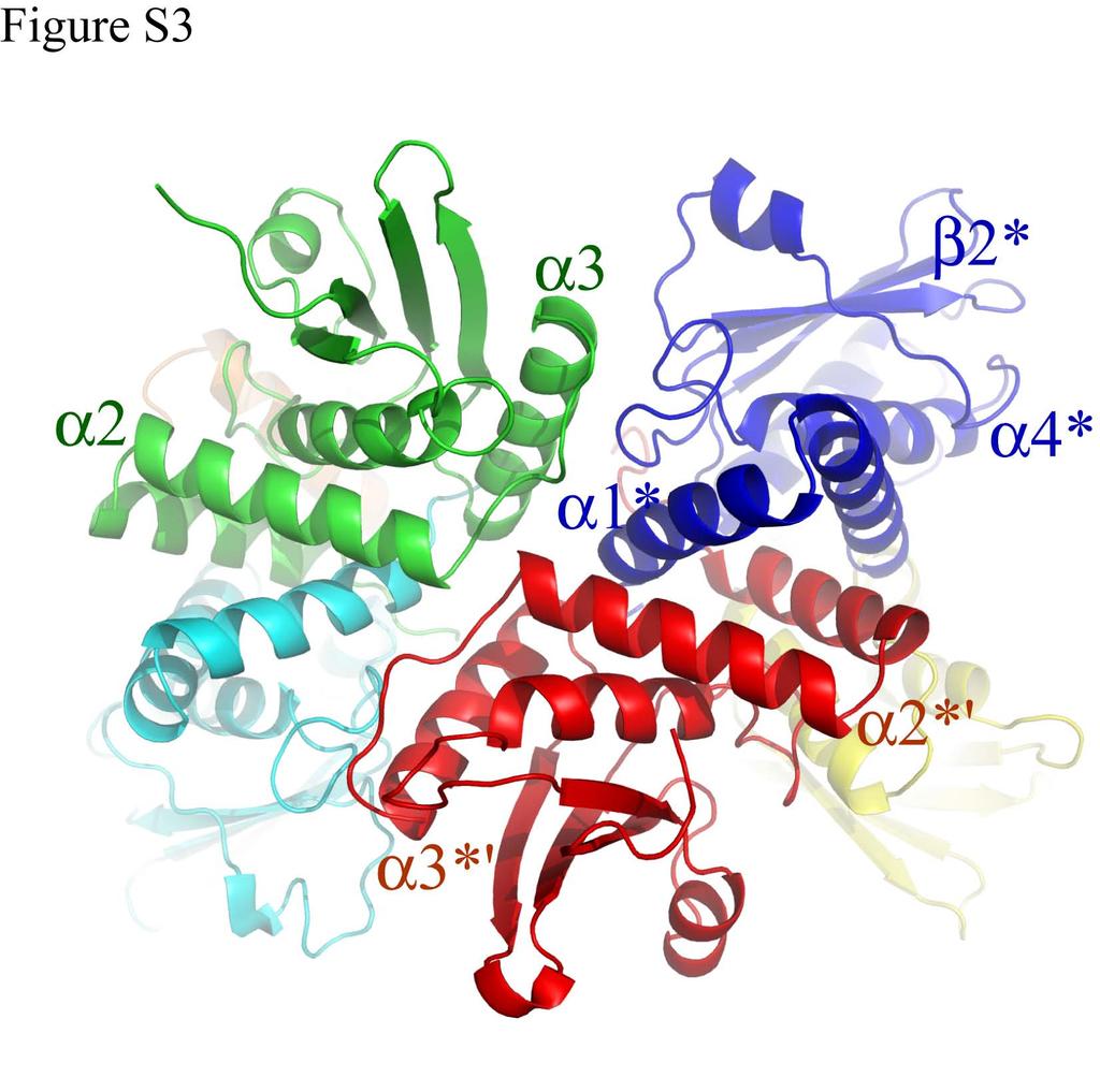
8 Figure Legends Figure S1. Molecular weight determination of AxCeSD in solution (a) The result of the gel filtration experiment using a column of Hi-load 26/60 Superdex 200.(Amersham Biosciences) (b) The standard molecules were Vitamin B 12 (13.5 kda), Myoglobin (17 kda), Ovalbumin (44 kda), γ-globulin (158 kda), and Thyrogllobulin (670 kda). The calculated molecular weight of AxCeSD in solution was kda, corresponding to AxCeSD ocatamer. Figure S2. Plot of small angle X-ray scattering The logarithm of the scattering intensity of N_AxCeSD (Rg = 34.4 Å) is plotted by black dots against the momentum transfer S = 4π sinθ/λ, where 2θ is the scattering angle and λ = 1.0 Å is the wavelength. The red (Rg = 34.4 Å, χ = 0.085) and green (Rg = 34.0 Å, χ = 0.059) curves are theoretical data calculated from crystal structure of octamer N_AxCeSD and full-length octamer N_AxCeSD model (with His 6 -tag and linker of 5 residues), respectively. Figure S3. Interaction between dimers A ribbon representation of the AxCeSD structure is shown from the side view with each monomer A (labeled with *), B (labeled with * ), C (labeled), and D (unlabeled) in blue, red, green, and cyan, respectively. The secondary structures contributing to 7
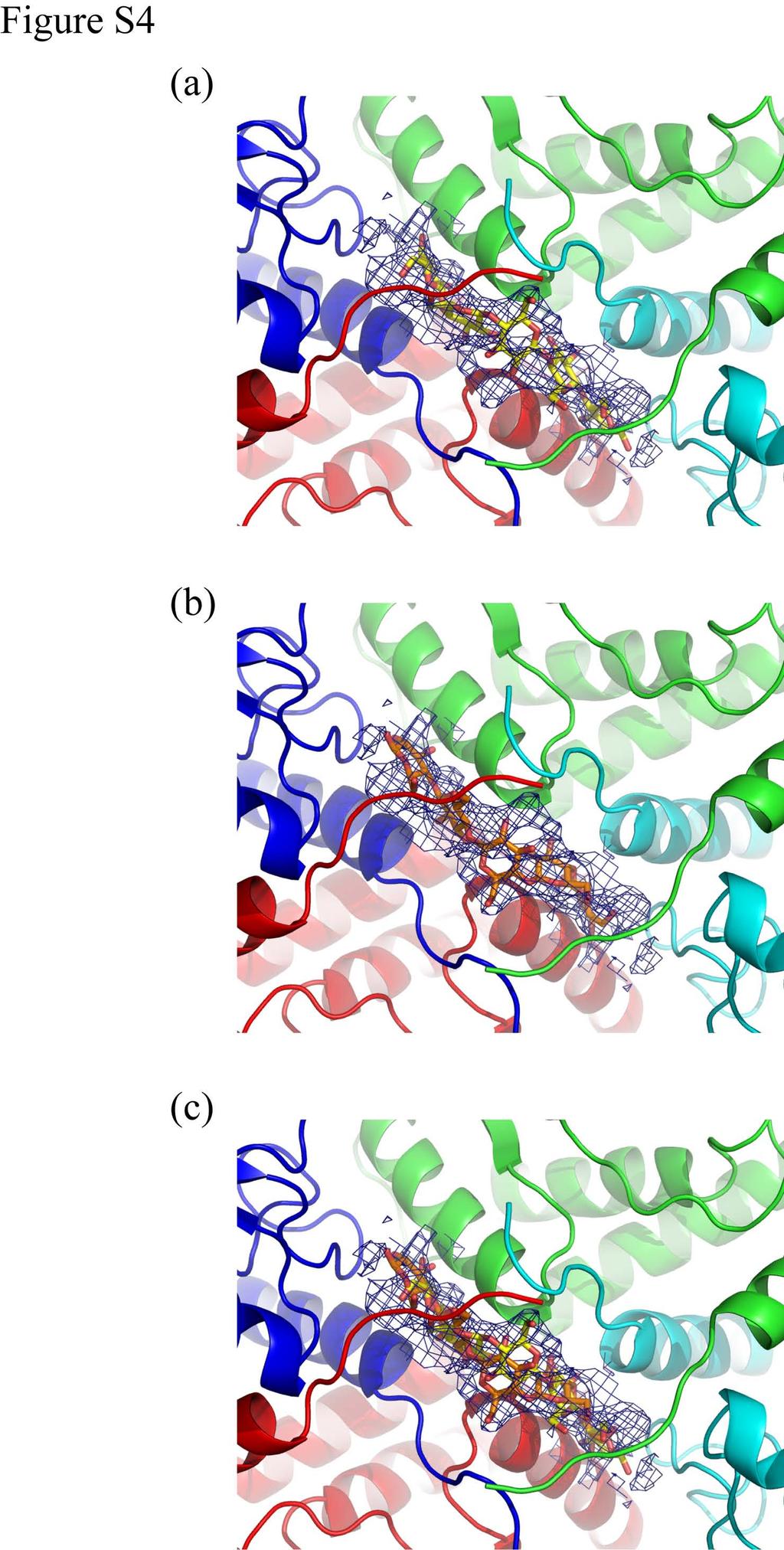
9 dimer dimer interaction are labeled in the same color of their ribbon model. Figure S4. The CPTs with omit maps from the inner view of the AxCeSD cylinder The four copies of C_AxCeSD (A, B, C, and D) are shown in ribbon representation in blue, red, green, and cyan, respectively (the same in all structure figures in this paper). The CPTs are shown as stick models (oxygen atoms: red, carbon atoms: yellow or orange), and omit maps are contoured at 1.6 σ. One of two CPTs in a pair is shown in (a) and the other is shown in (b). (c) A pair of CPTs. Figure S5 Confirmation of axcesd gene deletion (a) The result of PCR for checking axcesd gene deletion by insertion of antibiotic-registant gene into a genomic DNA. (b) The result of Western-blotting for checking axcesd gene deletion. The lanes are same in both experiments, Lane 1: Marker; Lane 2: wild-type; Lane 3: DBCD. 8
10
11
12
13
 (b)")
14 Figure S5 (a) (a) (b) AxCeSD