Apoptosis And Anti-tumor Effect Induced By Mtor Inhibitor And Autophagy Inhibitor In Human Osteosarcoma Cells
|
|
|
- Collin Cain
- 5 years ago
- Views:
Transcription
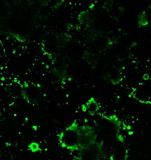
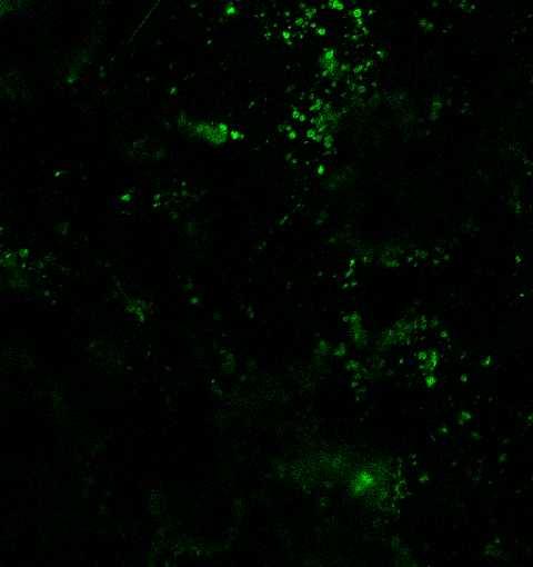
1 Apoptosis And Anti-tumor Effect Induced By Mtor Inhibitor And Autophagy Inhibitor In Human Osteosarcoma Cells Ryosuke Horie. Kagawa University of medecine, Kita-gun, Japan. Disclosures: R. Horie: None. Introduction: The mammalian Target of Rapamycin (mtor) regulates cellular functions, including cell growth, proliferation, migration, survival, and angiogenesis in many cancers. Not surprisingly, there is an intricate relationship between autophagy and apoptosis. Recent studies indicate that autophagy can function as a self-defense mechanism in cells that are exposed to antitumor agents, and that blocking autophagy can trigger activation of apoptosis. Recently, a potent small-molecule inhibitor of autophagy named spautin-1 (for Specific and Potent AUTophagy INhibitor-1) was discovered. This molecule promotes the degradation of Vps34-PI 3 kinase complexes by inhibiting two ubiquitin-specific peptidases, USP10 and USP13, that target the Beclin1 subunit of Vps34 complexes. Spautin-1 blocks the pro-survival autophagy pathway in cancer cells. The purpose of this study was to examine whether rapamycin affected suppression of phosphorylation of proteins in the mtor pathway, and whether a combination of rapamycin and spautin-1 induced apoptosis in MG63, a human osteosarcoma cell line. Methods: Chemical Reagents Both rapamycin and spautin-1 were purchased from Calbiochem (San Diego, CA, USA), dissolved in dimethyl sulfoxide (DMSO), and stored at -20 C. Cell lines and cell culture MG63 cells were grown in Dulbecco s Modified Eagle Medium (DMEM; Sigma-Aldrich, St. Louis, MO, USA) containing 10% fetal bovine serum (FBS; Sigma-Aldrich) and 100 U/ml penicillin. These cells were routinely maintained at 37 C in a humidified 5% CO 2 atmosphere, and cultures were used at mid-log phase. In vitro proliferation assay Cell proliferation was determined by the CellTiter 96 AQueous One Solution Cell Proliferation Assay (Promega, Madison, WI, USA). Briefly, cells were trypsinized and seeded at a density of approximately cells/well in 96-well cell culture plates containing 200 μl/well of growth medium. After 48 h, medium was replaced with fresh medium supplemented with rapamycin at a concentration of 0, 0.4, 2, 10, or 50 μm. After 24 h and 48 h, the medium was replaced with fresh medium containing 3-(4,5- dimethylthiazol-2-yl)-5-(3-carboxymethoxyphenyl)-2-(4-sulfophenyl)-2h-tetrazolium (MTS) reagent. The percent viability of each well was calculated. In the experiments testing the combined effect of rapamycin and spautin-1, cells were treated with 30 μm rapamycin and 100 μm spautin-1 for 24 h. The optical density was measured at 490 nm using an automatic microplate reader after 2 h of further incubation with the MTS reagent. Absorbance was directly proportional to the number of living cells. The percent viability of each well was calculated. At least three independent experiments were performed. Western blot analyses Cells were trypsinized and seeded at a density of approximately cells/well in 6-well cell culture plates with 2 ml/well of growth medium. After 48 h, cells were treated for 24 h with fresh medium supplemented (for separate experiments) with rapamycin at a concentration of 0, 0.4, 2, 10, or 50 μm; with 20 μm rapamycin and 100 μm spautin-1; or with 20 µm rapamycin or 100 μm spautin-1. Cell lysates were separated by sodium dodecyl sulfate polyacrylamide gel electrophoresis (SDS-PAGE) under reducing conditions. Gels then were electrophoretically transferred to nitrocellose membranes, which were incubated with primary and secondary antibodies overnight at 4 C. Bound antibodies were detected using the ECL plus Western blotting detection system (GE Healthcare Bio-Sciences, Piscataway, NJ, USA) and an LAS-1000 plus image analyzer (Fuji Film, Tokyo, Japan). Specific signals were quantified by densitometric analysis (NIH image J software). Fluorescence microscopy images of annexin-v-fitc-stained cells Cells were trypsinized and seeded at a density of approximately 1 x 10 6 cells/well on 25-mm circular coverslips (Matsunami Glass Ind. Ltd., Osaka, Japan) in 2 ml growth medium for 48 h. Next, cells were washed in phosphate-buffered saline and treated for 24 h with fresh medium supplemented with rapamycin and/or spautin-1. After treatment, cells were incubated with annexin-v- FITC and propidium iodide using the Annexin-V-Fluos Staining Kit (Roche Applied Science, Penzberg, Germany) for 15 min in a dark room. Cells then were imaged in a Attofluor cell chamber from Molecular Probes (Invitrogen) on the thermo-controlled stage of an inverted epifluorescence microscope as above. Results: Rapamycin inhibited proliferation of MG63 cells Rapamycin inhibited MG63 cell proliferation in a dose-dependent manner. The IC 50 for 24 h of rapamycin treatment in MG63 cells was μm.
, or 20 μm rapamycin and 100 μm spautin-1 (Rap-plus-Spa group). Cell proliferation was significantly lower in the Rap-plus-Spa group than in the Rap group.")
2 Rapamycin-induced MG63 cell death is enhanced by spautin-1 Based on the 24-h IC 50 of rapamycin, we examined proliferation of MG63 cells treated for 24 h with 20 μm rapamycin (Rap group), 100 μm spautin-1 (Spa group), or 20 μm rapamycin and 100 μm spautin-1 (Rap-plus-Spa group). Cell proliferation was significantly lower in the Rap-plus-Spa group than in the Rap group. These results indicated that spautin-1 enhanced rapamycinmediated suppression of MG63 cell proliferation. Inhibition of autophagy by spautin-1 increases rapamycin-induced apoptosis In cells treated with rapamycin or with rapamycin plus spautin-1, production of cleaved PARP was strongly increased. We observed multiple annexin-v-fitc-positive cells in the Rap-plus-Spa group, with the number of apoptotic cells strongly increased in the Rap-plus-Spa compared to the numbers in the control group, the spautin-1 group, or the Rap group. Discussion: This study demonstrated that rapamycin induced autophagy in MG63 cells by inhibiting phosphorylation of mtor pathway components, and that rapamycin-induced apoptosis was enhanced by spautin-1. These results suggest that selfprotective mechanisms involving mtor inhibitors in MG63 cells are prevented by inhibition of autophagy. The combination of an mtor inhibitor (e.g., rapamycin) and an autophagy inhibitor (e.g., spautin-1) may offer effective treatment for osteosarcoma, as this combination effectively induced apoptotic pathways in an osteosarcoma-derived cell line. Significance: The combination of an mtor inhibitor and an autophagy inhibitor may offer effective treatment for osteosarcoma, as this combination effectively induced apoptotic pathways in an osteosarcoma-derived cell line. Acknowledgments: none References: none
3
4

5 ORS 2014 Annual Meeting Poster No: 1990
6