ABSTRACT. The adaptive immune system s response to invading pathogens depends on the ability of T
|
|
|
- Kelley Snow
- 5 years ago
- Views:
Transcription
1 ABSTRACT McMILLAN, RUTH ERICA. Transcriptional Control of Dβ2. (Under the direction of Michael L. Sikes). The adaptive immune system s response to invading pathogens depends on the ability of T and B-lymphocytes to generate a diverse repertoire of antigen receptors. This diversity is created during lymphocyte development by somatic recombination of variable (V), diversity (D) and joining (J) gene segments. Transcription of unrearranged gene segments precedes their recombination. Given the correlation of transcription and recombination, it has been suggested that enhancers and promoters within each locus may regulate the recombinational accessibility of nearby gene segments. For example, in developing T cells, two stages of recombination (first D to J, then V to DJ) are seen in the T cell receptor beta gene (TCRβ). Both steps require the TCRβ enhancer, Eβ, while a promoter associated with the first D segment (PDβ1) only controls its recombination. In mutant cells that lack PDβ1, the second Dβ rearranges normally. To investigate the regulation of Dβ2 recombination, we have characterized the transcriptional profile of DJCβ2 gene cassette and identified separate promoter activities positioned 5 and 3 of Dβ2. The relatively simple downstream promoter (3 PDβ2) contains no discernable initiator or TATA elements, is located 400 bases downstream of Dβ2, and is driven by two NFkB binding sites. Ribonuclease protection assay and 5 RACE PCR revealed diffused transcription start sites throughout the entire 150 bp promoter region. Transcription from 3 PDβ2 occurs only when the TCRβ locus is in its germline configuration. In-vivo germline transcription of the upstream promoter (5 PDβ2) is detected in DNA with DβJβ2 joints. When a luciferase reporter plasmid bearing Eβ and sequential truncations of the DJβ2 sequence was transiently transfected into a RAG1
2 deficient cell line in-vitro, transcription from 5 PDβ2 was detected when a 290-bp region immediately downstream of Dβ2 was deleted. Therefore, germline transcription from 5 PDβ2 repressed while 3 PDβ2 is active. Our report indicates that 3 PDβ2 is positioned downstream of its associated D segment, a position that does not efficiently promote recombination. We hypothesize that this unique positioning plays an essential role in delaying Dβ2 recombination, thereby providing the locus with a second recombination opportunity should the recombination with the upstream Dβ1 fail.
3 Transcriptional Control of Dβ2 by Ruth Erica McMillan A dissertation submitted to the Graduate Faculty of North Carolina State University In partial fulfillment of the Requirements for the degree of Doctor of Philosophy Microbiology Raleigh, NC 2007 Approved by: Dr. Linda K. Hanley-Bowdoin Dr. James W. Brown Dr. Scott M. Laster Co-chair of Advisory Committee Dr. Michael L. Sikes Chair of Advisor Committee
4 DEDICATION I would like to dedicate this work to my late aunt, Enid Harrison, you gave me the motivation and courage to pursue my dreams. ii
5 BIOGRAPHY Ruth was born and raised in Shirley Castle, a small mountainous village in rural Jamaica. As a child, she had a keen interest in science. Upon graduation from Holy Childhood High School, Ruth moved to Florida in search of employment. After countless denials, she joined the United States Air Force where she spent the next four years and met her husband. While serving in the Air Force, Ruth never lost sight of her aspiration of becoming a biologist. Upon completion of her military duty, she pursued this interest in college and in 1981 she earned B.S. in biology from the University of North Carolina at Wilmington. It was during this time her first son born. After graduation Ruth worked as a medical technologist and raised her family, but had a burning desire to fulfill her academic dreams. In 2001, she enrolled in college at North Carolina Central University. In 2003 she received a M.S. in biology and also found a love for immune system. She continued her education by pursuing a Ph.D in Microbiology department at North Carolina State University, under the direction of Dr. Michael Sikes. iii
6 ACKNOWLEDGEMENTS First, I would like to thank God for giving me patience and endurance. Next, I would specially like to thank Dr. Michael Sikes for training me in his lab over the last four years. He has been very patient, supportive, and always available for advice. He is always optimistic, and encouraging. I am grateful to Dr. Scott Laster, Dr. James Brown and Dr. Linda Hanley-Bowdoin who served as my committee members. I would also like to thank our undergraduate student Kristy Lamb, and graduate students Tim Orcutt and Justin Bradshaw for their help. I am very thankful to all my family for giving me the opportunity to further my education, even if it meant giving up some good times. They have been my inspiration and I would not have made it without their constant encouragement and support. Thanks to the Microbiology department for accepting me into the graduate program and to the many friends in the department that helped me to attain my goal. iv
7 TABLE OF CONTENTS Page LIST OF TABLES...viii LIST OF FIGURES... ix CHAPTER 1: Literature Review Overview of the Immune system Overview of antigen receptor structures Organization of antigen receptor genes V(D)J Recombination Gene segment utilization The role of RSS in recombination The recombinase DNA repair proteins involved in V(D)J recombination Control of V(D)J Recombination Regulation of the recombinase Regulation of the substrate: The accessibility model Enhancers Promoters Other cis-acting regulatory elements Transcription Chromatin Structure Modulating DNA architecture Applications to V(D)J recombination:acetylation Methylation Chromatin remodeling Thymic Development Transcriptional Regulation of Dβ References CHAPTER 2: Differential Activation Of Dual Promoters Alters Dβ2 Germline Transcription During Thymocyte Development Abstract Introduction v
8 2.2 Materials and Methods Cell-sorting RT-PCR Transcript analysis Plasmids EMSA Luciferase assay Results Developmental timing of Dβ2 recombination Initiation of Dβ2 germline transcription Promoter activity associated with Dβ NFκB regulates promoter activity downstream of Dβ GATA-3, Runx1, and USF1 bind multiple sites 5 Dβ Organization of promoter activity upstream of Dβ Discussion References CHAPTER 3: The Expression of Runx Transcriptional Factors During Thymocyte Development Abstract Introduction Materials and Methods Antibodies and cell-sorting Quantitative real-time PCR Nuclear extracts Results Discussion References CHAPTER 4: Recombination Accessibility Of Dβ2 Gene Segment In Developing Thymocytes Abstract Introduction vi
9 4.2 Materials and Methods Analysis of RSS PCR/ RT-PCR analysis of sorted thymocyte populations Results Differences in RSS Developmental timing of Dβ2 recombination Discussion References CONCLUSION vii
10 LIST OF TABLES CHAPTER 4: Recombination Accessibility Of Dβ2 Gene Segment In Developing Thymocytes Page Table 4.1 Alignment of Recombination Signal Sequences Table 4.2 Analysis of DJβ RSS quality viii
11 LIST OF FIGURES CHAPTER 1: Literature Review Page Figure 1.1 A comparison of the monomeric B-cell and T-cell receptor... 4 Figure 1.2 Maps of B-cell receptor loci... 7 Figure 1.3 Maps of T-cell receptor loci... 9 Figure 1.4 Schematic diagram of somatic gene recombination Figure 1.5 Schematic diagram of recombination signal sequences Figure 1.6 DNA rearrangement by RAG proteins Figure 1.7 Processing of coding ends generated during V(D)J recombination CHAPTER 2: Differential Activation Of Dual Promoters Alters Dβ2 Germline Transcription During Thymocyte Development Figure 2.1 Development timing of DJβ germline transcription Figure 2.2 Initiation of Dβ2 germline transcripts Figure 2.3 Promoter activities associated with Dβ Figure 2.4 Organization of the Dβ2 regulatory region Figure 2.5 Sp1 and GATA-3 bind downstream of Dβ Figure 2.6 NFκB binds downstream of Dβ Figure 2.7 NFκB contributes to promoter activity of 3 of Dβ Figure 2.8 GATA-3 binds multiple sites upstream of Dβ Figure 2.9 Runx1 binds upstream of Dβ Figure 2.10 USF1 binds upstream of Dβ Figure 2.11 Promoter activity of 5 of Dβ Figure 2.12 Multiple transcription factors contribute to 5 PDβ2 promoter activity Figure 2.13 Model for differential transcription within Dβ2 cassette CHAPTER 3: The Expression Runx Transcriptional Factors During Thymocyte Development Figure 3.1 Transcription of Runx during T cell development Figure 3.2 Expression of Runx proteins during thymocyte development CHAPTER 4: Recombinational accessibility of Dβ2 gene segment in developing thymocyte Figure 4.1 Timing of DJβ recombination during thymopoiesis ix
12 CONCLUSION Figure 5.1 The advantage of an initial Vβ-to-Dβ1rearrangement x
13 CHAPTER 1 Literature Review 1
14 1.1 Overview of immune system The immune system defends against a wide variety of foreign pathogens, such as bacteria, viruses, parasites, and abnormalities such as cancer cells. There are two branches of the immune system: the innate or natural immune system, and the adaptive or acquired immune system. The innate system includes host mechanisms that are encoded in the germline genes of the host. Recognition molecules are broadly expressed on many different cells and are always poised and ready to rapidly defend the host against invading pathogens. This allows for a proficient early anti-microbial response, but lacks specificity for the antigen. Adaptive response, on the other hand, mounts a specific response to a diverse selection of antigen, which improves with subsequent infections. Unlike the germline encoded recognition molecules of the innate system, somatic rearrangement of germline genes form the antigen-specific receptors of adaptive immune cells. It is this rearrangement of genes that gives the adaptive immune cells the enormous diversity of antigen receptors needed to mount a specific response to virtually any pathogen encountered. T and B-lymphocytes are the primary immune cells of the adaptive system. These cells are derived from a common haematopoetic stem cell precursor and are morphologically similar. For B cells, the molecules responsible for specific antigen recognition are the various classes of antibodies, which are expressed as cell surface receptors called B-cell receptors (BCRs), or a secreted form, referred to as antibody molecules. Antibody production is very important to a variety of processes including the clearance of particulate pathogens and initiating complement mediated destruction. The comparable recognition molecule on T cells is the membrane bound T-cell antigen receptor (TCR), which recognizes 2
15 antigen presented by the major histocompatibiliy complex (MHC). T-lymphocytes are either cytotoxic or helper-t cells and mediate cellular immunity. Recognition of surface antigens by the cytotoxic T-cell receptors results in direct lysis of target cell. Helper T-cells on the other hand, help to propagate the immune response by antigen mediated cell-cell interactions and the production of cytokines. The main focus of this introduction is to give an overview of the structure of the antigen receptor and discuss the mechanism and regulation of somatic gene rearrangement Overview of antigen receptor structures Antibodies belong to the class of serum proteins called the γ-globulins or immunoglobulins (Ig) and are built from four polypeptide chains. The monomeric Ig molecule is made up of two identical larger polypeptide chains (Heavy chain) of molecular weight 50,000 kda and two identical smaller polypeptide chains (Light chain) of molecular weight 25,000 kda, which can be either kappa or lambda (Igκ, Igλ respectively). Each light chain is covalently attached to each heavy chain by a disulfide bond, and the two heavy chains are attached together by disulfide bonds. Both the light and heavy chains contain several homologous units of approximately 110 amino acid residues in length. Within each unit, an intra-chain disulfide bond forms a globular domain (Ig domain) that results in a loop of about 60 amino acids. Light chains contain one variable domain (V L ) and one constant (C L ) domain and heavy chains contain one variable domain (V H ) and three or four constant domains (C H 1- C H 4), depending on the class of antibody (Figure 1.1A). The amino acid variation within the variable domains of the light and heavy chains accounts for the capacity of different antibodies to bind an infinite number of structurally diverse antigens. It is the constant 3
16 A B Figure 1.1: A comparison of the monomeric B-cell and the αβ T-cell receptor. (A). The migm consist of a heavy chain (H) (blue) and a light chain (L) (black). (B). The αβ T-cell receptor consists of a β-chain (blue) that is analogous to the heavy chain and an α-chain analogous to the light chain. Both the light and heavy chain and the α- and β-chains contain amino-terminal variable (V) region and constant region (s). The IgM and αβ T-cell receptor contain Ig domains that consist of approximately amino acids linked by disulfide bonds. Disulfide bonds also link L-H, H-H, and the α-β chains together. The constant region defines the light chain subtype and the heavy chain isotype of the Ig molecule. 4
17 domain that defines the classes of antibodies. All antibodies of a given class share identical constant domains. Immunoglobulins are divided in five classes or isotypes and each class differs in effector immune function. They are immunoglobulin A (IgA), immunoglobulin D (IgD), immunoglobulin E (IgE), immunoglobulin G (IgG), and immunoglobulin M (IgM). Their heavy chains symbolized by the following Greek letters (α, δ, ε, γ, and, µ respectively). The TCR is a heterodimer that consists of two transmembrane polypeptide chains, covalently linked by disulfide bonds. There are four TCR chains: alpha (α), beta (β), delta (δ), and gamma (γ). Once rearranged, the TCR chains combine to form one of two possible TCR heterodimers, αβ or γδ and each mature T cell expresses only one combination. The TCRαβ is the most common form. Each α chain and β chain consists of one Ig domain in the N-terminal region (V β /V α ) and one in the constant domain (C β /C α ). The N-terminal domain contains the variable region of the α chain and β chain and represents the antigen-binding site for peptides presented on MHC molecules. The carboxyl-terminal Ig domain is anchored to the cell by a hydrophobic transmembrane region and extends within the cell as a short cytoplasmic tail (Figure 1.1B). Each TCR chain, like the BCR heavy and light chains is encoded by multiple gene segment that undergo somatic gene rearrangement to create antigen binding sites that are capable of recognizing different antigens. The TCR and BCR are similar structurally; however there are significant functional differences between the receptors. One notable difference is that the TCR, unlike the BCR, is not produced in a secreted form and is incapable of directly binding antigens, but is rather dependent on cell-to-cell interactions through the peptide-mhc complex. Also, unlike the Ig, the C-terminal of T cell chains cannot be changed to produce different classes, and hence cannot change effector function. 5
18 1.1.2 Organization of antigen receptor genes In the germline configuration, antigen receptor genes are composed of separate gene segments separated by intervening non-coding DNA segments. During T cell and B cell maturation, these gene segments must recombine by gene rearrangement to form functional genes for receptors. The organization of the Ig and TCR gene loci is essentially the same in all mammals, although the numbers and sequence of gene segments and the location their chromosomal location may vary. The BCR is encoded by three separate loci namely the Ig heavy chain (IgH), the κ light chain (Igκ) and the λ light chain (Igλ) loci (Figure 1.2). The IgH locus is on chromosome 14 in humans and on chromosome 12 in mice. In the germline configuration, the Ig locus is composed of multiple copies of three gene segments: the V (variable), J (joining) and C (constant) regions. In addition, the IgH locus has diversity (D) gene segments located between the V and J segments. In human, the locus spans approximately 1.5 Mb and contains from a few hundred to a few thousands VH segments at the 5 end, 13 DH segments downstream, followed by 4 JH segments and ends with 8 CH segments. In Mice, the IgH locus is similarly organized and spans about a 200 kb region. Many cis-acting transcription elements have been identified in the IgH locus. These include three enhancer elements: the 5 DQ52 enhancer located just upstream the most 3 D gene segment; the intronic Eµ enhancer, located in the JH-Cµ intron; and the 3 EH enhancer located 16 kb downstream of Cα. Promoters flanking VH and CH gene segments are also present. The human Igλ locus is located on chromosome 22 and spans approximately 1000 kb. There are about 100 Vλ gene segments located in the 5 portion of the locus and seven Jλ-Cλ clusters 3 of the most distal Vλ gene segment. In the mouse, the Igλ locus is situated on 6
19 A 5 L VH DH JH Eµ Cµ Cγ Cγ2 Cγ2b Cγ2 (134) a C ε Cα 3 E 3 7 kb 4.5 kb 55 kb 39 kb 21 kb 15 kb 14 kb 12 kb B 5 L Vκ (85) J κ ie κ Cκ 3 E κ 3 23 kb 2.5 kb C L Vλ2 Jλ2 Cλ2 Jλ4 Cλ4 3 E L Vλ1 Jλ3 Cλ3 Jλ1 Cλ1 λ kb 1.2 kb 2.0 kb 1.3 kb 1.4 kb 1.7 kb 1.3 kb Figure 1.2: Maps of B-cell receptor loci. Immunoglobulin Loci. A, B, and C murine IgH, Igκ, and Igλ loci, respectively. Solid circles represent enhancer regions, while open circles are matrix attachment regions. The V, D and J gene segments are shown by solid rectangles. Hatched bars mark the switch regions in the IgH locus. The horizontally striped bars mark leader segments. 7
20 chromosome 16 and contains only two V genes located in the 5 portion of the locus and two Jλ-Cλ clusters in the 3 portion. Several cis-regulatory elements have been identified with the Igλ locus which include the Vλ promoter, an enhancer 3 of Cλ4, a silencer and two antisilencers located between Vλ and Jλ gene segments. In the mouse, the Igκ locus is located on chromosome 6 and spans at least 1Mb. At the 5 region there are approximately 300 Vκ gene segments and five Jκ and one Ck gene segment in the 3 end of the locus. Unlike the IgH and Igλ locus, Igκ contains only one Ck gene segment, so there are no subtypes of the κ-light chain. In humans, the Igκ locus is very similar to that of the mouse; however there are fewer Vk gene segments. The Igκ locus contains an intronic enhancer located in the Jκ- Ck intron. There is also another enhancer (3 Eκ) located 9 kb downstream of Ck. The TCR is encoded by three separate loci: the TCRβ, α/δ, and γ (Figure 1.3). In the mouse, the TCRβ locus is on chromosome 6 and spans over 800 kb. Approximately 25 to 30 Vβ gene segments is situated within a 450 kb region located 300 kb upstream of two clusters of D, J, and C gene segments. Each clusters contains one Dβ, six or seven Jβ and one Cβ. A Vβ segment is located about 10 kb downstream of the most distal Cβ (Cβ2) (1, 2). TCRβ contains a single transcriptional enhancer element (Eβ), which is located between the Cβ2 and Vβ14 (3). In mice and humans, the TCR α/δ locus is on chromosome 14 and spans over 1Mb. The δ chain gene segments are located between the Vα and Jα segments. Mouse germ-line DNA contains about 100 Vα at the 5 region of the locus. The δ chain gene family is found downstream of Vα and contains about 10 Vδ gene segments, which are mostly distinct from Vα gene segments, however some sharing has been observed in rearranged α and δ genes. In the 120 kb region 3 of Vδ gene segments there are two Dδ segments 8
21 A B C Figure 1.3: Maps of murine T-cell receptor. (A). TCR α/δ locus. (B). TCRβ locus. (C). TCRγ locus. Black, dark and light gray boxes mark the V, D and J gene segments, respectively. Solid black circles mark enhancers. Hatched bars mark leader regions. Solid black rectangles represent the constant regions. Checkered boxes mark the silencers. 9
22 followed by two Jδ segments, a single Cδ segment, the T-early alpha TEA (TEA) exon, 61 Jα and one Cα segment. The TCR α/δ locus contains numerous cis-acting elements which include several promoters, Eα, silencer elements, and a locus control region (LCR) 3 of Eα. Lastly, the mouse γ chain on chromosome 13 consists of three functional clusters, each containing V, D, and J segments. The most 5 cluster contains five Vγ segments, one Jγ and one constant Cγ1 segment. The downstream clusters have one to two Vγ, one functional Jγ and one constant region. Two enhancers have been identified, one 3 of Cγ1 and the other at the most 3 end of the locus, downstream of Cγ V(D)J Recombination The ability of the immune system to respond to nearly any possible pathogen is dependent on the capability of developing B and T cells to generate a diverse repertoire of antigen receptors. This remarkable diversity is achieved through the somatic rearrangement of the Ig and TCR loci. This process is called V(D)J recombination, named after the germline gene segments that recombines to form the clonally unique receptor structure expressed on each developing T and B lymphocyte (2, 4-7). During lymphocyte development, Variable (V), Diversity (D), and Joining (J) gene segments undergo a specific site somatic recombination to create a unique receptor gene. During transcription, this uniquely combined gene is spliced to a constant region of the locus, which does not undergo somatic recombination, and the resulting mrna encodes for the full length V(D)JC polypeptide chain (Figure 1.4). Studies from the Tonegawa group (8) provided direct proof that the variable gene segments had rearranged at the DNA level. They isolated germline DNA gene segments from mouse embryos that contained part or all the λ chain as well as recombined λ chain from mouse myeloma. They used restriction enzymes, a new technique 10
23 at that time, to cut the DNA into fragments, hybridized to λ light chain and analyzed for R loops. This technique detected V gene segments that were not attached to a C gene segment Gene segment utilization All antigen receptors contain multiple V and J gene segments, however only one V and one J segment will be used in the recombined product. In the IgH, TCRβ, TCRδ gene rearrangement involves one D gene segment joining to one J segment and the recombined D- J segment recombining to an upstream V segment to form a contiguous coding exon for the antigen receptor gene (9). An exception to this is the TCRδ D gene, where more than one D gene segment may be used to yield a Vδ-D-δD-δJδ-Cδ recombined gene. In Igλ, Igκ, TCRα, and TCRγ, V gene segments join directly to J segments because there are no D gene segments present The role of RSS in recombination Each V, D, and J gene segment is flanked by a recombination signal sequence (RSS), which serves as recognition site for the recombinase and directs site-specific cleavage (Figure 1.5). RSSs are required and sufficient for recombination of extrachromosomal substrates (10-12). An RSS consists of a conserved palindromic heptamer (5 -CACAGTG- 3 ), and an A/T rich nonamer (5 -ACAAAAACC-3 ), which is separated by poorly conserved spacer sequence of either 12 or 23 bases in length. There is much heterogeneity in the RSS (13) flanking the individual gene segments, with some RSS being closer to the consensus sequence than others (14). The RSS are referred to as either a 12 RSS or a 23 RSS depending on the length of the intervening spacer. V(D)J recombination occurs only between two gene segments whose flanking RSSs contain a 12 RSS and a 23 RSS, referred to as the 12/23 rule (15). The 12/23 restrictions 11
24 Figure 1.4: Schematic diagram of somatic gene recombination. The DNA of T-cell α and β loci are recombined and transcripts are processed to generate a functional αβ T-cell receptor. First D-J joining occurs on the β-chain, followed by V to D-J joining. After transcription and splicing the β-chain is expressed, and V to J joining occurs on the α-chain. Both the β-chain and the α-chain are expressed on the surface of the T cell. 12
25 Figure 1.5: Schematic diagram of the recombination signal sequences. (A). The RSS is made up of a conserved seven bases (heptamer) and nine bases (nonamer), separated by either 23 or 12 bases of semi-conserved sequence, and flanked by V, D or J coding sequences. (B). 12RSS are denoted by open triangles, whereas 23 RSS are denoted by black triangles. 13
26 help to ensure the proper assembly of Ig and TCR variable gene segments, preventing wasteful V-to-V and J-to-J rearrangements. For example, within the IgH locus, the VHs are flanked with 23 RSS, DHs with 5 and 3 12 RSS, and JH with 23 RSS. Since VH to JH gene segment joining is prohibited, the configuration of the Ig RSS forces the use of DH segments, and further adds to the diversity of the IgH chain. At the TCRβ locus, the influence of the RSS is even more specific. For instance, Vβ are flanked with 23 RSS and Jβ with 12 RSS, while Dβ have 5 12 RSS and 3 23 RSS, however Vβ to Jβ rearrangement does not occur, despite the 12/23 compatibility between the Vs and the Js. This is because the 12 RSS 5 Dβ1, and not the 12 RSS 5 Jβ, specifically targets upstream Vβ gene segments (16, 17). This is referred to as the beyond 12/23 restriction, and is believed to be limited by nicking at Jβs (18) although the molecular mechanisms remains unknown. This beyond 12/23 restriction has also been confirmed in nonlymphoid cells by using transiently expressed recombination substrates and purified RAG proteins (19-21). The RSS sequences and the adjacent coding sequences are important for efficient V(D)J recombination, as shown by substrate recombination studies (9, 12). This means that the quality of the RSS may contribute to the efficiency of RAG binding, synapsis, and subsequent cleavage; skewing the usage of particular V, D, and J segments (12). Gene segment usage is not dependent on the distances between RSS, as gene segments in the IgH locus that are 2000 to 3000 kb apart can efficiently recombine. V(D)J recombination is initiated by DNA double-strand breaks (DSB) between V, D, and J segments and the adjacent RSS. The two signal ends (RSS ends) are precisely joined, whereas the coding ends are modified by nucleotide addition or deletion. The orientation of 14
27 the recombining RSS determines if recombination results in deletion or inversion. When two RSS are in opposing orientation the intervening sequence between the two RSS is deleted from the chromosome, but when the two RSS are in the same orientation the intervening sequence is inverted, but not deleted The recombinase Two lymphoid cell-specific proteins encoded by the recombination activating genes RAG1 and RAG2 (22, 23) are both necessary and sufficient to support cleavage of individual RSS or coupled cleavage of 12/23 RSS in vitro (24, 25). The RAG (RAG1 and RAG2) proteins were identified by their ability to initiate V(D)J recombination in non-lymphoid cells (23). RAG1 and RAG2 proteins are absolutely required for V(D)J recombination. In RAG1 and RAG2 deficient mice, V(D)J recombination of the receptor locus does not occur, and B and T cell development is completely block (26, 27). In addition, McBlane has demonstrated that RAG proteins bind to RSS and initiate V(D)J recombination on DNA substrates in vitro (24). Ubiquitous DNA-bending factors, high mobility group proteins-1 and -2 (HMG1/2) are also important to RAG mediated binding and cleavage at the 23 RSS (28, 29). The biochemical reaction responsible for the recombination of gene segments can be divided into two phases (Figure 1.6). The first phase of V(D)J recombination begins with the recognition of signal sequences by RAG proteins. The two gene segments undergoing recombination are juxtaposed one to another, forming a synaptic complex, and the DNA is cleaved precisely at the border of the hepatmer of the RSS and the flanking coding sequence. In a divalent cation dependent reaction, RAG makes a single-strand nick of the phosphodiester bond between the last nucleotide of the coding sequence and the first nucleotide of the heptamer of the top strand. This nicking creates a free 3 OH at the end of 15
28 Figure 1.6: DNA rearrangement by RAG proteins. Initiation of V(D)J recombination requires two recombining gene segments with RSS of different lengths. RAG proteins (grey ovals) bind to the 12 RSS (black arrow) and 23 RSS (white arrow) and form a synaptic complex or paired complex (PC). In the absence of DNA repair proteins, DNA is cleaved by a RAG-mediated transesterification reaction that generates blunt double strand breaks (DSB) at the signal ends and hairpin ends at the coding ends. The signal and hairpin ends are held in a cleaved signal complex (CSC) by the RAG proteins. The ends are processed and ligated together by ubiquitously expressed DNA repair enzymes to form signal and coding joints. 16
29 the coding sequence and a 5 phosphate at the heptamer. Nicking is followed by a transesterification reaction where the 3 OH attacks the opposing phosphodiester bond of the antiparallel strand of DNA forming hairpin ends. This cleavage reaction results in four free ends, two 5 -phosphorylated blunt double strand breaks (DSB) at the RSS, or signal end; and two covalently closed, hairpins at the gene segment, or coding ends (30-33). RAG remains associated with the free ends in a stable postsynaptic complex (PSC) (34). The nicking step can occur in the absence of a synaptic complex, but an efficient transesterification reaction needs 12/23 paired RSS (35, 36). The RAG proteins are also important in the joining phase of the reaction (37). The second half of the V(D)J recombination reaction is not as well understood, but involves the processing of coding and signal ends by RAG proteins, as well as ubiquitous DNA repair proteins. The joining of signal ends is usually precise without additions or deletions of nucleotides (12). However, the coding joints are rarely exact and the added nontemplate nucleotides substantially increase antigen receptor diversity. The formation of the coding joint is initiated by first nicking the hairpin loops, followed by joining and ligation of the ends (4, 5, 12). If the nicking of the hairpin occurs in the middle of the loop, blunt ends are generated. However, if the nick occurs along any other point of the loop a 5 or 3 overhang occurs which is filled in by template derived palindromic (P) nucleotides (Figure 1.7). Nicking can also lead to deletion of nucleotides by exonuclease activity or the addition of non-germline or non-template (N) nucleotides by terminal deoxynucleotidyl transferase (TdT) (38). TdT, although not essential for recombination, further diversifies the viability of antigen receptors binding sites (39, 40). Ubiquitous DNA repair proteins, which include three components of the DNA- 17


 nucleotides by the")
30 Figure 1.7: Processing of coding ends generated during V(D)J recombination. Adapted from Immunity: The Immune Response in Infectious and Inflammatory Disease by Defranco, Locksley and Robertson, New Science Press. RAG-mediated cleavage produces covalently closed hairpin ends at each RSS. These hairpins may be nicked at any point, which creates gaps in the nucleotide sequence. These gaps may be filled by the addition of pallindromic (P) nucleotides by the polymerase enzyme, or by non-template (N) nucleotides by a terminal deoxynucleotide transferase (TDT) before joining. 18
31 dependent protein kinase (DNAPK): Ku70, Ku80, and the kinase catalytic subunit DNAPKcs; XRCC4, and DNA ligase IV proteins are responsible for joining and ligation of the processed coding ends (41). Recent evidence suggests that Artemis, an endonuclease which is activated by DNA-PK, catalyzes the hairpin opening and that the ubiquitous nonhomologous end-joining (NHEJ) machinery catalyzes the joining of coding ends to form a coding joint (42-45). The blunt-ended signal ends are also joined to form a signal joint (46). A deficiency in any of these factors result in an early block in T and B cell development. RAG1 and RAG2 proteins expressed only in T and B cells are the only lymphoidspecific factors needed for V(D)J recombination. The enzymatic core regions of RAG1 (47-49) and RAG2 (50) were determined by gradually truncating the full length RAG1 and RAG 2 proteins to their smallest active sizes. These core regions are capable of mediating complete V(D)J recombination of extrachromosomal substrates in nonlymphoid mammalian cells (7, 22, 23). Hence all other necessary factors must be ubiquitously expressed in the cell. Mice lacking either RAG gene are completely defective in V(D)J recombination (26, 51) because they are unable to initiate rearrangement of their TCR or Ig antigen receptors and consequently lack mature B or T cells. Mice in which full length RAG 1 is replaced with the core region produce reduced, but normal circulating T and B cells (52). This is the only defect noted in these mice, which strongly suggestive that RAG proteins are only essential for the generation of T and B cells. Mutations in RAG1 and RAG2 are also responsible for immune defects in humans, in which both T and B cells are depleted. Mutations such as frame-shifts that destroy RAG 19
32 activity result in complete immunodeficiency, while point mutations cause only partial loss in activity and result in severe combined immune deficiency (SCID) and Omenn s syndrome (OS) (53, 54). Despite the indications that RAG proteins were necessary for recombination, direct proof was delayed due to repeated failed attempts to purify the large insoluble proteins (42). It was not until Oettinger (23) and colleagues identified an active soluble core domain of each protein that the field of V(D)J recombination was revolutionized (49, 50, 55). The biochemistry of V(D)J cleavage was only understood when purified RAG1 and RAG2 proteins together bind the RSS (56) and DSBs are generated (24). RAG proteins are relatively large. The murine RAG1 contains 1040 residues, while RAG2 contains 527 residues. The majority of biochemical studies involving RAG proteins are done using the truncated form of the RAG proteins known as the core regions, which includes residues for RAG1 (47), and only the first 383 amino acids for RAG2 (50, 55). Together core RAG1 and core RAG2 are sufficient for cleavage in vitro and in vivo. Initially, studies from surface plasmon resonance and one-hybrid system demonstrated that the N-terminal portion of core RAG1 is homologous to the bacterial invertase, Hin, and directly binds to the nonamer RSS (57, 58). More recently, combination interference assays and footprinting have revealed that RAG1 binds the 12 RSS from the third base of the spacer to the sixth base of the nonamer (59). In addition, a central domain within core RAG1 binds specifically to the heptamer of the RSS (60, 61). Although RAG2 is necessary for DNA cleavage, its direct role in V(D)J recombination remains elusive. In the absence of RAG 1, RAG2 binds weakly and without sequence specificity (62) or not at all (63) to the RSS. However, the RAG1/RAG2 complex 20
33 binds to the RSS with more affinity (63, 64) and specificity (63) than RAG 1 alone. A possible explanation of this phenomenon is that RAG2 does not bind the RSS per se, but is bound to RAG1, resulting in a conformational change in RAG1 (65, 66) that increases its affinity for the RSS. Further support for this explanation comes from computational analysis of RAG2 that predicts the core region to contain a six-bladed beta-propeller structure (67) that is known to mediate protein-to-protein assembly (68). The active site of core RAG1 required for cleavage contains a triad of acidic residues D600, D708, and E962 collectively called the DDE motif that serves as a binding site for divalent metal (34, 69-71). These residues are important for nicking and hairpin formation. A mutation in any of these residues abolishes recombination in vivo and blocks cleavage by purified RAG1 protein but permits binding to the RSS (34, 69-71). In vitro experiments have demonstrated a direct role for RAG proteins in cleaving substrates and generating DSB at isolated RSS (24, 25). In the presence of Mn +, RAG1 and RAG2 efficiently cleave individual 12 RSS or 23 RSS to yield blunt 5 phosphorylated signal ends and hairpin coding ends, without loss of the coding sequence (24). However, in the presence of Mg +, the 12/23 rule is enforced and coupling of 12/23 RSS becomes necessary for cleavage and hairpin formation (25, 72). Together these in vitro studies have confirmed that RAG 1 and RAG 2 alone are responsible for RSS recognition, binding, initial cleavage, and hairpin formation during V(D)J recombination. While it is demonstrated that RAG1 and RAG2 are sufficient for the cleavage reaction, it seems likely that the initiation of V(D) J recombination of endogenous V, D, and J gene segments, which may be separated by several hundred of kilobases would require other proteins to bend the DNA in order to bring the gene segment closer together. Studies 21
34 have shown that HMG1and 2, ubiquitous DNA binding and bending proteins, may be added to RAG 1 and RAG 2 proteins to enhance binding and cleavage (28, 28, 29, 73-76). However, this effect is more pronounced at the 23 RSS stable complex (SC). Binding and bending is very inefficient at the 23 RSS unless supplemented with HMG1 and 2. Formation of a 23 SC improves by 10 fold with the addition of HMG1 and 2, while the 12 SC complex is slightly improved (28). Hence, HMG1 and 2 strongly enhance RAG binding to the 23 RSS and mediate strict 12/23-coupled cleavage in vitro (74, 76-78). However, a recent study has shown that RAG proteins are capable of inducing a 60 0 bend in the RSS, which does not increase with the addition of HMG 1 and 2 (73). HMG proteins have no sequence specific DNA binding sites, but instead recognize structures, such as DNA bends (79, 80). It is proposed that RAG binds and bends the RSS and the HMG1 and 2 proteins recognize this bent region, bind and in a clamp-like fashion stabilize the bent conformation. Currently, there are two proposed models that seek to explain 12/23 RSS coupling. In the first model, RAG proteins bind the heptamer and nonamer ends of each RSS and mediate synapsis through protein-to-protein interactions. The second model proposes that RAG proteins first preferentially load on one RSS and recruit the second RSS. Recent evidence suggests that RAG first loads on the 12 RSS (81). In addition, the 12RSS has been shown to have a higher affinity for RAG protein complexes (82), and nicked 12RSS but not 23 RSS was detected in vivo (83). These results are consistent with the observations that the 23 stable SC is less stable than the 12 (SC) (56) DNA repair proteins involved in V(D)J recombination As mentioned above, the initial recognition of RSS by RAG proteins is followed by the formation of a synaptic complex and subsequent cleavage by the RAG proteins. In vitro 22
35 experiments have confirmed the presence of both a pre-synaptic complex and a post-synaptic complex containing RAG proteins (23, 23, 84-86). Furthermore, the formation of a hybrid, which is a coding end joined to the opposite signal end, is indicative of cleavage in a synaptic complex (85). In spite of the importance of RAG proteins in mediating recombination of a substrate in vitro (87, 88), the resolution of the coding and signal joint in vivo is not mediated exclusively by RAG proteins. Notwithstanding the various types of ends produced by mechanisms such as: TdT, N, and P nucleotides, as well as exonuclease activity, the repair pathway is dependent on the same ubiquitous DNA repair factors that are a part of the nonhomologous end-joining (NHEJ) pathway. The NHEJ proteins join double strand breaks in DNA by a sequence homology independent mechanism. The NHEJ proteins are expressed in the G1 phase of the cell cycle (89, 90); the same stage at which DSB are generated during V(D)J recombination (91). There are at least six NHEJ proteins. First, the DSB is recognized by a DNA dependent protein kinase (DNA-PK) which is composed of two DNA-binding subunits, Ku70 and Ku 80, which binds DNA as a heterodimer with particular affinity for free double stranded DNA ends and single-strand nicks. This is followed by recruitment of DNA-dependent protein kinase catalytic subunit (DNA-PKcs) (92). Artemis, a nuclease that is phosphorylated by DNA-PKcs, opens the hairpin structures at the coding ends. The broken DNA ends are repaired by DNA ligase IV and XRCC4. The first link between DNA repair proteins and V(D)J recombination came from studies of severe immunodeficient (SCID) mice. Cells from SCID mice were shown to have increased sensitivity to ionizing radiation (93-96). Further support came from studies using Chinese hamster ovary (CHO) cell mutants and by molecular characterization of radiosensitive T-B-SCID patients (RS-SCID). 23
36 The SCID mouse model was discovered in It was first observed that these mice completely lacked serum Ig and had an absence of mature T and B cells (97). SCID mice have a deficiency in V(D)J recombination that affects coding joint formation but not signal joints (30, 98). These mice have an accumulation of B and T cells with broken signal ends and covalently sealed hairpin structures. The SCID mice were discovered to have an autosomal recessive mutation in the coding region of DNA-PKcs (96, 99, 100). As mentioned above, the DNA-PKcs activates Artemis for hairpin opening prior to ligation of coding ends. Studies involving cdna complementation cloning of the ionizing radiation hypersensitive CHO cells have identified four complementation groups: X-ray crosscomplementing (XRCC) groups numbered 4 through 7. XRCC5 and 6 were found to have mutations in the Ku auto-antigen ( ), which is the regulatory subunit of DNA-PK that binds to DNA ends, nicks and gaps. In Ku70, Ku 80 and DNA-PKcs deficient mice, each deletion showed defective coding and signal joint formation in either T or B cells. Furthermore, covalently sealed hairpin ends accumulate in the thymii of these mice, indicating a direct role for hairpin opening in vivo ( ) XRCC4 partners DNA ligase IV to complete recombination by ligation of the two coding ends or RSS ends (41, ). There are several lines of evidence that suggest a direct role for DNA ligase IV and XRCC4. The association with XRCC4 has been shown to stimulate DNA ligase IV in vitro (86). Two hybrid assays have also indicated that DNA ligase IV and XRCC4 coimmunoprecipitate together, suggesting direct interaction with each other. Gene targeted mutations of ligase IV or XRCC4 have also clearly demonstrated a role for both in V(D)J recombination. XRCC4 deficient cells lines showed virtually no coding joints, although 24
37 hybrid joints formed with normal efficiency (110, 111). Immature lymphocytes lacking DNA ligase IV showed a defect in recombination that could not be restored by over expression of ligase I or 3 (112), implying that DNA ligase IV is necessary. Moreover, homozygous mutations of both genes result in embryonic lethality and the deficient embryos show a complete block in lymphocyte development (106, 108). 1.3 Control of V(D)J Recombination V(D)J recombination is a tightly regulated process, which can be broadly divided into three layers. The first level is tissue-specific, as rearrangement only occurs in developing lymphocytes. The second level of regulation of V(D)J recombination is lineage-specific in that only B cells rearrange BCR genes, and T cells rearrange TCR genes. D to J joining of the IgH chain can occur in both T and B cells; however complete recombination of the IgH chain, which is dependent on V to DJ joining, only occurs in B cells. In both lineages, antigen receptor gene assembly is an ordered process. In α/β T cells, TCRβ genes are assembled before the TCRα genes, whereas, in B cells, assembly of the IgH occurs before Igκ and Igλ light chain. Although α and β chains rearrangements are limited just to T cells; IgH, Igκ and Igλ light chain assembly is restricted to B cells. Despite the similarity of the gene segment organization of the Ig and TCR gene loci and the conserved RSS, lineage specificity is maintained. The third level of regulation is stage-specific. In general, the stages of lymphocyte development can be broadly subdivided into pro-, pre-, immature and mature stages, each defined by cell surface markers, cell size, and the rearrangement status of the antigen receptor loci. Stage specificity simply means that the antigen receptor loci do not all undergo V(D)J recombination at the same developmental stage, but rather proceeds in an orderly 25
38 fashion that is linked to further development progression ( ). In the case of B cells, the heavy chain locus rearranges at the pro-b cell stage, while the light chain loci rearrange at the pre-b cell stage, with Igκ chains rearranging before Igλ chains. A similar situation occurs in TCR α/β cells, with beta chain rearrangement occurring at the pro-t cell stage before alpha chain rearrangement in pre-t cell. By contrast, TCRγ and TCRδ rearrangements do not exhibit the same order of assembly because TCRγ and TCRδ rearrangements occur in the pro-t cell stage before the alpha chain. Stage specificity can also be viewed in the context of the cell cycle. Evidence for cell cycle dependence comes from studies of neonatal thymocytes that were sorted into two fractions, a G0/G1 and S-G2M fraction. Ligation-mediated polymerase chain reaction (LMPCR) was performed on purified DNA from these fractions to detect DSB. The results revealed that the vast majority of DBSs occur in the G1 phase (33). These results are consistent with the expression patterns of RAG2 during the cell cycle: high in G1 and decreasing by about a 20 fold in S, G2, and M phases (91). Analysis of the rearrangement process has revealed a further level of stage specific regulation. In 1994, studies by Alt revealed that in the IgH locus D to J rearrangement preceded V to DJ rearrangement (116). This ordered process is also true for the TCRβ locus. However, an interesting exception to this order is seen in the TCRδ locus. While the TCRδ locus has an ordered rearrangement process, it is unique in that it progresses by V to D joining first, followed by VD to J. As expected, given that Ig light chains, TCRα and γ loci do not contain D gene segments, rearrangements occur in one step, with direct V to J joining. These studies not only explained the order of rearrangement, but also explained the clonal nature of expressed antigen surface receptors. Gene assembly occurs on only one of 26
39 two chromosome copies. For example in the IgH locus, V to DJ rearrangement occurs on one allele at a time. If rearrangement is out of frame, due to the introduction of stop codons, or fails to produce a functional protein that can assemble with the surrogate light chain, then the second allele undergoes V to DJ rearrangement. However, if the rearrangement resulted in a functional joining, then V to DJ rearrangement would be turned off on the second allele; ensuring only one functional IgH chain is synthesized. At the TCRβ locus, the exclusion of the second allele is not due to a differential accessibility, but is rather regulated by an intraallelic mechanism (117). This process results in the clonal expression of antigen receptors on lymphocytes, and is referred to as allelic exclusion Regulation of the recombinase The easiest explanation of lineage and stage specificity of V(D)J recombination is the restriction of RAG expression to immature lymphocytes. RAG proteins are only expressed in two developmental windows during lymphocyte development (9, 118). In the first window, pro-b and double negative T cells perform IgH or TCRβ V-D-J rearrangement, respectively. Productive rearrangement leads to the expression of pre-bcr and TCR and signals a down-regulation of RAG expression. After a burst of proliferation the cells transition to the pre-b or double positive T cell, where RAG proteins are again expressed, and the Ig light chain or TCRα rearranges. After expression of BCR or TCR; RAG expression is turned off. However, in the case of B cells, re-induction or continued expression of RAG proteins in newly generated B cells permit replacement rearrangement of endogenous light chain, and possibly IgH chain, of potentially self-reactive cells (119, 120). In both T and B cell lineage, the timing of V(D)J rearrangement coincides with RAG expression. 27
40 Although RAG expression defines the stage at which recombination of B or T cells occurs, it does not fully account for all the restrictions of V(D)J recombination. Even within RAG expressing cells there is additional regulation. For instance, fibroblasts transfected with RAG-1 and RAG-2 recombine plasmid substrates, yet the endogenous antigen receptor loci remain in the germline configuration (7). This suggests that the endogenous alleles are inaccessible to the recombinase. Within endogenous substrates, there are many additional levels of regulation, which cannot be explained by RAG expression alone. First, B cells do not recombine their TCR genes and Ig genes are not fully recombined in T cells (121). Second, IgH chain rearranges before light chain and TCRβ rearranges before TCRα in B and T cells respectively (122, 123). Third, V(D)J recombination progresses by a highly ordered and regulated mechanism at each locus. For example, D-to-J rearrangement occurs before V-to-DJ assembly within the IgH and TCRβ locus (124). Allelic exclusion ensures that V-to-DJ is the last step, preventing further wasteful rearrangements productive (117). Last, allelic exclusion of V(D)J recombination occurs on only one allele. In summary, while RAG expression is important for controlling V(D)J recombination, it is not sufficient to determine which specific gene segment or allele is targeted for recombination by itself. Regulation of the substrate is also involved Regulation of the substrate: The accessibility model. Yancopolous and Alt (125) proposed that the substrate may be regulated by developmental changes in the accessibility of the chromatin. In the accessibility model, the differential accessibility of RSS embedded in the chromatin determines which segments undergo recombination in a cell expressing RAG-1 and RAG-2. Stanhope-Baker, Schissell 28
41 and co-workers (126) provided strong experimental support for the accessibility hypothesis. These researchers introduced RAG proteins into isolated nuclei derived from RAG deficient pro-b and pro-t cells at differing developmental stages. Recombination analysis by LMPCR revealed that DNA from pro-b cell nuclei could rearrange at the IgH locus, but not at the TCRδ locus. Conversely, pro-t cells could undergo cleavage at the TCRδ locus. In addition, cleavage of the Igκ locus was only detected in DNA derived from pre-b cells, indicating that cleavage of RSS was specific to the lineage and developmental stage of the isolated nuclei, while naked DNA showed no substrate specificity. These results clearly demonstrates that developmental changes in the chromatin structure are necessary for the control of V(D)J recombination. In many antigen receptor loci and recombination substrates rearrangement is preceeded by the transcription of unrearranged gene segments (6, 127). These transcripts have been described as sterile, that is, incapable of encoding protein products. It is not fully understood whether the transcription of an unrearranged gene segment serves to directly open the locus or simply represents an open locus. Therefore the function of transcription is still in question, and has been the source of intense study. In a recently reported study, Bolland (128) has discovered extensive genic and intergenic antisense transcription in the unrearranged V region of the IgH locus. These transcripts were primarily detected in the pro-b stage, and not in earlier or later stages. The authors proposed that the process of transcription serves to open the locus which facilitates V(D)J recombination. They also suggested that concomitant sense and antisense transcription could form a double stranded RNA duplex that could recruit chromatinmodifying proteins. 29
42 The locus-specific, chromatin-dependent regulation suggests a model that regulates the availability of each RSS to the recombinase (38). Because of the correlation between transcription of unrearranged gene segments (germline transcription) and the onset of rearrangement at the same gene segments, it has been suggested that cis-acting regulatory elements may play an important role in establishing accessible chromatin. Each antigen receptor locus contains at least one enhancer in the vicinity of the constant region and multiple promoters have been found near V, D, and J gene segments Enhancers It is clear that transcriptional regulatory elements influence accessibility to the V(D)J recombinase. Enhancers, for example, are required for efficient recombination of chromosomal substrates. Inclusion of Eβ in recombination substrates resulted in both Dβ to Jβ and V to DJβ joining in transgenic mice (129, 130). Targeted deletion of transcriptional enhancer in the TCRβ, TCRα, TCRδ, Igκ and IgH loci all lead to a marked decrease in transcription and recombination (6). By analysis of the effects of targeted deletion of Eβ, the only known enhancer within the TCRβ locus, it was demonstrated that efficient and complete VDJ recombination within the TCRβ locus is dependent upon a functional transcriptional enhancer. In T cells from both heterozygous (Eβ +/- ) and homozygous (Eβ -/- ) mutants, deletion of Eβ substantially blocked V-D-Jβ rearrangement on targeted alleles ( ). A more detailed analysis of Eβ -/- mice revealed that the RSS flanking Dβ and Jβ gene segments were cut with less frequency when compared to wildtype mice. The level of DJβ coding joints was drastically reduced, by up to 300 fold (134). This suggest that Eβ can control accessibility of Dβ gene segments that are located up to 20 kb away, as well as the more distant Vβ gene segments, suggesting a global control. 30
43 The effects of targeted deletions of the enhancer elements on transcription and V(D)J recombination are not only seen in the TCRβ locus, but also in TCRα, TCRδ, Igκ and IgH loci as well. In the TCRα locus, elimination of Eα blocks Vα to Jα rearrangements (135), while deletions of both the intronic Eκ and 3 Eκ blocks Vκ to Jκ recombination (136). Taken together these results suggest that Eβ is critical for recombination. Enhancers are also important in establishing lineage and development stage specificity of V(D)J recombination. Experiments using transgenic gene V(D)J recombination reporter construct have shown that inclusion of an enhancer can drive developmentally appropriate transgene rearrangement. For example, Eβ and Eδ drive rearrangement in double negative thymocytes, while Eα drives rearrangement in double positive thymocytes (129, 137, 138). Analysis of the 3 Igκ enhancer also gives a clear indication of the importance of enhancers in lineage and stage specificity of Igκ recombination (139). Inclusion of the enhancer in a transgenic substrate restricted Igκ rearrangement to B cells at the pre-b cell stage, whereas deletion of the enhancer resulted in loss of lineage, as well as stage specificity, as the transgene recombined in the pro-b cell stage. Analysis of chimeric mice in which the Eβ was replaced by the IgH intronic enhancer, Eµ, revealed that lineage control of TCRβ was maintained in that Dβ to Jβ rearrangement only occurred in T cells (131). Although germline transcription of Cβ could be detected in B cells, indicating that the locus was accessible, no DJβ rearrangement could be detected. When taken together, these studies indicate that the enhancer is necessary for rearrangement, but suggests that gene segment utilization is regulated by additional localized cis-acting control elements. 31
44 1.3.4 Promoters Similar studies have indicated that transcriptional promoters are also essential for the regulation of V(D)J recombination. Studies from the Oltz laboratory have characterized one such promoter positioned within a 400 bp region 5 of Dβ1 (140). By replacing the endogenous promoter, PDβ1, with a heterologous promoter such as a synthetic promoter tetracycline inducible promoter, they were able to show Dβ1-Jβ recombination in a minilocus (141). Supporting evidence from Chen and colleagues (142) has established that PDβ1 is required for recombination of Dβ1-Jβ1 gene segments, but not for the entire locus, as transcription and rearrangement at the down-stream Dβ2-Jβ2 gene segments were unaffected. This suggests that promoters have a limited or local range of control. When PDβ1 promoter is deleted in mice, both transcription and recombination involving the Dβ1-Jβ1 segment is reduced. If PDβ1 was progressively moved 3 of the Dβ1, transcription and rearrangement of Dβ1 was decreased (143). All the above studies clearly demonstrate that neither the promoter nor the enhancer is sufficient to drive recombination, but rather enhancers and promoters work in concert to target V(D)J recombination. In fact, deletion of enhancers or promoters leads to a profound reduction in V(D)J recombination (131, 132, 142, 144, 145). These interpretations are also supported by the observation that promoters direct preferential gene segment utilization within the TCRα locus. TEA is a promoter positioned immediately 5 of the Jα gene segments in the TCRα locus. Deletion of TEA affects only rearrangements of Jα gene segments that are within a 15 kilobases (kb) region immediately downstream of the promoter (146). However, Jα gene segments that lies further downstream are unaffected by a deletion of TEA. J49α, another TCRα locus promoter controls transcription and recombination of the nearby 3 Jα gene segments (147). 32
45 How can cis-acting elements such as promoters and enhancers, which are separated by large distances, communicate with one another to target V(D)J recombination? Currently, there are two models that seek to explain this crosstalk ( ). One model, proposes that the enhancers open a restricted area of chromatin, which expands through the locus and allows transcription factors to bind at distal promoters. The other model proposes that enhancers and promoters come in direct contact through shared interactions with common proteins, forming a holocomplex (151, 152). Recently, Oestreich (153) demonstrated that Eβ opens the chromatin over a long distance that includes the DβJβ cluster; however, accessibility of Dβ1 is protected. Dβ1 becomes accessible only after the formation of a stable holocomplex between Eβ and PDβ1, suggesting that enhancers and promoters communicate through direct physical contact Other cis-acting regulatory elements Besides promoters and enhancers, the V(D)J loci contains other cis-acting regulatory elements, but to date their function is less characterized. One such cis-acting element is the nuclear matrix attachment regions (MAR) that are often found juxtaposed to antigen receptor genes including the IgH chain ( ), the Igκ chain (157), the TCRδγ, (158) and the TCRβ locus (159). MARs are AT-rich regions located throughout the genome and define the points of attachment of chromatin to the inner surface of the nuclear envelope (160). They have been shown to organize chromatin into topological loops by anchoring the DNA to nonhistone nuclear matrix proteins (157, 161). MARS are also closely associated with enhancers and promoters (162, 163). Depending on the framework, MARs have been shown to have transcriptional regulatory functions and can either enhance (164, 165) or suppress transcription (155, 159). The role of 33
46 these elements in regulating recombination has yet to be defined. The two MAR sequences flanking the H chain intronic enhancer, Eµ, have been shown to expand the Eµ-mediated accessible region (166, 167), but had no effect on recombination (168). While deletion of ieκ associated MAR resulted in increased recombination of the Jκ gene segments that were closest to the deleted MAR (169). Furthermore, deletion of the ieκ associated MAR in mice shows a premature onset of Vκ to Jκ joining (170). The Eδ associated MAR has been shown to be required for complete recombination within a TCRδ minilocus, but only in single copy lines (158). Studies from the chicken Igλ light chain locus carrying various deletions have revealed a silencer that acts on both transcription and recombination (171, 172). This silencer is shown to be position and orientation independent. In B cells, Lui (173) have identified a silencer located between the Vκ-Jκ called silencer in the intervening sequence (Sis). Deletion of Sis drastically enhanced Vκ-Jκ recombination without affecting the developmental timing. Sis is the first B cell specific silencer identified that negatively regulates Vκ-Jκ joining during B cell development. In pre-b cells, Sis targets transgenes to centromeric heterochromatin where it associates with a repressor (174). Locus control regions are similar to enhancers in that they have strong transcription promoter activity. Most of what we have learned about LCRs has come from studies of the human and murine β-globulin, as well as from the chicken LCR. Chromatin unfolding of the chicken β locus requires the presence of both the LCR and the promoter (175). LCRs are associated with a cluster of DNase 1 hypersensitive sites (HS) that serves as binding sites for many ubiquitous and cell specific transcription factors. They are often greater than 20 kb and can direct transcription of neighboring genes, however not all genes are transcribed at the 34
47 same time, but the LCRs activate one promoter at a time (176). The effects of LCRs are strictly copy-number dependent and position independent (177). Within the TCRγ locus, there are two regions that have LCR activity: a 3 E (Cγ1) and a region called HsA that is located between the Vγ5 and Vγ2 gene segments (178). Research has shown that the 3 E (Cγ1) alone can drive positional dependent transcription in mature and immature T cells. However, HsA can only support position independent transcription in mature but not immature cells. This copy number-dependence and position independence suggests that HsA serves to make the chromosome accessible Transcription The above studies clearly indicate that transcriptional cis-regulatory elements play a crucial role in the regulation of V(D)J recombination. It has been clearly shown that enhancers are required for V(D)J recombination, but promoters are not absolutely essential. Although transcriptional cis-regulatory elements play fundamental roles in making the chromosome accessible to the RAG proteins, the exact mechanism is unknown. The link between transcription and recombination suggests that the act of transcription itself may be required to open the locus (125). However, it is not known if transcription causes the locus to open or results from the locus opening. In some cases there is no correlation between germline transcription and recombination. Sometimes rearrangement occurs in the absence of transcription, and in other cases transcription occurs, yet the locus remains refractive to the recombinase (143, ). The inconsistency between transcription and recombination suggests that transcription per se may not be necessary for chromatin accessibility or may not be sufficient to open the locus for recombination. Opening of the locus may be analogous to 35
48 transcriptional activation, where the promoter and enhancers may serve to recruit factors that can alter the local and distal chromatin structure rather than their ability to induce transcription. This idea was further strengthened by the observation that in an active chicken β-globulin gene, a 33 kb region of the chromatin is in an open conformation as measured by DNase I sensitivity and histone hyperacetylation (183). However, these changes were insufficient for transcription of all the included genes. The requirement for transcriptional active gene and recombination may require different DNA architecture that is sometimes, but not necessarily, coincidental. Much of the recent attention has been focused on how the chromatin structure can be altered to increase the accessibility of a specific gene segment to the recombination machinery, and in doing so regulate gene expression. 1.4 Chromatin Structure To understand the mechanisms by which cis-acting elements, such as enhancers and promoters regulate chromatin accessibility and recombination, it is important to address a central question. What biochemical changes cause the locus to be accessible? To determine the answer to this question, we must define the structure of chromatin and take a look at the possible alterations. DNA in the nucleus of eukaryotic cells is not found in the 2- dimensional linear structure as is found in vitro, but is rather packaged into chromatin by histones and other nuclear proteins (184). The basic unit of chromatin is organized into repeat units, called nucleosomes, which occurs every bases and consists of 146 bp of DNA that is wrapped around an octomer of histone molecules (185, 186). In early electron microscopy studies, this was observed as beads on a string motif (187). More than 80% of the DNA within the nucleus is in this conformation (188). The histone octamer, or core nucleosome, is made up of four 36
49 core histones: H2A, H2B, H3 and H4. Each contains one H3-H4 tetramer and two H2A-H2B dimers. A fifth histone protein, H1, binds to the exterior of the nucleosome core and the linker DNA. The wrapping of DNA around the histone, compacts or shortens the DNA, allowing it to fit into the nucleus. However, the DNA can be further compacted by folding the beaded fiber to form a thicker fiber. These thick fibers are about 30 nm in diameter and can be further folded to form 130 nm diameter fibers, which is visible during metaphase. In addition to the folded histone core, each histone forms extensions of N-terminal and C-terminal tails that juts out from the nucleosome in a non-structured fashion. The C- terminal tails mediate histone-to-histone and histone-to-dna interaction, which are necessary for structural integrity (189). On the other hand, trypsin digestion of the N- terminal tails has shown that these tails are not necessary for the integrity of the nucleosome structure (190, 191). These tails are free to interact with DNA, histones and other proteins. These flexible interactions are thought to be crucial for allowing dynamic structural changes, hence accessibility of the DNA (192, 193). The organization and compaction of DNA within nucleosomes makes it inaccessible to DNA binding proteins. For instance, bending of DNA around the histone core could distort binding sites, or the histones proteins may sterically block interactions of the DNA with other proteins. In addition, DNA sequences may be occluded by highly compacted chromatin. The occlusion of the minor groove by the histone tails of H3 and H4 can further restrict access of DNA binding proteins. Highly compacted chromatin is referred to as heterochromatin, while the more open conformation is called euchromatin. Generally, DNA in heterochromatin is in a closed conformation and is inaccessible for transcription and recombination, while euchromatin is 37
50 accessible. It has been observed that purified RAG proteins are unable to bind or cleave RSS that are assembled in mononucleosomes. Moreover, the addition of HMG-1 does not improve binding or cleavage (194, 195). This suggests that the mononucleosome, which is the most basic unit of chromatin, is capable of blocking accessibility to the recombinase Modulating DNA architecture Accessibility to chromatin can be achieved in many different ways, including modifications of the N- terminal tails of histones (196, 197). But, how do the N-terminal tails allow modification of the chromatin architecture? One-way is that the N-terminal tails are targets for post-translational covalent modifications such as acetylation, methylation, phosphorylation and ubiquitinylation that may alter the contacts between DNA and histones ( ). Another possibility is that they are modulated through the action of adenosinetriphosphate (ATP)-dependent remodeling complexes. These modifications are reversible and therefore can act as an on/off switch that may regulate accessibility of DNA. An alteration in chromatin configuration that results in differential gene expression without changing the DNA sequence is called epigenetic modification and is the source of intense research. Recently, there are many studies, which demonstrate that specific modifications tend to be associated with open or closed states (196, 197, ). For example, acetylation of H3 and H4 is well known to associate with transcriptional active genes, while methylation of H3 on lysine 4 (methyl H3-K4) marks active loci in yeast (206) and chicken (207). However, methylation of H3 histone on lysine 9 (methyl H3-K9) is often correlated with regions of silent genes or inactive chromatin (208, 209). The extent of modification, as in mono-, di-, and tri-methylation, further adds to the levels of complexity and allows for 38
51 differential regulation of chromatin (208, 209). In the TCR-γ locus, IL-7 signaling increases histone acetylation and creates an open chromatin configuration. When IL-7 signaling is absent the TCR-γ locus is repressed and the histones are hypermethylated and hypoacetylated (210). Chromatin remodeling complexes are important regulators of chromatin accessibility. The best-characterized classes of chromatin remodeling complexes are the SWI/SNF and ISWI families (199, 201). These complexes can create accessible DNA by moving the nucleosome along the DNA, changing the translational position of the nucleosome. SWI/SNF can also create access for DNA binding proteins while the DNA is associated with the nucleosome. In addition, several chromatin modifiers can travel with the elongating RNA polymerase II to create accessible DNA. Chromatin modifiers traveling with RNA polymerase II may also be helpful in explaining the link between transcription and recombination Applications to V(D)J recombination: Acetylation Accessibility of chromatin may be regulated by many different mechanisms. One of the first studied and most well known chromatin modification in V(D)J recombination is the acetylation of lysines in the N-terminal of histones H3 and H4. Support for a direct role of acetylation comes from in vitro studies of an RSS within a mononucleosome that became susceptible to cleavage upon the addition of hyperacetylated mononuclosomes (82). However, this effect became debatable when two other groups showed that mononucleosomes substrates supplemented with hyperacetylated histones were refractile to RAG cleavage (195). But, the discrepancy may be explained by the synergistic effect of 39
52 SWI/SNF and hyperacetylated histones demonstrated in the first study, and not the latter (211). McMurry and Krangel (212) showed a remarkable correlation between hyperacetylation and rearrangement. By the use of chromatin immunoprecipitation assays, they were able to map the hyperacetylated histones on the endogenous TCRα and δ loci and transgenic recombination substrates and demonstrate a correlation between the distributions of hyperacetylation and active rearrangements. In addition, acetylation was dependent on the TCR δ enhancer. Many other groups substantiated these results at several different loci, including TCRβ (133, 143, 213), Igκ (214) and the IgH loci (215). When cells are treated with histone deacetylase (HDAC) inhibitors, which cause an accumulation in acetylation, it was found that recombination is activated in loci that would be expected to be inactive. HDAC inhibitor treatments result in Vγ to Jγ joining in the absence of IL-7 signaling (216) and Dβ2 to Jβ2 joining in Eβ negative cells (133). These studies suggest that hyperacetylation causes chromatin accessibility and hence recombination. Studies in Rag -/- cells, shows acetylation of the DJ region, which suggests that acetylation precedes rearrangement. However, recombination is not necessarily only dependent on acetylation. In the case of a TCRβ minilocus, the enhancer can induce acetylation, but recombination is dependent on the presence of the promoter (141). In addition, in T cells, the entire Jα array is hyperacetylated, yet recombination only occurs in the most 5 gene segments that are closest to the promoter (212, 217, 218) Methylation Another epigenetic feature that is important in regulating V(D)J recombination is DNA methylation. In early lymphoid cells, immune receptor loci are methylated and 40
53 undergo enhancer-induced demethylation as the cell progress through development (219). The methylation state of CpG dinucleotides can regulate V(D)J recombination. In vivo and in vitro studies have demonstrated that methylation inhibits gene rearrangement. For example, deletion of PDβ1 from TCRβ locus results in hypermethylation of Dβ1-Jβ1cluster, reduced germline transcription and Dβ1 rearrangements (142). A detailed analysis of cleavage and methylation levels surrounding the RSS revealed that cleavage preferably occurs at the hypomethylated alleles. Furthermore, methylation of a specific CpG site within the heptamer renders the RSS refractive to cleavage. Several studies have corroborated the finding that methylated reporter substrates are refractory to the recombinase (220, 221). In a recent report, Osipovich (222) has shown that methylation of histone 3 lysine 9 can result in inaccessible chromatin. By targeting histone methyltransferase (HMT) enzyme G9a, to chromosomal recombination substrates that recapitulate normal endogenous enhancer and promoter accessibility dependence, they showed that recruitment of HMT inhibited transcription of neighboring gene segments and rendered the chromatin inaccessible. Demethylation of the antigen receptors is developmentally programmed and correlates well with recombination (223). However, it remains unclear whether hypomethylation is necessary for accessibility of the endogenous RSS to the recombinase Chromatin remodeling What other trans-acting factors are important for V(D)J recombination beside the HAT and HMT? One of the key factors that may be required for V(D)J recombination is nucleosome remodeling, Recently Spicuglia (224) examined the effects of transcriptional factors, histone acetylation, and chromatin remodeling complexes on Eβ induced activation of PDβ1 at the endogenous TCRβ locus. They found Eβ associated changes in methylation 41
54 and acetylation at PDβ1, as well as the recruitment of SWI/SNF subunit BRG1 to PDβ1. Recently Osipovich (225) showed that deletion of Eβ or PDβ1 reduced the association of the BRG1 to the Dβ1 gene segments and PDβ1 function could be replaced by recruitment of BRG1. These results suggest that PDβ1and Eβ act synergistically to recruit SWI/SNF BRG1 remodeling complex. At the TCRβ and IgH locus BRG1 distribution spans the entire accessible region and correlates well with the H3 acetylated and demethylated regions (226). Furthermore, in vitro studies have demonstrated that the addition of SWI/SNF nucleosome remodeling complexes can reverse the inhibitory effects of nucleosome on RAG mediated cleavage at the RSS, but only if the histones are first acetylated (211). These studies suggest interplay between histone modifications and nucleosomal remodeling in opening the locus. A need for recruitment of chromatin modeling activities specifically to the promoter may be supported by observation that a TCRβ minilocus transgene is acetylated in the presence of an active enhancer, yet recombination is not achieved unless the PDβ1 is present in close proximity to the Dβ1 RSS (143). This reasoning can be further supported by the fact that BRG1 is recruited to the PDβ1 site (224). A similar scenario is seen at the TCRα locus, where initial rearrangement is restricted to the most 5 Jα gene segments, although the entire Jα array is acetylated (212, 217, 218). The preference for the 5 Jα gene segments may be due to the proximity of the TEA promoter and its ability to recruit chromatin-remodeling activities that facilitate recombination (144). TEA knockout studies proved the requirement of TEA for accessibility and recombination of the most 5 Jα gene segments (146). 1.5 Thymic development Regulation of V(D)J recombination within TCRβ locus is the primary focus of our work. 42
55 T cells develop in the thymus from uncommitted progenitors that are influenced by many soluble and membrane associated factors that regulate the migration, survival, proliferation, and differentiation of developing thymocytes. The earliest progenitors that migrate to the thymus are not committed to the T cell lineage, because they have not recombined the V, D, and J gene segments of the TCR loci. They also lack expression of RAG 1, which is required for TCR rearrangements (227). There are two subsets of T lymphocytes that express heterodimeric antigen receptors composed of either α and β chains or γ and δ chains. These two subsets are phenotypically different, have distinct antigen recognition specificities, and play different roles in an immune response. However, these two subsets are derived from common progenitor cells. During thymic development, T cells pass through a series of development stages that are characterized, in part, by the expression of cell surface markers ( ). The earliest immature thymocytes express low levels of CD4 (CD4 low ), but these cells lose expression of CD4 and transit through the CD4 - CD8 - double negative (DN) stage to CD4 + CD8 + double positive (DP) stage. It is during the DN stage and the transition from DN to DP that αβ versus γδ lineage commitment is made. Cells committed to the γδ lineage will express a γδ TCR on their surface and remain as DN, while αβ committed cells express αβ TCR and will further differentiate to become DP cells (231). DN cells have been divided into several stages based on the surface expression of CD25 (Interleukin-2 receptor α chain) and CD44 (Pgp-1). During development, DN cells transition through CD44 + CD25 - (DN 1), CD44 + CD25 + (DN 2), CD44 - CD25 + (DN3), and CD44 - CD25 - (DN 4) (232, 233, 233). TCR β, γ and δ gene rearrangement may begin as early as DN1 (234). However, the majority of DN2 thymocytes maintain their TCR loci in 43
56 germline configuration until the DN3 stage. TCR α rearrangement begins during the DP stage ( ). The branch for commitment to αβ cells happens at the DN3 stage, however the potential to commit to γδ cells is maintained until the DN4 stage (234). CD4 + CD8 + DP cells are committed to the αβ lineage. During T cell development, T cells are required to pass through at least two developmental checkpoints at which the cells are rescued from death and allowed to proceed into distinct lineages. The first check point occurs in the DN3 to DN4 transition stage and requires productive rearrangement of the TCR receptor β chain or γ and δ chains. Developmental progression is determined by an in-frame rearrangement that produces a functional TCR β chain or γ and δ chains. The γ and δ chains are expressed together as a heterodimeric γδ TCR. However, in the case TCR β chain, a pre-t cell receptor is expressed that consists of the β chain and the pre-t α glycoprotein, which is encoded by nonrearranging DNA (236). At the first checkpoint, T cells that failed to produce a functional receptor γδ receptor or the pre-t cell receptor will not receive a survival signal and as a result will die by apoptosis. Cells that express either a functional γδ or the pre-t cell receptor will receive a survival signal, which shuts down any further rearrangement and allows commitment to the γδ or αβ T cell lineage. For αβ T cell, signaling through the pre-t cell receptor results in a transient decrease in RAG-1 and RAG-2 expression, TCRβ allelic exclusion, increased TCR α germline transcription (237), and a decrease in TCRγ rearrangement. As DN4 cells shut down recombination of the TCRβ, they undergo intense proliferation and become double positive cells (DP, CD4 +, CD8 + ). Although RAG proteins are transcriptionally active during proliferation, TCR α chain rearrangement cannot occur because the RAG proteins are rapidly 44
57 degraded. Rearrangement of TCR α chain occurs when proliferation ceases and RAG protein levels increase. The proliferation of DP cells further diversifies the repertoire of αβ TCR because it allows for the clonal generation of T cells with a single TCR β-chain rearrangement. Each of these cells can rearrange a different α-chain and therefore will express a unique αβ TCR, allowing for greater diversity. Committed αβ cells are required to pass through a second checkpoint upon rearrangement and expression of their α chain genes, as well as the CD4 and CD8 coreceptors. This checkpoint occurs between the DP and SP stage. Cell surface expression of a functional αβ TCR complex is required for the DP to SP transition as deletion of the TCR α gene results in developmental arrest at the DP stage (26, 238). DP cells are programmed to die unless they are rescued by the interaction of their αβ T-cell receptor and CD4 or CD8 coreceptor with thymic major histocompatibiliy complex (MHC) molecules. This checkpoint ensures that the thymocytes generated are useful in the defense of the host. It determines if expressed TCR/co-receptor complex can interact with self-mhc molecules and at the same time avoid interactions with self-antigens. Thymocytes with a functional TCR that binds with low avidity or weakly to self-mhc molecules are positively selected by receiving a survival signal, which allows differentiation to either CD4 + or CD8 + subset of SP mature thymocytes (239). Thymocytes that are unable to interact with MHC molecules die by neglect, because they failed to receive a TCR signal. This process is referred to as positive selection. TCR-peptide/MHC complex is needed for selection as deletion of MHC genes inhibits the transition ( ). The population of T cells that survives positive selection may bear antigen receptors that are capable of interacting with self antigen presented by self MHC molecules on antigen presenting cells in the periphery. These TCRs have the potential 45
58 to react with self-antigens. Upon strong binding with self-mhc molecules, a TCR signal is sent that results in death by apoptosis. Potential self-reactive cells may edit their receptor by replacing the TCRα chain (243). Commitment to αβ lineage or γδ lineage takes place at the first checkpoint, whereas commitment to either CD4+ CD8- or CD4-CD8+ takes place at the second checkpoint. 1.6 Transcriptional regulation of Dβ2 This dissertation focuses on the transcriptional regulation of DJCβ2 gene cassette during thymocyte development. In this study we have characterized the transcriptional profile of DJCβ2 gene cassette during T cell development. We have identified separate promoter activities positioned 5 and 3 of Dβ2. Germline transcription of the upstream promoter was only detected in DNA harboring DβJβ2 joints and was driven by GATA3, Runx1, and USF sites, while transcriptional activity of the downstream promoter was localized to the Jβ region, 400bp downstream from Dβ2 and driven by NFκB sites. We propose that the differential activation of the two promoters may explain the inefficiency of recombination of the Dβ2 gene segment. Recombination inefficiency of the Dβ2 gene segment would be advantageous as it allows non-productive Dβ1 rearrangements to be rescued and maximizes the opportunity for a functional TCRβ gene to be assembled. This work expands the current knowledge of transcriptional and recombinational regulation of the TCRβ locus. 46
59 1.6 REFERENCES 1. Gascoigne SM. Genomic organization of T cell receptor genes in the mouse. In T cell receptors. M.J. Owen and E Simpson, eds. (Oxford university press). 1995: Alt FW, Blackwell TK, Yancopoulos GD. Development of the primary antibody repertoire. Science Nov 20;238(4830): McDougall S, Peterson CL, Calame K. A transcriptional enhancer 3' of C beta 2 in the T cell receptor beta locus. Science Jul 8;241(4862): Alt FW, Oltz EM, Young F, Gorman J, Taccioli G, Chen J. VDJ recombination. Immunol Today Aug;13(8): Gellert M. Molecular analysis of V(D)J recombination. Annu Rev Genet. 1992;26: Sleckman BP, Gorman JR, Alt FW. Accessibility control of antigen-receptor variableregion gene assembly: Role of cis-acting elements. Annu Rev Immunol. 1996;14: Schatz DG, Oettinger MA, Schlissel MS. V(D)J recombination: Molecular biology and regulation. Annu Rev Immunol. 1992;10: Brack C, Hirama M, Lenhard-Schuller R, Tonegawa S. A complete immunoglobulin gene is created by somatic recombination. Cell Sep;15(1): Hesslein DG, Schatz DG. Factors and forces controlling V(D)J recombination. Adv Immunol. 2001;78: Akira S, Okazaki K, Sakano H. Two pairs of recombination signals are sufficient to cause immunoglobulin V-(D)-J joining. Science Nov 20;238(4830): Hesse JE, Lieber MR, Gellert M, Mizuuchi K. Extrachromosomal DNA substrates in pre- B cells undergo inversion or deletion at immunoglobulin V-(D)-J joining signals. Cell Jun 19;49(6): Lewis SM. The mechanism of V(D)J joining: Lessons from molecular, immunological, and comparative analyses. Adv Immunol. 1994;56: Foster SJ, Brezinschek HP, Brezinschek RI, Lipsky PE. Molecular mechanisms and selective influences that shape the kappa gene repertoire of IgM+ B cells. J Clin Invest Apr 1;99(7): Williams GS, Martinez A, Montalbano A, Tang A, Mauhar A, Ogwaro KM, et al. Unequal VH gene rearrangement frequency within the large VH7183 gene family is not due to recombination signal sequence variation, and mapping of the genes shows a bias of 47
60 rearrangement based on chromosomal location. J Immunol Jul 1;167(1): Tonegawa S. Somatic generation of antibody diversity. Nature Apr 14;302(5909): Bassing CH, Alt FW, Hughes MM, D'Auteuil M, Wehrly TD, Woodman BB, et al. Recombination signal sequences restrict chromosomal V(D)J recombination beyond the 12/23 rule. Nature Jun 1;405(6786): Sleckman BP, Bassing CH, Hughes MM, Okada A, D'Auteuil M, Wehrly TD, et al. Mechanisms that direct ordered assembly of T cell receptor beta locus V, D, and J gene segments. Proc Natl Acad Sci U S A Jul 5;97(14): Drejer-Teel AH, Fugmann SD, Schatz DG. The beyond 12/23 restriction is imposed at the nicking and pairing steps of DNA cleavage during V(D)J recombination. Mol Cell Biol Sep;27(18): Jung D, Bassing CH, Fugmann SD, Cheng HL, Schatz DG, Alt FW. Extrachromosomal recombination substrates recapitulate beyond 12/23 restricted VDJ recombination in nonlymphoid cells. Immunity Jan;18(1): Tillman RE, Wooley AL, Khor B, Wehrly TD, Little CA, Sleckman BP. Cutting edge: Targeting of V beta to D beta rearrangement by RSSs can be mediated by the V(D)J recombinase in the absence of additional lymphoid-specific factors. J Immunol Jan 1;170(1): Olaru A, Patterson DN, Villey I, Livak F. DNA-rag protein interactions in the control of selective D gene utilization in the TCR beta locus. J Immunol Oct 1;171(7): Schatz DG, Oettinger MA, Baltimore D. The V(D)J recombination activating gene, RAG-1. Cell Dec 22;59(6): Oettinger MA, Schatz DG, Gorka C, Baltimore D. RAG-1 and RAG-2, adjacent genes that synergistically activate V(D)J recombination. Science Jun 22;248(4962): McBlane JF, van Gent DC, Ramsden DA, Romeo C, Cuomo CA, Gellert M, et al. Cleavage at a V(D)J recombination signal requires only RAG1 and RAG2 proteins and occurs in two steps. Cell Nov 3;83(3): van Gent DC, Ramsden DA, Gellert M. The RAG1 and RAG2 proteins establish the 12/23 rule in V(D)J recombination. Cell Apr 5;85(1): Mombaerts P, Iacomini J, Johnson RS, Herrup K, Tonegawa S, Papaioannou VE. RAG- 1-deficient mice have no mature B and T lymphocytes. Cell Mar 6;68(5): Shinkai Y, Koyasu S, Nakayama K, Murphy KM, Loh DY, Reinherz EL, et al. Restoration of T cell development in RAG-2-deficient mice by functional TCR transgenes. 48
61 Science Feb 5;259(5096): van Gent DC, Hiom K, Paull TT, Gellert M. Stimulation of V(D)J cleavage by high mobility group proteins. EMBO J May 15;16(10): Sawchuk DJ, Weis-Garcia F, Malik S, Besmer E, Bustin M, Nussenzweig MC, et al. V(D)J recombination: Modulation of RAG1 and RAG2 cleavage activity on 12/23 substrates by whole cell extract and DNA-bending proteins. J Exp Med Jun 2;185(11): Roth DB, Menetski JP, Nakajima PB, Bosma MJ, Gellert M. V(D)J recombination: Broken DNA molecules with covalently sealed (hairpin) coding ends in scid mouse thymocytes. Cell Sep 18;70(6): Roth DB, Nakajima PB, Menetski JP, Bosma MJ, Gellert M. V(D)J recombination in mouse thymocytes: Double-strand breaks near T cell receptor delta rearrangement signals. Cell Apr 3;69(1): Roth DB, Zhu C, Gellert M. Characterization of broken DNA molecules associated with V(D)J recombination. Proc Natl Acad Sci U S A Nov 15;90(22): Schlissel M, Constantinescu A, Morrow T, Baxter M, Peng A. Double-strand signal sequence breaks in V(D)J recombination are blunt, 5'-phosphorylated, RAG-dependent, and cell cycle regulated. Genes Dev Dec;7(12B): Fugmann SD, Lee AI, Shockett PE, Villey IJ, Schatz DG. The RAG proteins and V(D)J recombination: Complexes, ends, and transposition. Annu Rev Immunol. 2000;18: Yu K, Lieber MR. The nicking step in V(D)J recombination is independent of synapsis: Implications for the immune repertoire. Mol Cell Biol Nov;20(21): Eastman QM, Schatz DG. Nicking is asynchronous and stimulated by synapsis in 12/23 rule-regulated V(D)J cleavage. Nucleic Acids Res Nov 1;25(21): Yarnell Schultz H, Landree MA, Qiu JX, Kale SB, Roth DB. Joining-deficient RAG1 mutants block V(D)J recombination in vivo and hairpin opening in vitro. Mol Cell Jan;7(1): Alt FW, Baltimore D. Joining of immunoglobulin heavy chain gene segments: Implications from a chromosome with evidence of three D-JH fusions. Proc Natl Acad Sci U S A Jul;79(13): Komori T, Okada A, Stewart V, Alt FW. Lack of N regions in antigen receptor variable region genes of TdT-deficient lymphocytes. Science Aug 27;261(5125): Gilfillan S, Dierich A, Lemeur M, Benoist C, Mathis D. Mice lacking TdT: Mature animals with an immature lymphocyte repertoire. Science Aug 27;261(5125):
62 41. Critchlow SE, Bowater RP, Jackson SP. Mammalian DNA double-strand break repair protein XRCC4 interacts with DNA ligase IV. Curr Biol Aug 1;7(8): Gellert M. V(D)J recombination: RAG proteins, repair factors, and regulation. Annu Rev Biochem. 2002;71: Ma Y, Pannicke U, Schwarz K, Lieber MR. Hairpin opening and overhang processing by an Artemis/DNA-dependent protein kinase complex in nonhomologous end joining and V(D)J recombination. Cell Mar 22;108(6): Moshous D, Callebaut I, de Chasseval R, Corneo B, Cavazzana-Calvo M, Le Deist F, et al. Artemis, a novel DNA double-strand break repair/v(d)j recombination protein, is mutated in human severe combined immune deficiency. Cell Apr 20;105(2): Rooney S, Sekiguchi J, Zhu C, Cheng HL, Manis J, Whitlow S, et al. Leaky scid phenotype associated with defective V(D)J coding end processing in artemis-deficient mice. Mol Cell Dec;10(6): Lewis S, Gifford A, Baltimore D. DNA elements are asymmetrically joined during the site-specific recombination of kappa immunoglobulin genes. Science May 10;228(4700): Sadofsky MJ, Hesse JE, McBlane JF, Gellert M. Expression and V(D)J recombination activity of mutated RAG-1 proteins. Nucleic Acids Res Dec 11;21(24): Silver DP, Spanopoulou E, Mulligan RC, Baltimore D. Dispensable sequence motifs in the RAG-1 and RAG-2 genes for plasmid V(D)J recombination. Proc Natl Acad Sci U S A Jul 1;90(13): Kirch SA, Sudarsanam P, Oettinger MA. Regions of RAG1 protein critical for V(D)J recombination. Eur J Immunol Apr;26(4): Sadofsky MJ, Hesse JE, Gellert M. Definition of a core region of RAG-2 that is functional in V(D)J recombination. Nucleic Acids Res May 25;22(10): Shinkai Y, Rathbun G, Lam KP, Oltz EM, Stewart V, Mendelsohn M, et al. RAG-2- deficient mice lack mature lymphocytes owing to inability to initiate V(D)J rearrangement. Cell Mar 6;68(5): Dudley DD, Sekiguchi J, Zhu C, Sadofsky MJ, Whitlow S, DeVido J, et al. Impaired V(D)J recombination and lymphocyte development in core RAG1-expressing mice. J Exp Med Nov 3;198(9): Villa A, Santagata S, Bozzi F, Giliani S, Frattini A, Imberti L, et al. Partial V(D)J recombination activity leads to omenn syndrome. Cell May 29;93(5):
63 54. Villa A, Sobacchi C, Notarangelo LD, Bozzi F, Abinun M, Abrahamsen TG, et al. V(D)J recombination defects in lymphocytes due to RAG mutations: Severe immunodeficiency with a spectrum of clinical presentations. Blood Jan 1;97(1): Cuomo CA, Oettinger MA. Analysis of regions of RAG-2 important for V(D)J recombination. Nucleic Acids Res May 25;22(10): Hiom K, Gellert M. A stable RAG1-RAG2-DNA complex that is active in V(D)J cleavage. Cell Jan 10;88(1): Spanopoulou E, Zaitseva F, Wang FH, Santagata S, Baltimore D, Panayotou G. The homeodomain region of rag-1 reveals the parallel mechanisms of bacterial and V(D)J recombination. Cell Oct 18;87(2): Difilippantonio MJ, McMahan CJ, Eastman QM, Spanopoulou E, Schatz DG. RAG1 mediates signal sequence recognition and recruitment of RAG2 in V(D)J recombination. Cell Oct 18;87(2): Swanson PC, Desiderio S. V(D)J recombination signal recognition: Distinct, overlapping DNA-protein contacts in complexes containing RAG1 with and without RAG2. Immunity Jul;9(1): Arbuckle JL, Fauss LA, Simpson R, Ptaszek LM, Rodgers KK. Identification of two topologically independent domains in RAG1 and their role in macromolecular interactions relevant to V(D)J recombination. J Biol Chem Oct 5;276(40): Peak MM, Arbuckle JL, Rodgers KK. The central domain of core RAG1 preferentially recognizes single-stranded recombination signal sequence heptamer. J Biol Chem May 16;278(20): Mo X, Bailin T, Sadofsky MJ. RAG1 and RAG2 cooperate in specific binding to the recombination signal sequence in vitro. J Biol Chem Mar 12;274(11): Akamatsu Y, Oettinger MA. Distinct roles of RAG1 and RAG2 in binding the V(D)J recombination signal sequences. Mol Cell Biol Aug;18(8): Godderz LJ, Rahman NS, Risinger GM, Arbuckle JL, Rodgers KK. Self-association and conformational properties of RAG1: Implications for formation of the V(D)J recombinase. Nucleic Acids Res Apr 1;31(7): Swanson PC, Desiderio S. RAG-2 promotes heptamer occupancy by RAG-1 in the assembly of a V(D)J initiation complex. Mol Cell Biol May;19(5): Eastman QM, Villey IJ, Schatz DG. Detection of RAG protein-v(d)j recombination signal interactions near the site of DNA cleavage by UV cross-linking. Mol Cell Biol May;19(5):
64 67. Callebaut I, Mornon JP. The V(D)J recombination activating protein RAG2 consists of a six-bladed propeller and a PHD fingerlike domain, as revealed by sequence analysis. Cell Mol Life Sci Aug;54(8): Adams J, Kelso R, Cooley L. The kelch repeat superfamily of proteins: Propellers of cell function. Trends Cell Biol Jan;10(1): Rice P, Craigie R, Davies DR. Retroviral integrases and their cousins. Curr Opin Struct Biol Feb;6(1): Kim DR, Dai Y, Mundy CL, Yang W, Oettinger MA. Mutations of acidic residues in RAG1 define the active site of the V(D)J recombinase. Genes Dev Dec 1;13(23): Landree MA, Wibbenmeyer JA, Roth DB. Mutational analysis of RAG1 and RAG2 identifies three catalytic amino acids in RAG1 critical for both cleavage steps of V(D)J recombination. Genes Dev Dec 1;13(23): Eastman QM, Leu TM, Schatz DG. Initiation of V(D)J recombination in vitro obeying the 12/23 rule. Nature Mar 7;380(6569): Aidinis V, Bonaldi T, Beltrame M, Santagata S, Bianchi ME, Spanopoulou E. The RAG1 homeodomain recruits HMG1 and HMG2 to facilitate recombination signal sequence binding and to enhance the intrinsic DNA-bending activity of RAG1-RAG2. Mol Cell Biol Oct;19(10): Mo X, Bailin T, Noggle S, Sadofsky MJ. A highly ordered structure in V(D)J recombination cleavage complexes is facilitated by HMG1. Nucleic Acids Res Mar 1;28(5): West RB, Lieber MR. The RAG-HMG1 complex enforces the 12/23 rule of V(D)J recombination specifically at the double-hairpin formation step. Mol Cell Biol Nov;18(11): Kim DR, Oettinger MA. Functional analysis of coordinated cleavage in V(D)J recombination. Mol Cell Biol Aug;18(8): Nagawa F, Kodama M, Nishihara T, Ishiguro K, Sakano H. Footprint analysis of recombination signal sequences in the 12/23 synaptic complex of V(D)J recombination. Mol Cell Biol Oct;22(20): Swanson PC. Fine structure and activity of discrete RAG-HMG complexes on V(D)J recombination signals. Mol Cell Biol Mar;22(5): Bianchi ME, Beltrame M, Paonessa G. Specific recognition of cruciform DNA by nuclear protein HMG1. Science Feb 24;243(4894 Pt 1):
65 80. Pil PM, Lippard SJ. Specific binding of chromosomal protein HMG1 to DNA damaged by the anticancer drug cisplatin. Science Apr 10;256(5054): Jones JM, Gellert M. Ordered assembly of the V(D)J synaptic complex ensures accurate recombination. EMBO J Aug 1;21(15): Kwon J, Imbalzano AN, Matthews A, Oettinger MA. Accessibility of nucleosomal DNA to V(D)J cleavage is modulated by RSS positioning and HMG1. Mol Cell Dec;2(6): Curry JD, Geier JK, Schlissel MS. Single-strand recombination signal sequence nicks in vivo: Evidence for a capture model of synapsis. Nat Immunol Dec;6(12): Agrawal A, Schatz DG. RAG1 and RAG2 form a stable postcleavage synaptic complex with DNA containing signal ends in V(D)J recombination. Cell Apr 4;89(1): Hiom K, Gellert M. Assembly of a 12/23 paired signal complex: A critical control point in V(D)J recombination. Mol Cell Jun;1(7): Grawunder U, Lieber MR. A complex of RAG-1 and RAG-2 proteins persists on DNA after single-strand cleavage at V(D)J recombination signal sequences. Nucleic Acids Res Apr 1;25(7): Ramsden DA, Paull TT, Gellert M. Cell-free V(D)J recombination. Nature Jul 31;388(6641): Leu TM, Eastman QM, Schatz DG. Coding joint formation in a cell-free V(D)J recombination system. Immunity Aug;7(2): Takata M, Sasaki MS, Sonoda E, Morrison C, Hashimoto M, Utsumi H, et al. Homologous recombination and non-homologous end-joining pathways of DNA doublestrand break repair have overlapping roles in the maintenance of chromosomal integrity in vertebrate cells. EMBO J Sep 15;17(18): Rothkamm K, Kruger I, Thompson LH, Lobrich M. Pathways of DNA double-strand break repair during the mammalian cell cycle. Mol Cell Biol Aug;23(16): Lin WC, Desiderio S. V(D)J recombination and the cell cycle. Immunol Today Jun;16(6): Smith GC, Jackson SP. The DNA-dependent protein kinase. Genes Dev Apr 15;13(8): Fulop GM, Phillips RA. The scid mutation in mice causes a general defect in DNA repair. Nature Oct 4;347(6292):
66 94. Biedermann KA, Sun JR, Giaccia AJ, Tosto LM, Brown JM. Scid mutation in mice confers hypersensitivity to ionizing radiation and a deficiency in DNA double-strand break repair. Proc Natl Acad Sci U S A Feb 15;88(4): Hendrickson EA, Qin XQ, Bump EA, Schatz DG, Oettinger M, Weaver DT. A link between double-strand break-related repair and V(D)J recombination: The scid mutation. Proc Natl Acad Sci U S A May 15;88(10): Blunt T, Finnie NJ, Taccioli GE, Smith GC, Demengeot J, Gottlieb TM, et al. Defective DNA-dependent protein kinase activity is linked to V(D)J recombination and DNA repair defects associated with the murine scid mutation. Cell Mar 10;80(5): Bosma MJ, Carroll AM. The SCID mouse mutant: Definition, characterization, and potential uses. Annu Rev Immunol. 1991;9: Danska JS, Holland DP, Mariathasan S, Williams KM, Guidos CJ. Biochemical and genetic defects in the DNA-dependent protein kinase in murine scid lymphocytes. Mol Cell Biol Oct;16(10): Kirchgessner CU, Patil CK, Evans JW, Cuomo CA, Fried LM, Carter T, et al. DNAdependent kinase (p350) as a candidate gene for the murine SCID defect. Science Feb 24;267(5201): Boubnov NV, Weaver DT. Scid cells are deficient in ku and replication protein A phosphorylation by the DNA-dependent protein kinase. Mol Cell Biol Oct;15(10): Taccioli GE, Cheng HL, Varghese AJ, Whitmore G, Alt FW. A DNA repair defect in chinese hamster ovary cells affects V(D)J recombination similarly to the murine scid mutation. J Biol Chem Mar 11;269(10): Smider V, Rathmell WK, Lieber MR, Chu G. Restoration of X-ray resistance and V(D)J recombination in mutant cells by ku cdna. Science Oct 14;266(5183): Errami A, Smider V, Rathmell WK, He DM, Hendrickson EA, Zdzienicka MZ, et al. Ku86 defines the genetic defect and restores X-ray resistance and V(D)J recombination to complementation group 5 hamster cell mutants. Mol Cell Biol Apr;16(4): Gu Y, Seidl KJ, Rathbun GA, Zhu C, Manis JP, van der Stoep N, et al. Growth retardation and leaky SCID phenotype of Ku70-deficient mice. Immunity Nov;7(5): Zhu C, Bogue MA, Lim DS, Hasty P, Roth DB. Ku86-deficient mice exhibit severe combined immunodeficiency and defective processing of V(D)J recombination intermediates. Cell Aug 9;86(3):
67 106. Gao Y, Sun Y, Frank KM, Dikkes P, Fujiwara Y, Seidl KJ, et al. A critical role for DNA end-joining proteins in both lymphogenesis and neurogenesis. Cell Dec 23;95(7): Grawunder U, Wilm M, Wu X, Kulesza P, Wilson TE, Mann M, et al. Activity of DNA ligase IV stimulated by complex formation with XRCC4 protein in mammalian cells. Nature Jul 31;388(6641): Frank KM, Sekiguchi JM, Seidl KJ, Swat W, Rathbun GA, Cheng HL, et al. Late embryonic lethality and impaired V(D)J recombination in mice lacking DNA ligase IV. Nature Nov 12;396(6707): Gao Y, Chaudhuri J, Zhu C, Davidson L, Weaver DT, Alt FW. A targeted DNA-PKcsnull mutation reveals DNA-PK-independent functions for KU in V(D)J recombination. Immunity Sep;9(3): Li Z, Otevrel T, Gao Y, Cheng HL, Seed B, Stamato TD, et al. The XRCC4 gene encodes a novel protein involved in DNA double-strand break repair and V(D)J recombination. Cell Dec 29;83(7): Han JO, Erskine LA, Purugganan MM, Stamato TD, Roth DB. V(D)J recombination intermediates and non-standard products in XRCC4-deficient cells. Nucleic Acids Res Aug 15;26(16): Grawunder U, Zimmer D, Fugmann S, Schwarz K, Lieber MR. DNA ligase IV is essential for V(D)J recombination and DNA double-strand break repair in human precursor lymphocytes. Mol Cell Oct;2(4): Rajewsky K. Clonal selection and learning in the antibody system. Nature Jun 27;381(6585): Gorman JR, Alt FW. Regulation of immunoglobulin light chain isotype expression. Adv Immunol. 1998;69: Kisielow P, von Boehmer H. Development and selection of T cells: Facts and puzzles. Adv Immunol. 1995;58: Alt FW, Yancopoulos GD, Blackwell TK, Wood C, Thomas E, Boss M, et al. Ordered rearrangement of immunoglobulin heavy chain variable region segments. EMBO J Jun;3(6): Khor B, Sleckman BP. Intra- and inter-allelic ordering of T cell receptor beta chain gene assembly. Eur J Immunol Mar;35(3): Bassing CH, Swat W, Alt FW. The mechanism and regulation of chromosomal V(D)J recombination. Cell Apr;109 Suppl:S
68 119. Nemazee D. Receptor selection in B and T lymphocytes. Annu Rev Immunol. 2000;18: Casellas R, Shih TA, Kleinewietfeld M, Rakonjac J, Nemazee D, Rajewsky K, et al. Contribution of receptor editing to the antibody repertoire. Science Feb 23;291(5508): Born W, White J, Kappler J, Marrack P. Rearrangement of IgH genes in normal thymocyte development. J Immunol May 1;140(9): Wilson A, Held W, MacDonald HR. Two waves of recombinase gene expression in developing thymocytes. J Exp Med Apr 1;179(4): Petrie HT, Livak F, Burtrum D, Mazel S. T cell receptor gene recombination patterns and mechanisms: Cell death, rescue, and T cell production. J Exp Med Jul 1;182(1): Strominger JL. Developmental biology of T cell receptors. Science May 26;244(4907): Yancopoulos GD, Alt FW. Developmentally controlled and tissue-specific expression of unrearranged VH gene segments. Cell Feb;40(2): Stanhope-Baker P, Hudson KM, Shaffer AL, Constantinescu A, Schlissel MS. Cell type-specific chromatin structure determines the targeting of V(D)J recombinase activity in vitro. Cell Jun 14;85(6): Lansford R, Okada A, Chen J, Oltz EM, Blackwell TK, Alt FW, et al. Mechanism and control if immunoglobulin gene rearrangement. Oxford: IRL Press. 1996: Bolland DJ, Wood AL, Johnston CM, Bunting SF, Morgan G, Chakalova L, et al. Antisense intergenic transcription in V(D)J recombination. Nat Immunol Jun;5(6): Capone M, Watrin F, Fernex C, Horvat B, Krippl B, Wu L, et al. TCR beta and TCR alpha gene enhancers confer tissue- and stage-specificity on V(D)J recombination events. EMBO J Nov;12(11): Okada A, Mendelsohn M, Alt F. Differential activation of transcription versus recombination of transgenic T cell receptor beta variable region gene segments in B and T lineage cells. J Exp Med Jul 1;180(1): Bories JC, Demengeot J, Davidson L, Alt FW. Gene-targeted deletion and replacement mutations of the T-cell receptor beta-chain enhancer: The role of enhancer elements in controlling V(D)J recombination accessibility. Proc Natl Acad Sci U S A Jul 23;93(15):
69 132. Bouvier G, Watrin F, Naspetti M, Verthuy C, Naquet P, Ferrier P. Deletion of the mouse T-cell receptor beta gene enhancer blocks alphabeta T-cell development. Proc Natl Acad Sci U S A Jul 23;93(15): Mathieu N, Hempel WM, Spicuglia S, Verthuy C, Ferrier P. Chromatin remodeling by the T cell receptor (TCR)-beta gene enhancer during early T cell development: Implications for the control of TCR-beta locus recombination. J Exp Med Sep 4;192(5): Hempel WM, Stanhope-Baker P, Mathieu N, Huang F, Schlissel MS, Ferrier P. Enhancer control of V(D)J recombination at the TCRβeta locus: Differential effects on DNA cleavage and joining. Genes Dev Aug 1;12(15): Sleckman BP, Bardon CG, Ferrini R, Davidson L, Alt FW. Function of the TCR alpha enhancer in alphabeta and gammadelta T cells. Immunity Oct;7(4): Inlay M, Alt FW, Baltimore D, Xu Y. Essential roles of the kappa light chain intronic enhancer and 3' enhancer in kappa rearrangement and demethylation. Nat Immunol May;3(5): Hernandez-Munain C, McMurry MT, Krangel MS. Regulation of chromatin accessibility for V(D)J recombination. Cold Spring Harb Symp Quant Biol. 1999;64: Lauzurica P, Krangel MS. Enhancer-dependent and -independent steps in the rearrangement of a human T cell receptor delta transgene. J Exp Med Jan 1;179(1): Hiramatsu R, Akagi K, Matsuoka M, Sakumi K, Nakamura H, Kingsbury L, et al. The 3' enhancer region determines the B/T specificity and pro-b/pre-b specificity of immunoglobulin V kappa-j kappa joining. Cell Dec 29;83(7): Sikes ML, Gomez RJ, Song J, Oltz EM. A developmental stage-specific promoter directs germline transcription of D beta J beta gene segments in precursor T lymphocytes. J Immunol Aug 1;161(3): Sikes ML, Suarez CC, Oltz EM. Regulation of V(D)J recombination by transcriptional promoters. Mol Cell Biol Apr;19(4): Whitehurst CE, Chattopadhyay S, Chen J. Control of V(D)J recombinational accessibility of the D beta 1 gene segment at the TCR beta locus by a germline promoter. Immunity Mar;10(3): Sikes ML, Meade A, Tripathi R, Krangel MS, Oltz EM. Regulation of V(D)J recombination: A dominant role for promoter positioning in gene segment accessibility. Proc Natl Acad Sci U S A Sep 17;99(19): Krangel MS. Gene segment selection in V(D)J recombination: Accessibility and beyond. Nat Immunol Jul;4(7):
70 145. Mostoslavsky R, Alt FW, Bassing CH. Chromatin dynamics and locus accessibility in the immune system. Nat Immunol Jul;4(7): Villey I, Caillol D, Selz F, Ferrier P, de Villartay JP. Defect in rearrangement of the most 5' TCR-J alpha following targeted deletion of T early alpha (TEA): Implications for TCR alpha locus accessibility. Immunity Oct;5(4): Hawwari A, Bock C, Krangel MS. Regulation of T cell receptor alpha gene assembly by a complex hierarchy of germline jalpha promoters. Nat Immunol May;6(5): Bulger M, Groudine M. Looping versus linking: Toward a model for long-distance gene activation. Genes Dev Oct 1;13(19): Dorsett D. Distant liaisons: Long-range enhancer-promoter interactions in drosophila. Curr Opin Genet Dev Oct;9(5): Engel JD, Tanimoto K. Looping, linking, and chromatin activity: New insights into beta-globin locus regulation. Cell Mar 3;100(5): Hatzis P, Talianidis I. Dynamics of enhancer-promoter communication during differentiation-induced gene activation. Mol Cell Dec;10(6): Tolhuis B, Palstra RJ, Splinter E, Grosveld F, de Laat W. Looping and interaction between hypersensitive sites in the active beta-globin locus. Mol Cell Dec;10(6): Oestreich KJ, Cobb RM, Pierce S, Chen J, Ferrier P, Oltz EM. Regulation of TCRbeta gene assembly by a promoter/enhancer holocomplex. Immunity Apr;24(4): Cockerill PN. Nuclear matrix attachment occurs in several regions of the IgH locus. Nucleic Acids Res May 11;18(9): Cockerill PN, Yuen MH, Garrard WT. The enhancer of the immunoglobulin heavy chain locus is flanked by presumptive chromosomal loop anchorage elements. J Biol Chem Apr 15;262(11): Webb CF, Das C, Eneff KL, Tucker PW. Identification of a matrix-associated region 5' of an immunoglobulin heavy chain variable region gene. Mol Cell Biol Oct;11(10): Cockerill PN, Garrard WT. Chromosomal loop anchorage sites appear to be evolutionarily conserved. FEBS Lett Aug 11;204(1): Zhong XP, Carabana J, Krangel MS. Flanking nuclear matrix attachment regions synergize with the T cell receptor delta enhancer to promote V(D)J recombination. Proc Natl Acad Sci U S A Oct 12;96(21):
71 159. Chattopadhyay S, Whitehurst CE, Chen J. A nuclear matrix attachment region upstream of the T cell receptor beta gene enhancer binds Cux/CDP and SATB1 and modulates enhancer-dependent reporter gene expression but not endogenous gene expression. J Biol Chem Nov 6;273(45): Allen GC, Hall GE,Jr, Childs LC, Weissinger AK, Spiker S, Thompson WF. Scaffold attachment regions increase reporter gene expression in stably transformed plant cells. Plant Cell Jun;5(6): Mirkovitch J, Mirault ME, Laemmli UK. Organization of the higher-order chromatin loop: Specific DNA attachment sites on nuclear scaffold. Cell Nov;39(1): Blasquez VC, Sperry AO, Cockerill PN, Garrard WT. Protein:DNA interactions at chromosomal loop attachment sites. Genome. 1989;31(2): Berezney R, Coffey DS. Identification of a nuclear protein matrix. Biochem Biophys Res Commun Oct 23;60(4): Rampalli S, Kulkarni A, Kumar P, Mogare D, Galande S, Mitra D, et al. Stimulation of tat-independent transcriptional processivity from the HIV-1 LTR promoter by matrix attachment regions. Nucleic Acids Res Jun 15;31(12): Wang Z, Goldstein A, Zong RT, Lin D, Neufeld EJ, Scheuermann RH, et al. Cux/CDP homeoprotein is a component of NF-muNR and represses the immunoglobulin heavy chain intronic enhancer by antagonizing the bright transcription activator. Mol Cell Biol Jan;19(1): Jenuwein T, Forrester WC, Fernandez-Herrero LA, Laible G, Dull M, Grosschedl R. Extension of chromatin accessibility by nuclear matrix attachment regions. Nature Jan 16;385(6613): Jenuwein T, Forrester WC, Qiu RG, Grosschedl R. The immunoglobulin mu enhancer core establishes local factor access in nuclear chromatin independent of transcriptional stimulation. Genes Dev Oct;7(10): Sakai E, Bottaro A, Davidson L, Sleckman BP, Alt FW. Recombination and transcription of the endogenous ig heavy chain locus is effected by the ig heavy chain intronic enhancer core region in the absence of the matrix attachment regions. Proc Natl Acad Sci U S A Feb 16;96(4): Hale MA, Garrard WT. A targeted kappa immunoglobulin gene containing a deletion of the nuclear matrix association region exhibits spontaneous hyper-recombination in pre-b cells. Mol Immunol Jul;35(10): Yi M, Wu P, Trevorrow KW, Claflin L, Garrard WT. Evidence that the igkappa gene MAR regulates the probability of premature V-J joining and somatic hypermutation. J Immunol May 15;162(10):
72 171. Lauster R, Reynaud CA, Martensson IL, Peter A, Bucchini D, Jami J, et al. Promoter, enhancer and silencer elements regulate rearrangement of an immunoglobulin transgene. EMBO J Dec;12(12): Bulfone-Paus S, Reiners-Schramm L, Lauster R. The chicken immunoglobulin lambda light chain gene is transcriptionally controlled by a modularly organized enhancer and an octamer-dependent silencer. Nucleic Acids Res Jun 11;23(11): Liu ZM, George-Raizen JB, Li S, Meyers KC, Chang MY, Garrard WT. Chromatin structural analyses of the mouse igkappa gene locus reveal new hypersensitive sites specifying a transcriptional silencer and enhancer. J Biol Chem Sep 6;277(36): Liu Z, Widlak P, Zou Y, Xiao F, Oh M, Li S, et al. A recombination silencer that specifies heterochromatin positioning and ikaros association in the immunoglobulin kappa locus. Immunity Apr;24(4): Reitman M, Lee E, Westphal H, Felsenfeld G. Site-independent expression of the chicken beta A-globin gene in transgenic mice. Nature Dec 20-27;348(6303): Wijgerde M, Grosveld F, Fraser P. Transcription complex stability and chromatin dynamics in vivo. Nature Sep 21;377(6546): Fraser P, Grosveld F. Locus control regions, chromatin activation and transcription. Curr Opin Cell Biol Jun;10(3): Baker JE, Kang J, Xiong N, Chen T, Cado D, Raulet DH. A novel element upstream of the Vgamma2 gene in the murine T cell receptor gamma locus cooperates with the 3' enhancer to act as a locus control region. J Exp Med Sep 6;190(5): Fernex C, Capone M, Ferrier P. The V(D)J recombinational and transcriptional activities of the immunoglobulin heavy-chain intronic enhancer can be mediated through distinct protein-binding sites in a transgenic substrate. Mol Cell Biol Jun;15(6): Tripathi RK, Mathieu N, Spicuglia S, Payet D, Verthuy C, Bouvier G, et al. Definition of a T-cell receptor beta gene core enhancer of V(D)J recombination by transgenic mapping. Mol Cell Biol Jan;20(1): Angelin-Duclos C, Calame K. Evidence that immunoglobulin VH-DJ recombination does not require germ line transcription of the recombining variable gene segment. Mol Cell Biol Nov;18(11): Oettinger MA. V(D)J recombination: On the cutting edge. Curr Opin Cell Biol Jun;11(3):
73 183. Hebbes TR, Clayton AL, Thorne AW, Crane-Robinson C. Core histone hyperacetylation co-maps with generalized DNase I sensitivity in the chicken beta-globin chromosomal domain. EMBO J Apr 15;13(8): Khorasanizadeh S. The nucleosome: From genomic organization to genomic regulation. Cell Jan 23;116(2): Kornberg RD. Chromatin structure: A repeating unit of histones and DNA. Science May 24;184(139): Kornberg RD, Thomas JO. Chromatin structure; oligomers of the histones. Science May 24;184(139): Williamson R. Properties of rapidly labelled deoxyribonucleic acid fragments isolated from the cytoplasm of primary cultures of embryonic mouse liver cells. J Mol Biol Jul 14;51(1): Noll M. Subunit structure of chromatin. Nature Sep 20;251(5472): Wolffe AP. Packaging principle: How DNA methylation and histone acetylation control the transcriptional activity of chromatin. J Exp Zool Sep-Oct 1;282(1-2): Whitlock JP,Jr, Simpson RT. Localization of the sites along nucleosome DNA which interact with NH2-terminal histone regions. J Biol Chem Sep 25;252(18): Ausio J, Dong F, van Holde KE. Use of selectively trypsinized nucleosome core particles to analyze the role of the histone "tails" in the stabilization of the nucleosome. J Mol Biol Apr 5;206(3): Chodaparambil JV, Edayathumangalam RS, Bao Y, Park YJ, Luger K. Nucleosome structure and function. Ernst Schering Res Found Workshop. 2006;(57)(57): Luger K. Dynamic nucleosomes. Chromosome Res. 2006;14(1): McBlane F, Boyes J. Stimulation of V(D)J recombination by histone acetylation. Curr Biol Apr 20;10(8): Golding A, Chandler S, Ballestar E, Wolffe AP, Schlissel MS. Nucleosome structure completely inhibits in vitro cleavage by the V(D)J recombinase. EMBO J Jul 1;18(13): Jenuwein T, Allis CD. Translating the histone code. Science Aug 10;293(5532): Fischle W, Wang Y, Allis CD. Histone and chromatin cross-talk. Curr Opin Cell Biol Apr;15(2):
74 198. Berger SL. An embarrassment of niches: The many covalent modifications of histones in transcriptional regulation. Oncogene May 28;20(24): Narlikar GJ, Fan HY, Kingston RE. Cooperation between complexes that regulate chromatin structure and transcription. Cell Feb 22;108(4): Becker PB, Horz W. ATP-dependent nucleosome remodeling. Annu Rev Biochem. 2002;71: Lusser A, Kadonaga JT. Chromatin remodeling by ATP-dependent molecular machines. Bioessays Dec;25(12): Wang W. The SWI/SNF family of ATP-dependent chromatin remodelers: Similar mechanisms for diverse functions. Curr Top Microbiol Immunol. 2003;274: Lachner M, Jenuwein T. The many faces of histone lysine methylation. Curr Opin Cell Biol Jun;14(3): Cheung P, Allis CD, Sassone-Corsi P. Signaling to chromatin through histone modifications. Cell Oct 13;103(2): Turner BM. Cellular memory and the histone code. Cell Nov 1;111(3): Noma K, Allis CD, Grewal SI. Transitions in distinct histone H3 methylation patterns at the heterochromatin domain boundaries. Science Aug 10;293(5532): Litt MD, Simpson M, Gaszner M, Allis CD, Felsenfeld G. Correlation between histone lysine methylation and developmental changes at the chicken beta-globin locus. Science Sep 28;293(5539): Rice JC, Briggs SD, Ueberheide B, Barber CM, Shabanowitz J, Hunt DF, et al. Histone methyltransferases direct different degrees of methylation to define distinct chromatin domains. Mol Cell Dec;12(6): Peters AH, Kubicek S, Mechtler K, O'Sullivan RJ, Derijck AA, Perez-Burgos L, et al. Partitioning and plasticity of repressive histone methylation states in mammalian chromatin. Mol Cell Dec;12(6): Masui N, Tani-Ichi S, Maki K, Ikuta K. Transcriptional activation of mouse TCR Jgamma4 germline promoter by STAT5. Mol Immunol Jul Kwon J, Morshead KB, Guyon JR, Kingston RE, Oettinger MA. Histone acetylation and hswi/snf remodeling act in concert to stimulate V(D)J cleavage of nucleosomal DNA. Mol Cell Nov;6(5): McMurry MT, Krangel MS. A role for histone acetylation in the developmental 62
75 regulation of VDJ recombination. Science Jan 21;287(5452): Tripathi R, Jackson A, Krangel MS. A change in the structure of vbeta chromatin associated with TCR beta allelic exclusion. J Immunol Mar 1;168(5): Maes J, O'Neill LP, Cavelier P, Turner BM, Rougeon F, Goodhardt M. Chromatin remodeling at the ig loci prior to V(D)J recombination. J Immunol Jul 15;167(2): Chowdhury D, Sen R. Stepwise activation of the immunoglobulin mu heavy chain gene locus. EMBO J Nov 15;20(22): Huang J, Durum SK, Muegge K. Cutting edge: Histone acetylation and recombination at the TCR gamma locus follows IL-7 induction. J Immunol Dec 1;167(11): Yannoutsos N, Wilson P, Yu W, Chen HT, Nussenzweig A, Petrie H, et al. The role of recombination activating gene (RAG) reinduction in thymocyte development in vivo. J Exp Med Aug 20;194(4): Guo J, Hawwari A, Li H, Sun Z, Mahanta SK, Littman DR, et al. Regulation of the TCRalpha repertoire by the survival window of CD4(+)CD8(+) thymocytes. Nat Immunol May;3(5): Mostoslavsky R, Singh N, Kirillov A, Pelanda R, Cedar H, Chess A, et al. Kappa chain monoallelic demethylation and the establishment of allelic exclusion. Genes Dev Jun 15;12(12): Cherry SR, Baltimore D. Chromatin remodeling directly activates V(D)J recombination. Proc Natl Acad Sci U S A Sep 14;96(19): Hsieh CL, Lieber MR. CpG methylated minichromosomes become inaccessible for V(D)J recombination after undergoing replication. EMBO J Jan;11(1): Osipovich O, Milley R, Meade A, Tachibana M, Shinkai Y, Krangel MS, et al. Targeted inhibition of V(D)J recombination by a histone methyltransferase. Nat Immunol Mar;5(3): Mostoslavsky R, Bergman Y. DNA methylation: Regulation of gene expression and role in the immune system. Biochim Biophys Acta Aug 8;1333(1):F Spicuglia S, Kumar S, Yeh JH, Vachez E, Chasson L, Gorbatch S, et al. Promoter activation by enhancer-dependent and -independent loading of activator and coactivator complexes. Mol Cell Dec;10(6): Osipovich O, Cobb RM, Oestreich KJ, Pierce S, Ferrier P, Oltz EM. Essential function for SWI-SNF chromatin-remodeling complexes in the promoter-directed assembly of TCRβ 63
76 genes. Nat Immunol Aug;8(8): Morshead KB, Ciccone DN, Taverna SD, Allis CD, Oettinger MA. Antigen receptor loci poised for V(D)J rearrangement are broadly associated with BRG1 and flanked by peaks of histone H3 dimethylated at lysine 4. Proc Natl Acad Sci U S A Sep 30;100(20): Blom B, Res P, Noteboom E, Weijer K, Spits H. Prethymic CD34+ progenitors capable of developing into T cells are not committed to the T cell lineage. J Immunol Apr 15;158(8): Scollay R, Shortman K. Identification of early stages of T lymphocyte development in the thymus cortex and medulla. J Immunol Jun;134(6): Crispe IN, Moore MW, Husmann LA, Smith L, Bevan MJ, Shimonkevitz RP. Differentiation potential of subsets of CD4-8- thymocytes. Nature Sep 24-30;329(6137): Wu L, Scollay R, Egerton M, Pearse M, Spangrude GJ, Shortman K. CD4 expressed on earliest T-lineage precursor cells in the adult murine thymus. Nature Jan 3;349(6304): Livak F, Tourigny M, Schatz DG, Petrie HT. Characterization of TCR gene rearrangements during adult murine T cell development. J Immunol Mar 1;162(5): Pearse M, Wu L, Egerton M, Wilson A, Shortman K, Scollay R. A murine early thymocyte developmental sequence is marked by transient expression of the interleukin 2 receptor. Proc Natl Acad Sci U S A Mar;86(5): Godfrey DI, Zlotnik A. Control points in early T-cell development. Immunol Today Nov;14(11): Godfrey DI, Kennedy J, Mombaerts P, Tonegawa S, Zlotnik A. Onset of TCR-beta gene rearrangement and role of TCR-beta expression during CD3-CD4-CD8- thymocyte differentiation. J Immunol May 15;152(10): Mallick CA, Dudley EC, Viney JL, Owen MJ, Hayday AC. Rearrangement and diversity of T cell receptor beta chain genes in thymocytes: A critical role for the beta chain in development. Cell May 7;73(3): Saint-Ruf C, Ungewiss K, Groettrup M, Bruno L, Fehling HJ, von Boehmer H. Analysis and expression of a cloned pre-t cell receptor gene. Science Nov 18;266(5188):
77 237. Levelt CN, Carsetti R, Eichmann K. Regulation of thymocyte development through CD3. II. expression of T cell receptor beta CD3 epsilon and maturation to the CD4+8+ stage are highly correlated in individual thymocytes. J Exp Med Dec 1;178(6): Philpott KL, Viney JL, Kay G, Rastan S, Gardiner EM, Chae S, et al. Lymphoid development in mice congenitally lacking T cell receptor alpha beta-expressing cells. Science Jun 5;256(5062): Sebzda E, Wallace VA, Mayer J, Yeung RS, Mak TW, Ohashi PS. Positive and negative thymocyte selection induced by different concentrations of a single peptide. Science Mar 18;263(5153): Koller BH, Marrack P, Kappler JW, Smithies O. Normal development of mice deficient in beta 2M, MHC class I proteins, and CD8+ T cells. Science Jun 8;248(4960): Zijlstra M, Bix M, Simister NE, Loring JM, Raulet DH, Jaenisch R. Beta 2- microglobulin deficient mice lack CD4-8+ cytolytic T cells. Nature Apr 19;344(6268): Grusby MJ, Johnson RS, Papaioannou VE, Glimcher LH. Depletion of CD4+ T cells in major histocompatibility complex class II-deficient mice. Science Sep 20;253(5026): Huang CY, Sleckman BP, Kanagawa O. Revision of T cell receptor {alpha} chain genes is required for normal T lymphocyte development. Proc Natl Acad Sci U S A Oct 4;102(40):
78 CHAPTER 2 Differential Activation Of Dual Promoters Alters Dbeta2 Germline Transcription During Thymocyte Development 66
79 ABSTRACT Antigen receptor genes are assembled through somatic rearrangements of V, D, and J gene segments. This process is directed in part by transcriptional enhancers and promoters positioned within each gene locus. Whereas enhancers coordinate reorganization of large chromatin stretches, promoters are predicted to facilitate the accessibility of proximal downstream gene segments. In TCRβ, rearrangement initiates at two D-J cassettes, each of which exhibits transcriptional activity coincident with DJ rearrangement in CD4/CD8 DN pro-t cells. Consistent with a model of promoter-facilitated recombination, assembly of the DJβ1 cassette is dependent on a PDβ1 promoter positioned immediately 5 of the D. Assembly of DJβ2 proceeds independent from that of DJβ1, albeit with less efficiency. To gain insight into the mechanisms that selectively alter D usage, we have defined transcriptional regulation at Dβ2. We find that both DJβ cassettes generate germline messages in murine CD44 + CD25 - DN1 cells. However, transcription of unrearranged DJβ2 initiates at multiple sites 400 to 550 bp downstream of the Dβ2. Unexpectedly, loci from which germline promoter activity has been deleted by DJ rearrangement redirect transcription to sites immediately 5 of the new DJβ2 joint. Our analyses suggest that 3 PDβ2 activity is largely controlled by two NFκB binding sites, while Runx1, GATA-3, and USF1 function in concert to direct 5 PDβ2 activity at initiator elements 76 bp upstream of the Dβ2 5 RSS. The unique organization and timing of Dβ2 promoter activity is consistent with a model in which promoter placement selectively regulates the rearrangement potential of Dβ2 during TCRβ assembly. 67
80 2.1 Introduction The immunoglobulin and T cell receptor genes are assembled through a series of highly regulated somatic rearrangements in developing B and T lymphocytes, respectively. Despite cell-specific segregation of Ig and TCR rearrangements, both types of antigen receptor genes are assembled via a conserved mechanism. For each locus, the antigen recognition domain is assembled from arrays of segments termed variable (V), diversity (D), and joining (J), by a single enzymatic complex, which targets recombination signal sequences (RSS) that flank individual V, D, and J segments. During rearrangement, select RSS are bound by the lymphocyte-specific components of recombinase, encoded by the Recombination Activating Genes (Rag-1 and 2) (1, 2), which introduce double-stranded DNA breaks precisely at the boundaries between the RSS and their coding sequences (3). Processed coding ends are then ligated by ubiquitous ds break-repair machinery to form unique coding joints. As they mature in the thymus, T lymphocytes transition through a series of developmental stages identified by differential expression of numerous cell surface proteins including CD4, 8, 25, and 44. Rearrangement and expression of individual T cell receptor genes serve as obligate checkpoints during this maturation process. For example, before thymocytes express the CD4 and CD8 co-receptors, so-called double negative (DN) cells must complete the assembly of a functional TCRβ gene (4). Cells that survive this period of β selection (5) progress to the CD4/CD8 double positive (DP) stage of development where TCRα genes are assembled (6). Recombination of the TCRβ locus occurs in a stepwise fashion in DN cells, involving initial D-to-J joining at each of two cassettes that contain one D and 6 or 7 J s, followed by assembly of one DJ joint with one of approximately 20 distal V elements. Recombination of the upstream Dβ1 to its associated J elements has been detected in the earliest stage of DN thymocyte maturation, termed DN1 (CD ) (7, 8), and 68
81 accumulates through DN development. In contrast, rearrangement of the second DJ cassette was not detected in granulocyte/myelocyte clones derived from the DN1 or DN2 (CD ) cells of an IL-2Rβ transgenic mouse (7). The potentiality that Dβ2 rearrangement initiates later in thymocyte development than does Dβ1 assembly correlates with the long-standing observation that germline Dβ2 sequences persist in the endogenous loci of TCRβ transgenic mice (9) and fetal thymocytes (10-12). Whether the persistence of germline Dβ2 sequences stems from a delay in the onset of Dβ2 rearrangement or a relative inefficiency with which the DJβ2 cassette gains recombinational accessibility, its consequence is to limit the availability of DJβ2 substrates for initial V-to-DJ recombination in DN3 (CD44 -,low 25 + ) cells. The strict developmental programming of V(D)J recombination has been proposed to reflect programmed decondensation of the chromatin surrounding individual gene segments, thereby making them more accessible to recombinase. In support of this accessibility model, recombination has long been thought to require enhancer-driven chromatin decondensation and germline transcription (13, 14). Indeed, enhancer deletion impairs rearrangement to varying degrees in each of the antigen receptor loci (14). In TCRβ, recombination is strictly dependent upon the lone enhancer, Eβ (15, 16), which has been shown to modulate the chromatin organization of a 35 kb domain that spans both DJC cassettes (17). Recombination of the first DJβ cassette holds an additional requirement for the activation of an associated germline promoter, PDβ1 (18, 19). Similar requirements for germline promoters in the TCRα (20, 21), and IgH loci (22) have suggested a general role for promoters in regulating the accessibility of neighboring gene segments. When PDβ1 was repositioned between Dβ1 and Jβ1.1, recombination of a chromatinized TCRβ minilocus was severely attenuated (23). This sensitivity of DJβ1 recombination to moving the promoter downstream of the D, together with the role of transcription from individual TCRα germline promoters in targeting rearrangement of downstream Jα elements (24), implies that the 69
82 placement and timing of germline promoter activity relative to individual antigen receptor gene segments may profoundly impact patterns of gene segment usage. To better understand the role of germline promoters in coordinating the differential usage of antigen receptor gene segments, we sought to characterize the elements that regulate germline transcription within the DJCβ2 cassette. In this study, we describe two regions of promoter activity flanking the Dβ2 gene segment. Prior to rearrangement, germline transcription in CD DN1 thymocytes is initiated at a diffuse array of start sites 400 to 550 bp 3 of Dβ2. The sites of transcription initiation overlap a poorly organized 3 promoter driven largely by two NFκB sites immediately upstream of Jβ2.1. The unexpected placement of PDβ2 between Dβ2 and Jβ2.1 precludes transcription through the Dβ2 RSS, suggesting a potential model for the inefficiency of DJβ2 assembly (9-12). We identified an additional promoter element 5 of the Dβ2, activated by a complex assembly of binding sites for the Runx1, GATA-3, and USF1 transcription factors. This 5 PDβ2 was only revealed when sequences between the two promoters were deleted, and mirrored the redirection of transcription to consensus initiator elements immediately upstream of Dβ2 upon assembly of a DJβ2 joint in developing thymocytes. In light of this unique functional architecture, we speculate that differential promoter usage within the DJCβ2 cassette may play a key role in coordinating the scope and timing of individual TCRβ rearrangements. 70
83 2.2 Materials and Methods Cell sorting. FITC-conjugated CD8a (53-6.7) and CD25 (7D4) Abs, as well as PE-conjugated CD4 (RM4-5) and CD44 (IM7) Abs were purchased from BD-Pharmingen. Thymii were isolated from 5 wk old C57Bl/6 mice, and red blood cells were removed by hypotonic lysis. Thymocytes were subsequently mixed with antibodies to CD4 and CD8, and labeled cells were removed on magnetic beads coated with sheep antibody to rat IgG (Dynal). The remaining cells were labeled with FITC-conjugated anti-cd25 and PE-conjugated anti-cd44 Abs before sorting into CD44 - /CD25 -, CD44 + /CD25 - (DN1), CD44 + /CD25 + (DN2), and CD44 - /CD25 + (DN3) cell populations on a 3 laser MoFlo cell sorter (Cytomation). Post-sort evaluation of individual populations revealed purities of 93% for each of three experiments. All mouse studies described here were reviewed and approved by the institutional animal care and use committee at North Carolina State University RT-PCR Total RNA was isolated using TriReagent (Sigma) according to the manufacturer s recommendations. RNAs (0.5 µg) were reverse transcribed using MMLV-RT (100U, New England Biolabs) and oligo d(t) primers. The resultant cdnas were amplified with 1 µmol each of primer pairs for Dβ1 (forward: 5 -GGCTACCTCACTTTGATG-3 ; reverse: 5 - CCCCAGGCCTCTGCACTGATGTTCTGTGTG-3 ), Dβ2 (forward: 5 - CAGTTCTGGAGGTAGATGGAGAATG-3 ; reverse: 5 - CTGTGTGACAGGTTTGGGTGAGCCCTCTG-3 ), and β-actin (18) in either 10 mm Tris- Cl (ph 9), 50 mm KCl, 2 mm MgCl2, 200 µm dntps, and 1 U Taq or 1X iq SYBR Green Supermix (Bio-RAD). For semi-quantitative PCR, reaction mixtures were amplified (94 o C, 1 min.; 57 o C, 1 min.; 72 o C, 1.5 min.) for 27 (actin) or 30 cycles (TCRβ), and the products were assessed using gel electrophoresis and Southern blotting. For real-time PCR, reaction 71
84 mixtures were cycled 50 times. Quantitation of Dβ cdnas was achieved by comparison of sample C T values with those of serially diluted C57Bl/6 thymus cdna. Variations in sample loading were controlled by normalizing Dβ values to values obtained for β actin Transcript analyses For northern blotting, 10 µg total RNA was electrophoresed on 1% agarose formaldehyde gels. Duplicate Zeta-Probe (Bio-RAD) membranes were hybridized with probes 5 of Dβ2 (1 kb StuI/AlwNI fragment) or 3 of Dβ2 (650 bp AflIII/EcoNI fragment). Relative amounts of RNA in each lane were estimated by hybridization with a probe to GAPDH. For ribonuclease protection, single-stranded RNA probes were generated from a plasmid harboring a 1 kb PCR fragment spanning from 96 bp 5 of Dβ2 to the Jβ2.1 RNA splice donor site. The plasmid was linearized with SmaI, and radiolabeled antisense probes were generated using the T7 polymerase in the presence of α 32 P-CTP. Full length probes were purified on a 5% polyacrylamide gel, and hybridized with 50 µg total RNA using the RPA III kit (Ambion). Hybridization products were separated by electrophoresis on a 5% polyacrylamide gel, and visualized by autoradiography. The 5 -RACE was performed using a BD Smart Race kit (BD Biosciences) according to the manufacturer s instructions. Touchdown PCR of individual cdnas was performed according to the manufacturer s instructions using a Jβ2.1-specific 3 primer (5 - TAGGACGGTGAGTCGTGTCCC-3 ), and replacing polymerase activity with the Phusion polymerase blend (New England Biolabs). Amplification products were cloned into pbluescript (Stratagene), and individual clones were sequenced Plasmids. To construct luciferase plasmids, individual restriction fragments were isolated from p5 D2JJ-BS, which harbors a 2.5 kb PCR fragment spanning the Dβ2-Jβ2.1-Jβ2.2 region of 72
85 murine TCRβ. Fragments were purified by gel electrophoresis and Qiaquick gel extraction columns (Qiagen). Purified fragments were blunted and inserted into the Sma I site of the pgl3-eβ or pgl2-enhancer vectors (McMillan and Sikes, unpublished data). The pgl3-eβ vector carries a 570 bp Stu I/Nco I fragment containing the mouse TCRβ enhancer, Eβ, inserted into a BamHI site downstream of the firefly luciferase gene. Site-specific mutations were introduced into individual reporter constructs using the Quickchange II site-directed mutagenesis kit (Stratagene), according to the manufacturer s recommendations. Sequences for the oligonucleotide primers used to introduce each mutation (shown in bold) are as follows: mg GCAGTTAGGTCTCCAGAGGAGGG-3 ; mg AGACAAAATTCGCCAAAATCAAG-3 ; mg AGGAATGATGGAGGTTTCTGAGG-3 ; mg GGGACCTCTCCACTTCACTCC-3 ; mg GGTCTTATAACATCCGAGCATCTT-3 ; mg TTCAGCCCTTGCGATGTTAA-3 ; mg GAATAGATGGGCTTCCGTTCCC-3 ; mκb-1 5 -TGTGGAGGAGCCTGGTAGGG-3 ; mκb ; mκb AGGAAGCGCAGGAAAGAGG-3 ; mrunxa 5 -ACTAAGTTACAGCTTTCAAG-3 ; mrunxb 5 -GAGTGAGATTTCATTCAGAACAG-3 ; musf-a 5 - CAGTCTAAGATGGTGGGCAC-3 ; musf-b 5 -TCTGCCACAACGTCTCCCTGC-3 ; msp1 5 -AACATGTGAGGATATGTCTATG-3 ; mtata 5 - TTTCTCTGCGAATGCCAGTC EMSA. Potential transcription factor binding sites were predicted using TESS (Transcription Elements Search Software, U. of Pennsylvania). Gelshift probes to each potential site were generated by annealing equimolar amounts of complementary single-stranded oligonucleotides in 10mM Tris-Cl (ph 7.4), 1mM EDTA, and 50mM NaCl. Each annealed oligonucleotide (1ng) was labeled by Klenow-mediated fill-in of 4-6 base 5 overhangs using 73
86 α 32 P dctp and α 32 P datp radionucleotides. Nuclear protein extracts were prepared as previously described (25) from untreated P5424 cells, or cells treated 4 hours with either phorbol myristrate acetate (20 ng/ml) and ionomycin (1µM), or overnight with LPS (1 µg/ml). Nuclear extracts were quantitated by Bradford protein assay (Bio-Rad), and stored at C. For binding reactions, each ds oligonucleotide probe (1 ng) was incubated with 20 µg of the indicated nuclear extract for min. on ice in a binding cocktail (20 µl) containing ds poly(di-dc) (2 µg), and BSA (10 µg) buffered in 20 mm HEPES (ph 7.9), 5% glycerol, 1 mm EDTA, 1% Nonidet P-40, and 5 mm DTT. For cold competition or supershift, reaction mixtures were pretreated with either unlabeled ds competitor (100 ng) or antibody (1 µg) for 30 minutes on ice prior to addition of the radiolabeled probe. Antibodies against Runx-1 (sc- 8563), Sp1 (sc-59), GATA-3 (sc-268), USF-1 (sc-229), PU.1 (sc-352), NFκB p65 (sc-372), and NFκB p50 (sc-8414), were purchased from Santa Cruz Biotechnology. Reactions were separated on 6% non-denaturing polyacrylamide gel, and visualized by autoradiography. Sense strand sequences for wildtype oligonucleotides are as follows: G CAGTTAGGTTATCAGAGGAGGGC-3 ; G CGTGAAAGAGACAAAATTAATC-3 ; G GCAACAGGAATGATGATAGTTTC-3 ; G TAGGGACCTTATCACTTC-3 ; G GGTCTTATAACATCTATGCATC-3 ; G ; PDβ1 G3 (25); κb TTCTGTGGAGTCTCCTGG-3 ; κb-2 5 -GAGAATGTGAGTAACC-3 ; κb TTGAGGAAGGTGAGGAAAGAG-3 ; RunxA 5 -GGTAACTAAACCACAGCT-3 ; RunxB 5 -GAGTGAGACCACATTCAG-3 ; USF-A 5 -CAGTCTAAGCAGGTGGGCAC-3 ; USF- B 5 -TCTGCCACCTGGTCT-3 ; Sp1 5 -AACATGTGAGGAGGAGTCTAT-3. The IL2Rα κb ds oligonucleotide was purchased from Santa Cruz (sc-2511). 74
87 2.2.6 Luciferase assays. The RAG1 -/-, p53 -/- pro-t cell line, P5424 (26), the BW5147 mature T (27), and M12 mature B cell lines (28) have been previously described. All cell lines were cultured at 37 o C/5% CO 2 in RPMI 1640 media supplemented with 10% fetal calf serum, 2 mm L- glutamine, 0.01% penicillin/streptomycin, and 50 µm β-mercaptoethanol. For luciferase assays, endotoxin-free (Promega PureYield) reporter plasmids were electroporated (260V/950µF) in P5424 cells as described. Briefly, 10 7 cells were washed and resuspended in 0.3 ml serum-free RPMI 1640 along with 10 µg of individual pgl3-eβ luciferase reporter plasmids and 0.5 µg of the control plasmid, psv-rl (Promega). All transfections were performed 3 or more times with each of two independent plasmid preparations. Transfected cells were cultured for 24 hrs hours, and 50 µg total protein from each transfectant was assayed for luciferase activity using the Dual-Luciferase Reporter system (Promega) and a Centro LB960 luminometer (Berthold). 75
88 2.3 Results Developmental timing of Dβ2 recombination TCRβ recombination in developing thymocytes correlates with the appearance of sterile germline transcripts within the two DJCβ gene segment cassettes. Despite transcription within both cassettes, Dβ2-Jβ2 rearrangements have long been shown to accumulate more slowly than Dβ1-Jβ rearrangements (9-12), raising the possibility that accessibility of the Dβ2 cassette may lag behind that of the Dβ1 cassette during thymocyte development (7). To determine if differences in DJβ recombination derive from delayed Dβ2 expression relative to Dβ1, we assessed the germline transcription status of each cassette in sorted DN subpopulations (Fig. 1). DN thymocytes account for <5% of the normal mouse thymus. After immunodepletion of DP and CD4 or 8 SP T cells, a pool of enriched DN cells from 30 C57Bl6 thymii were sorted into CD44 + /CD25 - (DN1), CD44 + /CD25 + (DN2), and CD44 - /CD25 + (DN3) fractions (Fig 1A). Qualitative RT-PCR readily detected germline transcription at both DJCβ cassettes in unsorted thymocytes from either RAG1 -/- or RAG2 -/- mice (Fig. 1B, lanes 1-2), but not in control RNA from the BW5147 T cell line (lane 7), in which both TCRβ alleles are rearranged, or in RNA from the M12 B cell line (lane 8). The majority of RAG-deficient thymocytes arrest as DN3 cells (29), well after the point at which Dβ1-Jβ1 recombination is first observed in wildtype DN1 cells (8). Consistent with such early Dβ1 recombination, we found that Dβ1 germline transcription was also present in wildtype DN1 cells (upper panel, lane 3), and continued throughout development (upper panel, lanes 3-6). Significantly, DN1 cells also supported transcription through the unrearranged Dβ2 cassette (lower panel, lane 3), suggesting that DJCβ2, like DJCβ1, is accessible to transcription machinery during the earliest stages of T cell development. When normalized to a B cell negative control, realtime amplification levels for each Dβ cassette (Fig. 1C) showed a peak in transcription through 76
89 Figure 2.1: Developmental timing of DJβ germline transcription. (A). Flow cytometry CD44 (Y axis) and CD25 (X axis) expression in DN thymocytes following immunodepletion of CD4/CD8 DP and SP cells. Gates used to sort DN1 cells (CD44 + CD25 - ), DN2 cells (CD44 + CD25 + ), and DN3 cells (CD44 - CD25 + ) are shown. Preparation of 30 thymii yielded approximately 10 9 total thymocytes, and sorted DN numbers of x 10 6 DN1 (0.1% total), x 10 6 DN2 (0.07% total), and 2-6 x 10 6 DN3 (0.4% total). Post-sort evaluation revealed purities of 93-95% for DN2 and 96-98% for DN1 and DN3 for each of 3 independent sorts. (B). Schematic diagram of the TCRß locus showing the location of gene segments and PCR primers used for amplification. (C). RT-PCR analysis of germline transcripts that initiate 5 of Jβ1.1 (upper panel) or 5 of Jβ2.1 (middle panel), and splice to Cβ1 or Cβ2, respectively. R2-, total Rag2 -/- thymocytes; R1-, total Rag1 -/- thymocytes; T, BW5147; and B, M12 cell lines. PCR of the unrelated β-actin message (lower panel) served as a loading control. (D). Quantitative realtime RT-PCR of Dβ1 (black bars) and Dβ2 germline transcription (white bars). Amplification signals were normalized for each sample (n=4 replicates) to those obtained for β-actin, and expressed as fold-induction above that obtained for the M12 B cell line (ctrl), THY, and unsorted C57Bl/6 thymocytes. 77
90 78
91 both Dβ cassettes at the DN2 stage of development. Consistent with the relative inefficiency of DJβ2 recombination, germline DJβ2 transcription remained constant in later stages of development, while DJβ1 transcription markedly declined Initiation of Dβ2 germline transcription The Dβ1 promoter, PDβ1, induces strong hypersensitivity to DNAse I digestion (19, 30). No corresponding DNAse hypersensitive sites have been detected proximal to Dβ2 (31). Nonetheless, the persistence of 1.0 kb germline Dβ2 transcripts in mice lacking PDβ1 (30), strongly suggests the presence of a Dβ2 promoter. We initially used Northern analyses to assess the origin of Dβ2 germline transcription in DN thymocytes (Fig. 2A). Despite abundant transcription through Cβ1 in both DN and DP thymocytes (McMillan and Sikes, unpublished observations), transcripts reading through sequence immediately 5 of Dβ2 were only detected in DP thymocytes (lane 2). In contrast, a Dβ2 3 probe readily detected message in RNA harvested from either wildtype or PDβ1-deficient DN thymocytes (lanes 3 and 4). Unrearranged transcripts initiating at or upstream of Dβ2 would yield an expected size of at least 1.6 kb. Indeed, trace amounts of 1.6 kb transcripts have previously been reported in CD25+CD44+ thymocyte RNA (4). However, the enrichment of 1.0 kb transcripts in RAG1 -/- P5424 (DN, lane 3) and RAG2 -/- PDβ1 -/- RNA (P1-, lane 4), together with previous studies in Rag-deficient animals (30) suggest that the bulk of Dβ2 germline transcripts initiate bp 3 of Dβ2. We used Ribonuclease protection (Fig. 2C) and 5 RACE (Fig. 2D and E) to separately map transcription start site within the Dβ2 cassette. An internally-labeled 727 base antisense probe that extended from 94 bases 5 of Dβ2 to the splice donor site of Jβ2.1 (Fig. 2A) protected an array of transcription start sites in separate DN RNAs (lanes 3 and 4). Sequence analysis of cloned 5 RACE products from P5424 RNA (Fig. 2D, lane 1) corroborated this diffuse pattern of transcript initiation in DN cells. 79
92 Figure 2.2: Initiation of Dβ2 germline transcripts. (A) Schematic of Dβ2 region. Probes used in B (bars above schematic) and C (bar below schematic). S, StuI; A, AlwNI; L, AflIII; and N, EcoNI. (B) Autoradiograms of Northern hybridization with the indicated probes to RNAs from M12 (B) pooled DP and SP thymocytes (DP/SP), P5424 (DN), and Rag1 -/- PDβ1 - /- thymocytes (P1-). Levels of GAPDH expression serve as a loading control (lower panel). (C) Ribonuclease protection of a 750 base antisense probe by Dβ2 germline transcripts in RNA isolated from separate Rag1-deficient DN sources (P5424 cells or Rag1 -/- thymocytes) or pooled DP/SP thymocytes. Asterisks mark protected species enriched in DP/SP RNA. (D) Agarose gel electrophoresis of 5 RACE products obtained from the indicated RNA sources, or from wildtype thymus in the presence of the 5 RACE primer alone (lane 4) or the 3 primer alone (lane 5). (E) Map of the 5 ends from individual RACE clones harboring germline (filled circles) or Dβ2Jβ2.1 joined sequences (open circles). Dβ2 and Jβ2.1 coding sequences are underlined. 80
93 A B C D E 81
94 Consistent with size expectations from the Northern blots, the great preponderance of cloned start sites mapped to a 150 nucleotide region flanking Jβ2.1 (Fig. 2E, filled circles). A tight cluster of start sites was mapped within the Jβ2.1 coding sequence. None of the cloned sites were associated with consensus TATA, initiator (inr), or downstream promoter elements (32) The pattern of RNase protection in DN RNA was conserved in RNA from pooled DP and SP C57Bl/6 thymocytes (lane 5). Unlike transcripts observed in RAG-deficient thymocytes (Fig. 2A and (30)), early analyses of 1.0 kb Dβ2 germline transcripts in wildtype mice suggested that they contained joined Dβ2Jβ2 segments (8). Our RPA revealed enrichment of multiple ~45 and ~90 base species (Fig. 2C, asterisks) predicted when transcripts that contain a DJβ2 joint protect the individual Dβ2 and Jβ2.1 components of our internally-labeled probe. Analysis of 5 RACE products in DP/SP RNA confirmed the presence of DJ-joined segments as well as segments harboring germline and V(D)J joints. Moreover, the bulk of start sites identified in segments carrying a Dβ2Jβ2.1 joint were positioned 100 and 104 bp 5 of Dβ2 within overlapping near-consensus initiator sequences 5 -PyPyA +1 N T / A PyPy-3 (32), while a single more distal start site was positioned 30 bp downstream from a perfect consensus TATA Promoter activity associated with Dβ2 The differential use of start sites between germline and DJ-joined segments suggested that promoter elements might also be found both 5 and 3 of Dβ2. To identify such promoter activities, we tested the ability of fragments upstream and downstream of Dβ2 to direct expression of a luciferase reporter in the presence of the TCRβ enhancer, Eβ (Fig. 3). When 82
95 Figure 2.3: Promoter activities associated with Dβ2. The indicated DNA fragments were inserted upstream of the luciferase cassette in pgl3-eβ. Fragment positioning is relative to the first base of the Dβ2 coding region (+1). Each plasmid was co-transfected with psv-rl into P5424 cells, and protein extracts were assayed for luciferase activity 24 hours after transfection. Values from 4 independent transfections were normalized to renilla controls. Bars represent mean normalized luciferase activity +SD relative to a promoterless control. Activity from the upstream PDβ1 promoter is provided for comparison. Deletion or substitution of sequences between 61 and +230 in the full-length promoter fragment (- 1104/+563) are respectively indicated (-61/+230) or (R-61/+230). 83
96 normalized to a promoterless control, transient transfection of a 1.65 kb StuI/EcoRI fragment into RAG1 -/- P5424 DN cells yielded only 2-fold promoter activity (-1104/+563 relative to the first base of Dβ2); significantly less than that observed for PDβ1. However, 3 deletion of as little as 147 bp containing the majority of identified transcription start sites (- 1104/+416) reduced promoter activity below that of the promoterless control. In contrast, sequential 5 deletion of all but the 147 bp start site fragment more than doubled promoter activity (+416/+563). Similar results were obtained using the SV40 enhancer or a variety of cell lines including the 2017 pro-t and BW5147 mature T lines (McMillan and Sikes, unpublished data). To mimic the impact of D-to-J recombination on promoter activity, plasmids bearing further truncations to +230, +98, and 61 were assessed. In fact, a fragment entirely 5 of Dβ2 (-1104/-61) exhibited promoter activity ~7.5 fold above the promoterless control. When sequence between this upstream promoter activity and the downstream 147 bp promoter was deleted or replaced with a 310 bp fragment of DNA from the bacteriophage φx173, activity from the two promoters appeared additive. From these functional assays, we conclude that Dβ2 is flanked by two promoter elements (5 PDβ2 and 3 PDβ2), the upstream of which may be repressed until germline sequences are deleted by D-to-J recombination NFκB regulates promoter activity downstream of Dβ2 Potential binding sites for multiple ubiquitous and lineage-restricted transcription factors were predicted within the 1.7 kb StuI/EcoR1 Dβ2 fragment (summarized in Fig. 4). Since germline promoter activity was restricted to sequences 3 of Dβ2, we focused our initial analyses to this region that contained potential binding sites for Sp1, GATA-3, and 84
97 Figure 2.4: Organization of the Dβ2 regulatory region. Potential transcription factor binding sites (underscored) and Dβ2 and Jβ2.1 coding sequences (bold) are shown. Numbering is relative to the first base of the Dβ2 coding sequence (+1). USF (USF1), G3 (GATA-3), κb (NFκB). 85
98 NFκB. A duplex oligonucleotide probe spanning the predicted Sp1 binding site 105 bp 3 of Dβ2 bound two distinct protein complexes in a P5424 nuclear protein extract (Fig. 5A, lane 1). Both complexes were specifically inhibited by competition with 100-fold excess unlabeled probe (lane 2), while mutation of the Sp1 motif severely impaired its ability to compete for protein binding (lane 3). The presence of Sp1 in each of the nucleoprotein complexes was confirmed by their supershift in the presence of an Sp1-specific Ab (lane 4), not in the presence of nonspecific IgG (lane 5). Binding of the T lineage-specific GATA transcription factor, GATA-3, has been demonstrated previously in both PDβ1 (25, 33) and Eβ (34, 35). Three separate GATA binding sites were predicted between 145 and 415 bp 3 of Dβ2, though the third site contained mismatches in both bases flanking the core GATA. Separate oligonucleotide probes spanning the G3-5 and G3-6 sites formed multiple nucleoprotein complexes with P5424 protein extracts (Fig. 5B, lane 1), while the G3-7 site failed to demonstrate protein binding. In each case, one major complex was fully competed and another weakly competed by an excess of unlabeled probe (lane 2) or a GATA-3 binding site within PDβ1 (lane 3). Neither oligonucleotide was effective at competing the major binding activities when its GATA core was mutated (lanes 4 and 5). The specificity of both primary protein complexes with G3-5 and G3-6 was confirmed by their supershift in the presence of a GATA-3 Ab, while other minor complexes were not specifically attenuated by the anti-gata-3 Ab (lane 6). Luciferase analyses suggested that the bulk of promoter activity 3 of Dβ2 lies within a
 or G3-5 and G3-6 sites (B) alone (lane 1), in the presence of 100-fold molar excess of the indicated")
99 A B Figure 2.5: Sp1 and GATA-3 bind downstream of Dβ2. Nuclear extracts from the P5424 cell line were incubated with radiolabeled probes to the Sp1 (A) or G3-5 and G3-6 sites (B) alone (lane 1), in the presence of 100-fold molar excess of the indicated unlabeled competitors (5A, lanes 2-3; 5B, lanes 2-5), or in the presence of the indicated antibodies (5A, lanes 4-5; 5B, lanes 6-7). Nucleoprotein complexes were resolved on a 5% acrylamide gel, and visualized by autoradiography. Specific protein-dna complexes (arrowheads) and antibody supershifted complexes (asterisks) are indicated. 87
 were incubated with nuclear extracts from untreated (6A, lane 1; 6B, lanes 1, 7, 9, 11, and 13) or")
100 A B Figure 2.6: NFκB binds downstream of Dβ2. Radiolabeled probes to two potential NFκB binding sites 3 of Dβ2 (A, κb-1; and B, κb-2) were incubated with nuclear extracts from untreated (6A, lane 1; 6B, lanes 1, 7, 9, 11, and 13) or PMA/Ionomycin activated P5424 cells (6A, lanes 2-9; 6B, lanes 2-6, 8, 10, and 12) alone (6A, lane 1; 6B, lanes 1-2), or in the presence of 100-fold molar excess of the indicated unlabeled competitors (6A, lanes 2-6; 6B, lanes 3-5), or in the presence of the indicated antibodies (6A, lanes 7-9; 6B, lanes 6-13). Nucleoprotein complexes were resolved on a 5% acrylamide gel, and visualized by autoradiography. 88
101 bp span bounded by sites for the EcoRV and EcoRI restriction enzymes (Fig. 3). Though this fragment contains no identifiable TATA, inr, or DPE elements, two potential binding sites for the NFκB family of transcription factors are positioned proximal to the Jβ2.1 RSS (Fig. 4). Initial gelshifts detected only minimal protein binding to probes for either of the predicted NFκB sites using P5424 nuclear extracts (Fig. 6, lane 1). Strong binding to each putative binding site was readily induced after P5424 cells were treated for 4 hours with either PMA/ionomycin (lane 2) or overnight with LPS (data not shown). In each case, nucleoprotein complexes were specifically inhibited by incubation with 100-fold excess unlabeled probe from either site (6A, lanes 3, 5; 6B, lanes 3-4). Binding at the downstream κb-2 probe was more effectively competed by unlabeled κb-1 than by unlabeled κb-2 selfoligonucleotides. Competition by either unlabeled probe was abolished by mutation of the 3 half-site within each putative κb motif (6A, lanes 4, 6; 6B, lane 5). The specificity of each site for p65 (Rel-A) was confirmed by supershift of each nucleoprotein complex in the presence of p65-specific Ab (6A, lane 7; 6B, lane 10). Antibodies to a second NFκB protein, p50, failed to alter the binding pattern at either κb-1 or κb-2 (lane 8). Likewise, anti-pu.1 Ab failed to supershift the complexes that bound κb-2 (6B, lane 6), despite the presence of a consensus PU.1 motif. Collectively, gelshift results indicate a complex array of transcription factor bindings sites arranged between Dβ2 and Jβ2.1. In our initial reporter studies, a 320 bp region containing the Sp1, G3-5, and G3-6 binding sites was dispensable for 3 PDβ2 activity (Fig. 3). The G3-7, κb-1, and κb-2 sites are positioned within the 147 bp fragment that exhibited peak promoter activity 3 of Dβ2. To assess the contributions of these 3 sites to promoter 89
 were selectively mutated to assess their contribution to downstream promoter activity.")
102 Figure 2.7: NFκB contributes to promoter activity 3 of Dβ2. Individual transcription factor binding sites within the minimal downstream PDβ2 fragment (+416/+563) were selectively mutated to assess their contribution to downstream promoter activity. P5424 transfectants of the indicated mutant pgl3-eβ(+416/+563) reporters were assessed. Results from 6 independent co-transfections with psv-rl were normalized to Renilla levels, and expressed as percent of that obtained for wildtype pgl3-eβ(+416/+563). Bars represent mean (+SEM). 90
103 function, we selectively mutated each binding motif in the +416/+563 promoter fragment. Consistent with its failure to bind nuclear proteins in EMSA studies, mutation of the G3-7 site had no impact on promoter activity in transfected P5424 cells (Fig. 7). Mutation of either NFκB binding site however, dramatically impaired promoter function, with luciferase activity reduced to 38 and 49% of the wildtype fragment, respectively. When both κb sites were destroyed, resultant promoter function was less than 20% of wildtype. While we cannot exclude the possibility that the G3-5, G3-6, or Sp1 sites play essential roles in directing transcription through the DJβ2 cassette in developing thymocytes, our studies strongly suggest that promoter activity in the germline sequences downstream of Dβ2 is primarily dependent on the actions of NFκB GATA-3, Runx1, and USF1 bind multiple sites 5 of Dβ2 Although the GATA binding sites downstream of Dβ2 appear dispensable for germline promoter activity, GATA-3 transactivation is essential for activation of the PDβ1 promoter (25) and Eβ (8). Motif searches indicated a cluster of four GATA binding sites within a 415 bp stretch of sequence 5 of Dβ2, three of which conform to the canonical GATA motif, WGATAR (36), while the fourth (G3-2; TGATTA) is mismatched at a single nucleotide. Despite the mismatch in G3-2, radiolabeled probes to each of the 4 sites generated a series of complexes with nuclear protein from the P5424 cell line, though binding patterns differed between the separate sites (Fig. 8, lanes 1 and 6). The ability of unlabeled probe to compete for binding at each site was impaired by mutation of the central ATA trinucleotide (lanes 2-3, and unpublished data). That each site bound the GATA-3 transcription factor was confirmed when the major binding complexes at each site were supershifted in the presence of a GATA-3 Ab (lanes 4 and 7). The Runx family of transcription factors play essential and diverse roles in T cell development, binding to canonical RACCRCA sites within the enhancers of all four TCR 91
, in the presence of 100-fold molar excess of")
104 Figure 2.8: GATA-3 binds multiple sites upstream of Dβ2. Nuclear extracts from the P5424 cell line were incubated with radiolabeled probes to 4 potential binding sites for GATA-3 5 of Dβ2 alone (lanes 1 and 6), in the presence of 100-fold molar excess of the indicated unlabeled competitors (G3-1 and G3-2, lanes 2-3), or in the presence the indicated antibodies (lanes 4, 5, 7, and 8). Nucleoprotein complexes were resolved on a 5% acrylamide gel, and visualized by autoradiography. Specific protein-dna complexes (arrowheads) are indicated. 92
 or Runx1-B (lower panel) alone (lane 1), in the presence of 100-fold molar excess of the")
105 Figure 2.9: Runx1 binds upstream of Dβ2. Nuclear extracts from the P5424 cell line were incubated with radiolabeled probes to Runx1-A (upper panel) or Runx1-B (lower panel) alone (lane 1), in the presence of 100-fold molar excess of the indicated unlabeled competitors (lanes 2-5), or in the presence of the indicated antibodies (lanes 6-8). Each probe was also incubated with nuclear extracts from the M12 mature B (lane 9) or BW5145 mature T cell line (lane 10). Specific protein-dna complexes with P5424 (filled arrowheads) or BW5147 nuclear extracts (open arrowhead) and antibody supershifted complexes (asterisks) are indicated. Nucleoprotein complexes were resolved on a 5% acrylamide gel, and visualized by autoradiography. 93
106 loci (37). Recent chromatin immunoprecipitation suggested that in addition to binding known sites in Eβ, the Runx1 protein also recognizes unidentified sites flanking both Dβ1 and Dβ2 (38). Like the recently described Dδ2 promoter (39), the promoter region upstream of Dβ2 contains multiple potential E boxes, as well as consensus Runx binding sites at 948 and 219 (Fig. 4). Radiolabeled probes to both sites detected a complex pattern of binding activities when mixed with nuclear proteins from P5424 cells (Fig. 9, lane 1); a binding pattern markedly different than that seen when probes were mixed with proteins from mature B or T cell lines (lanes 9-10). Unlabeled Runx1-A or Runx1-B oligonucleotides were equally effective at inhibiting all of the binding complexes at both sites (lanes 2-3), whereas mutation of core ACC trinucleotide completely abolished each probe s competitive ability (lanes 4-5). Expression of the three Runx proteins varies during early thymopoiesis. Whereas Runx1 peaks in DN3 cells, Runx2 and Runx3 expression declines throughout DN development (40). Consistent with the pattern of Runx expression, nucleoprotein complexes formed between Runx1-A or Runx1-B and proteins from the DN3-staged P5424 cells were exclusively supershifted by Runx1 Ab (lane 6), and not by antibodies to Runx3 (lane 7) or Runx2 (data not shown). Subsequent supershift analyses of the binding patterns at Runx1-A and B with B and T cell protein extracts (lanes 9-10) suggested that both sites are occupied by Runx3 in mature B cells and by Runx2 in mature T cells (data not shown). Radiolabeled probes of the two E boxes flanking Dβ2 each formed three specific nucleoprotein complexes with P5424 extracts that were abolished upon competition with unlabeled probes to either site (Fig. 10, lanes 1-3). Mutation of the E box 3 half-site in either probe eliminated its ability to compete for binding (lanes 4-5). Despite the diversity of bhlh-zipper proteins that can recognize the canonical E box sequence, CANNTG, binding at the two Dβ2 E boxes was not inhibited by incubation with antibodies to E47 (data not shown). Incubation with antibodies to the USF1 protein but not nonspecific IgG attenuated 94
 or USF1-B (lower panel) alone (lane 1), in the presence of 100-fold molar excess of the")
107 Figure 2.10: USF1 binds upstream of Dβ2. Nuclear extracts from the P5424 cell line were incubated with radiolabeled probes to USF1-A (upper panel) or USF1-B (lower panel) alone (lane 1), in the presence of 100-fold molar excess of the indicated unlabeled competitors (lanes 2-5), or in the presence of the indicated antibodies (lanes 6-7). Specific protein-dna complexes (arrowheads) are indicated. Nucleoprotein complexes were resolved on a 5% acrylamide gel, and visualized by autoradiography. 95
108 complex formation at both sites (lanes 6-7), suggesting that in P5424 cells, the E boxes upstream of Dβ2 are predominantly bound by USF1-containing assemblies Organization of promoter activity upstream of Dβ2 Taken together, our gelshift analyses indicate a variety of transcription factor binding sites within the 1104/-61 promoter fragment 5 of Dβ2. To identify the minimal 5 PDβ2 promoter, we transfected a panel of pgl3-eβ luciferase vectors that contained progressive deletions of the 1104/-61 promoter fragment into cultured P5424 cells. Initial deletion of a fragment containing the USF1-B site and all but one of the upstream transcription start sites identified by 5 RACE reduced promoter activity to 37% of the 1104/-61 fragment (Fig. 11, /-203). Deletion of an additional 372 bp containing all but the Runx1-A and G3-1 sites or 520 bp that retained only the Runx1-A site, further attenuated promoter activity to 18% and 2% of the 1104/-61 fragment, respectively. Conversely, progressive 5 deletions had no negative effect on activity of the 5 PDβ2 promoter. A 280/-61 fragment that lacked the Runx1-A site and all four upstream GATA-3 sites exhibited slightly higher promoter activity than the parental fragment. Deletion assays suggested only minimal roles for the GATA-3 and Runx1-A binding sites in 5 PDβ2 activity. To assess the potential contribution of individual binding sites to activity of the upstream promoter, we selectively eliminated each site in the pgl3-eβ /-61 reporter by site-directed mutagenesis. Consistent with the deletion studies, disruption of the upstream Runx1-A site had only a modest impact on promoter activity (70% of wildtype levels), while mutation of the downstream Runx1-B site had significantly more impact (49% of wildtype). Mutation of either the G3-1, G3-2, or G3-3 site also reduced activity to 57%, 55%, and 51% of wildtype, respectively, suggesting a role for each in 5 PDβ2 activity. Unexpectedly, mutation of the 3 most GATA-3 site, G3-4, dramatically attenuated promoter activity (6% of wildtype), despite the ability of 5 deletion fragments that 96
109 Figure 2.11: Promoter activity 5 of Dβ2. The indicated DNA fragments were inserted upstream of the luciferase cassette in pgl3-eβ. Fragment positioning is relative to the first base of the Dβ2 coding region (+1). Each plasmid was co-transfected with psv-rl into P5424 cells, and protein extracts were assayed for luciferase activity 24 hours after transfection. Results from 6 independent co-transfections with psv-rl were normalized to Renilla levels, and expressed as percent of that obtained for pgl3-eβ (-1104/-61). Bars represent mean (+SEM). 97
110 Figure 2.12: Multiple transcription factors contribute to Dβ2 upstream promoter activity. Individual transcription factor binding sites within the upstream PDβ2 fragment (- 1104/-61) were selectively mutated to assess their contribution to downstream promoter activity. P5424 transfectants of the indicated mutant pgl3-eβ (-1104/-61) reporters were assessed. Results from 6 independent co-transfections with psv-rl were normalized to Renilla levels, and expressed as percent of that obtained for wildtype pgl3-eβ (-1104/-61). Bars represent mean (+SEM). 98
111 lacked G3-4 to direct levels of reporter expression equal to or above the 1104/-61 fragment (Fig. 11, -723/-61 and 280/-61). Similar to this seeming inequality of the GATA-3 sites, mutation of USF1-A severely reduced promoter activity, while USF1-B mutation had little impact (22% and 86% of wildtype 1104/-61, respectively). The simplified 280/-61 fragment leaves the 5 PDβ2 core promoter region containing the paired inr sequences under control of the USF1 and Runx1-B sites, and may not accurately model regulation of the endogenous promoter. Indeed, our mutational analyses would suggest that activity of the 5 PDβ2 promoter is coordinately regulated by Runx1, USF1, and GATA-3 bound to multiple sites 5 Dβ2. Consistent with the paucity of transcription start sites immediately downstream of the consensus TATA element at 297, constructs that carried a mutation in the TATA box exhibited 81% of wildtype promoter activity. 99
112 2.4 Discussion A growing body of evidence has established a role for germline promoters in regulating the recombinational accessibility of discrete domains within individual antigen receptor loci. Promoters are proposed to provide a conduit through which enhancerdependent chromatin alterations drive accessibility of promoter-proximal gene segments. Such promoter-dependent activities as recruitment of SWI/SNF chromatin remodeling complexes (41), initiation of germline transcription (24), and changes in CpG methylation (30) have all been implicated in efficient gene segment assembly. In this study we have characterized the developmental profile of Dβ2 transcriptional activation, and have identified separate promoter activities positioned 5 and 3 of Dβ2. Significantly, while the downstream promoter appeared to direct transcription of germline Jβ2 sequences throughout DN thymocyte development, transcription from the upstream promoter was only detected in DNA harboring Dβ2Jβ2 joints (Fig 2.13). We suggest that differential activation of the two Dβ2 promoters may play a central role in coordinating the relative efficiencies with which the two DJβ cassettes are assembled. The two Dβ2 promoters are structurally and functionally distinct from one another. Indeed, the 3 PDβ2 bears little homology to any of the identified TCR germline promoters, being poorly organized and with no discernable core promoter elements. The structural simplicity of 3 PDβ2 may account for the decentralized manner of transcriptional initiation downstream of germline Dβ2. In sharp contrast, transcription directed by the 5 PDβ2 is tightly clustered around two potential initiator elements positioned 38 bp downstream of a TTAGATT palindrome that is both a perfect match to the canonical inr and a single base mismatch to the canonical TATA. Moreover, while activity of the upstream promoter appears to require a complex assembly of GATA-3, Runx1, and USF1 regulatory proteins, 100
 and after Dβ2-Jβ recombination (bottom).")
113 Figure 2.13: Model for differential transcription within Dβ2 cassette. Schematic depiction of transcript initiation proximal to Dβ2 before (top) and after Dβ2-Jβ recombination (bottom). Arrows indicate the location of transcription initiation (pointed arrow). 101
114 REL-A-containing NFκB dimers expressed throughout early T cell development (42) may be sufficient to drive germline transcription from the downstream promoter. A role for germline promoters in controlling the recombination of downstream gene segments has been supported in the TCRα (20, 21, 24), TCRβ (19), Tcrd (39), and IgH loci (22). Deletion of the J49α promoter selectively impaired assembly of J segments 3 of the deletion without significantly affecting the recombination potential of upstream J s (21). Likewise, D-to-J recombination in stably transfected substrates was progressively attenuated by the repositioning of PDβ1 at increasing distances 3 of Dβ1 (23). In light of the localization of germline promoter activity between Dβ2 and Jβ2.1, the effects of PDβ1 repositioning on DJ recombination bear a marked similarity to the relative inefficiency with which Dβ2Jβ2 joints are made in vivo (9-12). Alternatively, Dβ2 recombination levels relative to Dβ1 could reflect the presence of less efficient RSS sites within the DJCβ2 cassette, or a reduced ability of the Dβ2 region to recruit enhancer-dependent chromatin alteration. Our analyses of germline transcription argue against the possibility that Dβ2 accessibility is developmentally delayed until after T lineage commitment in DN2 cells (7). What evolutionary advantage would drive the unique organization of Dβ2 promoter activity? D-to-J joints in TCRβ are rarely out of frame. By contrast, 2 in every 3 V-to-DJβ joints will contain a frameshift that renders the assembly nonfunctional. In New Zealand white mice that lack the DJβ2 gene segments, the frameshift potential of V-to-DJβ joining would theoretically block ~44% of early thymocytes from progressing through the αβ lineage beyond β selection. However, the positioning of two complete DJC cassettes downstream of the Vβ elements would offer each allele an attempt to rescue nonproductive V(D)J joints involving Dβ1. Such a selective advantage to αβ development would only be realized if the initial DJ substrates for Vβ recombination were preferentially assembled from the upstream DJ cassette, since V-to-DJβ2 rearrangements delete the Dβ1 sequences. 102
115 Therefore, positioning of the germline promoter downstream of Dβ2 could provide the programmed inefficiency in DJβ2 rearrangement necessary to maximize the opportunities to assemble a functional TCRβ gene. The dependence of Vβ recombination on formation of a DJβ joint has limited study of the role promoters might play in V-to-DJ assembly. Nonetheless, the breadth of sequence that separates Vβ elements from the DJCβ cassettes, together with the dependence of TCRα assembly on Jα promoters (24), strongly suggests an analogous role for Dβ germline promoter activity in Vβ-to-DJβ assembly. Consequently, rearrangement of DJβ2 joints with upstream Vβ elements would require that a second Dβ2 promoter reside upstream of Dβ2, and be active only after DJ assembly. Our finding that transcription initiates predominantly downstream of Dβ2 before recombination and upstream of DJβ2 joints (Fig. 2) corroborates long-standing Northern analyses in wildtype (4, 43) and RAG-deficient thymocytes (30), and strongly supports such a model of dual promoters. It remains unclear how activity of the 5 PDβ2 promoter is limited prior to DJβ recombination. Deletion studies suggest that elements within a bp region between the two promoters selectively repress activity of the upstream promoter without affecting the downstream promoter (Fig. 3). Indeed, a similar cryptic repressive activity immediately downstream of Dβ1 was suggested to modestly impair PDβ1 activity (33). EMSA confirmed binding sites for the Sp1 and GATA-3 transcription factors within the region of the putative 5 PDβ2 repressor (Fig. 5). While Sp1 and GATA-3 have both been defined primarily as transcriptional activators, each has the capacity to drive transcriptional repression (44, 45). However, reporter constructs from which fragments containing the Sp1 and G3-5 and 6 sites had been deleted retained their ability to repress 5 PDβ2 activity (Fig. 3, 1104/+98). It remains to be seen whether either element contributes to repression of 5 PDβ2 activity in a chromatin setting. Indeed, many or all of the sites surrounding Dβ2 could function within 103
116 the endogenous chromatin as part of a single complex that preferentially targets transcription start sites 3 of Dβ2 until they are eliminated by D-to-J recombination. Alternatively, other as yet undefined elements between Dβ2 and Jβ2.1 may contribute to repression of the upstream promoter, or DJ recombination could relieve topological restraints that prevent occupancy of the upstream promoter. Oltz and colleagues have recently shown that both Runx1 and TATA binding protein are recruited to targets upstream of the unrearranged Dβ2 sequences in RAG-deficient thymocytes, and coordinate physical interaction between the Dβ2 and Eβ elements (38). Their findings, coupled with the general openness of chromatin within the DJβ2 cassette of P5424 cells (46) argue against a model in which the upstream promoter is epigenetically silenced by chromatin condensation. Future studies will be necessary to unravel the complex regulatory schemes that govern differential promoter activation at Dβ2, and to assess the potential impact of each element on TCRβ assembly. Acknowledgements We gratefully thank Drs. Michael Krangel, Jianzhu Chen, and Pierre Ferrier for providing the C57Bl/6, PDβ1 -/-, and Eβ -/- mice and the P5424 cell line, Drs. Scott Laster and Jonathon Olson for key reagents, and Janet Dow at the Flow Cytometry and Cell Sorting Facility at North Carolina State University College of Veterinary Medicine. 104
117 2.5 REFERENCES 1. Schatz, D. G The V(D)J recombination activating gene, RAG-1. Cell. 59: Oettinger, M. A RAG-1 and RAG-2, adjacent genes that synergistically activate V(D)J recombination. Science. 248: Gellert, M V(D)J recombination: RAG proteins, repair factors, and regulation. Annu Rev Biochem. 71: Godfrey, D. I., J. Kennedy, P. Mombaerts, S. Tonegawa, and A. Zlotnik Onset of TCR-beta gene rearrangement and role of TCR-beta expression during CD3-CD4- CD8- thymocyte differentiation. J Immunol. 152: Dudley, E. C., H. T. Petrie, L. M. Shah, M. J. Owen, and A. C. Hayday T cell receptor beta chain gene rearrangement and selection during thymocyte development in adult mice. Immunity. 1: Petrie, H. T., F. Livak, D. Burtrum, and S. Mazel T cell receptor gene recombination patterns and mechanisms: cell death, rescue, and T cell production. J Exp Med. 182: King, A. G., M. Kondo, D. C. Scherer, and I. L. Weissman Lineage infidelity in myeloid cells with TCR gene rearrangement: a latent developmental potential of prot cells revealed by ectopic cytokine receptor signaling. Proc Natl Acad Sci U S A. 99: Porritt, H. E., L. L. Rumfelt, S. Tabrizifard, T. M. Schmitt, J. C. Zuniga-Pflucker, and H. T. Petrie Heterogeneity among DN1 prothymocytes reveals multiple progenitors with different capacities to generate T cell and non-t cell lineages. Immunity. 20: Uematsu, Y., S. Ryser, Z. Dembic, P. Borgulya, P. Krimpenfort, A. Berns, H. von Boehmer, and M. Steinmetz In transgenic mice the introduced functional T cell receptor beta gene prevents expression of endogenous beta genes. Cell. 52: Born, W., J. Yague, E. Palmer, J. Kappler, and P. Marrack Rearrangement of T-cell receptor beta-chain genes during T-cell development. Proc Natl Acad Sci U S A. 82: Haars, R., M. Kronenberg, W. M. Gallatin, I. L. Weissman, F. L. Owen, and L. Hood Rearrangement and expression of T cell antigen receptor and gamma genes 105
118 during thymic development. J Exp Med. 164: Lindsten, T., B. J. Fowlkes, L. E. Samelson, M. M. Davis, and Y. H. Chien Transient rearrangements of the T cell antigen receptor alpha locus in early thymocytes. J Exp Med. 166: Krangel, M. S Gene segment selection in V(D)J recombination: accessibility and beyond. Nat Immunol. 4: Cobb, R. M., K. J. Oestreich, O. A. Osipovich, and E. M. Oltz Accessibility control of V(D)J recombination. Adv Immunol. 91: Bories, J. C., J. Demengeot, L. Davidson, and F. W. Alt Gene-targeted deletion and replacement mutations of the T-cell receptor beta-chain enhancer: the role of enhancer elements in controlling V(D)J recombination accessibility. Proc Natl Acad Sci U S A. 93: Bouvier, G., F. Watrin, M. Naspetti, C. Verthuy, P. Naquet, and P. Ferrier Deletion of the mouse T-cell receptor beta gene enhancer blocks alphabeta T-cell development. Proc Natl Acad Sci U S A. 93: Hempel, W. M., P. Stanhope-Baker, N. Mathieu, F. Huang, M. S. Schlissel, and P. Ferrier Enhancer control of V(D)J recombination at the TCRβeta locus: differential effects on DNA cleavage and joining. Genes Dev. 12: Sikes, M. L., C. C. Suarez, and E. M. Oltz Regulation of V(D)J recombination by transcriptional promoters. Mol Cell Biol. 19: Whitehurst, C. E., S. Chattopadhyay, and J. Chen Control of V(D)J recombinational accessibility of the D beta 1 gene segment at the TCR beta locus by a germline promoter. Immunity. 10: Villey, I., D. Caillol, F. Selz, P. Ferrier, and J. P. de Villartay Defect in rearrangement of the most 5' TCR-J alpha following targeted deletion of T early alpha (TEA): implications for TCR alpha locus accessibility. Immunity. 5: Hawwari, A., C. Bock, and M. S. Krangel Regulation of T cell receptor alpha gene assembly by a complex hierarchy of germline Jalpha promoters. Nat Immunol. 6: Afshar, R., S. Pierce, D. J. Bolland, A. Corcoran, and E. M. Oltz Regulation of IgH gene assembly: role of the intronic enhancer and 5'DQ52 region in targeting DHJH recombination. J Immunol. 176:
119 23. Sikes, M. L., A. Meade, R. Tripathi, M. S. Krangel, and E. M. Oltz Regulation of V(D)J recombination: A dominant role for promoter positioning in gene segment accessibility. Proc Natl Acad Sci U S A. 99: Abarrategui, I., and M. S. Krangel Regulation of T cell receptor-alpha gene recombination by transcription. Nat Immunol. 7: Sikes, M. L., R. J. Gomez, J. Song, and E. M. Oltz A developmental stagespecific promoter directs germline transcription of D beta J beta gene segments in precursor T lymphocytes. J Immunol. 161: Mombaerts, P., C. Terhorst, T. Jacks, S. Tonegawa, and J. Sancho Characterization of immature thymocyte lines derived from T-cell receptor or recombination activating gene 1 and p53 double mutant mice. Proc Natl Acad Sci U S A. 92: Lee, N. E., and M. M. Davis T cell receptor beta-chain genes in BW5147 and other AKR tumors. Deletion order of murine V beta gene segments and possible 5' regulatory regions. Journal of Immunology. 140: Kim, K. J., C. Kanellopoulos-Langevin, R. M. Merwin, D. H. Sachs, and R. Asofsky Establishment and characterization of BALB/c lymphoma lines with B cell properties. J Immunol. 122: Malissen, M., A. Gillet, L. Ardouin, G. Bouvier, J. Trucy, P. Ferrier, E. Vivier, and B. Malissen Altered T cell development in mice with a targeted mutation of the CD3-epsilon gene. Embo J. 14: Whitehurst, C. E., M. S. Schlissel, and J. Chen Deletion of germline promoter PD beta 1 from the TCR beta locus causes hypermethylation that impairs D beta 1 recombination by multiple mechanisms. Immunity. 13: Chattopadhyay, S., C. E. Whitehurst, F. Schwenk, and J. Chen Biochemical and functional analyses of chromatin changes at the TCR-beta gene locus during CD4-CD8- to CD4+CD8+ thymocyte differentiation. Journal of Immunology. 160: Smale, S. T., and J. T. Kadonaga The RNA polymerase II core promoter. Annu Rev Biochem. 72: Doty, R. T., D. Xia, S. P. Nguyen, T. R. Hathaway, and D. M. Willerford Promoter element for transcription of unrearranged T-cell receptor beta-chain gene in pro-t cells. Blood. 93:
120 34. Gottschalk, L. R., and J. M. Leiden Identification and functional characterization of the human T-cell receptor beta gene transcriptional enhancer: common nuclear proteins interact with the transcriptional regulatory elements of the T-cell receptor alpha and beta genes. Mol Cell Biol. 10: Marine, J., and A. Winoto The human enhancer-binding protein Gata3 binds to several T-cell receptor regulatory elements. Proc Natl Acad Sci U S A. 88: Merika, M., and S. H. Orkin DNA-binding specificity of GATA family transcription factors. Mol Cell Biol. 13: Rothenberg, E. V., and T. Taghon Molecular genetics of T cell development. Annu Rev Immunol. 23: Oestreich, K. J., R. M. Cobb, S. Pierce, J. Chen, P. Ferrier, and E. M. Oltz Regulation of TCRbeta gene assembly by a promoter/enhancer holocomplex. Immunity. 24: Carabana, J., E. Ortigoza, and M. S. Krangel Regulation of the murine Ddelta2 promoter by upstream stimulatory factor 1, Runx1, and c-myb. J Immunol. 174: Taghon, T., M. A. Yui, R. Pant, R. A. Diamond, and E. V. Rothenberg Developmental and molecular characterization of emerging beta- and gammadeltaselected pre-t cells in the adult mouse thymus. Immunity. 24: Patenge, N., S. K. Elkin, and M. A. Oettinger ATP-dependent remodeling by SWI/SNF and ISWI proteins stimulates V(D)J cleavage of 5 S arrays. J Biol Chem. 279: Voll, R. E., E. Jimi, R. J. Phillips, D. F. Barber, M. Rincon, A. C. Hayday, R. A. Flavell, and S. Ghosh NF-kappa B activation by the pre-t cell receptor serves as a selective survival signal in T lymphocyte development. Immunity. 13: Siu, G., M. Kronenberg, E. Strauss, R. Haars, T. W. Mak, and L. Hood The structure, rearrangement and expression of D beta gene segments of the murine T-cell antigen receptor. Nature. 311: Won, J., J. Yim, and T. K. Kim Sp1 and Sp3 recruit histone deacetylase to repress transcription of human telomerase reverse transcriptase (htert) promoter in normal human somatic cells. J Biol Chem. 277:
121 45. Usui, T., R. Nishikomori, A. Kitani, and W. Strober GATA-3 suppresses Th1 development by downregulation of Stat4 and not through effects on IL-12Rbeta2 chain or T-bet. Immunity. 18: Morshead, K. B., D. N. Ciccone, S. D. Taverna, C. D. Allis, and M. A. Oettinger Antigen receptor loci poised for V(D)J rearrangement are broadly associated with BRG1 and flanked by peaks of histone H3 dimethylated at lysine 4. Proc Natl Acad Sci U S A. 100:
122 CHAPTER 3 The Expression Runx Transcriptional Factors During Thymocyte Development 110
123 ABSTRACT T cell development depends on the ability of thymocytes to pass through distinctive stages of differentiation. At each differentation step, transcriptional factors are pivotal to the cells destiny. Runx proteins play an indispensible role in T cell development. There are three isoforms of Runx transcription factors: Runx1 (AML1, Cbfα2, and PEBP2αB), Runx2 (AML3, Cbfα1, Pebp2αA), and Runx3 (AML2, Cbfα3, and Pebp2αC). We have previously identified a TCR Dβ2 germline promoter, 5 PDβ2, which is activated after a DβJβ2 joint is formed and before Vβ-to-DβJβ2 joining. 5 PDβ2 has two functional Runx transcription factor binding sites. Because of the critical importance of Runx1 to the activation of 5 PDβ2 and the transition of thymoctes from the DN to the DP stage, we characterized Runx expression during thymocyte development by PCR and electomobility shift assay (EMSA). We detected all three Runx transcripts in DN thymocytes. In agreement with previous reports, Runx1 transcripts peaked in DN2 thymocytes. Runx1 mrna expression was consistent with the protein expression and Vβ-to-Dβ2Jβ2 joining. The highest level of Runx3 transcripts was detected in DN1 cells. Both Runx1 and Runx3 were drastically decreased in DN3 cells. Runx 2 transcripts were barely detectable throughout thymocyte development. 111
124 3.1 Introduction T cells develop from undifferentiated stem cells that reside in the bone marrow. Cells that are destined to become T cells migrate to the thymus as double negative (DN) thymocytes that lack CD4 and CD8 co-receptors. These CD4 - /CD8 - DN thymocytes are subdivided into DN1, DN2, and DN3 subpopulations based on the expression of cell surface markers (1). TCRβ recombination begins late in DN1 and is completed by the DN3 stage. Upon the expression of a functional TCRβ chain, thymocytes undergo a proliferation phase that ends in the double positive (DP) stage and CD4 and CD8 co-receptors are expressed. The TCRα chain is rearranged in the DP cells (2). In the single positive (SP) stage, the expression of CD4 or CD8 co-receptor is repressed and thymocytes mature as SP/CD4 + or SP/CD8 + cells. The development of mature immunocompetent lymphocytes is a gradual process that strictly depends on a successful passage through multiple distinctive stages of differentiation. At each developmental stage lymphocytes must face choices that ultimately decides whether they will live or die, their lineage specificity, or their specific effector function. Transcriptional factors that control changes in gene expression ultimately dictate a cell s fate. Runx proteins, although not expressed exclusively in lymphocytes, play an indispensable role in T and B cell development (3). The Runx family of transcription factors consists of three members, Runx1 (AML1, Cbfα2, and PEBP2αB), Runx2 (AML3, Cbfα1, Pebp2αA), and Runx3 (AML2, Cbfα3, and Pebp2αC) (4, 5). Runx proteins, particularly Runx1 have been the targets of several acute leukemic-associated chromosomal translocations (6, 7). All three proteins share a conserved C-terminal VWRPY domain and a conserved DNA-binding motif, runt domain, that binds to 112
125 the consensus recognition sequence, ACCACA. Independently, Runx proteins bind poorly to DNA. When binding cofactors such as CBF, which lack intrinsic DNA binding activity, partners with the runt domain, the affinity for DNA is increased (5). Runx proteins can be transcription activators or repressors. Activation or repression of gene expression occurs in a context-dependent manner and depends on the organization of promoter and enhancer elements. Many isoforms of the three families of Runx transcription factors are expressed due to differential promoter usage and alternative splicing (8-10). The multiplicities of the Runx proteins and the variation in binding partners have made it difficult to resolve their functions in T cell development. During thymocyte development, Runx1 regulates the transition of DN1 to DP thymocytes (11). Reduction of Runx1 in DN1 thymocytes results in increased DN and reduced DP cells (12). Runx1 expression is detected in DN1-DN3, but is most intense in DN3 thymocytes (13, 14). After the DN3 stage, Runx1 dramatically decreases (14). Runx1 mrna and protein levels are also detected in DP and SP, but expression levels are higher in CD4 + than CD8 + thymocytes (11, 14, 15). In the B cell lineage, Runx1 can be detected in all stages of development, but expression levels are higher in the pro-and pre-b cell populations than in the more mature cells (14). Runx2 is expressed in mature B cells (6). Runx2 mrna expression is also detected in DN, DP, CD4 + and CD8 + SP T cells (11, 16, 17); however, Runx2 protein is expressed only in DN and CD8 + SP populations (18). While mrna expression of Runx3 is detected in DN, DP, CD4 + and CD8 + populations (11, 19), protein could be only detected in DN and CD8 + SP and expression is highest in the CD8 + population. 113
126 We previously identified a promoter, 5 PDβ2, that is positioned immediately 5 of Dβ2 and is activated by a complex assembly of transcription factors that includes T cellspecific GATA-3, Runx-1 and ubiquitous USF. Transcription from 5 PDβ2 is only detected in DNA harboring Dβ2Jβ2 joints. This implies that this promoter is active after a Dβ2Jβ2 joint has been formed, and before Vβ-to-Dβ2Jβ2 joining occurs. Vβ-to-Dβ2Jβ2 joining occurs in DN3 stage of thymocyte development. Because the activation of 5 PDβ2 occurs concurrently with the transition of thymocytes from the DN to the DP stage and the critical importance of Runx1 to this transition, we wanted to determine the expression profile of each Runx factor during thymocyte development. To determine expression of Runx transcription factors in wild-type DN thymocytes and compare to Runx protein binding our in cell lines, we used real-time PCR and gelshift assay. We detected all three Runx transcripts in DN thymocytes. However, levels of Runx2 were greatly reduced when compared to Runx1 and Runx3. The expression of Runx1 peaked in DN2 and sharply declined in DN3 thymocytes, while expression of Runx3 peaked during DN1 thymocytes and was barely detected in DN3 thymocytes. Runx1 protein binding was detected in nuclear extract from DN1 cells. Consistent with previous reports, Runx2 protein was detected in nuclear extracts from SP CD8 + thymocytes. Runx1 mrna expression was consistent with protein expression in our cell lines and occurred within the same developmental window as Vβ-to-Dβ2Jβ2 joining. 114
127 3.2 Materials and Methods Antibodies and cell sorting Thymii were removed from 30 C57Bl/6 mice and emulsified between the frosted ends of two glass slides to generate a single cell suspension. Erythrocytes were removed by hypotonic lysis, and remaining cells (10 9 ) were incubated with FITC-conjugated CD8a (53-6.7) and PE-conjugated CD4 (RM4-5) antibodies purchased from BD-Pharmingen. Cells were washed and incubated with magnetic beads precoated with rat anti-mouse CD4 and CD8a Abs in PBS supplemented with 3% FBS (15 ml) for 1 hr at 4 o C according to the manufacturer s recommendations (Dynal). After magnetic depletion of CD8 + CD4 + DP/SP cells, the negatively selected DN cells were washed and stained with rat anti-mouse CD25- FITC and CD44-PE Abs (BD-Pharmingen), and electronically sorted into CD44 + /CD25 - (DN1), CD44 + /CD25 + (DN2), and CD44 - /CD25 + (DN3) fractions on a MoFlo high-speed cell sorter (Cytomation). Post-sort evaluation revealed purities of greater than 93% for each population Quantitative Real-time PCR. Total RNA was extracted from sorted cells using the TriReagent (Sigma) according to the manufacturer s recommendations. RNAs (0.5 µg) were reverse transcribed using oligo d(t) primers. For real-time PCR, reactions were run in triplicate using 0.2 µl of each cdna product as the template, 50 ng of each primer set, and iq SYBR green supermix (Bio-RAD) in a total reaction of 50 µl. Primer sets sequence are as follows: RUNX1 foward 5 CCGCAGCATGGTGGAGGTA 3 and reverse 5 AGCGATGGGCAGGGTCTTG 3 ; RUNX2 forward 5 CCGTGGCCTTCAAGGTTGT 3 and reverse 5 CGGCCATGACGGTAACCA 3 ; Runx3 forward 5 TAAACCTGAACCCCTTCTCCG 3 ; 115
128 β-actin forward 5 AGAGCTATGAGCTGCCTGACGGCC 3 and reverse 5 AGTAATCTCCTTCTGCATCCTGTC 3. Reaction mixtures were amplified (94 o C, 1 min.; 55 o C, 1 min.; 72 o C, 1.5 min.) for either 30 cycles for Runx or 27 cycles for loading control. Reactions were pwerformed and analyzed on a BioRAD detection system. PCR products detected by SYBRGreen were compared to a standard curve for each primer set generated from serially diluted C57Bl/6 thymus cdna. To control for sample loading, each sample was amplified with primers for β-actin and these values were used to normalize Runx transcripts. The highest value in each panel was arbitrarily set, and all other relative values within each panel were adjusted accordingly Nuclear extracts All cell lines were cultured to confluency in RPMI media supplemented with 10% heat inactivated calf serum, 2 mm L-glutamine, 0.01 % penicillin/streptomycin, and 50 µm 2-mercaptoethanol by incubation at 37 0 C in 5% CO2. Cells were washed in phosphate buffered saline (PBS), and then hypotonically lysed for two minutes on ice in five times the packed cell volume of buffer A (10 mm HEPES, 10 mm KCL, 1.5 mm MgCl 2 and 0.1% Nonidet P-40,) ph 7.9. The lysates were centrifuged at rpm for 30 seconds. After removal of the supernatant, the nuclei were extracted in two times the packed cell volume of high salt buffer B (20 mm HEPES, 420 mm NaCl, 1.5 mm MgCl 2 and 25% glycerol) on ice for 30 minutes with frequent mixing. After a 20-minute centrifugation at 4 C, nuclear proteins were quantitated by Bradford protein assay and frozen at C. Both buffers A and B were supplemented with the proteolytic inhibitors dithiotheritol (DTT), 0.5 mm phenylmethylsulfonyl (PMSF), 5µg/ml each of leupeptin, pepstatin, chymostatin, and 116
129 aprotinin before using. Gelshift probes to each Runx transcription factor-binding site were prepared by annealing synthetic complimentary sense and antisense oligonucleotides in 10mM Tris-CL (ph 7.4), 1mM EDTA, and 50mM NaCl. The reaction mixture was heated to 83 0 C for 3 minutes, and then slowly cooled to room temperature. Annealed oligonucleotides (1ng) were labeled using α 32 P dctp and α 32 P datp, dttp, dgtp and the klenow fragment of the DNA polymerase. The sense oligonucleotides are as follows: RunxA 5 - GGTAACTAAACCACAGCT-3 ; RunxB 5 -GAGTGAGACCACATTCAG-3. For binding reaction, either 20 µg of BW5147 (mature CD8+ T cell), or M12 (mature B cell) or 2017 (DN1) nuclear extract was incubated with each radiolabeled probe for 30 minutes on ice in a final volume of 20 µl containing 2 mg ds poly(di-dc), and 10 mg BSA, 20 mm HEPES (ph 7.9), 5% glycerol, 1 mm EDTA, 1% Nonidet P-40, and 5 mm DTT. For super-shifts, the relevant antibody was preincubated with nuclear extract for 30 minutes on ice before addition of the radiolabeled probe. Samples were electropheresed on 6% non-denaturing polyacrylamide gel for 2.5 hours at 190V in 1X TE, and analyzed by radiography. 117
130 3.3 Results To determine the expression profile of each Runx transcription factor during thymocyte development, we first used RT-PCR. For these experiments, thymii were pooled from multiple 5 week-old C57Bl/6 mice, and the resultant thymocytes were magnetically immunodepleted of DP and SP mature T cells sorted in DN subpopulations. Runx1 mrna expression was detected in DN1 and continued throughout thymocyte development (Fig 1B). However, detection of Runx1 expression peaked in DN2, drastically declined by over 80% in DN3 and then remained constant in DP. The expression levels detected in DP thymocytes were comparable to that of total thymocytes. This is consistent with the fact that the thymus consists of 85% DP and less than 5% of total DN thymocytes. Runx2 expression was detected in DN1 and to a lesser degree in DN2 thymocytes, but not after DN2 stage. In DN1 thymocytes, the expression of Runx3 doubled that of Runx1, but was reduced by 50% in DN2. Runx3 expression was barely detected after the DN2 stage of thymocyte development. The detected levels of Runx1, Runx2, and Runx3 in DN3 wildtype thymocytes were very similar to that from RAG deficient thymocytes. This is expected as the development of RAG deficient thymocytes are arrested at the DN3 stage. All Runx transcripts were detected in DN1 thymocytes (Fig.1C). For Runx3 (lower panel) and Runx2 (middle panel) detection was highest in DN1 thymocytes and was decreased by at least 2-fold in DN2 thymocytes. However, the Runx1 (upper panel) transcripts levels peaked in DN2 thymocytes. 118
131 Figure 3.1: Transcription of Runx during T cell development. A. A schematic diagram of T cell development. B. Quantitative realtime RT-PCR of Runx1 (black bars), Runx2 (white bars), and Runx3 (gray bars) germline transcripts in DN1-DP, RAG deficient, wildtype thymocytes and mature B cells. C. Transcripts of Runx1 (upper panel), Runx2 (middle panel), Runx3 (lower panel). Amplification signals were normalized to signals from β-actin, the highest value was arbitrarily set and all relative values within each panel were adjusted accordingly. 119
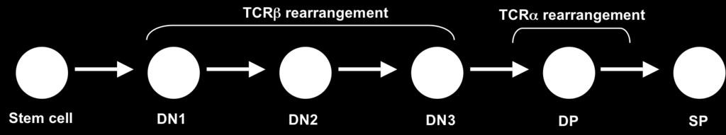
132 A B C 120
133 Previously we had shown that nucleoprotein complexes formed when each Runx radiolabeled probe was incubated with the nuclear extract from P5424 T-cell line (DN3) were supershifted by Runx1 antibody, and not by Runx2 or Runx3 antibodies. At the TCRβ locus, Dβ-to-Jβ joining begins during the DN1 and is completed by the DN3 stage of development. The 5 PDβ2 becomes active only after a Dβ-Jβ joint has been formed. To determine how Runx factors are regulated during thymocyte development, we used nuclear extract from 2017 T-cell line (DN1), BW5147 mature T-cell line (SP CD8+), and mature B cell line (M12) to test the ability of each Runx protein to bind the consensus binding sites within 5 PDβ2. Multiple nucleoprotein complexes were detected when radiolabeled probes to each Runx site found within 5 Dβ2 were incubated with nuclear extracts from DN T-cell line (Fig 2A). These nucleoprotein complexes were competed by addition of 100-fold excess of unlabeled RunxA, RunxB probes (lane 2 and 3). To further test the specificity of each binding site, we made CC TT mutations of the core binding sites and tested the ability of unlabeled mutant probes to compete with the labeled probe. In each case, the mutant probe failed to compete for binding (lane 4 and 5). To determine the specific Runx factor involved in binding, we pretreated the nuclear extract with antibodies against Runx1, Runx2, Runx3, and IgG prior to the addition of the radiolabeled probe. Supershift analysis revealed that nucleoprotein complexes of both Runx-A and Runx-B probes were specifically supershifted by anti-runx1, but not Runx2, Runx3 or IgG (lanes 5-9). To determine if the RunxA binding site can bind different isoforms of the Runx proteins during lymphocyte development, we tested the ability of Runx-A binding site to form nucleoprotein complexes with nuclear extracts from BW5147 T-cell line and M12 121
134 A B Figure 3.2: Expression of Runx proteins during thymocyte development. A. Nuclear extracts from 2017 T-cell line were incubated with radiolabeled probes that contained binding sites for Runx-A (upper panel) and Runx-B (lower panel). Nuclear extracts were added alone (lane1) or were pre-incubated with 100-fold excess of cold competitor (lanes 2-3), mutated competitor (lanes 4-5), or antibodies against Runx1 (lane 6), Runx2 (lane 7), Runx3 (lane 8) or non-specific IgG (lane 9). Each Runx radiolabel probe was also incubated with nuclear extracts from mature B-cell (lane 11) and T- cell (lane 12). Supershifts are represented by asterisks. B. Nuclear extracts from the mature B-cell line, M12, (left) and a mature T cell line, BW5147, (right) were pre-incubated without antibody (lanes 1) or with antibodies against Runx1 (lanes 2), Runx2 (lanes 3), Runx3 (lanes 4) or non-specific IgG (lanes 5). 122
135 mature B-cell line (Fig 2B). We detected Runx-A binding in the presence of nuclear extract from M12 and BW5147 cell-lines (lanes 1). Nucleoprotein complexes formed in the presence of BW5147 nuclear extract were specifically supershifted by anti-runx2, but not anti-runx1 or anti-runx3. While nucleoprotein complexes in the presence of M12 nuclear extracts were supershifted by anti-runx3 (lanes 2-5). 123
136 3.4 Discussion We have identified two functional Runx binding sites within 5 PDβ2. The 5 PDβ2 is activated after a DβJβ2 joint is formed. In developing T cell, DβJβ2 joining of the TCRβ locus begins in DN1 stage and continues till the DN3 stage of development. The placement and activation timing of 5 PDβ2 suggest that 5 PDβ2 may be important in Vβ-DβJβ joining. Expression of all three Runx proteins varies during T cell development. Consistent with the idea that Runx1 regulates the transition of DN to DP cells (11), we showed that Runx1 mrna expression is highest in DN2 subpopulation and gradually decreases as thymocyte matures. In addition, Runx1 protein binding was only detected when DN1 and DN3 nuclear extracts were used. Knockdown Runx1 experiments have shown that inactivation of Runx1 in DN1 thymocytes results in decreased numbers of DP thymocytes and an accumulation of DN3 thymocytes (20). Furthermore, the expression of the TCRβ chain was decreased. This effect became more pronounced when both Runx1 and Runx3 were inactivated. While we did not detect Runx3 binding from the DN1 or DN3 nuclear extracts in our gelshift assay, high levels of Runx3 transcripts were detected by the PCR. The progression of thymocytes from the DN to the DP stage requires complete VβDβJβ rearrangement and expression of a functional TCRβ chain. Development of thymocytes that fail to express a functional TCRβ chain is blocked at the DN3 stage (21). The placement and activation timing of 5 PDβ2 suggest that 5 PDβ2 may be important in Vβ- DβJβ joining. While the roles of Runx1 and Runx3 have not been tested in Vβ- to-dβjβ rearrangement, it is may be insightful in delineating the regulation of thymocyte transition from DN to DP. 124
137 3.5 REFERENCES 1. Godfrey, D. I. and A. Zlotnik Control points in early T-cell development. Immunol. Today 14: Petrie, H. T., F. Livak, D. Burtrum, and S. Mazel T cell receptor gene recombination patterns and mechanisms: cell death, rescue, and T cell production. J. Exp. Med. 182: Rothenberg, E. V Regulatory factors for initial T lymphocyte lineage specification. Curr. Opin. Hematol. 14: Wheeler, J. C., K. Shigesada, J. P. Gergen, and Y. Ito Mechanisms of transcriptional regulation by Runt domain proteins. Semin. Cell Dev. Biol. 11: van Wijnen, A. J., G. S. Stein, J. P. Gergen, Y. Groner, S. W. Hiebert, Y. Ito, P. Liu, J. C. Neil, M. Ohki, and N. Speck Nomenclature for Runt-related (RUNX) proteins. Oncogene 23: Chimienti, G., M. Alaibac, F. Marzullo, A. Carbone, and G. Pepe The expression pattern of the AML1 gene in non-hodgkin's B-cell lymphomas and normal B lymphocytes. Blood Cells Mol. Dis. 26: Mitani, K., S. Ogawa, T. Tanaka, H. Miyoshi, M. Kurokawa, H. Mano, Y. Yazaki, M. Ohki, and H. Hirai Generation of the AML1-EVI-1 fusion gene in the t(3;21)(q26;q22) causes blastic crisis in chronic myelocytic leukemia. EMBO J. 13: Terry, A., A. Kilbey, F. Vaillant, M. Stewart, A. Jenkins, E. Cameron, and J. C. Neil Conservation and expression of an alternative 3' exon of Runx2 encoding a novel proline-rich C-terminal domain. Gene 336: Tsuji, K. and M. Noda Identification and expression of a novel 3'-exon of mouse Runx1/Pebp2alphaB/Cbfa2/AML1 gene. Biochem. Biophys. Res. Commun. 274: Bangsow, C., N. Rubins, G. Glusman, Y. Bernstein, V. Negreanu, D. Goldenberg, J. Lotem, E. Ben-Asher, D. Lancet, D. Levanon, and Y. Groner The RUNX3 gene-- sequence, structure and regulated expression. Gene 279: Taniuchi, I., M. Osato, T. Egawa, M. J. Sunshine, S. C. Bae, T. Komori, Y. Ito, and D. R. Littman Differential requirements for Runx proteins in CD4 repression and epigenetic silencing during T lymphocyte development. Cell 111: Egawa, T., R. E. Tillman, Y. Naoe, I. Taniuchi, and D. R. Littman The role of the Runx transcription factors in thymocyte differentiation and in homeostasis of naive T cells. J. Exp. Med. 204:
138 13. Lorsbach, R. B., J. Moore, S. O. Ang, W. Sun, N. Lenny, and J. R. Downing Role of RUNX1 in adult hematopoiesis: analysis of RUNX1-IRES-GFP knock-in mice reveals differential lineage expression. Blood 103: North, T. E., T. Stacy, C. J. Matheny, N. A. Speck, and M. F. de Bruijn Runx1 is expressed in adult mouse hematopoietic stem cells and differentiating myeloid and lymphoid cells, but not in maturing erythroid cells. Stem Cells 22: Lorsbach, R. B., J. Moore, S. O. Ang, W. Sun, N. Lenny, and J. R. Downing Role of RUNX1 in adult hematopoiesis: analysis of RUNX1-IRES-GFP knock-in mice reveals differential lineage expression. Blood 103: Satake, M., S. Nomura, Y. Yamaguchi-Iwai, Y. Takahama, Y. Hashimoto, M. Niki, Y. Kitamura, and Y. Ito Expression of the Runt domain-encoding PEBP2 alpha genes in T cells during thymic development. Mol. Cell. Biol. 15: Zhao, F. Q., Z. M. Sheng, M. M. Tsai, A. E. Hubbs, R. Wang, T. J. O'Leary, D. J. Izon, and J. K. Taubenberger Serial analysis of gene expression in murine fetal thymocyte cell lines. Int. Immunol. 14: Vaillant, F., K. Blyth, A. Terry, M. Bell, E. R. Cameron, J. Neil, and M. Stewart A full-length Cbfa1 gene product perturbs T-cell development and promotes lymphomagenesis in synergy with myc. Oncogene 18: Ehlers, M., K. Laule-Kilian, M. Petter, C. J. Aldrian, B. Grueter, A. Wurch, N. Yoshida, T. Watanabe, M. Satake, and V. Steimle Morpholino antisense oligonucleotidemediated gene knockdown during thymocyte development reveals role for Runx3 transcription factor in CD4 silencing during development of CD4-/CD8+ thymocytes. J. Immunol. 171: Egawa, T., R. E. Tillman, Y. Naoe, I. Taniuchi, and D. R. Littman The role of the Runx transcription factors in thymocyte differentiation and in homeostasis of naive T cells. J. Exp. Med. 204: Shinkai, Y., G. Rathbun, K. P. Lam, E. M. Oltz, V. Stewart, M. Mendelsohn, J. Charron, M. Datta, F. Young, and A. M. Stall RAG-2-deficient mice lack mature lymphocytes owing to inability to initiate V(D)J rearrangement. Cell 68:
139 CHAPTER 4 Recombinational Accessibility Of Dβ2 Gene Segment In Developing Thymocytes 127
140 ABSTRACT A healthy immune system depends on the ability of developing B and T cells to express diverse antigen receptors. During lymphocyte development, the Variable (V), Diversity (D), and Joining (J) gene segments of the Ig and TCR loci undergo site-specific somatic recombinations to create unique antigen receptors. Each V, D, and J gene segment is flanked by recombination signal sequences (RSS), which serve as recognition sites for the recombinase and direct site-specific cleavage. In this report we address the elements that may be responsible for the preferential usage of Dß1 during recombination. Analysis of both Dß 23 RSS and all Jß 12 RSS reveal no apparent difference in the functionality between different Dß1 and Dß2, or Jß1 and Jß2 RSS. Recombination analysis suggests that the differential usage of Dß1 and Dß2 does not stem from variations in the timing of recombination or the accessibility of each DJ cassettes. However, rearrangements of Dß2 were less efficient than Dß1. This may be due to differences in promoter positioning at each Dß gene segment. 128
141 4.1 Introduction The ability of the immune system to respond to pathogens is dependent on the capability of developing B and T cells to generate a diverse repertoire of antigen receptors. This remarkable diversity is achieved through the somatic rearrangement of the Ig and TCR loci. This process is called V(D)J recombination. During lymphocyte development, Variable (V), Diversity (D), and Joining (J) gene segments undergo a site-specific somatic recombination to create unique antigen receptor genes in each cell (1). The TCRß locus contains 20 V segments followed by the Dß1, its six associated J segments and a constant region (referred to as the Dß1 cassette). Dß2, its associated seven J s (Dß2 cassette) and a second constant region, Cß2, are located 3 of Cß1. Recombination of the TCRß locus occurs in a stepwise fashion involving initial D-to-J joining at each of the two Dß cassettes that contain one D and 6 or 7 J s, and then subsequent assembly of one DJ joint with one of the distal V elements. Each V, D, and J gene segment is flanked by a recombination signal sequence (RSS), which serves as recognition site for the recombinase and directs site-specific DNA cleavage. The RSS consists of a conserved palindromic heptamer (5 -CACAGTG-3 ), and an A/T rich nonamer (5 -ACAAAAACC-3 ) separated by poorly conserved spacer sequences of either 12 or 23 bases in length. Depending on the length of the intervening spacer, each RSS can be identified as either a 12 RSS or a 23 RSS. V(D)J recombination can only occur between two gene segments when one is flanked by a 12-RSS, and the other a 23-RSS (1). This is known as the 12/23 Rule. Despite similarity in the architecture of the Dß1 and Dß2 cassettes, Dß2Jß rearrangement has long been observed to accumulate more slowly than Dß1Jß (2). Gene 129
142 segment utilization is influenced by a number of factors that are found within each antigen receptor locus. First, gene segment usage may depend on the accessibility of the individual gene segment chromatin domains to the recombination machinery (3). Second, RSS sequence has been shown to influence preferential gene segment utilization, especially when accessibility of each gene segment is equal (4-10). Third, the orientation and distance between the two recombining segments, which can be hundreds of kilobases apart, impacts gene segment utilization (11, 12). Consequently, the differential rearrangement potentials of the two DJß cassettes could be derived from (i) differences in the developmental timing of transcription (ii), differences in RSS quality, (iii) differences in the levels of recombination accessibility. In this report, we sought to determine which of these possibilities is most likely responsible for the preferential Dß1 rearrangement. Analyses of each RSS quality revealed no apparent difference between the Dß1 and Dß2 23 RSS, or between the various D and J RSS, suggesting that preferential usage of Dß1 is independent of RSS sequence. Recombination analysis suggests that the differential usage of Dß1 and Dß2 is not imposed by variations in the timing of recombination or the accessibility of each DJ cassettes. Despite the similarity in Dß1and Dß2 architecture and the onset of rearrangements, Dß2 rearrangements were less efficient than Dß1 based on the persistence of germline Dß2 sequences in DN1 thymocytes. This may be due to differences in promoter positioning at each Dß gene segment as discusses earlier (Chapter 2). 130
143 4.2 Materials and Methods Analysis of RSS We obtained 23 RSS of Dß1 and Dß2 and 12RSS of each Jß at the TCRß locus from GenBank accession AE and AE The sequences of the nonamers and heptamers were aligned for comparison. The RS Information Content (RIC) algorithm ( program developed by Cowell and Kelsoe was used to predict the functionality of each RSS. The RS Information Content (RIC) score measures the similarity of a particular RSS sequence to a set of all the physiological RSS in the mouse genome, and assign a score based on similarity (19) PCR/RT-PCR analyses of sorted thymocyte populations For PCR, genomic DNA extracts were prepared from 1 x 10 4 cells. Briefly, cells were incubated at 56 o C for 90 min. in a 100 µl cocktail containing Tris-Cl ph 8.4 (10mM), KCl (50mM), MgCl 2 (2mM), NP-40 (0.45%), Tween-20 (0.45%), and Proteinase K (60µg/ml), followed by enzyme inactivation at 95 o C for 10 min. Resultant DNA (2-5 ng) extracts were assayed for Dß1 and Dß2 rearrangements by nested PCR by using Phusion polymerase (1U, New England Biolabs) and the following amplification conditions: initial denaturation at 98 o C for 30 sec., followed by cycling at 98 o C/10 sec., 62 o C/30 sec., and 72 o C/1.5 min., with a final extension at 72 o C for 5 min. Oligonucleotide primers were as follows: PCR1 Dß1 sense 5 -ACCTATGGGAGGGTCCTTTTTTGTATAAAG-3 reverse 5 -AAGACTCCTAGACTGCAGACTCAG-3 Dß2 forward 5 -CAGCCCCTCTCAGTCAGACAAACC-3 reverse 5 -CCCGGAGATTCCCTAACCCTGGTC-3 131
144 Cλ forward 5 -CAGAATTCACCTTCCYCTGARGAG-3 reverse 5 -GAGTCGACARACTCTTCTCCAC- 3 PCR2 Dß1 forward 5 -AAGGCTCGTACATTGTGGGGACAG-3 reverse 5 CCAGTTTGGTCCCATAGTTTACCT 3 Dß2 forward 5 CAGCCCCTCTCAGTCAGACAAACC reverse 5 -CTACTCCAGGGACCCAGGAATTTG-3. For each Dß assay, 2 µl of PCR 1 (15 cycles) were used as template for the second PCR (35 cycles). DNA content was normalized using PCR of the Igλ constant region (Cλ) as follows: 95 o C for 2 min., followed by 35 cycles of 95 o C/30 sec., 60 o C/30 sec., and 72 o C/30 sec., with a final extension at 72 o C for 3 min. We analyzed the reaction products by gel electrophoresis and Southern blotting. 132
145 4.3 Results Differences in RSS RSS are for the most part highly conserved. Despite this remarkable conservation amongst RSS sequences, only the first three nucleotides of the heptamer and the fifth and sixth position of the A-stretch of the nonamer show almost perfect conservation (13-16) and are essential for specificity and function. In fact, when any of these nucleotides are deleted, recombination is abolished (1, 13, 17). Only limited variability occurs within the other positions of the heptamer and nonamer (4-8, 18), which are 81 to 91% conserved. To address the possibility that variations in RSS sequence may be responsible for the differences in Dß1 and Dß2 recombination, we performed sequence alignment for both Dß 23 RSS and all Jß 12 RSS. As shown in Table 1, the heptamer has 100% conservation of CAC nucleotides within the first three bases and 93% conservation of CACA within the first four positions. Jß 1.1 and Jß2.1 heptamers are identical, as are Jß1.3 and Jß2.3. All the DJß heptamers contained a guanine in the fifth position except for Jß1.2 and Jß1.4. The nonamers were more variable than the heptamers. Despite the variability, all Dß and Jß nonamers contained an adenine in the fifth and sixth position. The only difference between Dß1 and Dß2 RSS is an A/T change in the eighth position of the nonamers. Given the strong identity between each RSS conserved regions, these empirical analyses would suggest that differential Dß usage is not encoded in the RSS heptamers or nonamers. To test the relative functionality of each Dß and Jß RS in recombination, we used the RSS information content or RIC algorith 133
146 Table 4.1: Alignment of Recombination Signal Sequences TCRβ Heptamer Nonamer Dβ1 cacaatg acaaaaaag Dβ2 cacaatg acaaaaatg Jβ1.1 cacagtg gtgaaaatc Jβ2.1 cacagtg ccagaattc Jβ1.2 cacataa tctaaaagg Jβ2.2 cacagtc gcacaaacc Jβ1.3 cacagcc ttcaaaacc Jβ2.3 cacagcc acagaaacc Jβ1.4 cacaaca aggaaaacc Jβ2.4 cacagcc acagaaact Jβ1.5 cacagtg ggcaaaccc Jβ2.5 cacagcc acaaaaacc Jβ1.6 cacagct ataaaaccc Jβ2.6 cacagcc gcaaaaacc Jβ Jβ2.7 cacggag atgcaaacc Sequences were taken from GenBank accession AE and AE Spacer sequences are not shown. 134
147 ( developed by Cowell and Kelsoe (19). The RS Information Content (RIC) score measures the similarity of a particular RSS sequence to a set of all the physiological RSS in the mouse genome, and assigns a score based on similarity. Sequences with a rare combination of nucleotides are assigned a low score, while sequences with a common combination of nucleotides receive a high score. RIC has been shown to correlate well with the efficiency of RAG binding, cleavage and rearrangement of RSS in extra-chromosomal recombination assays (19-21). Shown in Table 2, are RIC scores for each of the 12 and 23 RSS within each Dß cluster. All RSS, with the exception of the RSS associated with the Jß2.6 pseudogene, are well above threshold for functionality (-40 for a 12 RSS and 60 for a 23 RSS, respectively). Indeed, the Dß2 3 RSS (23 RSS) is <6 points below that of the Dß1 3 RSS, and all of the functional Jß2 RSS are within the range observed between Jß1.4 (-23.37) and Jß1.2 (-38.80). As such, while it remains possible that the pairing between the Dß2 3 RSS and Jß2 RSS is inferior to RSS pairing within the DJß1 cluster, the functionality of individual RSS is unlikely to account for the relative inefficiency of Dß2 rearrangement Developmental timing of Dß2 recombination Whereas Vß recombination is restricted to CD DN3 cells, Dß1Jß joints have been shown to accumulate throughout DN development. There is a strong correlation between the transcriptions of unrearranged within the two DJCß gene segment cassettes (germline transcription) and the onset of rearrangement at those same sites. The process of transcription is believed to provide differential accessibility of RSS embedded in the chromatin, and ultimately determines which segments undergo V(D)J recombination. We 135
148 Table 4.2: Analysis of DJβ RSS quality * The RSS of the Jβ2.6 is non-functional. 136
149 previously analyzed the germline transcription of both Dß1 and Dß2 cassettes throughout T cell development. Our findings suggest that the differences in rearrangement efficiency between Dß1 and Dß2 are not due to variations in the timing of gene segment accessibility during T cell development because DJß2, like DJß1, is transcribed during the earliest stage of T cell development. To determine if the differences in DJß recombination could be due to an uncoupling of recombination and transcription accessibility, we wanted to compare the developmental onset of recombination for Dß1 and Dß2 to the transcription accessibility. To this end, we assessed the recombination of each cassette in sorted DN1 through DP subpopulations, RAG deficient thymocytes, and a mature B cell line by PCR. The developmental stages of thymocyte maturation is depicted in Fig. 1A. Cells were gated on CD44 and CD25 expression (Fig 1C) and sorted as DN1 cells (CD44 + CD25 - ), DN2 cells (CD44 + CD25 + ), and DN3 cells (CD44 - CD25 + ). Post-sort purity was 96-98% for DN1 and DN3, and 93-95% for DN2. PCR primers were designed to anneal just 5 of the Dß gene segments and 3 of the fifth Jß gene segments (Fig1B). Dß1-to-Jß recombination is first detected in CD DN1 T cells. Consistent with the transcription findings, we readily detected Dß1 recombination with each tested Jß gene segment in all stages of T cell development (Fig. 1D, lanes 3-6). In contrast, only germline sequences were detected in DNA isolated from the mature B-cell line and RAG deficient thymocytes (lane 1-2). As with Dß1 joints, rearrangements between Dß2 and its 5 functional downstream Jß elements were readily detected at all stages of thymocyte development (middle panel, lanes 3-6), but were absent in B cells (lanes 1). While such a PCR-based approach does not afford a direct comparison of relative rearrangement efficiencies between the two DJß cassettes, the clear 137
150 Figure 4.1: Timing of DJß recombination during thymopoiesis. (A) Flowchart of thymocyte development. (B) Schematic diagram of the TCRß locus showing the location of gene segments and PCR primers used for amplification (C) Thymic distribution of CD44 (Y axis) and CD25 (X axis) following immunodepletion of cells expressing CD4 and/or CD8. Gates used to sort DN1 cells (CD44 + CD25 - ), DN2 cells (CD44 + CD25 + ), and DN3 cells (CD44 - CD25 + ) are shown. Post-sort purity was 96-98% for DN1 and DN3, and 93-95% for DN2. (D) PCR of each Dß segments in germline configuration (gl) or rearranged with its 5 most proximal Jß segments is shown for Dß1 (upper panel) and Dß2 cluster (middle panel). Control PCR for DNA content (Cλ) is shown in the bottom panel. 138

151 A B C D 139
152 appearance of Dß2Jß joints at the DN1 stage strongly suggests that the often-cited inefficiency of Dß2 recombination is not the result of delayed accessibility. 140
153 4.4 Discussion Dß1 rearranges more frequently than Dß2. We addressed two possibilities that have been proposed to explain preferential gene segment utilization. The first possibility is that the variation in RSS nonamers and heptamers is responsible for unequal V, D, and J rearrangement. The nonamers, heptamers, and the spacers to a lesser degree determine the recombination potential of the RSS. This recombination potential can influence which gene segments are chosen for rearrangement. We used the RIC score to predict the recombination potential of the 23 RSS for both Dß and the 12 RSS of downstream Jßs. RIC scores have been shown to accurately predict the efficiency of RAG binding, cleavage and recombination of RSS in extra-chromosomal assay (19-21). Based upon the RIC score, functionality of both Dß1 and Dß2 RSS appears even. This is not a surprising finding, considering the perfect conservation of the heptamers and only a single base-pair change in the eighth position of the nonamer. Similarly, V11 and V13 in the heavy chain have identical nonamer, heptamer and spacer, but only V11 gene segment recombines in-vivo (7). However, it is possible that the pairing of 23 and 12 RSS could influence the individual gene segment usage (22). Taken together these results suggest that the recombination potential of each individual RSS cannot account for all unequal gene segment utilization. There must be other factors contributing to preferential gene segment utilization. The second possibility is that the RSS are not accessible at the same time for RAG protein binding. Transcription of unrearranged germline gene segments is believed to open the locus, allowing RAG proteins to bind and gene segments to recombine. We previously showed that DJß1 and DJß2 gene cassettes are both transcribed in DN1 thymocytes, which is the earliest stage of thymocyte development. So, we performed rearrangement PCR assays 141
154 to determine if rearrangement occurs within the same stage that the gene segments were transcriptionally active. Our recombination assay detected rearrangement for both DJß1 and DJß2 in DN1 thymocyte, suggesting that recombination and transcription occur concurrently. While we cannot rule out the possibility that PCR amplification of each Dß is not equally efficient, our data clearly shows that recombination of Dß2 and Dß1 occurs at the earliest stage T-cell developmental. Therefore, the preferential usage of Dß1 gene segment is not likely due to a delay in the timing of recombination, nor is it due to recombination that is uncoupled from transcription accessibility. Other possibilities that seek to explain preferential gene segment utilization include: the proximity of recombining gene segments, the orientation of the gene segment, and the location of the gene segment relative to the transcription elements that controls locus accessibility. Gene segments that are close in proximity and in the same transcription orientation tends to recombine first, and more often. Within the TCRß locus, the two Dß-Jß- Cß clusters are spread over a 200kb region. However, the first Dß1 associated Jß1.1 is only 621bp away, while Jß2.1 lies only 591bp downstream of Dß2. The lone enhancer Eß, is 10 kb away from Cß2 and is nearest to the D2ßJß cluster (23). Considering the long distance of each gene segment cluster from the enhancer, the relatively small difference within each DßJß cluster would most likely not be responsible for the bias towards Dß1 usage. In addition, the preferred gene segments utilized were Jß1.5 and 2.5, the ones farthest from Dß. DJß1 and DJß2 gene segments are in the same transcription orientation as the enhancer. When the orientation of Eß was reversed recombination was unaffected (24). If in fact rearrangement were governed by the proximity to the enhancer, DJß2 would be expected to rearrange first, because it is closer than DJß1 to the enhancer. 142
155 Recently much attention has been focused on transcription regulation at individual gene segments usage. Previously a promoter that lies within a 300 bp region upstream of Dß1, PDß1, has been shown to regulate transcription and recombination of DJß1cassette, but not the downstream DJß2 cassette. When his promoter was moved 3 of Dß1, transcription and recombination of DJß1cassette drastically decreased, suggesting that promoter location can regulate differential accessibility. We have identified a Dß2 promoter, 3 PDß2 that is positioned 400bp downstream of the Dß2 gene segment. We propose that the DJß1 cassette rearranges more frequently because PDß1 is located 5 of Dß1, while Dß2 rearranges less frequently because 3 PDß2 is downstream of Dß2. However, mutational and cleavage assays would be necessary to decipher the roles of RSS and promoters play in preferential Dß1 usage. 143
156 4.5 REFERENCES 1. Tonegawa, S Somatic generation of antibody diversity. Nature 302: King, A. G., M. Kondo, D. C. Scherer, and I. L. Weissman Lineage infidelity in myeloid cells with TCR gene rearrangement: a latent developmental potential of prot cells revealed by ectopic cytokine receptor signaling. Proc. Natl. Acad. Sci. U. S. A. 99: Stanhope-Baker, P., K. M. Hudson, A. L. Shaffer, A. Constantinescu, and M. S. Schlissel Cell type-specific chromatin structure determines the targeting of V(D)J recombinase activity in vitro. Cell 85: Yu, K., A. Taghva, and M. R. Lieber The cleavage efficiency of the human immunoglobulin heavy chain VH elements by the RAG complex: implications for the immune repertoire. J. Biol. Chem. 277: Nadel, B., A. Tang, G. Escuro, G. Lugo, and A. J. Feeney Sequence of the spacer in the recombination signal sequence affects V(D)J rearrangement frequency and correlates with nonrandom Vkappa usage in vivo. J. Exp. Med. 187: Nadel, B., A. Tang, G. Lugo, V. Love, G. Escuro, and A. J. Feeney Decreased frequency of rearrangement due to the synergistic effect of nucleotide changes in the heptamer and nonamer of the recombination signal sequence of the V kappa gene A2b, which is associated with increased susceptibility of Navajos to Haemophilus influenzae type b disease. J. Immunol. 161: Feeney, A. J., A. Tang, and K. M. Ogwaro B-cell repertoire formation: role of the recombination signal sequence in non-random V segment utilization. Immunol. Rev. 175: Connor, A. M., L. J. Fanning, J. W. Celler, L. K. Hicks, D. A. Ramsden, and G. E. Wu Mouse VH7183 recombination signal sequences mediate recombination more frequently than those of VHJ558. J. Immunol. 155: VanDyk, L. F., T. W. Wise, B. B. Moore, and K. Meek Immunoglobulin D(H) recombination signal sequence targeting: effect of D(H) coding and flanking regions and recombination partner. J. Immunol. 157: Gerstein, R. M. and M. R. Lieber Coding end sequence can markedly affect the initiation of V(D)J recombination. Genes Dev. 7: Suzuki, H. and H. Shiku Preferential usage of JH2 in D-J joinings with DQ52 is determined by the primary DNA sequence and is largely dependent on recombination signal sequences. Eur. J. Immunol. 22:
157 12. Gauss, G. H. and M. R. Lieber The basis for the mechanistic bias for deletional over inversional V(D)J recombination. Genes Dev. 6: Hesse, J. E., M. R. Lieber, K. Mizuuchi, and M. Gellert V(D)J recombination: a functional definition of the joining signals. Genes Dev. 3: Akamatsu, Y., N. Tsurushita, F. Nagawa, M. Matsuoka, K. Okazaki, M. Imai, and H. Sakano Essential residues in V(D)J recombination signals. J. Immunol. 153: Ramsden, D. A., K. Baetz, and G. E. Wu Conservation of sequence in recombination signal sequence spacers. Nucleic Acids Res. 22: Ramsden, D. A., C. J. Paige, and G. E. Wu Kappa light chain rearrangement in mouse fetal liver. J. Immunol. 153: Akira, S., K. Okazaki, and H. Sakano Two pairs of recombination signals are sufficient to cause immunoglobulin V-(D)-J joining. Science 238: Larijani, M., C. C. Yu, R. Golub, Q. L. Lam, and G. E. Wu The role of components of recombination signal sequences in immunoglobulin gene segment usage: a V81x model. Nucleic Acids Res. 27: Cowell, L. G., M. Davila, T. B. Kepler, and G. Kelsoe Identification and utilization of arbitrary correlations in models of recombination signal sequences. Genome Biol. 3: RESEARCH Lee, A. I., S. D. Fugmann, L. G. Cowell, L. M. Ptaszek, G. Kelsoe, and D. G. Schatz A functional analysis of the spacer of V(D)J recombination signal sequences. PLoS Biol. 1: E Cowell, L. G., M. Davila, K. Yang, T. B. Kepler, and G. Kelsoe Prospective estimation of recombination signal efficiency and identification of functional cryptic signals in the genome by statistical modeling. J. Exp. Med. 197: Larijani, M., S. Chen, L. Cunningham, J. Volpe, L. Cowell, S. Lewis, and G. Wu The recombination difference between mouse κ and λ segments is mediated by a pair-wise regulation mechanism. Molecular Immunology 43: Glusman, G., L. Rowen, I. Lee, C. Boysen, J. C. Roach, A. F. Smit, K. Wang, B. F. Koop, and L. Hood Comparative genomics of the human and mouse T cell receptor loci. Immunity 15: Huang, F., O. Cabaud, C. Verthuy, A. O. Hueber, and P. Ferrier Alpha beta T-cell development is not affected by inversion of TCR beta gene enhancer sequences: polar enhancement of gene expression regardless of enhancer orientation. Immunology 109:
158 CONCLUSION In this study we have characterized the transcriptional profile of the DJCβ2 gene cassette during T cell development. We have identified separate promoter activities positioned 5 and 3 of Dβ2. The downstream promoter (3 PDβ2) is located within a 150 bp region immediately upstream of Jβ2.2 and 400 bases downstream of Dβ2. This promoter is unique in that it bears no homology to any previously reported gene segment germline promoter. It is a relatively simple promoter in that it is driven by two NFκB sites and contains no discernable initiator or TATA elements. Transcription start sites are diffused throughout the entire 150bp promoter region. Transcription from 3 PDβ2 only occurs when the TCRβ locus is in its germline configuration. Invivo germline transcription of the upstream promoter (5 PDβ2) can only be detected in DNA that contains DβJβ2 joints. When a luciferase reporter plasmid bearing Eβ and sequential truncations of the DJβ2 sequence was transiently transfected into a RAG1 deficient cell line, transcription from 5 PDβ2 could only be detected upon deletion a 290 bp region immediately downstream of Dβ2 was deleted. Therefore, germline transcription from 5 PDβ2 is repressed while 3 PDβ2 is active. However after DJβ2 joining, the repressive region and the downstream promoter are deleted and transcription is directed from 5 PDβ2. The 5 PDβ2 is located within a one kb region immediately upstream of the Dβ2 gene segment, but the bulk of promoter activity is limited to the 219 bp sequence most proximal to Dβ2. Transcription is driven by GATA3, Runx1, and USF1 transcription factors. Start sites are tightly clustered about 100 bases 5 of the Dβ gene segment at two overlapping initiator elements. The transcription control of this promoter bears homology to the previously 146
159 described TCRβ Dβ1 gene segment promoter and the Dδ2 promoter of the TCRδ locus. Based on transcription and recombination lessons learned from the Dβ1promoter, we believe that the placement of the poorly organized 3 PDβ2 between Dβ2 and Jβ2.1 would result in inefficient Dβ2 transcription and Dβ2-to Jβ2 recombination. Recombination inefficiency of the Dβ2 gene segment would be advantageous as it allows non-productive Dβ1 rearrangements to be rescued and maximizes the opportunity for a functional TCRβ gene to be assembled. The TCRβ locus has four possible attempts to produce a functional TCRβ gene. Due to the frequency with which stop codons and frameshift mutations are introduced during Vβ- DJβ joining, only one in three rearrangements is likely to produce a functional TCRβ gene. A non-functional VDJβ1 rearrangement can be replaced with a rearrangement of an upstream Vβ gene segment to the downstream Dβ2-Jβ2 (Fig 5.1 A). An initial Vβ-to-Dβ2 rearrangement would delete Dβ1 and its associated Js, preventing any further rearrangements and reducing the number of attempts to produce a functional TCRβ gene (Fig 5.1B). Therefore, restricting initial D-to-J recombination to Dβ1 would delay Dβ2 rearrangements and maximize the developing cell s opportunities of making a functional TCRβ gene. Inclusion of DJβ2 gene segments in the overall TCRβ repertoire is important for proper immune function. The Jβ1.5 and 2.5 are the most unique and commonly expressed Jβ gene segments in the thymus. Without the presence of these unique sequences variability of the TCRβ repertoire is hampered. The TCRβ locus in New Zealand White mice has a natural 8.8 kb deletion that removes the Dβ2 to Jβ2 regions. New Zealand White mice serve as a natural model for assessing the impact of using a single Dβ gene segment. These mice are 147


160 A B Figure 5.1: The advantage of an initial Vβ-to-Dβ1rearrangement. A non-functional VDJβ1 rearrangement can be replaced with a rearrangement of an upstream Vβ gene segment to the downstream Dβ2-Jβ2. 148