HLA typing techniques
|
|
|
- Kelly Hodge
- 5 years ago
- Views:
Transcription
1 HLA typing techniques D R. S A R A A S S A D I A S L M D. P H D O F I M M U N O L O G Y A S S I S TA N T P R O F E S S O R T E H R A N U N I V E R S I T Y O F M E D I C A L S C I E N C E M O L E C U L A R I M M U N O L O G Y R E S E A R C H C E N T E R
2 HLA typing methods: 1- serological HLA typing 2- PCR-sequence specific primers (SSP) 3- PCR-sequence specific oligonucleotide (SSO) Luminex 5- PCR-sequence-based typing (SBT) Illumina
3 Serological HLA typing A complement-dependent cytotoxicity (CDC) test Or microlymphocytotoxicity assay Has been developed in the 60 s Serology is still the method of choice in many laboratories for low resolution typing of HLA-A and HLA-B Simplicity and low cost
4 Serological HLA typing Lymphocytes are tested with a panel of sera containing well characterized HLAspecific alloantibodies Each serum is placed in a microtiter well of a Terasaki plate (60-72 wells) After a short incubation, rabbit serum is added as a source of complement and the cells that have bound the alloantibody are lysed making them permeable to the fluorochrome ethidium bromide, eosin or trypan blue The wells containing the lysed cells are easily discriminated by microscopy

5 Serological HLA typing

6 Serological HLA typing
7 Serological HLA typing HLA-A, B HLA-C, DQ, DR Molecular typing has replaced the CDC for the other HLA-loci (HLA-C, DR and DQ) for which serology is not precise enough Typing of HLA II by purified B lymphocytes as targets Human monoclonal antibodies : more suitable for typing

8
 2.")
9 PCR PCR based methods used in HLA laboratory are classified under two categories: 1. Polymorphism is identified directly as a part of the PCR process (SSP) 2. PCR generates a product containing internally located polymorphisms, detected by a second technique (SSO or SBT)
10 PCR- SSP PCR-sequence specific primers Based on the specificity of primer extension following hybridization of nucleotide primers directed toward a polymorphic sequence motif defining an allele or group of alleles Extension of the primer will only occur when there is exact matching with the test template 1. A SSP tray contains multiple pairs of PCR primers that are designed to anneal within DNA regions present in certain alleles or groups of alleles 2. If corresponding alleles are present, the presence of their specific amplicons can be detected by gel electrophoresis 3. Software analysis of gel image permits allele identification


11 PCR-SSP The method is rapid and ideally suited for small numbers of samples The method can be applied for low resolution typing as well as for high resolution typing Low resolution HLA-A/B/DR typing can be achieved by a combination of 96 primer mixes Depending on the HLA phenotype, a combination of different primer mixes are necessary to determine the HLA-A/B/C/DRB1/DQB1 alleles Combi tray A/B/DR Combi tray A/B/C HLA DQ high res HLA A02 high res


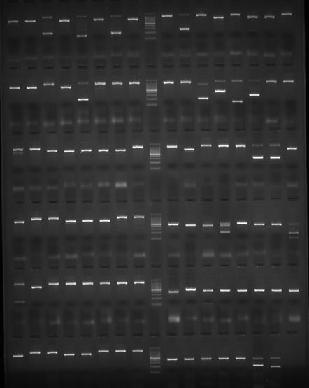

12 PCR- SSP
13 Ambiguity-PCR typing Positive wells: B* B* B* B* B* B*


14 PCR-SSP
 Hybridization is carried out under stringent conditions to allow only specific annealing of short synthetic oligonucleotide probes complementary to specific HLA sequences present in an allele or a")
15 PCR-SSO(P) PCR-sequence specific oligonucleotide (probe) PCR amplified test DNA is blotted onto a solid support (nylon membrane, color-coded microspheres,..) Hybridization is carried out under stringent conditions to allow only specific annealing of short synthetic oligonucleotide probes complementary to specific HLA sequences present in an allele or a group of certain alleles The hybridization can be detected through: Radioautography of 32P labeled probes Enzyme linked probes Chemiluminescent systems Mean fluorescence intensity


16 PCR-SSO SSO HLA typing: Low resolution Intermediate resolution High resolution The higher the number of probes the better the resolution level Membrane-based SSO: Dynal, Inno-lipa, Bead-base SSO: Luminex
17 PCR-SSO (membrane-based)


18 PCR-SSO

19 Luminex (bead-based SSO) Bead-based assay HLA typing is performed with a set of polystyrene microspheres containing different fluorochromes, coated with probes The red classification laser excites the fluorochromes in beads The green reporter laser excites the fluorescence of the PE molecules bound target on each bead The combination of the two signals defines target and its intensity The fluorescence signal of PE label is expressed as a mean fluorescence intensity (MFI)


20 Luminex (bead-based SSO)
21 PCR-SBT PCR-sequence-based typing High resolution HLA typing Uses 2,3 dideoxynucleotides as substrates: When it is incorporated at the 3 end of the growing chain, chain elongation is stopped selectively at A, C, G, T because the chain lacks a 3 hydroxyl group Many DNA fragments of differing size are generated DNA fragments are separated by capillary electrophoresis Four different dyes are used to identify the A, C, G, and T extension reaction Four bases at fragment ends are detected through specific fluorescence emission Exon 2 for HLA II Exons 2 and 3 for HLA I

22
")
23 PCR-SBT (Sanger)

24 PCR-SBT
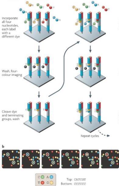


25 SBT (Illumina)
26

27

28 THANKS FOR YOUR ATTENTION