THE EFFECTS OF POLYETHYLENE PARTICLE PHAGOCYTOSIS ON THE VIABILITY OF MATURE HUMAN MACROPHAGES BY PETER ASPINALL, KYRA BURNETT, JOE MAHER, CAT TJAN
|
|
|
- Jodie Lawrence
- 5 years ago
- Views:
Transcription
1 THE EFFECTS OF POLYETHYLENE PARTICLE PHAGOCYTOSIS ON THE VIABILITY OF MATURE HUMAN MACROPHAGES BY PETER ASPINALL, KYRA BURNETT, JOE MAHER, CAT TJAN


2 THE AUTHORS S. Xing Institute of Biomaterials and Biomedical Engineering and Faculty of Dentistry, University of Toronto J. P. Santerre and and Institute of Biomaterials Biomedical Engineering Faculty of Dentistry, University of Toronto Rosalind Labow University of Ottawa Heart Institute E.L. Boynton Institute of Biomaterials and Biomedical Engineering, University of Toronto Department of Surgery, Mt Sinai Hospital
3 INTRODUCTION Look at the effects of HDPE particles on macrophages Using mature human macrophages Goal: Better understand the effects related to implant loosening Discussion Question: So the researchers discussed how other studies were poor because they utilized macrophages that were still differentiating rather than the mature MDMs as they did. Why do you think differentiating macrophages degrade the quality of the study?
4 MATERIALS AND METHODS: PARTICLE PREPARATION AND CHARACTERIZATION Particles of high density polyethylene (HDPE) Particles had similar chemical characteristics to ultra high molecular weight polyethylene (UHMWPE) and were free of endotoxin The particles were suspended into 0.01% collagen type I solution at a concentration of 10 X 10 ^6 particles/ml

5 /cells/scale/
6 MATERIALS AND METHODS: COVERSLIP PREPARATION Microscope coverslips were sterilized by dry heat at 200 o C for 2 hours Each coverslip was coated with 10 ul of particle suspension or about 100,000 particles The coverslips covered with collagen alone were used as negative controls
7 MATERIALS AND METHODS: CELL CULTURES Healthy human blood was collected from and monocytes were isolated by density gradient centrifugation Monocytes were put onto well plates and cultured for 10 days On the 10 th day the MDMs were harvested with 0.25% trypsin and 16.7 mm of EDTA About 500,000 MDMs were seeded on each coverslip and were maintained up to 31 days
8 Discussion Question: This study is performed in vitro, with HDPE introduced to cells from coverslips, with no further addition. In what ways is this model different from what we would expect in an in vivo environment?
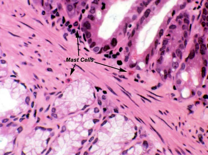
9 MATERIALS AND METHODS: HISTOLOGY Cultures were terminated at 2 hours, 24 hours and at 31 days Washed with PBS buffer mixed with methanol and formaldehyde for 2 hours Stained with hematoxylin eosin (H&E) Coverslips were mounted on glass slides and photographed under regular and polarized light
10
11 MATERIALS AND METHODS: HISTOLOGY Cell incubated for 2 and 24 hours were examined in an scanning electron microscope (SEM) Cells incubated for 24 hours were sectioned and examined using a transmission electron microscope (TEM)


12 MATERIALS AND METHODS: HISTOLOGY SEM TEM
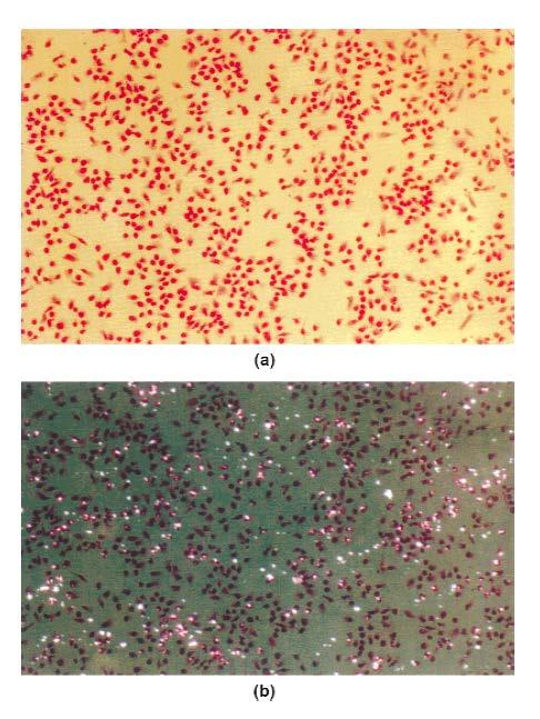
13 METHODS AND MATERIALS: CELL VIABILITY Live/dead cell staining kit utilized two probes Calcein and Ethidium Detect intracellular esterase and membrane integrity 1E6 MDMs were washed with PBS and stained in 200 µl of ethidium for 10 min The samples were then examined by FACS analysis After 24 h of incubation, the cells were rinsed with PBS and double stained with the probes Viable cells appear green, necrotic cells are red
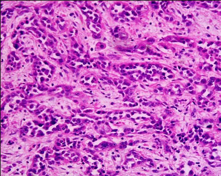
14 FIGURE 1
15 METHODS AND MATERIALS: DNA ANALYSIS Cells were harvested at time zero, day 7, and day 31 They were then rinsed with PBS and combined with a lysis buffer A flourometric assay was used to characterize genetic material by Labow et al. s method
16 METHODS AND MATERIALS: STATISTICAL ANALYSIS Microsoft excel was used to conduct a t test Used to analyze the cell viability at 24 h against the controls Blood donor variability led to large differences in DNA content throughout the experiment A scheffe s test was used to characterize these differences using Statistical Analysis Software (SAS) α = 0.03 because of the small sample size
17 FIGURE 2


18 FIGURE 3 Discussion Question: From part c, how do you think this interaction causes aseptic loosening?
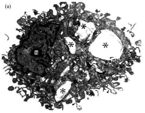
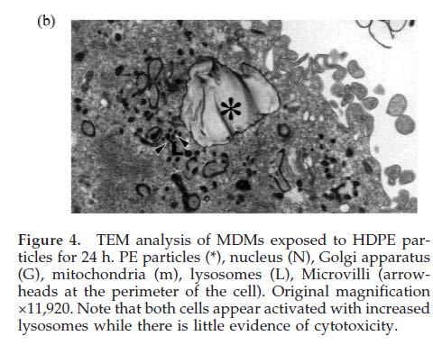
19 FIGURE 4
20 Discussion Question: Toxicity is determined by visual interpretation (TEM) even though other methods such as MTS assay could have made this process much easier. Why do you think they decided against using these other methods?
21 There was no chromatin condensation or mitochondrial dilation, which are features of apoptosis MDMs have greater phagocytic and degradative capacity compared to differentiating human monocytes (DHM) DHM have a smooth surface tomography, encroached nuclei, and fewer lysosomes when having the same amount of HDPE particles
22 FIGURE 5
23 FIGURE 6
24 TABLE 1 DNA content was measured in order to estimate cell number Why?
25 There was relatively higher cell viability in the presence of HDPE in long term culture. There were significantly more cells in the HDPE cultures versus the collagen controls at day 31. This cannot be explained in by differences in MDM proliferation because they have little or no proliferation capacity because they are fully differentiated. Therefore, prolonged MDM survival can be explained by the greater cell number in HDPE cultures than in collagen controls at day 31.
26 CONCLUSIONS MDMs investigated the long term effect of PE phagocytosis on cell viability in vitro. Ultimately determined HDPE particles increased cell viability Discussion Question: The researchers mention the ability to manipulate cytokine secretion with varying surface chemistry. Why is this beneficial?