CHALLENGES OF 3D BIOPRINTING IN CLINICAL PRACTICE
|
|
|
- Marsha Morgan
- 5 years ago
- Views:
Transcription


1 CENTRE DE THÉRAPIE TISSULAIRE & CELLULAIRE CHALLENGES OF 3D BIOPRINTING IN CLINICAL PRACTICE Pr. D. Dufrane MD, PhD
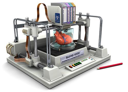
2 3D-BIOPRINTING: MYTH OR REALITY? 2

3 REGENERATIVE MEDICINE FOR ORGAN AND TISSUE A LARGE AND EMERGENCY MEDICAL NEED - There are currently 123,175 people waiting for lifesaving organ transplants in the U.S. - Of these, 101,170 await kidney transplants. - Nearly 3,000 new patients are added to the kidney waiting list each month people die each day while waiting for a lifesaving kidney transplant - Every 14 minutes someone is added to the kidney transplant list - In 2013, 4,453 patients died while waiting for a kidney transplant. 3


 Small")


4 Tools & Reagent Companies Cell Therapy (CT) 15% REGENERATIVE MEDICINE FOR ORGAN AND TISSUE A LARGE MARKET FOR A LARGE NUMBER OF INDICATIONS Total Regenerative Total Regenerative Medicine Market: Medicine Per cent Market: Segmentation, Total Regenerative Global, Medicine 2013 Market: Per cent Breakdown of Companies by Industry Sectors, Breakdown of Companies by Therapeutic Global, 2013 Approach, Global, 2013 Regenerative Medicine Market Gene Therapy (GT) Therapeutic Companies 40% Tissue Engineering Tissue Companies Engineering 27% (TE) Small Molecules & Biologics Cell-based immunotherapy Stem cell therapeutics Cell & Tissue Banks 33% Service Companies 12% Synthetic Small materials Molecules & Biologics Companies Bio-based 5% materials Gene Therapy Companies 8% Cell Therapy Companies 60% Note: All figures are rounded. The base year is Source: Frost & Sullivan 4


5 REGENERATIVE MEDICINE FOR ORGAN AND TISSUE MEDICAL DISCIPLINES FOR TISSUE ENGINEERING Cell Therapy Regenerative Medicine Segment: Per cent Breakdown of Number of Commercially Available Products by Therapeutic Area, Global, 2013 Cancer 10% Ocular 7% Cardiovascular 2% Nonhealing wounds/ Skin 46% Tissue Engineering Regenerative Medicine Segment: Per cent Breakdown of Commercially Available Products by Therapeutic Area, Global, 2013 Per cent Breakdown (%) % 21.0% 9.0% 8.0% 6.0% 4.0% 3.0% 3.0% 2.0% 2.0% 2.0% 2.0% Therapeutic Area Musculoskeletal 35% Per cent Breakdown (%) 23.0% 11.0% 3.0% 16.0% 7.0% 9.0% 5.0% 3.0% Source: Alliance for Regenerative Medicine; Frost & Sullivan Tissue Engineering Regenerative Medicine Segment: Per cent Breakdown of Products in Pre-clinical and Clinical Development by Therapeutic Area, Global, % 9.0% 9.0% 0 Therapeutic Area Source: Alliance for Regenerative Medicine; Frost & Sullivan 5


6 BIOPRINTING AND RESPECT OF THE HUMAN BODY BALANCE FOR 3D RECONSTRUCTION BY TISSUE ENGINEERING Bio-Implant! Host Tissue/Organ! Tissue remodelling Anatomy Functionality Physiology Biocompatibility Cellular engraftment Cellular viability Angiogenesis Proof of concept Pre clinical 6
7 3D-BIOPRINTING A CLINICAL CHALLENGE 3D printing is moving in diverse directions and it is expected that in the coming future it will further expand its horizons. => There is still a much larger scope for 3D printing in the area of medical field. => Recent advances have enabled 3D printing of biocompatible materials, cells and supporting components into complex 3D functional living tissues. 3D bioprinting is being applied to regenerative medicine to address the need for tissues and organs suitable for transplantation. 7

8 AN INTEGRATED APPROACH Compared with non-biological printing, 3D bioprinting involves additional complexities, such as the choice of materials, cell types, growth and differentiation factors, and technical challenges related to the sensitivities of living cells and the construction of tissues. Addressing these complexities requires the integration of technologies from the fields of engineering, biomaterials science, cell biology, physics and medicine. Step 1 Step 2 Step 3 Step 4 Step 5 Step 6 Imaging Design approach Material selection Cell selection Bioprinting Application Murphy SV & Atala A; Nature Biotechnology 2014 X-ray Biomimicry Synthetic polymers Differentiated cells Inkjet Maturation CT Self-assembly Natural polymers Plurpotent stem cells Microextrusion Implantation MRI Mini-tissues ECM Multipotent stem cells Laser-assisted In vitro testing Figure 1 A typical process for bioprinting 3D tissues. Imaging of the damaged tissue and its environment can be used to guide the design of 8


9 SCAFFOLD LEVEL Complex combinations or gradients to achieve desired functional, mechanical and supportive properties modified or designed to facilitate bioprinter deposition, while also exhibiting desired postprinting properties Use of decellularized tissue-specific ECM scaffolds to study ECM compositions, and/or as printable material 9
10 SELECTED MATERIALS The selection of appropriate materials for use in bioprinting and their performance in a particular application depend on several features: Printability: Properties that facilitate handling and deposition by the bioprinter may include viscosity, gelation methods and rheological properties. Biocompatibility: Materials should not induce undesirable local or systemic responses from the host and should contribute actively and controllably to the biological and functional components of the construct. Degradation kinetics and byproducts: Degradation rates should be matched to the ability of the cells to produce their own ECM; degradation byproducts should be nontoxic; materials should demonstrate suitable swelling or contractile characteristics. Structural and mechanical properties: Materials should be chosen based on the required mechanical properties of the construct, ranging from rigid thermoplastic polymer fibers for strength to soft hydrogels for cell compatibility. Material biomimicry: Engineering of desired structural, functional and dynamic material properties should be based on knowledge of tissue-specific endogenous material compositions. 10
11 CELLULAR LEVEL Cells are the smallest functional units of life. Tissue, comprising of different cells, are arranged in specific 3D orientation depending on the functions they perform. Tissue engineering technology has used different fabrication methods for bringing cells together to generate appropriate tissue. => Well-characterized and reproducible source of cells required => Combinations of cell phenotypes with specific functions => Greater understanding required of the heterogeneous cell types present in the tissues => Direct control over cell proliferation and differentiation with small molecules or other factors 11
12 for crosslinking often slows the bioprinting process and involves chemical modification of naturally occurring ECM materials, which changes both their chemical and material properties. Additionally, some crosslinking mechanisms require products or conditions that are toxic to cells, which results in decreased viability and functionality 66. Another limitation encountered by users of inkjet-based bioprinting technology is the difficulty in achieving biologically relevant cell densities. Often, low cell concentrations (fewer than 10 million cells/ml) 42 are used to facilitate droplet formation, avoid nozzle clogging and reduce shear stress 60. Higher cell concentrations may also inhibit some of the hydrogel crosslinking mechanisms 67. Table 1 Comparison of bioprinter types BIOPRITNING TECHNOLOGY electrospinning inkjet bioprinting technique enabled the fabrication of a layered construct that supported cell function and maintained suitable mechanical and structural properties. Inkjet bioprinters have also been used to fabricate bone constructs 75, matured in vitro before implantation into mice. These constructs continued to mature in vivo and formed highly mineralized tissues with similar density as endogenous bone tissue. => Compatible with physiologically relevant materials and cells Increased resolution and speed => Scale up for commercial applications => Combining bioprinter technologies to overcome technical challenges Microextrusion bioprinting. The most common and affordable nonbiological 3D printers use microextrusion. Microextrusion bioprinters usually consist of a temperature-controlled material-handling and Bioprinter type Inkjet Microextrusion Laser assisted Refs. Material viscosities mpa/s 30 mpa/s to > mpa/s mpa/s 48,63,78,107 Gelation methods Chemical, photo-crosslinking Chemical, photo-crosslinking, sheer Chemical, photo-crosslinking 64,85,106,110 thinning, temperature Preparation time Low Low to medium Medium to high 38,64,94,107 Print speed Fast (1 10,000 droplets per second) Slow (10 50 m/s) Medium-fast (200 1,600 mm/s) 49,58,76,90 Resolution or droplet size <1 pl to >300 pl droplets, 50 m wide 5 m to millimeters wide Microscale resolution 49,68,69,76 Cell viability >85% 40 80% >95% 42,54,80,104 Cell densities Low, <10 6 cells/ml High, cell spheroids Medium, 10 8 cells/ml 42,49,88,89 Printer cost Low Medium High VOLUME 32 NUMBER 8 AUGUST 2014 NATURE BIOTECHNOLOGY eads of material and/or cells. (c) Laser-assisted printers use lasers focused on an absorbing substrate to 12


13 IN VIVO CONNEXION VASCULARIZATION Well-developed vascular tree required for large tissues May have to be engineered in the bioprinted construct Capillaries and microvessels required for tissue perfusion Suitable mechanical properties for physiological pressures and for surgical connection INNERVATION Innervation is required for normal tissue function May be inducible after transplantation using pharmacologic or growth factor signaling Simulation before transplantation could be achieved using bioreactors 13


14 MATURATION IN VITRO MATURATION Time required for assembly and maturation Bioreactors may be used to maintain tissues in vitro Provide maturation factors as well as physiological stressors Potential for preimplantation testing of constructs 14

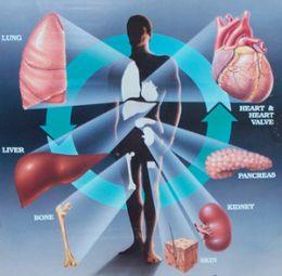
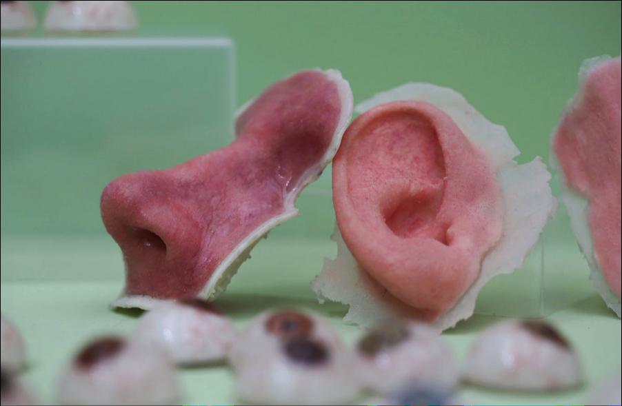
15 PRELIMINARY RESULTS 3D bioprinting has already been used for the generation and transplantation of several tissues, including multilayered skin, bone, vascular grafts, tracheal splints, heart tissue and cartilaginous structures. Other applications include developing highthroughput 3D-bioprinted tissue models for research, drug discovery and toxicology. Skin Cartilage Trachea Heart valve Vasculature Two-dimensional tissue Skin Cartilage Hollow tubes Vasculature Aortic valve Tracheal splint Solid organs Kidney Kidney 15
16 PRELIMINARY RESULTS AND COMBINATIONS Table 1: Tissue engineering applications using bioprinting technology Tissue Techniques Cell types Growth factors Materials Heart valve Extrusion-based bioprinting Aortic valve interstitial cell Aortic root sinus smooth muscle cell Hyaluronic acid Gelatin Alginate Myocardial tissue Extrusion-based bioprinting Cardiomyocyte progenitor cell Alginate Blood vessel Jetting-based bioprinting Endothelial cell Fibrin Smooth muscle cell Mesenchymal stem cell Extrusion-based bioprinting Endothelial cell Cardiac cell Smooth muscle cell Fibroblast Collagen Agarose Alginate Musculo-skeletal tissue Jetting-based bioprinting Muscle-derived stem cells Myoblast Mesenchymal fibroblast Extrusion-based bioprinting Bone marrow stromal cell Endothelial progenitor cell Endogeneous stem cell Nerve Jetting-based bioprinting Embryonic motorneuron cell Hippocampal cell Cortical cell Neuronal precursor cell Neural stem cells Extrusion-based bioprinting Bone marrow stem cell Schwann cells Skin Jetting-based bioprinting Dermal fibroblast Epidermal keratinocyte BMP-2 FGF-2 TGF-β CNTF VEGF Fibrin Agarose Agarose Alginate Hydroxyapatite Polycaprolactone Soy agar Collagen Fibrin Collagen 16

![(Fig. 5 and Table 1) [52, 54, 56, 57].](/docs-images/96/127269841/images/17-4.jpg "Bioprinting has been used to 3D-BIOPRINTING FOR generate ORGAN/TISSUE 2D and 3D structures for various purposes, including fabrication of scaffolds and tissue constructs for tissue regeneration.")

 Blood vessel construct fabricated by extrusion-based bioprinting [38]. (B) Aortic valve conduit fabricated by extrusion-based bioprinting [47].")
17 GRADUAL EVOLUTION Figure 4: Computer-aided design and manufacturing (CAD/CAM) process for bioprinting technology to fabricate biomimetic-shaped tissue or organ. Recently, bioprinting technology has gained increasing attention due to its ability to spatially control the placement of cells, biomaterials and biological molecules for tissue or organ regeneration (Fig. 5 and Table 1) [52, 54, 56, 57]. Bioprinting has been used to 3D-BIOPRINTING FOR generate ORGAN/TISSUE 2D and 3D structures for various purposes, including fabrication of scaffolds and tissue constructs for tissue regeneration. Patients with valvular heart disease often require valve replacement, either with mechanical or with biological prostheses. However, these prosthetic valves are frequently associated with complications, such as mechanical failure and calcification. As such, various approaches have been proposed to improve the Two-dimensional Hollow tubes Hollow organs Solid organs Figure 5: Tissue engineering applications using the bioprinting technology. (A) Blood vessel construct fabricated by extrusion-based bioprinting [38]. (B) Aortic valve conduit fabricated by extrusion-based bioprinting [47]. (C) Confocal microscopic image of multilayered skin structure fabricated by jetting-based bioprinting [28]. (D) Fluorescent image of microvascular structure fabricated by jetting based-bioprinting [35]. (E) Three-dimensional cardiac tissue with biomimetic shape fabricated by jetting-based bioprinting [25]. (F) Alkaline phosphate staining of cultured muscle stem cells on spatially controlled BMP-2 by jetting-based bioprinting [33]. Reproduced with permission from references [25, 28, 33, 35, 38, 47]. Short-term Mid-term Long-term Figure 4 Timeframe for the development of various types of 3D bioprinted tissues. There are four main types of tissues that can be ranked from simple to complex; 2D tissues, such as skin; hollow tubes, such as blood vessels; hollow nontubular organs, such as the bladder; and solid organs, such as the kidney. As the complexity of tissues increases, new approaches will be needed to overcome the challenges of creating them by bioprinting. 2D organs have already been fabricated and tested, and these will likely be one of the first types of bioprinted tissues to be transplanted in patients. Hollow tubes, including blood vessels, tracheas and urethras are currently in development and are likely to closely follow 2D tissues in clinical application. Hollow organs are more complex and printing may in healthcare. take longer Picture Credit: Frost & Sullivan 17 to develop. Solid organs are the most complex, and there are still many challenges to overcome, especially in achieving vascularization Futuretech Alert Exhibit 1 depicts the drivers and restraints along with their impact in 3D MILESTONE ACHIEVEMENTS IN HEALTHCARE WITH 3D PRINTING A few achievements of 3D printing technology in healthcare are given below:


18 LIMITATIONS REGULATION : ATMP ETHICAL ASPECTS 18