Lighting research Toulouse team (France) Ludovic VANQUIN Ikbal MARGHAD Lydie AREXIS BOISSON
|
|
|
- Marsha Barrett
- 6 years ago
- Views:
Transcription

1 1 Lighting research Toulouse team (France) Ludovic VANQUIN Ikbal MARGHAD Lydie AREXIS BOISSON
2 Plan 2 INTRODUCTION I. Medical Imaging for the diagnosis of the Alzheimer s disease (Ludovic) II.Lighting display: Organic LED technology (Ikbal) III.Light and vision research Flickering (Lydie) CONCLUSION


3 INTRODUCTION 3 10 Departments 90 Research Laboratories 28,000 Students 2,200 Teaching & Research staff 1,250 Administrative & Technical staff Pharmacy 318 research units 52 clinical investigation centers 12 population health and clinical research networks 59 federative research institutes 8,000 Inserm employees 3,000 members of university hospital personnel 1,450 foreign research scientists Medicine Sport & Languages Sciences Natural Sciences Physics Mathematics Computer Science Civil Engineering
4 4 I-Medical Imaging for the diagnosis of the Alzheimer s disease
 Nuclear medical imaging technique Use of radioactive tracers introduced into the body Magnetic")

5 I-Medical Imaging for the diagnosis of the Alzheimer s disease 1/6 5 Positron emission tomography (PET) Nuclear medical imaging technique Use of radioactive tracers introduced into the body Magnetic Resonance Imaging (MRI) Used in radiology to visualize internal structures of the body Use of the property of nuclear magnetic resonance of water molecules to make pictures of organs and structures of the body
 Interest for the")

6 I-Medical Imaging for the diagnosis of the Alzheimer s disease 2/6 6 Optical Imaging (fluorescence imaging) Imaging modility using light to obtain information on tissue composition and biomolecular processes in the living body Fluorescent molecules that emit light when activated by an external light source (ex : laser) Interest for the diagnosis of Alzheimer s disease Alzheimer's disease is characterized by the accumulation of β-amyloid peptides, which form senile plaques in the brain : Use near infrared fluorescent agent for specific plaque targeting Use fluorescence optical imaging to localize senile plaques Enable definitive preclinical diagnosis of Alzheimer's disease

7 I-Medical Imaging for the diagnosis of the Alzheimer s disease 3/6 J.Skoch, A.Dunn, B.T. Hyman, B.J. Bacskai, Development of an optical approach for noninvasive imaging of Alzheimer s disease pathology, Journal of Biomedical Optics 10(1) (2005); Subjects and methods: Transgenic and normal mouse brains fluorescent agents : BAM-10 conjugated Alexa Fluor 750 ICG (indocyanine Green) + Thioflavin S Laser Scanning Microscope: excitation 750 nm laser fluorescence detected by an avalanche photodiode NIR microscopy of labeled plaques in mouse tissue

8 I-Medical Imaging for the diagnosis of the Alzheimer s disease 4/6 8 LI-COR Odyssey : NIR Imaging system hole brain imaging (in vivo and ex vivo) high resolution (21 micrometer/pixel) laser diodes 780-nm excitation /800-nm emission Tissue sections were cut from a transgenic mouse brain. Sections were stained with the NIR fluorescent compound ICG followed by thioflavin S

9 I-Medical Imaging for the diagnosis of the Alzheimer s disease 5/6 9 explore Optix Imaging: NIR Imaging system hole brain imaging 0.5-mm resolution Pulsed diode laser at 750 nm Tissue sections were cut from mouse brains. Sections were stained with the the fluorescent agent (Alexa Fluor 750-labeled BAM-10 antibody) Left brain : transgenic mouse Right brain : agematched nontransgenic control
10 I-Medical Imaging for the diagnosis of the Alzheimer s disease 6/6 Conclusion and prospects: 10 Fluorescence from within a whole mouse brain can be measured quantitatively with near infrared laser scanning devices These images illustrate the potential for noninvasive, whole animal detection of specifically labeled plaques in the brains of mouse models of Alzheimer s disease using optical fluorescent imaging Non invasive applications for the diagnosis of Alzheimer s disease in human patients
11 11 II- Lighting display: OLED technology (Organic Light-Emitting Diode )

 Charge combination")


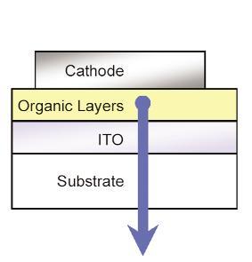
12 II- Lighting display: Organic LED technology 1/5 12 Principle: OLED: Organic Light-Emitting Diode 1) Charge Injection 2) Charge transport 3) Charge combination Structure : ETL: Electron transport Layer EML:Emissive Layer HTL:Hole transport Layer HIL: Hole injection Layer Advantages: Low consumption of power Could be: transparent, flexible, Stretchable (new application) Very thin, wider viewing angles, improved brightness Holst center


13 II- Lighting display: Organic LED technology 2/5 OLED displays in automobiles 13 Integrating OLEDs with electronic textile Multimedia displays Samsung's prototype of smartphone OLED challenges Holst center My PhD research: Synthesis of molecules Their incorporation within OLED Characterization of the OLED device


14 II- Lighting display: Organic LED technology 3/5 OLED process: 14 Substrate cleaning Preparating figure Deposition layer Encapsulation R R N N N N R R Small molecule Metal-organic complexe PPV Polymer


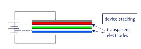
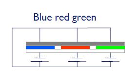
15 II- Lighting display: Organic LED technology 4/5 15 What is responsible for the luminescence? -Luminescence: Fluorescent/ Phosphorescent -Color:.RGB.Combination :WOLED HTL EML ETL OSRAM Press Release April 2008
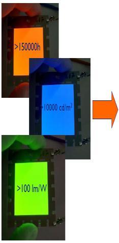
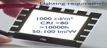
16 II- Lighting display: Organic LED technology 5/5 16 Summary -OLED is a thin, efficient and bright display. -OLED technology has great potential and a very wide range of applications. -Perspectives: the price and lifetime. -OLED s display are a reality, lighting to come (with WOLED)
17 17 III- Light and vision research Flickering
 Optics Physiology Neurophysiologie Cognitive psychology Visual comfort Sociology Figure 1 : modelization of an ammetropic human eye")
18 III- Light and vision research Flickering 1/6 18 Subject : «Study and optimization of energy efficient lighting and adapted to the needs of its users (health, safety and quality of life) Pluridisciplinary subject : Light sources (photometry colorimetry) Optics Physiology Neurophysiologie Cognitive psychology Visual comfort Sociology Figure 1 : modelization of an ammetropic human eye with ZEMAX-SE
19 III- Light and vision research Flickering 2/6 19 Experiments on light flickering visual perception Flickering : light vibration temporal contrast conscious or unconscious perception Critical Fusion Frequency (CFF): limit of perception Health impact: Discomfort Headaches Decrease in visual performance Photosensitive epileptic seizures Objective: To find a lighting using consuming less energy and allowing a good visual quality by limiting discomfort system.
 Figure 2 : Experimental scheme Flickering created by : frequency duty cycle changes (new parameter) LEDs controled by electronic boxes and software Recording")
20 III- Light and vision research Flickering 3/6 A> Expérimental Protocol on flickering perception Technical parameters : A black box in a dark room Different color background 20 Indirect and diffused light (never perceived directly) Figure 2 : Experimental scheme Flickering created by : frequency duty cycle changes (new parameter) LEDs controled by electronic boxes and software Recording responses by Push Button
21 III- Light and vision research Flickering 4/6 21 Power regulation average illuminance regulation Human parameters : 25 observers/test Age : between 20 and 40 years old Emmetropia vision or with optical correction First studies on central vision ( diract angle with the fovéa) The question : Figure 3 : transmitted signal with power control :L=f(t) Do you perceive a light vibration? Proper to the individual feels Like
 White background : more categorical perception Larger range of perception")
22 III- Light and vision research Flickering 5/6 B> First results : 22 For the white an dark backgroung : FFC : 65Hz Same kind of curves more frequencies increase and less flickering is perceived Impact of duty cycle on the same frequency perception More the duty cycle is little more it is perceived (higher temporal contrast) White background : more categorical perception Larger range of perception higher visual fatigue Influence by the scenarii order Increased detection performance with time Figure 4 : Results withc a) dark background b) white background
23 III- Light and vision research Flickering 6/6 23 C> Other flickering experiments realised : Study on higher ranges of frequencies (untill 100Hz) Study on the time influence on the response Order scenarii study Flicker experiments with color backgrounds : Red Green Blue Study in peripheral vision (90 and 45 ) Application : flickering for museum light de musée (Color degradation of paints) cf. FFC et sensation de brillance plus élevée en en lumière pulsé.
24 CONCLUSION 24 To learn more things about : Radiometry and photometry light and health impact indoor and outdoor specific light applications : lighting technologies and softwares lighting displays such as LED.

25 25
LUPAS Luminescent Polymers for in vivo Imaging of Amyloid Signatures
 LUPAS Luminescent Polymers for in vivo Imaging of Amyloid Signatures A research project for innovative diagnostics for neurodegenerative disorders Funded by the European Union under the 7 th Framework
Absorption of an electromagnetic wave
 In vivo optical imaging?? Absorption of an electromagnetic wave Tissue absorption spectrum Extinction = Absorption + Scattering Absorption of an electromagnetic wave Scattering of an electromagnetic wave
Biophotonics?? Biophotonics. technology in biomedical engineering. Advantages of the lightwave
 Biophotonics - Imaging: X-ray, OCT, polarimetry, DOT, TIRF, photon migration, endoscopy, confocal microscopy, multiphoton microscopy, multispectral imaging - Biosensing: IR spectroscopy, fluorescence,
Principles of translational medicine: imaging, biomarker imaging, theranostics
 Principles of translational medicine: imaging, biomarker imaging, theranostics Compiled by: Endre Mikus PhD, CEO Budapest, 21/9/2015 Imaging and imaging biomarkers An imaging biomarker is an anatomic,
CENTER FOR BRAIN EXPERIMENT
 CENTER FOR BRAIN EXPERIMENT Section of Brain Structure Associate Professor: ARII, Tatsuo, PhD 1967 Graduated from Tohoku University, Faculty of Science. Completed the doctoral course in Engineering, Nagoya
CBME/INSERM-Lille, France Mini-symposium
 CBME/INSERM-Lille, France Mini-symposium Contribution of Tau studies to Neuroregenerative Medicine & Development of Biomedical Engineering Tools March 6 th, 2018 8.45 am 14.00 pm Da An Campus, Room B204
1st Faculty of Medicine, Charles University in Prague Center for Advanced Preclinical Imaging (CAPI)
") ADVANTAGES Optical Imaging OI Optical Imaging is based on the detection of weak light by a highly sensitive and high resolution CCD camera DISADVANTAGES High sensitivity Limited penetration depth Easy
ADVANTAGES Optical Imaging OI Optical Imaging is based on the detection of weak light by a highly sensitive and high resolution CCD camera DISADVANTAGES High sensitivity Limited penetration depth Easy
Class 7: Methods in Research By: Ray
 Class 7: Methods in Research By: Ray Method in Brain Research 1. Non-Invasive (Human) o Imaging Computerized (Axial) Tomography (X-rays). Static pictures and high spatial resolution. Horizontal plane only.
Pre-Clinical Optical Molecular Imager
 Pre-Clinical Optical Molecular Imager ART Advanced Research Technologies Inc. 2300 Alfred-Nobel Blvd. Saint-Laurent, QC Canada, H4S 2A4 T 514.832.0777 / 1.888.278.7888 F 514.832.0778 E info@art.ca Table
BME101 Introduction to Biomedical Engineering Medical Imaging Özlem BİRGÜL Ankara University Department of Biomedical Engineering
 BME101 Introduction to Biomedical Engineering Medical Imaging Özlem BİRGÜL Ankara University Department of Biomedical Engineering Outline What is Medical Imaging? History of Medical Imaging X-Ray Imaging
Molecular imaging in vitro and in vivo
 Molecular imaging in vitro and in vivo Tony Lahoutte, MD PhD Free University Brussels Technology Day 18/09/2008, Brussels Molecular Imaging Definition: Molecular imaging is the visualization, the characterization
Positron Emission Tomography Present status and future prospects
 Positron Emission Tomography Present status and future prospects S. Tavernier VRIJE UNIVERSITEIT BRUSSEL July 2011 NDIP Lyon 1 What is PET Positron Emission Tomography is a non invasive method for imaging
Sapphire. Biomolecular Imager THE NEXT GENERATION OF LASER-BASED IMAGING
 Sapphire Biomolecular Imager THE NEXT GENERATION OF LASER-BASED IMAGING Breakthrough image capture and analysis The Sapphire Biomolecular Imager is a next generation laser scanning system that provides
Confocal Microscopy Analyzes Cells
 Choosing Filters for Fluorescence A Laurin Publication Photonic Solutions for Biotechnology and Medicine November 2002 Confocal Microscopy Analyzes Cells Reprinted from the November 2002 issue of Biophotonics
Spectroscopy and Imaging IV
 PROGRESS IN BIOMEDICAL OPTICS AND IMAGING Vol. 16 No. 55 Clinical and Biomedical Spectroscopy and Imaging IV J. Quincy Brown Volker Decked Edifors 22-24 June 2015 Munich, Germany Sponsored by SPIE (United
Rice/TCU REU on Computational Neuroscience. Fundamentals of Molecular Imaging
 Rice/TCU REU on Computational Neuroscience Fundamentals of Molecular Imaging June 2, 2009 Neal Waxham 713-500-5621 m.n.waxham@uth.tmc.edu Objectives Introduction to resolution in light microscopy Brief
NHS Imaging and Radiodiagnostic activity
 NHS Imaging and Radiodiagnostic activity NHS Imaging and Radiodiagnostic activity 2013/14 Release Version number: 1 First published: 6 th August 2014 Prepared by: NHS England Analytical Services (Operations)
Detecting Gene Expression In-Vivo Using Differential Laser. Absorption. Senior Thesis - Physics, May By Hermonta M Godwin
 Detecting Gene Expression In-Vivo Using Differential Laser Absorption Senior Thesis - Physics, May 2002 By Hermonta M Godwin Advisor: Professor William E. Cooke College of William and Mary Abstract: The
Development of an optical approach for noninvasive imaging of Alzheimer s disease pathology
 Journal of Biomedical Optics 10(1), 011007 (January/February 2005) Development of an optical approach for noninvasive imaging of Alzheimer s disease pathology Jesse Skoch Massachusetts General Hospital
NEWTON 7.0 BIOLUMINESCENCE & FLUORESCENCE IMAGING IN VIVO - IN VITRO IMAGING
 NEWTON 7.0 BIOLUMINESCENCE & FLUORESCENCE IMAGING IN VIVO - IN VITRO IMAGING The NEWTON s protocol driven image acquisition is as quick as it is intuitive: adjust your exposure, save, print or quantify.
Photoacoustic imaging of vascular networks in transgenic mice
 Photoacoustic imaging of vascular networks in transgenic mice J.G. Laufer 1, J.O. Cleary 1,2, E.Z. Zhang 1, M.F. Lythgoe 2, P.C. Beard 1 1. Department of Medical Physics and Bioengineering, University
Microscopy from Carl Zeiss
 Microscopy from Carl Zeiss LSM 710 In Tune with Your Application Enjoy new freedom in selecting fluorescent dyes with In Tune, the new laser system for the LSM 710. Whatever the wavelength, you can match
NEWTON 7.0 BIOLUMINESCENCE & FLUORESCENCE IMAGING IN VIVO - IN VITRO IMAGING
 NEWTON 7.0 BIOLUMINESCENCE & FLUORESCENCE IMAGING IN VIVO - IN VITRO IMAGING The NEWTON s protocol driven image acquisition is as quick as it is intuitive: adjust your exposure, save, print or quantify.
Master of Molecular Imaging Course Outline
 Master of Molecular Imaging Course Outline Graduate Outcomes On completion of the course, graduates will have achieved the following skills, knowledge and attributes: chemistry/pharmacy physics/engineering
Study Guide Imaging Physics and Biophysics for the Master-Study Programmes
 Study Guide Imaging Physics and Biophysics for the Master-Study Programmes Imaging Physics is one of the main areas of research of the Faculty for Physics and Astronomy at the Julius-Maximilians-University
A Brief History of Light Microscopy And How It Transformed Biomedical Research
 A Brief History of Light Microscopy And How It Transformed Biomedical Research Suewei Lin Office: Interdisciplinary Research Building 8A08 Email: sueweilin@gate.sinica.edu.tw TEL: 2789-9315 Microscope
Fluorescence Microscopy. Terms and concepts to know: 10/11/2011. Visible spectrum (of light) and energy
 and energy") Fluorescence Microscopy Louisiana Tech University Ruston, Louisiana Microscopy Workshop Dr. Mark DeCoster Associate Professor Biomedical Engineering 1 Terms and concepts to know: Signal to Noise Excitation
Fluorescence Microscopy Louisiana Tech University Ruston, Louisiana Microscopy Workshop Dr. Mark DeCoster Associate Professor Biomedical Engineering 1 Terms and concepts to know: Signal to Noise Excitation
Sapphire. Biomolecular Imager THE NEXT GENERATION OF LASER-BASED IMAGING
 Sapphire Biomolecular Imager THE NEXT GENERATION OF LASER-BASED IMAGING Breakthrough image capture and analysis The Sapphire Biomolecular Imager is a next generation laser scanning system that provides
Molecular Imaging. Professor Sir Michael Brady FRS FREng Department of Engineering Science Oxford University
 Molecular Imaging Professor Sir Michael Brady FRS FREng Department of Engineering Science Oxford University Over the past 20 years, we have developed new ways to image anatomy, new ways to see inside the
TARGETED IMAGING. Maureen Chan and Ruwani Mahathantila
 TARGETED IMAGING Maureen Chan and Ruwani Mahathantila Overview 2 Introduction to fluorescent imaging Fluorescent agents Quantum Dots Physical properties How QDs work In Vivo QD imaging Future Video What
BIOMEDICAL ENGINEERING (BME)
") Biomedical Engineering (BME) 1 BIOMEDICAL ENGINEERING (BME) BME 500 Introduction to Biomedical Engineering Introduction to the concepts and research in biomedical engineering. Provides an overview of current
Biomedical Engineering (BME) 1 BIOMEDICAL ENGINEERING (BME) BME 500 Introduction to Biomedical Engineering Introduction to the concepts and research in biomedical engineering. Provides an overview of current
Translational Multimodality Optical Imaging
 Translational Multimodality Optical Imaging Fred S. Azar Xavier Intes Editors 0 ARTECH H O U S E BOSTON LONDON artechhouse.com Contents Foreword Preface xv xvii CHAPTER1 Introduction to Clinical Optical
Experts in Femtosecond Laser Technology. DermaInspect. Non-invasive multiphoton tomography of human skin
 Experts in Femtosecond Laser Technology DermaInspect Non-invasive multiphoton tomography of human skin In vivo optical biopsies with subcellular spatial resolution based on near infrared femtosecond laser
NEWTON 7.0 BIOLUMINESCENCE & FLUORESCENCE IMAGING IN VIVO - IN VITRO IMAGING
 NEWTON 7.0 BIOLUMINESCENCE & FLUORESCENCE IMAGING IN VIVO - IN VITRO IMAGING SMART IMAGING SYSTEM The NEWTON 7.0 system combines high sensitivity with advanced animal-handling features and userfriendly
Atherosclerotic Plaque Targeting Mechanism of Long- Circulating Nanoparticles Established by Multimodal Imaging
 Atherosclerotic Plaque Targeting Mechanism of Long- Circulating Nanoparticles Established by Multimodal Imaging Mark E. Lobatto 1,2, Claudia Calcagno 1, Antoine Millon 1,3, Max L. Senders 1, Francois Fay
Optical Molecular Imaging Lab. David Hall, Ph.D. DABR Associate Professor, Department of Radiology, University of California, San Diego
 Optical Molecular Imaging Lab David Hall, Ph.D. DABR Associate Professor, Department of Radiology, University of California, San Diego Optical Imaging In Vivo - Clinical Hb/HbT HbT CTLM by Imaging Diagnostic
Molecular Imaging: Definition, Overview and Goals
 This tutorial will define what is currently considered molecular imaging. It will provide history and an overview, discuss the goals and the advantages of molecular imaging. It will clarify what is and
Course Code: BMEG5100 Course Title: Advanced Medical Robotics Course Code: BMEG5110 Course Title: Advanced Medical Devices and Sensor Networks
 Course Code: BMEG5100 Course Title: Advanced Medical Robotics Review of medical robotics fundamentals; introduction to robotics enabled endoscopic and laparoscopic surgeries; concepts of robotics based
Contents Preface xiii Introduction Fabrication and manufacturing technology for optical MEMS
 Contents Preface xiii 1 Introduction 1 1.1 Optical MEMS and optofluidics 1 1.2 History 1 1.2.1 Processes and materials 1 1.2.2 Early devices and systems 2 1.3 Progress in optical MEMS and optofluidics
Advanced preclinical optical imaging. Preclinical in vivo imaging. IVIS Spectrum
 IVIS Spectrum P R O D U C T N O T E Preclinical in vivo imaging Key Features High Sensitivity in vivo fluorescence and bioluminescence imaging 3D tomographic reconstruction Absolute calibration High throughput
10. OLEDs and PLEDs. Content
 Content 10. LEDs and PLEDs 10.1 Historical Development 10.2 Electroluminescent Molecules 10.3 Structure of LEDs and PLEDs 10.4 Working Principle of LEDs 10.5 Luminescence of Metal Complexes 10.6 Iridium
Nayar Prize I Quarterly Progress Report (Quarters 2&3) August, 2016
 August, 2016") Nayar Prize I Quarterly Progress Report (Quarters 2&3) August, 2016 Project: ADEPT Cancer Imager Team: Ken Tichauer, Jovan Brankov, Raju Mehta Students: Lagnojita Sinha, Xiaochun Xu Progress Summary Since
Nayar Prize I Quarterly Progress Report (Quarters 2&3) August, 2016 Project: ADEPT Cancer Imager Team: Ken Tichauer, Jovan Brankov, Raju Mehta Students: Lagnojita Sinha, Xiaochun Xu Progress Summary Since
PREPARED FOR: U.S. Army Medical Research and Materiel Command Fort Detrick, Maryland
 AD Award Number: W81XWH-07-1-0231 TITLE: Spectroscopic Photoacoustic Tomography of Prostate Cancer PRINCIPAL INVESTIGATOR: Xueding Wang CONTRACTING ORGANIZATION: University Of Michigan Ann Arbor, MI 48109-1274
Confocal Microscopy & Imaging Technology. Yan Wu
 Confocal Microscopy & Imaging Technology Yan Wu Dec. 05, 2014 Cells under the microscope What we use to see the details of the cell? Light and Electron Microscopy - Bright light / fluorescence microscopy
Basic principles of quantification using optical techniques
 Contents Basic principles of quantification using optical techniques Adrian Taruttis Helmholtz Zentrum München Chair for Biological Imaging Technische Universität München Light/ tissue interactions Planar
Design for Manufacturability (DFM) in the Life Sciences
 in the Life Sciences") T E C H N I C A L N O T E Design for Manufacturability (DFM) in the Life Sciences Fluorescence Spectroscopy Product Platform Realized with TracePro TM Suite of Opto-Mechanical Design Software Tools Authors:
T E C H N I C A L N O T E Design for Manufacturability (DFM) in the Life Sciences Fluorescence Spectroscopy Product Platform Realized with TracePro TM Suite of Opto-Mechanical Design Software Tools Authors:
FAQ on LED Lighting. Part 1: General Questions
 October 2017 FAQ on LED Lighting With the phasing-out of incandescent lamps in many countries, the introduction of new LED based light sources and luminaires sometimes raises questions by the public on
Direct visualization, sizing and concentration measurement of fluorescently labeled nanoparticles using NTA
 Direct visualization, sizing and concentration measurement of fluorescently labeled nanoparticles using NTA NANOSIGHT RANGE Visualize and Measure Nanoparticle Size and Concentration PARTICLE SIZE PARTICLE
Prof. Steven S. Saliterman
 Department of Biomedical Engineering, University of Minnesota http://saliterman.umn.edu/ Prof. Angela Panoskaltsis-Mortari s BMEn 5361, 3D Bioprinting Tissue engineering Bioprinting Design considerations
revolutionary flexibility
 revolutionary flexibility colorless The use of lasers in materials processing is growing rapidly. Speed, accuracy and flexibility are just a few of the benefits of this advanced manufacturing technique.
OUR WISH LIST RESEARCH EQUIPMENT
 OUR WISH LIST RESEARCH EQUIPMENT YOU CAN MAKE A TANGIBLE DIFFERENCE! The South Australian Health and Medical Research Institute (SAHMRI) is one of the most exciting developments in the field of health
Multiplexed 3D FRET imaging in deep tissue of live embryos Ming Zhao, Xiaoyang Wan, Yu Li, Weibin Zhou and Leilei Peng
 Scientific Reports Multiplexed 3D FRET imaging in deep tissue of live embryos Ming Zhao, Xiaoyang Wan, Yu Li, Weibin Zhou and Leilei Peng 1 Supplementary figures and notes Supplementary Figure S1 Volumetric
Translational & Molecular Imaging Institute
 Translational & Molecular Imaging Institute tmii.mssm.edu Summer 2015 CARDIOVASCULAR IMAGING The Imaging Research Center is the backbone of the Translational & Molecular Imaging Institute at Mount Sinai
OUR WISH LIST RESEARCH EQUIPMENT
 OUR WISH LIST RESEARCH EQUIPMENT WITH YOUR PHILANTHROPIC SUPPORT, WE CAN WORK TOGETHER TO COMPLETE OUR FULLY-FUNCTIONAL FACILITY BY PURCHASING THE CUTTING-EDGE EQUIPMENT AND RESOURCES TO SUPPORT SAHMRI
Analysis of Luminescence Properties of Phosphorescent Polyimides under Low Temperature and Vacuum Conditions
 SCIENTIFIC INSTRUMENT NEWS 2017 Vol. M A R C H 8 Technical magazine of Electron Microscope and Analytical Instruments. Article Analysis of Luminescence Properties of Phosphorescent Polyimides under Low
Review of techniques in imaging and cytometry. Peter Meeus Onze Lieve Vrouw Ziekenhuis, Aalst 7 may 2009, SCK/CEN Mol
 Review of techniques in imaging and cytometry Peter Meeus Onze Lieve Vrouw Ziekenhuis, Aalst 7 may 2009, SCK/CEN Mol Cytometry: The counting and measuring of cells,... Mosby's Medical Dictionary, 8th edition.
Resolution of Microscopes Visible light is nm Dry lens(0.5na), green(530nm light)=0.65µm=650nm for oil lens (1.4NA) UV light (300nm) = 0.13µm f
, green(530nm light)=0.65µm=650nm for oil lens (1.4NA) UV light (300nm) = 0.13µm f") Microscopes and Microscopy MCB 380 Good information sources: Alberts-Molecular Biology of the Cell http://micro.magnet.fsu.edu/primer/ http://www.microscopyu.com/ Approaches to Problems in Cell Biology
Microscopes and Microscopy MCB 380 Good information sources: Alberts-Molecular Biology of the Cell http://micro.magnet.fsu.edu/primer/ http://www.microscopyu.com/ Approaches to Problems in Cell Biology
Diagnostic Medical Image Processing
 Diagnostic Medical Image Processing Introduction WS 2010/11 Joachim Hornegger, Dietrich Paulus, Markus Kowarschik Lehrstuhl für Mustererkennung (Informatik 5) Friedrich-Alexander-Universität Erlangen-Nürnberg
Supplementary Information. Real-time imaging of oxidative and nitrosative stress in the liver of live animals for drug-toxicity testing
 Supplementary Information Real-time imaging of oxidative and nitrosative stress in the liver of live animals for drug-toxicity testing Adam J. Shuhendler 1*, Kanyi Pu 1*, Lina Cui 1, Jack P. Uetrecht &
Nanyang Technological University Division of Physics and Applied Physics
 Academic Year AY1819 Semester 1 Course Coordinator Asst. Prof (Adj) Dinish U. S Course Code PH4607/ CM4017 Course Title Biomedical Imaging and Sensing Pre-requisites PH2301 Physical Optics (for PHY) OR
Confocal Microscopes. Evolution of Imaging
 Confocal Microscopes and Evolution of Imaging Judi Reilly Hans Richter Massachusetts Institute of Technology Environment, Health & Safety Office Radiation Protection What is Confocal? Pinhole diaphragm
Intraoperative Fluorescence Surgical Goggle
 Intraoperative Fluorescence Surgical Goggle Ruogu Qin Abstract Background Tumor surgery is a great challenge to surgeons. Due to their disseminated characteristic, the small and varied foci and blurred
Simultaneous multi-color, multiphoton fluorophore excitation using dual-color fiber lasers
 Multiphoton Microscopy / Fiber Laser Simultaneous multi-color, multiphoton fluorophore excitation using dual-color fiber lasers Matthias Handloser, Tim Paasch-Colberg, Bernhard Wolfring TOPTICA Photonics
Dino-Lite knowledge & education. Fluorescence Microscopes
 Dino-Lite knowledge & education Fluorescence Microscopes Dino-Lite Fluorescence models Smallest fluorescence microscope in the world Revolution to biomedical and educational applications Flexible Easy
NV High Brightness Series
 PRODUCT SHEET Rev. 8/17, v9 NV High Brightness Series The NV-High Brightness Series features fluorescent nanodiamonds ranging in size from 20 nm up to 150µm containing nitrogen vacancy (NV) centers with
Cerenkov Imaging in Biomedicine
 Cerenkov Imaging in Biomedicine Nicole Ackerman Stanford Physics Department Department of Radiation Oncology Molecular Imaging Program at Stanford (MIPS) Bio-X 27 April 2011 Nicole Ackerman (Stanford)
High-Throughput In Vivo Bioluminescence Imaging System
 IVIS SpectrumBL P R O D U C T N O T E Pre-clinical in vivo imaging Key Features Ultra high-sensitivity to support in vivo bioluminescence, chemiluminescence and Cerenkov imaging High-throughput (10 mice)
MEDICAL PHYSICS (MED PHYS)
") Medical Physics (MED PHYS) 1 MEDICAL PHYSICS (MED PHYS) MED PHYS/PHYSICS 265 INTRODUCTION TO MEDICAL PHYSICS Primarily for premeds and other students in the medical and biological sciences. Applications
Medical Physics (MED PHYS) 1 MEDICAL PHYSICS (MED PHYS) MED PHYS/PHYSICS 265 INTRODUCTION TO MEDICAL PHYSICS Primarily for premeds and other students in the medical and biological sciences. Applications
Theremino DNA Meter. Fluorometer for DNA Measurements. Ana Rodriguez (MUSE) - Lodovico Lappetito (Theremino)
 - Lodovico Lappetito (Theremino)") Theremino DNA Meter Fluorometer for DNA Measurements Ana Rodriguez (MUSE) - Lodovico Lappetito (Theremino) Theremino_DNAMeter_Hardware_ENG - 11/01/2016 Pag. 1 Table of Contents Principle of Operation...
Theremino DNA Meter Fluorometer for DNA Measurements Ana Rodriguez (MUSE) - Lodovico Lappetito (Theremino) Theremino_DNAMeter_Hardware_ENG - 11/01/2016 Pag. 1 Table of Contents Principle of Operation...
FLUOPTICS. European leader in fluorescence imaging. The most gifted camera of its generation FLUOBEAM
 FLUOPTICS European leader in fluorescence imaging www.fluoptics.com The most gifted camera of its generation FLUOBEAM The most gifted fluorescence camera of its generation Thanks to several years of development
Optical Observation - Hyperspectral Characterization of Nano-scale Materials In-situ
 Optical Observation - Hyperspectral Characterization of Nano-scale Materials In-situ Research at the nanoscale is more effective, when research teams can quickly and easily observe and characterize a wide
average diameter = 3 nm, from PlasmaChem) was mixed in NLCs to produce QDembedded
 was mixed in NLCs to produce QDembedded") Electronic Supplementary Material (ESI) for RSC Advances. This journal is The Royal Society of Chemistry 2014 Supporting information Experimental Section The blended CLC-monomer materials used to fabricate
Electronic Supplementary Material (ESI) for RSC Advances. This journal is The Royal Society of Chemistry 2014 Supporting information Experimental Section The blended CLC-monomer materials used to fabricate
Biophotonics II general remarks
 general remarks BIOPHOTONICS I (WS 2017/18) I. Imaging Systems human vision microscopy II. Light Scattering Mie scattering light propagation in tissue BIOPHOTONICS II (SS 2018) III. Biospectroscopy Fluorescence
A legacy of innovation and discovery
 A legacy of innovation and discovery CellInsight CX7 LZR High Content Analysis Platform Quantifiably brilliant data Since the introduction of Thermo Scientific ArrayScan High Content Analysis (HCA) Readers
BIOMEDICAL SIGNAL AND IMAGE PROCESSING
 BIOMEDICAL SIGNAL AND IMAGE PROCESSING EE 5390-001 SYLLABUS Instructor: Wei Qian, Ph.D. Professor of Electrical and Computer Engineering Medical Signal and Image Computerized Processing Scheme for Medical
Overview: Unmet Need: 1 Cell Sense: Enabling in vivo cell tracking
 1 Cell Sense: Enabling in vivo cell tracking Overview: A challenge in the development and translation of new cellular therapies is effective tracking of cells post-transfer in both animal and human subjects.
Magnetic Resonance Spectroscopy from fundamental developments to improved noninvasive diagnosis and characterisation of children s brain tumours.
 Magnetic Resonance Spectroscopy from fundamental developments to improved noninvasive diagnosis and characterisation of children s brain tumours. Martin Wilson IOP Medical Physics Group Scientific and
d. Bands hidden by overexposure with chemiluminescence become clear when imaged with Odyssey.
 Core Equipment ID: 15922 Description: LI COR Biosciences, Odyssey Infrared Imaging System Room: B446 (Molecular & Biochemical Core) Champion: 1.0 Purpose Standardize the process for control, maintenance,
How are LED lighting products different from other lighting, like fluorescent or incandescent?
 Learn About LEDs What are LEDs? What is Solid-State Lighting? How are LED lighting products different from other lighting, like fluorescent or incandescent? Basic parts of LED lighting Aren t all LED lights
HYPERSPECTRAL MICROSCOPE PLATFORM FOR HIGHLY MULTIPLEX BIOLOGICAL IMAGING. Marc Verhaegen
 HYPERSPECTRAL MICROSCOPE PLATFORM FOR HIGHLY MULTIPLEX BIOLOGICAL IMAGING Marc Verhaegen CMCS, MONTREAL, MAY 11 th, 2017 OVERVIEW Hyperspectral Imaging Multiplex Biological Imaging Multiplex Single Particle
Geneva January 10 11, th Swiss Experimental. Surgery Symposium. MGY / 4thSESS /
 4th Swiss Experimental Surgery Symposium Geneva January 10 11, 2008 Source: Reflex issue 2, 2007 The 3 Rs Reduction Replacement Refinement NC3R http://www.nc3rs.org.uk/category.asp?catid =9 No specific
BIO 315 Lab Exam I. Section #: Name:
 Section #: Name: Also provide this information on the computer grid sheet given to you. (Section # in special code box) BIO 315 Lab Exam I 1. In labeling the parts of a standard compound light microscope
STUDY AND DEVELOPMENT OF NEW TECHNOLOGIES FOR INTERNAL LIGHTING COMPONENTS
 STUDY AND DEVELOPMENT OF NEW TECHNOLOGIES FOR INTERNAL LIGHTING COMPONENTS Isabella Luisa Vieira Aquino Cassimiro (CASSIMIRO, I. L.) Fiat Chrysler Automobiles isabella.aquino@external.fcagroup.com ABSTRACT
Mapping between Web of Science categories and 54 academic subjects
 Mapping between Web of Science categories and 54 academic subjects Academic Subjects Mathematics Mathematics Ecology Ecology Oceanography Atmospheric Science Mechanical Engineering Mechanical Engineering
BIO 315 Lab Exam I. Section #: Name:
 Section #: Name: Also provide this information on the computer grid sheet given to you. (Section # in special code box) BIO 315 Lab Exam I 1. In labeling the parts of a standard compound light microscope
August 15, 2018 $ " % ' & OMe. NEt2 MeO
 Taking a deep look: a near infrared fluorescent dye for long term bioimaging ~ Promising photostable tool for single molecule tracking and multicolor imaging ~ August 15, 2018 A team of researchers at
Edinburgh Imaging Academy online distance learning courses. Preclinical Imaging
 Preclinical Imaging Semester 1 / Autumn 10 Credits Each Course is composed of Modules & Activities. Modules: Introduction to Preclinical Imaging Legislation and ethics governing the use of animals in research
Principles of flow cytometry: overview of flow cytometry and its uses for cell analysis and sorting. Shoreline Community College BIOL 288
 Principles of flow cytometry: overview of flow cytometry and its uses for cell analysis and sorting Shoreline Community College BIOL 288 Flow Cytometry What is Flow Cytometry? Measurement of cells or particles
Fluorescence spectroscopy
 Fluorescence spectroscopy The light: electromagnetic wave Zoltán Ujfalusi Biophysics seminar Dept. of Biophysics, University of Pécs 14-16 February 2011 Luminescence: light is not generated by high temperatures!!!
Automatic symmetry based cluster approach for anomalous brain identification in PET scan image An Analysis
 Automatic symmetry based cluster approach for anomalous brain identification in PET scan image An Analysis A.Meena 1, K. Raja 2 1 Research Scholar, Dept. of CSE, Sathyabama University, Chennai 2 Principal,
Quantifying biological forces involved in the process of cell-mediated cytolysis
 Quantifying biological forces involved in the process of cell-mediated cytolysis Experimentation based on responses of living cells (Cell-based assays) represents an important stratum in biological and
Goat Anti Rabbit IgG Antibodies
 Goat Anti Rabbit IgG Antibodies Table 1. Contents and storage information. Material Amount Concentration Storage Upon Receipt Stability Whole antibodies 0.5 ml F(ab ) 2 fragments 250 µl 2 mg/ml in 0.1
Drug Discovery Research Clinical Screening. Comparison of ELISA and AlphaScreen Assay Technologies for Measurement of Protein Expression Levels
 Drug Discovery Research Clinical Screening FLAG Expression Measurement Application Note Comparison of ELISA and AlphaScreen Assay Technologies for Measurement of Protein Expression Levels ALPHASCREEN APPLICATION
Cell. Isolated Perfused Kidney. Isolated Perfused Tubule. Intact Kidney. Isolated Cells
 Bruce A. Molitoris i Departmentt of Medicine Indiana Center for Biological Microscopy Indiana University i Sh School of Medicine Mdii Functional and Therapeutic Studies in Vivo Cell Intact Kidney Isolated
QImaging Camera Application Notes Multicolor Immunofluorescence Imaging
 QImaging Camera Application Notes Multicolor Immunofluorescence Imaging In order to image localization of intracellular proteins with high specificity, it is frequently necessary to multiplex antibody
Picosecond Transient Absorption Spectroscopy System. picotas
 Picosecond Transient Absorption Spectroscopy System Can Easily Measure Short-Lived Intermediates. In most of light induced phenomena, intermediates (transient species) play important roles to determine
Special Techniques 1. Mark Scott FILM Facility
 Special Techniques 1 Mark Scott FILM Facility SPECIAL TECHNIQUES Multi-photon microscopy Second Harmonic Generation FRAP FRET FLIM In-vivo imaging TWO-PHOTON MICROSCOPY Alternative to confocal and deconvolution
More on fluorescence
 More on fluorescence Last class Fluorescence Absorption emission Jablonski diagrams This class More on fluorescence Common fluorophores Jablonski diagrams to spectra Properties of fluorophores Excitation
Medical Electronic Devices
 Medical Electronic Devices Basic Electronics (Electronic devices, Amplifiers, A/D, D/A) Electrodes EEG Deep brain stimulator ECG Cardiac Pacemakers External Defibrillators EMG Neuromuscular Electrical