Visualizing Cells Molecular Biology of the Cell - Chapter 9
|
|
|
- Jack Reginald Hood
- 6 years ago
- Views:
Transcription
1 Visualizing Cells Molecular Biology of the Cell - Chapter 9 Resolution, Detection Magnification Interaction of Light with matter: Absorbtion, Refraction, Reflection, Fluorescence Light Microscopy Absorbtion of Light (dyes, stains) Refraction methods Fluorescence Microscopy Green Fluorescent Protein Confocal Microscopy Total internal reflection fluorescence Electron microscopy Staining methods Scanning Electron Microscopy
 = 20x10-6")
 Large organelles, 2")
.")

2 Size > 2 meters (2x10 0 meters) Typical cell is 20 micrometers (um) = 20x10-6 meters 1.5 centimeter (1.5x10⁻² meters) Large organelles, 2 micrometers (um) = 2x10⁻⁶ meters Average human hair (diameter).1 mm = 100 micrometers (um) = 100x10-6 meters Macromolecular complexes, 0.2 micrometers (um) = 2x10⁻⁷ meters Figure 9-1

3 Resolution vs Magnification

4 Resolution Resolution is determined by the wavelength of light and the numerical aperture of the objective lens. Resolution IMPROVES if D gets SMALLER Resolution is directly proportional to the lens numerical aperture Resolution is inversely proportional to the wavelength used for imaging Visible light wavelength are between about 360 and 780 nm The resolution limit of the light microscope (using near UV illumination) is about.2µm (200 nm). (A red blood cell is 7µm across)

5 Long Wavelength Light Long wavelength light is less damaging and less easily scattered than short wavelength light and can illuminate deep structures Cy5 dyes emit nearinfrared light
 Actin (8nm) on myosin lawn (Fluorescent")
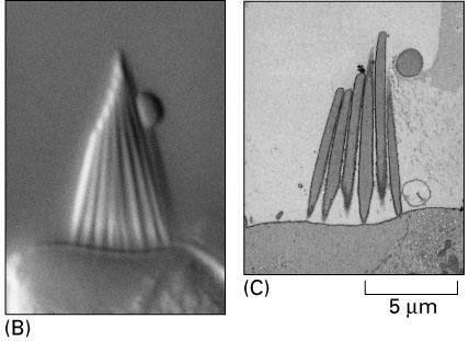
6 Resolution vs Detection Objects smaller than the resolution limit of a microscope can be detected MBoC supplemental video Microtubules24 nm diameter (DIC image) Actin (8nm) on myosin lawn (Fluorescent actin)

7 Objects smaller than the resolution limit can be detected but are not resolved


8 What is magnification good for? Imaging devices have discrete detector elements Rods and Cones in Eye Pixels in Digital Camera

9 Magnification The minimal adequate magnification is one that allows the smallest object you want to resolve to fall on 3 discrete elements of the imaging device.

10 Properties of Interaction Four properties of the interaction between light and matter influence the design of microscopes used to produce contrast.


11 Absorption methods: Stained tissue sections Stains are compounds that absorb light or electrons. Black stain absorbs all colors of light Colored stains absorb some colors of light, others to pass Kidney Collecting Duct Fig 9-11 Adipose Tissue Purkinje neuron


 Periodic acid Shiff s")
12 Different components of a cell can be selectively stained Chemical compounds of some stains bind preferentially to specific cellular components Hematoxylin (blue) stains nucleic acids Osmium stains lipids in neuron sheath Hematoxylin & eosin (proteins stain pink) Periodic acid Shiff s stains carbohydrates

13 Antibodies are used to detect specific cell components


14 Antibodies can be linked to enzymes that produce colored products Craniofacial neurons in E10.5 mouse (horseradish peroxidase labeling), Sahay et al., J. Neurosci : Delta I gene expression in developing somites (beta-gal staining)


15 Refraction Techniques in Light Microscopy Very little incident light is absorbed, reflected or refracted by a living biological specimen Brightfield Imaging: refracted light from the specimen is poorly detected


16 Methods for imaging refracted light I. Darkfield Imaging


17 Living Cells Living cells are seen clearly using phase contrast microscopy to image the difference between refracted and non-refracted light. Phase Contrast Imaging

18 Features of living cells can be seen clearly using phase contrast microscopy. Shelden

19 Fluorescence microscopy: Fluorescent molecules absorb high energy light and then emit less energetic, longer wavelength light. The shift in wavelength between absorbed and emitted light is called the Stokes shift. Excitation spectrum maxima Emission spectrum Rhodamine anti-map in cultured neurons
20 Fluorescence Microscopes Fluorescence microscopes use excitation, emission and dichroic filters to take advantage of the Stokes shift Fig 9-11
 stains actin DAPI (blue) stains nucleic acids")
21 Fluorescent Stains Chemical compounds of some fluorescent stains bind preferentially to specific cellular components Fluorescent phalloidin (red) stains actin DAPI (blue) stains nucleic acids Shelden

 Anti-neurofilaments")
22 Fluorophores Antibody linked fluorophores detect specific cellular antigens Anti-tubulin (green) Anti-neurofilaments (green)

23 FISH Fluorescence in situ hybridization (FISH) uses synthetic fluorescent RNA probes to detect compatible mrna in cells and tissue mrna Fluorescent RNA probe Figure 9-12


24 Light emitting dyes reveal changes in ion concentrations Figure 9.32 Calcium changes during fertilization visualized with aequorin Calcium signaling in renal epithelial cells (fluo-5 indicator) Shelden


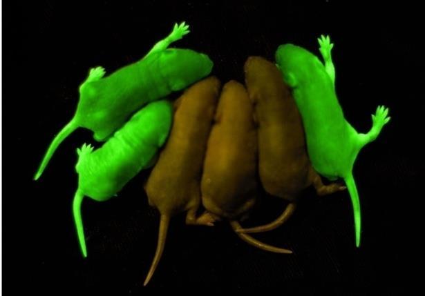
25 Fluorescent Protiens Green fluorescent proteins can be expressed in living organisms Aequorea victoria GFP

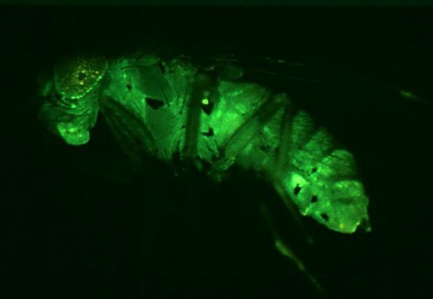
26 Gene Promoters Cell-type specific gene promoters can be used to express GFP in specific cells or tissues Neuron specific promoter GFP Coding Sequence Figure 9-25 (transgenic fruit fly larva)


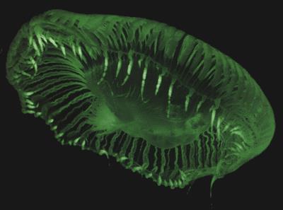
27 Fluorescent Fusion Protiens Green fluorescent fusion proteins can be used to label proteins in living cells Recombinant DNA Relocation of GFP-tagged proteins in muscle cells
")
28 Fluorescent Probes Dynamic studies of fluorescent probes can be conducted using Fluorescence Recovery After Photobleaching (FRAP) Fig 9-31

29 Fluorescent Probes Dynamic studies of fluorescent probes can be conducted using photo-activatable probes Fig 9-30

30 Electron Microscope The electron microscope uses electrons to resolve fine structure of the cell The wavelength of an electron can be.004 nanometers, so the theoretical limit of resolution of an electron microscope is 1/20 angstoms, or 1/20 the diameter of a hydrogen atom. Electrons pass through the specimen in TEM Fig 9-42

31 EM Stains Stains used for electron microscopy (EM) are very dense (metals) so they absorb electrons EM stains are generally soluble, reactive forms of metals such as lead, uranium, gold, silver and tungsten Water Osmium tetroxide Uranyl acetate Shelden


32 Gaseous metals can be directionally applied (shadowing) Sputter coatter Fig 9-52


33 Metal Shadowing Metal shadowing can be applies to surface structures or interior structures using freeze fracture and freeze etching methods Cryoelectron microscopy of freezefractured intestinal microvilli Cryoelectron microscopy of freezeetched skeletal muscle fibers Specimens are frozen, then split using a microtome and exposed surfaces prepared and imaged.


34 EM Negative Staining EM Negative staining allows isolated macromolecules to be seen Actin filaments Macromolecules on substrate Stain in excess Fig 9-54 Bacteriophage virus Gene Meyer University of S. Carolina Excess stain removed and dried Shelden


35 Computational Methods Computational methods can produce three dimensional structural inormation from multiple views of an object Figure 9-55 reconstruction of the Hepatitis B virus


36 Antibodies Antibodies can be attached to metal particles to detect specific cellular components Immuno-gold labeling of microtubules in an interphase cells Immuno-gold labeling of centromere proteins. Fig 9-46


37 Pulse Chase Radio-Labeling Dynamic studies of molecules can be conduced at the EM level by pulse chase radio-labeling. Fig 9-38
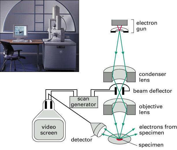

38 Reflection techniques in microscopy: Scanning EM: Fig 9-49 Fig 9-50
Resolution of Microscopes Visible light is nm Dry lens(0.5na), green(530nm light)=0.65µm=650nm for oil lens (1.4NA) UV light (300nm) = 0.13µm f
, green(530nm light)=0.65µm=650nm for oil lens (1.4NA) UV light (300nm) = 0.13µm f") Microscopes and Microscopy MCB 380 Good information sources: Alberts-Molecular Biology of the Cell http://micro.magnet.fsu.edu/primer/ http://www.microscopyu.com/ Approaches to Problems in Cell Biology
Microscopes and Microscopy MCB 380 Good information sources: Alberts-Molecular Biology of the Cell http://micro.magnet.fsu.edu/primer/ http://www.microscopyu.com/ Approaches to Problems in Cell Biology
Partha Roy
 Fluorescence microscopy http://micro.magnet.fsu.edu/primer/index.html Partha Roy 1 Lecture Outline Definition of fluorescence Common fluorescent reagents Construction ti of a fluorescence microscope Optical
Confocal Microscopes. Evolution of Imaging
 Confocal Microscopes and Evolution of Imaging Judi Reilly Hans Richter Massachusetts Institute of Technology Environment, Health & Safety Office Radiation Protection What is Confocal? Pinhole diaphragm
Chapter 10: Classification of Microorganisms
 Chapter 10: Classification of Microorganisms 1. The Taxonomic Hierarchy 2. Methods of Identification 1. The Taxonomic Hierarchy Phylogenetic Tree of the 3 Domains Taxonomic Hierarchy 8 successive taxa
Practical light microscopy: an introduction
 Practical light microscopy: an introduction Dr. Mark Leake, Oxford University www.physics.ox.ac.uk/users/leake Aim of today s talk: Explanation of the very (very) basics of how a light microscope works
Methods of Culturing Microorganisms. Chapter 3. Five Basic Techniques of Culturing Bacteria. Topics
 Chapter 3 Topics Methods of Culturing Microorganisms Microscope (History, Types, Definitions) Staining (Gram s) Methods of Culturing Microorganisms Five basic techniques of culturing Media Microbial growth
Baraa Ayed AL-Odat. Israa Ayed. Heba kalbouneh
 1 Baraa Ayed AL-Odat Israa Ayed Heba kalbouneh Introduction: "lecture #1" The name " histology " is derived from the Greek words: "histo" means a tissue and "logos" means the study of. So, Histology mean
Automated Digital Microscopy
 A p p l i c a t i o n G u i d e Peter Banks, Ph.D. and Peter J. Brescia, Applications Department, BioTek Instruments, Inc., Winooski, VT Table of Contents Introduction ----------------------------------------------------------------------------------------------------------------------
Fundamentals and Applications of Biofilms Analysis, Structure and Physiology of Bacterial Biofilms Ching-Tsan Huang ( 黃慶璨 ) Office: Agronomy
 Office: Agronomy") 1 Fundamentals and Applications of Biofilms Analysis, Structure and Physiology of Bacterial Biofilms Ching-Tsan Huang ( 黃慶璨 ) Office: Agronomy Building, Room 111 Tel: (02) 33664454 E-mail: cthuang@ntu.edu.tw
1 Fundamentals and Applications of Biofilms Analysis, Structure and Physiology of Bacterial Biofilms Ching-Tsan Huang ( 黃慶璨 ) Office: Agronomy Building, Room 111 Tel: (02) 33664454 E-mail: cthuang@ntu.edu.tw
Introduction to Fluorescence Jablonski Diagram
 ntroduction to Fluorescence Jablonski Diagram Excited Singlet Manifold S1 internal conversion S2 k -isc k isc Excited riplet Manifold 1 S0 k nr k k' f nr fluorescence k p phosphorescence Singlet round
Rice/TCU REU on Computational Neuroscience. Fundamentals of Molecular Imaging
 Rice/TCU REU on Computational Neuroscience Fundamentals of Molecular Imaging June 2, 2009 Neal Waxham 713-500-5621 m.n.waxham@uth.tmc.edu Objectives Introduction to resolution in light microscopy Brief
Simultaneous multi-color, multiphoton fluorophore excitation using dual-color fiber lasers
 Multiphoton Microscopy / Fiber Laser Simultaneous multi-color, multiphoton fluorophore excitation using dual-color fiber lasers Matthias Handloser, Tim Paasch-Colberg, Bernhard Wolfring TOPTICA Photonics
Azure Biosystems Western Blotting Workflow
 Azure Biosystems Western Blotting Workflow PROBE PLAN SEPARATE ANALYZE VISUALIZE PLAN Plan your experiment and choose your detection method Chemiluminescent Western Blotting The most common method for
Confocal Microscopy Analyzes Cells
 Choosing Filters for Fluorescence A Laurin Publication Photonic Solutions for Biotechnology and Medicine November 2002 Confocal Microscopy Analyzes Cells Reprinted from the November 2002 issue of Biophotonics
Innovations To Meet Your Needs
 Innovations To Meet Your Needs Cooled CCD Camera 1340 x 1037 pixel resolution for greatest image quality 12-bit precision provides 3 orders of linear dynamic range Windows and Power Macintosh Software
A legacy of innovation and discovery
 A legacy of innovation and discovery CellInsight CX7 LZR High Content Analysis Platform Quantifiably brilliant data Since the introduction of Thermo Scientific ArrayScan High Content Analysis (HCA) Readers
Super-resolution Microscopy
 Semr oc kwhi t epaperser i es : 1. Introduction Super-resolution Microscopy Fluorescence microscopy has revolutionized the study of biological samples. Ever since the invention of fluorescence microscopy
Fluorescent in-situ Hybridization
 Fluorescent in-situ Hybridization Presented for: Presented by: Date: 2 Definition In situ hybridization is the method of localizing/ detecting specific nucleotide sequences in morphologically preserved
QImaging Camera Application Notes Multicolor Immunofluorescence Imaging
 QImaging Camera Application Notes Multicolor Immunofluorescence Imaging In order to image localization of intracellular proteins with high specificity, it is frequently necessary to multiplex antibody
CENTER FOR BRAIN EXPERIMENT
 CENTER FOR BRAIN EXPERIMENT Section of Brain Structure Associate Professor: ARII, Tatsuo, PhD 1967 Graduated from Tohoku University, Faculty of Science. Completed the doctoral course in Engineering, Nagoya
Quantum Dot applications in Fluorescence Imaging for Calibration and Molecular Imaging
 Quantum Dot applications in Fluorescence Imaging for Calibration and Molecular Imaging Introduction In this application note, we will discuss the application of quantum dots in fluorescence imaging, both
Direct visualization, sizing and concentration measurement of fluorescently labeled nanoparticles using NTA
 Direct visualization, sizing and concentration measurement of fluorescently labeled nanoparticles using NTA NANOSIGHT RANGE Visualize and Measure Nanoparticle Size and Concentration PARTICLE SIZE PARTICLE
Optical Observation - Hyperspectral Characterization of Nano-scale Materials In-situ
 Optical Observation - Hyperspectral Characterization of Nano-scale Materials In-situ Research at the nanoscale is more effective, when research teams can quickly and easily observe and characterize a wide
Confocal Microscopy of Electronic Devices. James Saczuk. Consumer Optical Electronics EE594 02/22/2000
 Confocal Microscopy of Electronic Devices James Saczuk Consumer Optical Electronics EE594 02/22/2000 Introduction! Review of confocal principles! Why is CM used to examine electronics?! Several methods
Nodes of regulation in cellular systems
 Nodes of regulation in cellular systems cell membrane signal transduction ligands receptors oligomerization transport signal transduction modified protein Golgi transcription factor transport ER transport
Chapter 10 Analytical Biotechnology and the Human Genome
 Chapter 10 Analytical Biotechnology and the Human Genome Chapter Outline Enzyme tests and biosensors DNA-based tests DNA analysis technologies Human genome and genome-based analytical methods 1 Enzyme-based
Spectral Separation of Multifluorescence Labels with the LSM 510 META
 Microscopy from Carl Zeiss Spectral Separation of Multifluorescence Labels with the LSM 510 META Indians living in the South American rain forest can distinguish between almost 200 hues of green in their
Lecture 13: Analysis of 2D gels
 Lecture 13: Analysis of 2D gels A complete proteomic analysis aims at collecting quantitative information about all protein in a sample. A normal 2 Dimensional gel electrophoresis results are analyzed
Fluorescence Nanoscopy
 Fluorescence Nanoscopy Keith A. Lidke University of New Mexico panda3.phys.unm.edu/~klidke/index.html Optical Microscopy http://en.wikipedia.org/wiki/k%c3%b6hler_illumination 30 µm Fluorescent Probes Michalet
CHAPTER 20 DNA TECHNOLOGY AND GENOMICS. Section A: DNA Cloning
 Section A: DNA Cloning 1. DNA technology makes it possible to clone genes for basic research and commercial applications: an overview 2. Restriction enzymes are used to make recombinant DNA 3. Genes can
Final exam. Please write your name on the exam and keep an ID card ready.
 Biophysics of Macromolecules Prof. R. Jungmann and Prof. J. Lipfert SS 2017 Final exam Final exam First name: Last name: Student number ( Matrikelnummer ): Please write your name on the exam and keep an
CF Dyes Next Generation Fluorescent Dyes Secondary antibody
 CF Dyes Next Generation Fluorescent Dyes Secondary antibody OZYME 10 AVENUE AMPÈRE - CS 30268-78053 ST QUENTIN EN YVELINES CEDEX Tél. : 01 34 60 24 24 - Fax : 01 34 60 92 12 - www.ozyme.fr/info CF Dyes
BIOLOGICAL SAMPLE PREPARATION FOR TEM OBSERVATION. TEM Seminar Nov 16, 2017 Astari Dwiranti, Ph.D
 BIOLOGICAL SAMPLE PREPARATION FOR TEM OBSERVATION TEM Seminar Nov 16, 2017 Astari Dwiranti, Ph.D Why do we need EM for biological samples? (O'Connor and Adams, 2010) Why do we need EM for biological samples?
Flow Cytometry - The Essentials
 Flow Cytometry - The Essentials Pocket Guide to Flow Cytometry: 1. Know your Cytometer 2. Understanding Fluorescence and Fluorophores 3. Gating Process 4. Controls 5. Optimization 6. Panel Building 7.
ab CytoPainter ER Staining Kit Red Fluorescence
 ab139482 CytoPainter ER Staining Kit Red Fluorescence Instructions for Use Designed to detect Human endoplasmic reticulum by microscopy. This product is for research use only and is not intended for diagnostic
Electron microscopy technology of reticulocytes after sorting with
 Electron microscopy technology of reticulocytes after sorting with magnetic beads The Cell Analysis Center Scientific Bulletin Part 2 For efficient analysis of cells, sorting of the target cells is crucial.
Lab 1A: Microscopy I. Name: Section:
 Lab 1A: Microscopy I A response is required for each item marked: (# ). Your grade for the lab 1 report (1A and 1B combined) will be the fraction of correct responses on a 50 point scale[(# correct/# total)
Introduction to N-STORM
 Introduction to N-STORM Dan Metcalf Advanced Imaging Manager Outline Introduction Principles of STORM Applications N-STORM overview Biological Scale Mitochondrion Microtubule Amino Acid 1Å Kinesin 1nm
Genetically targeted all-optical electrophysiology with a transgenic Credependent
 Genetically targeted all-optical electrophysiology with a transgenic Credependent Optopatch mouse Short title: Transgenic Optopatch mouse Shan Lou 1, Yoav Adam 1, Eli N. Weinstein 1,4, Erika Williams 2,
Mystery microscope images
 Mystery microscope images Equipment: 6X framed microscopy images 6X cards saying what each microscope image shows 6X cards with the different microscopy techniques written on them 6X cards with the different
Reading for lecture 11
 Reading for lecture 11 1. Optical Tweezers, Myosin 2. Atomic Force Microscopy (AFM) 3. Single-Molecule Fluorescence Microscopy 4. Patch-Clamp 5. Genetic Techniques Key references are included in italics
CREOL, The College of Optics & Photonics, University of Central Florida
 Metal Substrate Induced Control of Ag Nanoparticle Plasmon Resonances for Tunable SERS Substrates Pieter G. Kik 1, Amitabh Ghoshal 1, Manuel Marquez 2 and Min Hu 1 1 CREOL, The College of Optics and Photonics,
Green Fluorescent Protein (GFP) Purification. Hydrophobic Interaction Chromatography
 Purification. Hydrophobic Interaction Chromatography") Green Fluorescent Protein (GFP) Purification Hydrophobic Interaction Chromatography What is the GFP gene? GFP is a green fluorescent protein that is normally found in jellyfish. It has been engineered
Green Fluorescent Protein (GFP) Purification Hydrophobic Interaction Chromatography What is the GFP gene? GFP is a green fluorescent protein that is normally found in jellyfish. It has been engineered
Fluorescence Imaging with One Nanometer Accuracy Lab
 I. Introduction. Fluorescence Imaging with One Nanometer Accuracy Lab Traditional light microscope is limited by the diffraction limit of light, typically around 250 nm. However, many biological processes
The Nuclear Area Factor (NAF): a measure for cell apoptosis using microscopy and image analysis
: a measure for cell apoptosis using microscopy and image analysis") The Nuclear Area Factor (NAF): a measure for cell apoptosis using microscopy and image analysis Mark A. DeCoster Department of Biomedical Engineering and Institute for Micromanufacturing, Louisiana Tech
The Nuclear Area Factor (NAF): a measure for cell apoptosis using microscopy and image analysis Mark A. DeCoster Department of Biomedical Engineering and Institute for Micromanufacturing, Louisiana Tech
over time using live cell microscopy. The time post infection is indicated in the lower left corner.
 Title of file for HTML: Supplementary Information Description: Supplementary Figures and Supplementary Table Title of file for HTML: Supplementary Movie 1 Description: Fusion of NBs. BSR cells were infected
Segments of the obstructed intestinal loops were fixed in 4% paraformaldehyde
 Supplementary text Supplementary materials and methods Histopathological examination Segments of the obstructed intestinal loops were fixed in 4% paraformaldehyde (PFA) and embedded in paraffin wax with
PREPARATION OF HISTOLOGICAL SPECIMENS
 PREPARATION OF HISTOLOGICAL SPECIMENS Histo-techniques Preparation of tissue for microscopic examination Series of processes Ultimate aim to make tissue visible as it is Pathology Vs Anatomy Steps vary
in-situ PCR Presented for: Presented by: Date:
 in-situ PCR Presented for: Presented by: Date: 2 in situ Hybridization - Definition in situ PCR is a method in which the polymerase chain reaction actually takes place in the cell on a slide, and the product
Nucleic Acid Staining. Fluorophores & Applica6ons
 Nucleic Acid Staining Fluorophores & Applica6ons Types of Nucleic Acid Stains Intercala)ng dyes- - ethidium bromide and propidium iodide Minor- groove binders- - DAPI and the Hoechst dyes Miscellaneous-
Multiphoton Microscopy: Seeing deeper and clearer
 Multiphoton Microscopy: Seeing deeper and clearer Since the invention of simple microscope by Leuwenhoek and Hooke in the 17th century, different types of light microscopy techniques (such as phase contrast,
TARGETED IMAGING. Maureen Chan and Ruwani Mahathantila
 TARGETED IMAGING Maureen Chan and Ruwani Mahathantila Overview 2 Introduction to fluorescent imaging Fluorescent agents Quantum Dots Physical properties How QDs work In Vivo QD imaging Future Video What
Stellaris RNA FISH Protocol for Simultaneous IF + FISH in Adherent Cells
 Stellaris RNA FISH Protocol for Simultaneous IF + FISH in Adherent Cells General Protocol & Storage Product Description A set of Stellaris RNA FISH Probes is comprised of up to 48 singly labeled oligonucleotides
Microscopy Reagents. for Immunocytochemistry and Immunohistochemistry. World-Class Quality Superior Customer Support Outstanding Value
 Microscopy Reagents for Immunocytochemistry and Immunohistochemistry BioLegend is ISO 9001:2008 and ISO 13485:2003 Certified Toll-Free Tel: (US & Canada): 1.877.BIOLEGEND (246.5343) Tel: 858.768.5800 biolegend.com
Gene Expression Technology
 Gene Expression Technology Bing Zhang Department of Biomedical Informatics Vanderbilt University bing.zhang@vanderbilt.edu Gene expression Gene expression is the process by which information from a gene
3.1.4 DNA Microarray Technology
 3.1.4 DNA Microarray Technology Scientists have discovered that one of the differences between healthy and cancer is which genes are turned on in each. Scientists can compare the gene expression patterns
Post-expansion antibody delivery, after epitope-preserving homogenization.
 Supplementary Figure 1 Post-expansion antibody delivery, after epitope-preserving homogenization. (a, b) Wide-field fluorescence images of Thy1-YFP-expressing mouse brain hemisphere slice before expansion
hfab Rhodamine Housekeeping Antibodies
 hfab Rhodamine Housekeeping Antibodies Catalog # Description 12004163 Anti-Actin hfab Rhodamine Antibody, 200 µl 12004164 Anti-Actin hfab Rhodamine Antibody, 40 µl 12004165 Anti-Tubulin hfab Rhodamine
CBI Toolbox Tour 2015
 CBI Toolbox Tour 2015 Thermophoresis (NanoTemper) NT.115 & NT.LabelFree Images: NanoTemper Circular Dichroism Jasco J-1500 Spectrometer Six Position Turreted Peltier Temperature Control System Automated
Monitoring and Optimizing the Lipopolysaccharides-plasmid DNA interaction by FLIM-FRET
 Transactions on Science and Technology Vol. 4, No. 3-3, 342-347, 2017 Monitoring and Optimizing the Lipopolysaccharides-plasmid DNA interaction by FLIM-FRET Nur Syahadatain Abdul Razak 1#, Clarence M.
Con-focal and Multi-photon Microscope Experiment Fundamental. Qian Hu, Lab of Laser Scanning Confocal & Two-Photon Microscopy, ION, CAS
 Con-focal and Multi-photon Microscope Experiment Fundamental Qian Hu, Lab of Laser Scanning Confocal & Two-Photon Microscopy, ION, CAS 1. Light is Electromagnetic Wave ν = c / λ 2. Image of a Point Source
Immunostaining Protocols
 Immunostaining Protocols Lula L. Hilenski, Ph.D. Director Microscopy in Medicine Core Emory University Variables in standard immunostaining protocol 2-step or indirect immunofluorescence 1. Substrate on
Nature Methods: doi: /nmeth Supplementary Figure 1. Retention of RNA with LabelX.
 Supplementary Figure 1 Retention of RNA with LabelX. (a) Epi-fluorescence image of single molecule FISH (smfish) against GAPDH on HeLa cells expanded without LabelX treatment. (b) Epi-fluorescence image
Simple method for formation of nanometer scale holes in membranes. E. O. Lawrence Berkeley National Laboratory, Berkeley, CA 94720
 Simple method for formation of nanometer scale holes in membranes T. Schenkel 1, E. A. Stach, V. Radmilovic, S.-J. Park, and A. Persaud E. O. Lawrence Berkeley National Laboratory, Berkeley, CA 94720 When
Lecture 25 (11/15/17)
") Lecture 25 (11/15/17) Reading: Ch9; 328-332 Ch25; 990-995, 1005-1012 Problems: Ch9 (study-guide: applying); 1,2 Ch9 (study-guide: facts); 7,8 Ch25 (text); 1-3,5-7,9,10,13-15 Ch25 (study-guide: applying);
Lecture 25 (11/15/17) Reading: Ch9; 328-332 Ch25; 990-995, 1005-1012 Problems: Ch9 (study-guide: applying); 1,2 Ch9 (study-guide: facts); 7,8 Ch25 (text); 1-3,5-7,9,10,13-15 Ch25 (study-guide: applying);
Determining fluorescence Limit of Detection with Nanoparticle Tracking Analysis (NTA)
") Determining fluorescence Limit of Detection with Nanoparticle Tracking Analysis (NTA) FLUORESCENCE DETECTION PARTICLE SIZE PARTICLE CONCENTRATION Introduction The ability to detect nanoparticle fluorescence
Determining fluorescence Limit of Detection with Nanoparticle Tracking Analysis (NTA) FLUORESCENCE DETECTION PARTICLE SIZE PARTICLE CONCENTRATION Introduction The ability to detect nanoparticle fluorescence
BCH 462. Western Blot
 BCH 462 Western Blot Blotting Immunoassay: A test that uses antibody and antigen complexes [immuno-complexes] as a means of generating measurable results. Antigens [Ag]: A substance that when introduced
Chapter 03 - Tools of the Laboratory: Methods for the Culturing of Microscopic Analysis of microorganisms
 Microbiology: A Systems Approach 4th Edition Cowan Test Bank Completed download: https://testbankreal.com/download/microbiology-systems-approach-4thedition-test-bank-cowan/ (Downloadable package TEST BANK
Dolphin-Chemi Plus. Aim: To visualise and evaluate the performance of chemiluminescent immunoblots using Wealtec s Dolphin-Chemi plus image system
 Application Note 03 Dolphin-Chemi plus 8/22/2007 Dolphin-Chemi Plus Aim: To visualise and evaluate the performance of chemiluminescent immunoblots using Wealtec s Dolphin-Chemi plus image system INTRODUCTION
Bioinstrumentation Light Sources Lasers or LEDs?
 Bioinstrumentation Light Sources Lasers or LEDs? A comprehensive analysis of all the factors involved in designing and building life sciences instrumentation reveals that lasers provide superior performance
Your Name: MID TERM ANSWER SHEET SIN: ( )
") MIDTERM EXAMINATION (October 23, 2008) BIOE150. Introduction to Bio-Nanoscience & Bio-Nanotechnology Professor Seung-Wuk Lee Fall Semester, 2008 0. Write down your name and the last digit of your SIN in
MIDTERM EXAMINATION (October 23, 2008) BIOE150. Introduction to Bio-Nanoscience & Bio-Nanotechnology Professor Seung-Wuk Lee Fall Semester, 2008 0. Write down your name and the last digit of your SIN in
A Survey of Laser Types. Gas Lasers
 Mihail Pivtoraiko Andrei Rozhkov Applied Optics Winter 2003 A Survey of Laser Types Laser technology is available to us since 1960 s, and since then has been quite well developed. Currently, there is a
CytoPainter Golgi Staining Kit Green Fluorescence
 ab139483 CytoPainter Golgi Staining Kit Green Fluorescence Instructions for Use Designed for the detection of Golgi bodies by microscopy This product is for research use only and is not intended for diagnostic
Microarray Industry Products
 Via Nicaragua, 12-14 00040 Pomezia (Roma) Phone: +39 06 91601628 Fax: +39 06 91612477 info@lifelinelab.com www.lifelinelab.com Microarray Industry Products Page 10 NBT / BCPIP Chromogenic phosphatase
FLUORESCENT PEPTIDES. Outstanding Performance and Wide Application Range
 FLUORESCENT PEPTIDES Peptides and amino acids labeled with and Tide Quencher TM We offer peptides and amino acids tagged with fluorescent dyes. They meet highest demands in fluorescence intensity and photo-stability,
Inoculate: Media. Physical State of Media: Liquid. The Five I s: Basic Techniques to Culture Microbes Tools of the Microbiology Laboratory
 The Five I s: Basic Techniques to Culture Microbes Tools of the Microbiology Laboratory 1. Inoculate 2. Incubate 3. Isolate 4. Inspect 5. Identify The Five I s: Inoculate Inoculate: Media Classified according
Methods of Biomaterials Testing Lesson 3-5. Biochemical Methods - Molecular Biology -
 Methods of Biomaterials Testing Lesson 3-5 Biochemical Methods - Molecular Biology - Chromosomes in the Cell Nucleus DNA in the Chromosome Deoxyribonucleic Acid (DNA) DNA has double-helix structure The
Immunohistochemistry. How does it look like? When do we need IHC? When do we need IHC? In clinic: In research:
 Introduction How does it look like? Immunohistochemistry Smooth muscle actin Parvalbumin Distrophyn Sandrine Bichet Head of Molecular Histology Platform Signal versus background 06.03.2012 IHC basics Introduction
Microendoscopes for imaging of the pancreas
 Microendoscopes for imaging of the pancreas Angelique Kano *, Andrew R. Rouse, Shona M. Kroto, Arthur F. Gmitro Optical Sciences Center& Radiology Research Lab, University of Arizona, Tucson 85724 ABSTRACT
Cryoplunge 3 with GentleBlot for cryo-em
 Cryoplunge 3 with GentleBlot for cryo-em Cryoplunge 3 with GentleBlot system GentleBlot technology provides very gentle 1- and 2-side blotting for preparing frozen hydrated specimens for cryo-em Ethane
Chapter 17: Immunization & Immune Testing. 1. Immunization 2. Diagnostic Immunology
 Chapter 17: Immunization & Immune Testing 1. Immunization 2. Diagnostic Immunology 1. Immunization Chapter Reading pp. 505-511 What is Immunization? A method of inducing artificial immunity by exposing
Technical Overview Cross-linking fixatives: What they are, what they do, and why we use them
 Technical Overview Cross-linking fixatives: What they are, what they do, and why we use them Focus on: Formaldehyde, Glutaraldehyde, and Osmium tetroxide M. Kuwajima/Kristen Harris Lab EM processing and
Cytomics in Action: Cytokine Network Cytometry
 Cytomics in Action: Cytokine Network Cytometry Jonni S. Moore, Ph.D. Director, Clinical and Research Flow Cytometry and PathBioResource Associate Professor of Pathology & Laboratory Medicine University
Analysis of receptor oligomerization by FRAP microscopy
 TIGP CBMB Student Seminar Analysis of receptor oligomerization by FRAP microscopy Dorsch S, Klotz KN, Engelhardt S, Lohse MJ, Bünemann M Nat Methods. 2009 Mar;6(3):225 30. K. Vijayasarathy March 10 th
NAME TA SEC Problem Set 4 FRIDAY October 15, Answers to this problem set must be inserted into the box outside
 MIT Biology Department 7.012: Introductory Biology - Fall 2004 Instructors: Professor Eric Lander, Professor Robert A. Weinberg, Dr. Claudette Gardel NAME TA SEC 7.012 Problem Set 4 FRIDAY October 15,
Advances in Intense Pulsed Light Solutions For Display Manufacturing. XENON Corporation Dr. Saad Ahmed Japan IDW 2016
 Advances in Intense Pulsed Light Solutions For Display Manufacturing XENON Corporation Dr. Saad Ahmed Japan IDW 2016 Talk Outline Introduction to Pulsed Light Applications in Display UV Curing Applications
Fig1: Melt pool size of LAMP vs. µlamp. The LAMP process s melt pool is x the area of the LAMP s melt pool.
 Proceedings of the 4th Annual ISC Research Symposium ISCRS 2010 April 21, 2010, Rolla, Missouri LOW COST IMAGING OF MELTPOOL IN MICRO LASER AIDED MANUFACTURING PROCESS (µlamp) ABSTRACT This paper describes
Qdot nanocrystal. wide range of biological investigations, Qdot nanocrystals are powerful complements
 Feature nanocrystal conjugates for flow cytometry Take the easy route to multicolor flow cytometry. With applications across a wide range of biological investigations, nanocrystals are powerful complements
Basic Principles in Flow Cytometry. Flow Cytometry
 Basic Principles in Flow Cytometry Flow Cytometry» Flow Cytometry is the technological process that allows for the individual measurements of cell fluorescence and light scattering. This process is performed
Cyfra 21-1 IRMA. Product information Information about other products is available at: Userś Manual DE52100
 Product information Information about other products is available at: www.demeditec.com Userś Manual Cyfra 21-1 IRMA The CYFRA 21.1 IRMA system provides a direct in vitro quantitative determination of
Application of Quantum Mechanics to Biology
 Application of Quantum Mechanics to Biology How can we apply quantum mechanics to biology? Polymers of nucleotides and amino acids - millions of atoms bounded into a large molecule Visual System Must turn
How small is a nanometer?
 How small is a nanometer? Purpose: The purpose of this activity is to learn about the size of a nanometer. Questions to think about: Could you see an object that measures 10 nanometers across with your
Cells and Tissues. Overview CELLS
 Cells and Tissues WIll The basic unit of structure and function in the human body is the cell. Each of a cell's parts, or organelles, as well as the entire cell, is organized to perform a specific function.
Developmental Biology BY1101 P. Murphy
 Developmental Biology BY1101 P. Murphy Lecture 7 Cellular differentiation and the regulation of gene expression. In this lecture we looked at two main questions: How is gene expression regulated? (revision
Figure S1. Figure S2. Figure S3 HB Anti-FSP27 (COOH-terminal peptide) Ab. Anti-GST-FSP27(45-127) Ab.
 Ab. Anti-GST-FSP27(45-127) Ab.") / 36B4 mrna ratio Figure S1 * 2. 1.6 1.2.8 *.4 control TNFα BRL49653 Figure S2 Su bw AT p iw Anti- (COOH-terminal peptide) Ab Blot : Anti-GST-(45-127) Ab β-actin Figure S3 HB2 HW AT BA T Figure S4 A TAG
/ 36B4 mrna ratio Figure S1 * 2. 1.6 1.2.8 *.4 control TNFα BRL49653 Figure S2 Su bw AT p iw Anti- (COOH-terminal peptide) Ab Blot : Anti-GST-(45-127) Ab β-actin Figure S3 HB2 HW AT BA T Figure S4 A TAG
Chapter 5 DNA and Chromosomes
 Chapter 5 DNA and Chromosomes DNA as the genetic material Heat-killed bacteria can transform living cells S Smooth R Rough Fred Griffith, 1920 DNA is the genetic material Oswald Avery Colin MacLeod Maclyn
A Level. A Level Biology. Cells, Microscopes, Cell Cycle and Immunity Questions. AQA, OCR, Edexcel. Name: Total Marks: Page 1
 AQA, OCR, Edexcel A Level A Level Biology Cells, Microscopes, Cell Cycle and Immunity Questions Name: Total Marks: Page 1 Q1.The diagram shows a eukaryotic cell. (a) Complete the table by giving the letter
The best and brightest
 Labeling and Detection The best and brightest Alexa Fluor 488 dye Labeling and Detection A superior alternative to FITC Brighter conjugate fluorescence Unequalled photostability Perfect spectral match
1. The microtubule wall is composed of globular proteins arranged in longitudinal rows called.
 Name: Quiz name: Quiz 7 ate: 1. The microtubule wall is composed of globular proteins arranged in longitudinal rows called. microfilaments protofilaments prototubules microtubular subunits 2. Which of
See the small world Subject Area(s) Associated Unit: Associated Lesson: Activity Title: Image 1 ADA Description: Image file name: Source/Rights:
 Associated Unit: Associated Lesson: Activity Title: Image 1 ADA Description: Image file name: Source/Rights:") See the small world Subject Area(s) Chemistry, Life science, Physical Science, Science & Technology, measurements Associated Unit: None Associated Lesson: Activity Title: See the small world Image 1 ADA
See the small world Subject Area(s) Chemistry, Life science, Physical Science, Science & Technology, measurements Associated Unit: None Associated Lesson: Activity Title: See the small world Image 1 ADA