Introduction to Histology
|
|
|
- Violet James
- 5 years ago
- Views:
Transcription
1 Introduction to Histology


2 The name "Histology" is derived from the Greek word for a tissue "Histos", and "-logos" = the study of It is tightly bounded to molecular biology, genetics, immunology and other basic sciences


3 Cells are the basic unit of structure and function in living things Tissues made up of cells that are similar in structure and function and which work together to perform a specific activity Organs made up of tissues that work together to perform a specific activity Systems are groups of two or more organs that work together to perform a specific function for the organism
4 Theory Practical Introduction to histology Cell overview Microscopes and Microtechniques Epithelium 1 Cell overview Epithelium 2 Epithelium-1 Epithelium 3 Epithelium-2 Connective tissue 1 Revision- Quiz Connective tissue 2 Connective tissue-1 Midterm exam Cartilage Connective tissue-2 Bone 1 Cartilage Bone 2 Bone 1 Muscular tissue 1 Bone 2 Muscular tissue 2 Revision/ Quiz Nervous tissue Muscular tissue Skin Skin
5 Practical sessions pm pm Group 4 Computer lab Histology lab Group 5 Histology lab Computer lab
6 Suggested Histology Reference Junqueira s Basic Histology Text and Atlas 13 th edition By Anthony L. Mescher
7 History Bichat is the first anatomist who defined the term TISSUE without the use of microscope Units used in microscopy One millimter = 1000 micrometer (µm) One micrometer = 1000 nanometer (nm)
8 Microtechniques Tissue preparation for microscopic examination There are different methods but the basic principles are similar Hardening and sectioning of the tissue Examples : paraffin and freezing techniques
9 Microtechniques 1. Fixation 2. Dehydration 3. Clearing 4. Impregnation (infiltration) 5. Embedding 6. Section cutting 7. Staining 8. Mounting


10 Fixation: Exposing the tissue to chemical agents called fixatives i.e paraformaldehyde
11 Dehydration The process to remove the water by using a graded series of alcohol Then the tissue can be filled with the paraffin or other embedding agent
12 Clearing Replacing the dehydrating fluid with a fluid that is totally miscible with both the dehydrating fluid and the embedding medium. i.e Xylene Infiltration with wax
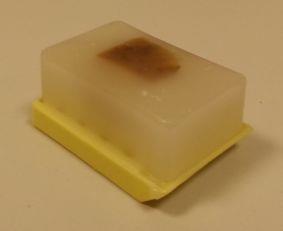
13 Embedding
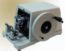
14 Sectioning
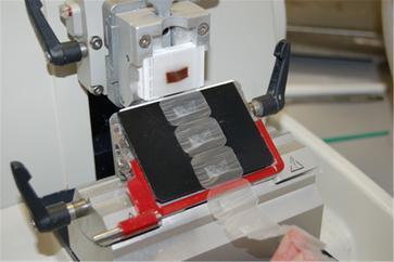
15
 the")

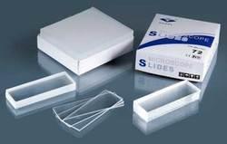
16 Mounting The process to place (mount) the tissue sections on the adhesive coated glass slides

17
18 Freezing technique Tissues are frozen using liquid nitrogen Frozen tissues are sectioned by cryostat It is faster and preserve tissue components The quality of the section is poor with more artifacts While paraffin technique produces intact tissue with less artifacts

19

20
21 Staining techniques The stain is a chemical substance which reacts with certain tissue components producing a color 1. Ordinary stains 2. Immunohistochemistry and Immunocytochemistry 3. Hybridization techniques
22 Immunohistochemistry Rely on the use of antibody directed against molecule of interest, usually protein The antibody is usually labeled with a colored substance


23 Direct methodprimary antibody only Goat anti-actin labeled with 594

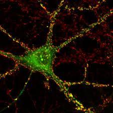
24 Indirect method primary and secondary antibodies Donkey anti-goat labeled with 488 Goat anti-actin
25 Hybridization techniques To detect and localize the presence or absence of specific DNA sequences on chromosomes to detect and localize specific RNA targets (ex. mrna) in cells a small oligonucleotide which is complementary to the target DNA/RNA sequence is used (ex. fluorescent probes) Can be applied to tissue sections, smears or chromosomes
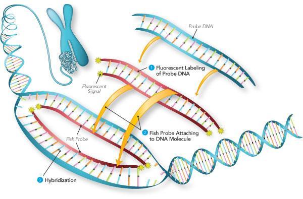
26 Hybridization techniques
27 Microscopy Light Microscopy Phase contrast Interference Fluorescence Polarizing Electron Microscopy Transmission EM Scanning EM
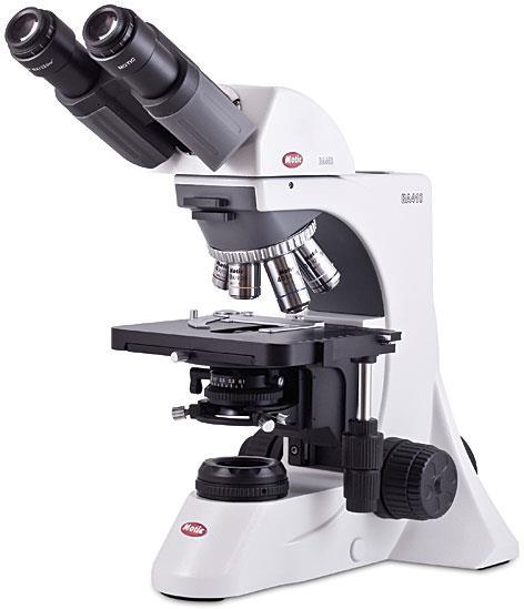
28 Light microscopy The basic functional unit consists of a tube; having an objective lens at one end and an ocular lens at the other The objective lens enlarges the image of the object in the direction of the ocular lens The ocular lens further magnifies this image toward the observer s eye The total magnification is obtained by multiplying the magnifying power of the objective and ocular lenses
29
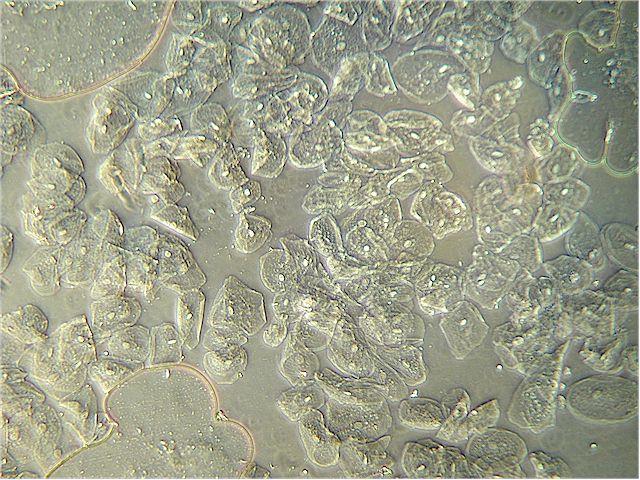
30 Phase Contrast Microscopy It uses a lens system that produces visible images from transparent objects The structures appear lighter or darker relative to each other The light changes its speed and direction when passing in different media Useful in tissue culture
31
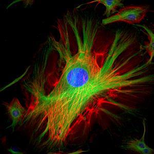
32 Fluorescence Microscopy Uses ultraviolet light When certain fluorescent substances are irradiated with ultra violet light, it emits light They appear as shiny particles on a dark background Placed in dark room
33
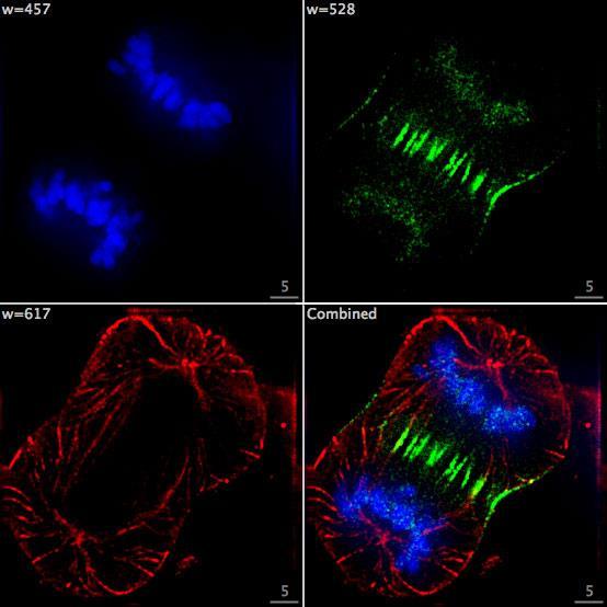
34 Confocal Microscopy Uses laser beams the laser can be moved (scanned) across the specimen as well as down into the specimen, it can produce 3D images Can be used in living and cultured cells and tissue sections
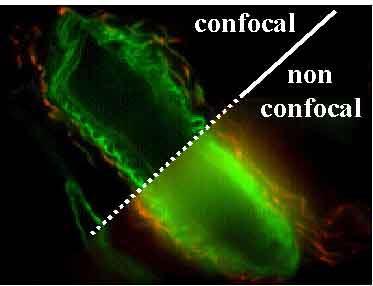
35
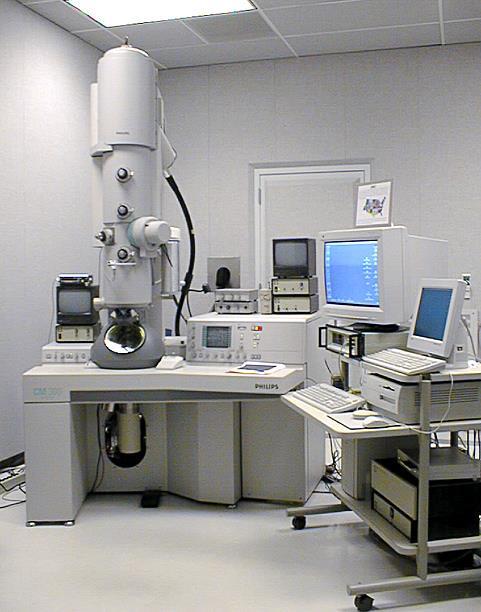
36 Electron Microscopy Uses electron beams instead of light Provides the highest resolution of subcellular structures Electromagnets to focus the electrons ( versus glass lenses to focus the light) Detect by fluorescent screen or photographic emulsion Requires ultrathin sections ( µm) Uses hard epoxy resin for embedding Ultrathin sections are produced by ultramicrotome ( Diamond or Glass knives)
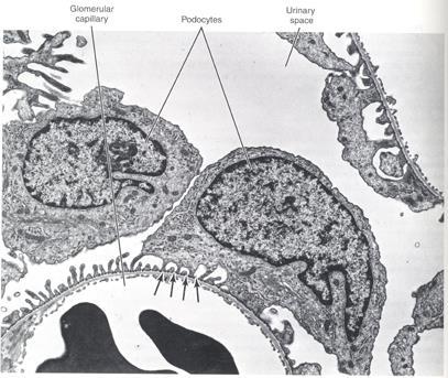
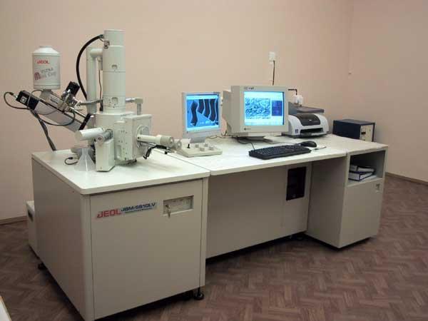
37 Types Transmission EM Views the ultrastructural details in shades of gray The bright areas of the images are unstained (the electrons passed through the sample) and the darker regions are areas which have taken up stain and either absorbed or scattered the electrons Scanning EM Provides information about the surface of a specimen Samples are coated with a gold-carbon film. The electron beam is then scanned across the specimen surface and the electrons that are reflected off of the surface are captured by the detector Views only the structure as a 3D image

38 SEM image TEM image
39
40
41 Light microscope Electron microscope Image Color images black and white images Images produced by Visible light rays Electron beam Magnification up to 1500x but a wider field of view and easier orientation Up to 2, x Resolution Resolving power to 0.25µm Resolving power to 0.2nm Time Frozen sections can yield an image within 20 minutes One day at least Section thickness Ranges from 1-30 µm Ranges from µm Specimen placed on Glass slide Copper mesh


42 Histology is a two dimensional study of a three dimensional reality.

43 Types of Tissue Sections (1) Longitudinal section tissue cut along the longest direction of an organ Cross section tissue cut perpendicular to the length of an organ Oblique section tissue cut at an angle between a cross & longitudinal section 43
44 Types of Tissue Sections (2) Would you classify the egg sections as longitudinal, cross, or oblique sections? How would the egg look if sectioned in the other two planes? 44 Practice at home.
45 Tissue Sectioning (2) Slices 1 & 5 miss the yolk / cell nucleus Cell nucleus is smaller in sections 2 & 4 45


46 Tissue Sectioning (3) A B Image A is a cross section of elbow macaroni, resembling a blood vessel, piece of gut, or other tubular organ. Image B is a longitudinal section of a sweat gland. Notice what a single slice could look like 46
; Specialized stains include PAS, Ag,")


47 Stains.. examples Standard stain (dye). H & E (Hematoylin & Eosin); Specialized stains include PAS, Ag, Aldehyde fuchsin, Orcein PAS- detects glycogen, glycoproteins, glycolipids and mucins in tissues 2 steps; CHO are oxidized with periodic acid to aldehyde groups. The Schiff reagent reacts with aldehyde groups to form a deep red-reaction (magenta) product Aldehyde fuchsin stains elastic fibres & ß-cells islets of pancreas Orcein stains elastic fibres dark brown Silver stain stains reticular fibres (type III collagen)
 Basophilic = Blue")
48 Basophilia Basophilic structures are stained by basic dyes: Basic dyes are positive Basophilic structures are negative (ex. DNA, RNA, ribosomes, RER) Basophilic = Blue

49 Acidophilia Acidophilic structures are stained by acid dyes: Acid dyes are negative Acidophilic structures are positive (ex. Proteins, collagen, cytoplasm) Eosinophilic = Pink
Baraa Ayed AL-Odat. Israa Ayed. Heba kalbouneh
 1 Baraa Ayed AL-Odat Israa Ayed Heba kalbouneh Introduction: "lecture #1" The name " histology " is derived from the Greek words: "histo" means a tissue and "logos" means the study of. So, Histology mean
COPYRIGHTED MATERIAL. Tissue Preparation and Microscopy. General Concepts. Chemical Fixation CHAPTER 1
 CHAPTER 1 Tissue Preparation and Microscopy General Concepts I. Biological tissues must undergo a series of treatments to be observed with light and electron microscopes. The process begins by stabilization
Introduction to Histology
 Introduction to Histology Histology The term "Histology" is derived from the Greek word for a tissue "Histos", and "-logos" = the study of Histology : Is the study of tissues and how they are arranged
Introduction to histology and its methods of study
 Introduction to histology and its methods of study Li shulei lishulei@tom.com Department of Histology & Embryology 1 What is histology Definition Cell: smallest units functions in the human body Tissue
Preparation of tissues for study
 Preparation of tissues for study HISTOLOGY : It is the branch of science which deals with the microscopic study of normal tissue HISTOPATHOLOGY : It is the branch of science which deals with the microscopic
PREPARATION OF HISTOLOGICAL SPECIMENS
 PREPARATION OF HISTOLOGICAL SPECIMENS Histo-techniques Preparation of tissue for microscopic examination Series of processes Ultimate aim to make tissue visible as it is Pathology Vs Anatomy Steps vary
Foundations in Microbiology Seventh Edition
 Lecture PowerPoint to accompany Foundations in Microbiology Seventh Edition Talaro Chapter 3 Tools of the Laboratory: The Methods for Studying Microorganisms Copyright The McGraw-Hill Companies, Inc. Permission
THE BASICS OF IMMUNOHISTOCHEMISTRY
 THE BASICS OF IMMUNOHISTOCHEMISTRY Introduction Immunohistochemistry (IHC) identifies specific tissue components by means of a specific antigen/antibody reaction tagged with a visible label. IHC makes
Cell Structure and Function
 Cell Structure and Function Dead White Men Who Discovered (and were made of) Cells: Anton Van Leeuwenhoek Robert Hooke Where the Magic Happened Schleiden Cell Theory All plants are made of cells Schwann
Methods of Culturing Microorganisms. Chapter 3. Five Basic Techniques of Culturing Bacteria. Topics
 Chapter 3 Topics Methods of Culturing Microorganisms Microscope (History, Types, Definitions) Staining (Gram s) Methods of Culturing Microorganisms Five basic techniques of culturing Media Microbial growth
Microscopy, Staining, and Classification
 CSLO CHECK CSLO1. Describe distinctive characteristics and diverse growth requirements of prokaryotic organisms compared to eukaryotic organisms. PowerPoint Lecture Presentations prepared by Mindy Miller-Kittrell,
Microbiology Chapter 2 Laboratory Equipment and Procedures 2:1 The Light Microscope MICROSCOPE: any tool with a lens to magnify and observe tiny
 Microbiology Chapter 2 Laboratory Equipment and Procedures 2:1 The Light Microscope MICROSCOPE: any tool with a lens to magnify and observe tiny details of specimens Micro tiny, small Scope to see SIMPLE
Preparation of thin slices for light microscopy
 Preparation of thin slices for light microscopy Optical light microscopy course 23.10.2012 Kirsi Rilla Shortly: Histological sample preparation for microscopy 1. Fixation: To fix the tissue components
Visualizing Cells Molecular Biology of the Cell - Chapter 9
 Visualizing Cells Molecular Biology of the Cell - Chapter 9 Resolution, Detection Magnification Interaction of Light with matter: Absorbtion, Refraction, Reflection, Fluorescence Light Microscopy Absorbtion
BIOLOGICAL SAMPLE PREPARATION FOR TEM OBSERVATION. TEM Seminar Nov 16, 2017 Astari Dwiranti, Ph.D
 BIOLOGICAL SAMPLE PREPARATION FOR TEM OBSERVATION TEM Seminar Nov 16, 2017 Astari Dwiranti, Ph.D Why do we need EM for biological samples? (O'Connor and Adams, 2010) Why do we need EM for biological samples?
Chapter 10: Classification of Microorganisms
 Chapter 10: Classification of Microorganisms 1. The Taxonomic Hierarchy 2. Methods of Identification 1. The Taxonomic Hierarchy Phylogenetic Tree of the 3 Domains Taxonomic Hierarchy 8 successive taxa
Module IB. Histochemistry. Martin Špaček, MD. (
 Module IB Histochemistry Martin Špaček, MD (E-mail: m.spacek@centrum.cz) http://www.lf3.cuni.cz/histologie What is histochemistry? It is a histological technique used for studying chemistry of tissues
Introduction to Histology and Basic Histological Techniques
 1 Introduction to Histology and Basic Histological Techniques Histology is that branch of anatomy that studies tissues of animals and plants. This textbook, however, discusses only animal, and more specifically
Electron microscopy technology of reticulocytes after sorting with
 Electron microscopy technology of reticulocytes after sorting with magnetic beads The Cell Analysis Center Scientific Bulletin Part 2 For efficient analysis of cells, sorting of the target cells is crucial.
EMS MICROSCOPY ACADEMY BIOLOGICAL TEM WORKSHOP: A COMPLETE PICTURE
 Examples of the endless possibilities in the field of Microscopy Bone Marrow: Transmission electron microscope image of a thin section cut through an area of bone marrow area near the cartilage/bone interface
Resolution of Microscopes Visible light is nm Dry lens(0.5na), green(530nm light)=0.65µm=650nm for oil lens (1.4NA) UV light (300nm) = 0.13µm f
, green(530nm light)=0.65µm=650nm for oil lens (1.4NA) UV light (300nm) = 0.13µm f") Microscopes and Microscopy MCB 380 Good information sources: Alberts-Molecular Biology of the Cell http://micro.magnet.fsu.edu/primer/ http://www.microscopyu.com/ Approaches to Problems in Cell Biology
Microscopes and Microscopy MCB 380 Good information sources: Alberts-Molecular Biology of the Cell http://micro.magnet.fsu.edu/primer/ http://www.microscopyu.com/ Approaches to Problems in Cell Biology
Name:
 Course No: DNTS2311 Course Title: General Histology I Date: 23/11/4104 No. of Questions: Time: 0hours Using Calculator (No) University of Palestine Midterm Exam 2014/2015 Total Grade: Instructor Name:
Staining Techniques. Staining Techniques. There are many dyes. Histochemical Stains: chemical reactions. Feulgen reaction -DNA
 Staining Techniques There are many dyes. http://medinfo.ufl.edu/~dental/denhisto/stains.html Examples: Sudan black -Lipids Myelinated axons- blue ihcworld.com/imagegallery/displayimage.php?al... Weigert
Hydroxystilbamidine Protocol
 ab138870 Hydroxystilbamidine Protocol Instructions for Use Staining of DNA and RNA in cells. This product is for research use only and is not intended for diagnostic use. 1 Table of Contents 1. Biological
Polymer Microscopy. Second edition LINDA C. SAWYER. and. DAVID T. GRUBB Cornell University Ithaca, NY USA. Hoechst Celanese Corporation Summit, NJ USA
 Polymer Microscopy Second edition LINDA C. SAWYER Hoechst Celanese Corporation Summit, NJ USA and DAVID T. GRUBB Cornell University Ithaca, NY USA CHAPMAN & HALL London Glasgow Weinheim New York Tokyo
The principles and practice of electron microscopy
 The principles and practice of electron microscopy Second Edition Ian M. Watt CAMBRIDGE UNIVERSITY PRESS Contents Preface tofirstedition page ix Preface to second edition xi 1 Microscopy with light and
Cell analysis and bioimaging technology illustrated
 Cell analysis and bioimaging technology illustrated The Cell Analysis Center Scientific Bulletin Part 1 Sysmex has been studying and exploring principles of automated haematology analysers, making full
Chapter 03 - Tools of the Laboratory: Methods for the Culturing of Microscopic Analysis of microorganisms
 Microbiology: A Systems Approach 4th Edition Cowan Test Bank Completed download: https://testbankreal.com/download/microbiology-systems-approach-4thedition-test-bank-cowan/ (Downloadable package TEST BANK
BIO 315 Lab Exam I. Section #: Name:
 Section #: Name: Also provide this information on the computer grid sheet given to you. (Section # in special code box) BIO 315 Lab Exam I 1. In labeling the parts of a standard compound light microscope
Methodology for Immunohistochemistry. Learning Objectives:
 Proteomics Methodology for Immunohistochemistry Methodology for Immunohistochemistry A staining process for identifying the proteins location in cells, tissues by using antigen-antibody property. Immuno
Fluorescent in-situ Hybridization
 Fluorescent in-situ Hybridization Presented for: Presented by: Date: 2 Definition In situ hybridization is the method of localizing/ detecting specific nucleotide sequences in morphologically preserved
Technical Note. Tissue Section Imaging. Published August The most recent version of this Technical Note is posted at licor.com/bio/support.
 Technical Note Tissue Section Imaging Published August 2017. The most recent version of this Technical Note is posted at licor.com/bio/support. Page 2 - Tissue Section Imaging Table of Contents Page I.
DNA/RNA MICROARRAYS NOTE: USE THIS KIT WITHIN 6 MONTHS OF RECEIPT.
 DNA/RNA MICROARRAYS This protocol is based on the EDVOTEK protocol DNA/RNA Microarrays. 10 groups of students NOTE: USE THIS KIT WITHIN 6 MONTHS OF RECEIPT. 1. EXPERIMENT OBJECTIVE The objective of this
Cell Growth and Reproduction
 Cell Growth and Reproduction Robert Hooke was the first person to describe cells, in the year 1665. He was looking through his microscope at a piece of cork when he noticed a lot of repeating honeycomb
Materials and Methods Materials Required for Fixing, Embedding and Sectioning. OCT embedding matrix (Thermo Scientific, LAMB/OCT)
") Page 1 Introduction Tissue freezing and sectioning is a rapid method of generating tissue samples (cryosections) for histological analysis, and obviates the need for wax embedding. The method is popular
Page 1 Introduction Tissue freezing and sectioning is a rapid method of generating tissue samples (cryosections) for histological analysis, and obviates the need for wax embedding. The method is popular
Fluorescent In Situ Hybridization (FISH) Assay
 Assay") Fluorescent In Situ Hybridization (FISH) Assay 1 What is FISH 2 Probes 3 FISH Procedure 4 Application Definition, Principle and Sample Types The core of FISH technology A quick and simple FISH protocol
Fluorescent In Situ Hybridization (FISH) Assay 1 What is FISH 2 Probes 3 FISH Procedure 4 Application Definition, Principle and Sample Types The core of FISH technology A quick and simple FISH protocol
Characterizing Phenotypes of Bacteria by Staining Method
 Experiment 3 Laboratory to Biology III Diversity of Microorganisms / Wintersemester / page 1 Experiment 3 Characterizing Phenotypes of Bacteria by Staining Method Advisor NN Reading Chapters in BBOM 9
Characterizing Phenotypes of Bacteria by Staining Method
 Experiment 3 Laboratory to Biology III Diversity of Microorganisms / Wintersemester / page 1 Experiment Characterizing Phenotypes of Bacteria by Staining Method Advisor Reading NN Chapters 3.1, 3.7, 3.8,
1. Carry the microscope in an upright position with both hands and place the base of the microscope 5cm from the edge of the bench
 The Microscope Operating the compound light microscope 1. Carry the microscope in an upright position with both hands and place the base of the microscope 5cm from the edge of the bench 2. Check that lenses
BIO 315 Lab Exam I. Section #: Name:
 Section #: Name: Also provide this information on the computer grid sheet given to you. (Section # in special code box) BIO 315 Lab Exam I 1. In labeling the parts of a standard compound light microscope
Monday: Y42 G53 Tuesday: Y42 G53 Wednesday: Y42 J11
 Locations: Irchel building 42, Level H and F Locations: Irchel building 42, Level H and F Self-study sessions: Monday: Y42 G53 Tuesday: Y42 G53 Wednesday: Y42 J11 1 Center for Microscopy and Image Analysis
Introduction to Electron Microscopy Andres Kaech
 Center for Microscopy and Image Analysis Introduction to Electron Microscopy Andres Kaech The types of electron microscopes Transmission electron microscope (TEM) Scanning electron microscope (SEM) 1 The
Materials and Methods Materials Required for Fixing, Embedding and Sectioning
 Page 1 Introduction Immunofluorescence uses the recognition of cellular targets by fluorescent dyes or antigenspecific antibodies coupled to fluorophores. Depending on the antibody or dye used, proteins,
Immunohistochemistry guide
 Immunohistochemistry guide overview immunohistochemistry Overview Immunohistochemistry is a laboratory technique utilized for the visual detection of antigens in tissue. When working with cells this technique
Transmission Electron Microscopy (TEM) Prof.Dr.Figen KAYA
 Prof.Dr.Figen KAYA") Transmission Electron Microscopy (TEM) Prof.Dr.Figen KAYA Transmission Electron Microscope A transmission electron microscope, similar to a transmission light microscope, has the following components along
Transmission Electron Microscopy (TEM) Prof.Dr.Figen KAYA Transmission Electron Microscope A transmission electron microscope, similar to a transmission light microscope, has the following components along
2. Know the parts of a light microscope and general rules for using and focusing a microscope, such as:
 SNC 2DI Exam Review: Biology Unit 1. Understand the meaning of the following terms. Be able to recognize their definitions: Biology Mounting medium Telophase Organelle Cell Theory Cell cycle Cytokinesis
DNA Microarray Technology
 2 DNA Microarray Technology 2.1 Overview DNA microarrays are assays for quantifying the types and amounts of mrna transcripts present in a collection of cells. The number of mrna molecules derived from
Observing Cells. 2.3 Explore
 1.1 Understand the 2.3 Challenge Explore 2.3 Explore Observing Cells What structure do all living things, whether they are germs or humans, have in common? The cell is the basic unit common to all living
Investigation of cellular uptake mechanisms by correlative TEM and SIM
 Collaborative Research Center (SFB 1278) Investigation of cellular uptake mechanisms by correlative TEM and SIM Rainer Heintzmann, Fengjiao Ma, Institute of Physical Chemistry Stephanie Höppener, Martin
Microscopy...Seeing the Unseen
 Technical Workshops Series 2013 Three Day Intensive Workshop on Venture Center Microscopy...Seeing the Unseen Organized by Venture Center Learn Organized by For whom When Principles and applications of
Chapter 1. A Preview of the Cell. Lectures by Kathleen Fitzpatrick Simon Fraser University Pearson Education, Inc.
 Chapter 1 A Preview of the Cell Lectures by Kathleen Fitzpatrick Simon Fraser University The Cell Theory: A Brief History Robert Hooke (1665) observed compartments in cork, under a microscope, and first
2-step or indirect immunofluorescence 1. Substrate on which cells are plated: plastic vs. glass; coating vs. non
 Variables in standard immunostaining protocol 2-step or indirect immunofluorescence 1. Substrate on which cells are plated: plastic vs. glass; coating vs. non 2. Plating density: sparse vs. confluent 3.
Fluorescence Microscopy. Terms and concepts to know: 10/11/2011. Visible spectrum (of light) and energy
 and energy") Fluorescence Microscopy Louisiana Tech University Ruston, Louisiana Microscopy Workshop Dr. Mark DeCoster Associate Professor Biomedical Engineering 1 Terms and concepts to know: Signal to Noise Excitation
Fluorescence Microscopy Louisiana Tech University Ruston, Louisiana Microscopy Workshop Dr. Mark DeCoster Associate Professor Biomedical Engineering 1 Terms and concepts to know: Signal to Noise Excitation
HistoMark Double Staining Procedures. Where Better Science Begins.
 HistoMark Double Staining Procedures Where Better Science Begins www.kpl.com HistoMark Double Staining Procedures Researchers often need the ability to visualize multiple proteins in one tissue sample.
Frozen tissue section
 IHC Protocol - Frozen Tissue Author : Dan Souw Immunohistochemistry on Frozen tissues IHC Protocol - Frozen Tissue: An introduction This is the second post in a series on immunohistochemistry (IHC). The
Segments of the obstructed intestinal loops were fixed in 4% paraformaldehyde
 Supplementary text Supplementary materials and methods Histopathological examination Segments of the obstructed intestinal loops were fixed in 4% paraformaldehyde (PFA) and embedded in paraffin wax with
Microscopy, Staining, and Classification. ~10 um. Red Blood Cells = mm 1500 um. Width of penny
 PowerPoint Lecture Presentations prepared by Mindy Miller-Kittrell, North Carolina State University C H A P T E R 4 Microscopy, Staining, and Classification Figure 3.4 Approximate size of various types
(A) Antigen is in excess. (B) Antibody is in excess. (C) Antibody is added to the antigen. (D) Antigen and antibody are at optimal concentrations.
 Antigen is in excess. (B) Antibody is in excess. (C) Antibody is added to the antigen. (D) Antigen and antibody are at optimal concentrations.") Amount of Antibody Precipitated Immunology - Problem Drill 21: Antigen-Antibody Interactions Question No. 1 of 10 1. When antigen and antibodies bind, maximal precipitation occurs when? Question #1 (A)
Amount of Antibody Precipitated Immunology - Problem Drill 21: Antigen-Antibody Interactions Question No. 1 of 10 1. When antigen and antibodies bind, maximal precipitation occurs when? Question #1 (A)
2. Know the parts of a light microscope and general rules for using and focusing a microscope, such as:
 SNC 2DI Exam Review: Biology Unit 1. Understand the meaning of the following terms. Be able to recognize their definitions: Biology Mounting medium Telophase Organelle Cell Theory Cell cycle Cytokinesis
Optical microscopy Theoretical background Galina Kubyshkina
 Optical microscopy Theoretical background Galina Kubyshkina Elektromaterial Lendava d.d., Slovenia Crystalline materials presence of a unit (cell), which is periodically repeated in space regular structure
NNIN Nanotechnology Education
 NNIN Nanotechnology Education Teacher s Preparatory Guide Powers of Ten with the Morpho Butterfly Purpose: This activity is designed to help students understand the concept of scale and magnification when
SNC 2DI Exam Review: Biology Unit 1. Understand the meaning of the following terms. Be able to recognize their definitions:
 SNC 2DI Exam Review: Biology Unit 1. Understand the meaning of the following terms. Be able to recognize their definitions: Apoptosis Cancer Cell membrane Cell specialization Cell wall Centriole Chloroplast
In Situ Hybridization
 In Situ Hybridization A Practical Approach Second Edition Edited by D. G. WILKINSON MMfl, London Oxford New York Tokyo OXFORD UNIVERSITY PRESS 1998 List of con trib u tors Abbreviations 1. The theory and
A Review of Imaging Techniques Compatible with Three Dimensional Culture of Cells Grown in Alvetex Scaffold
 Alvetex Scaffold technology represents a novel tool for the scientist working in the cell culture field by offering an opportunity for major advancements in cellular organisation over traditional 2D cultures.
NEW STAINS IN TISSUE DIAGNOSIS
 NEW STAINS IN TISSUE DIAGNOSIS CHARLES F. GESCHICKTER, M.D. (Prom the Surgical Pathological Lahoratory of the Johns Hopkins Hospital ami Univel'sily, Baltimore, Maryland) The use of stains has long been
in-situ PCR Presented for: Presented by: Date:
 in-situ PCR Presented for: Presented by: Date: 2 in situ Hybridization - Definition in situ PCR is a method in which the polymerase chain reaction actually takes place in the cell on a slide, and the product
Advanced SEM: ESEM and Cryo-SEM
 Advanced SEM: ESEM and Cryo-SEM Peter Harris www.reading.ac.uk/emlab Electron Microscopy Scanning electron microscope Transmission electron microscope Electron gun Condenser lens Objective lens Specimen
lumox & x-well Technology
 lumox & x-well Technology lumox lumox dish & lumox multiwell lumox cell culture products are characterized by their ultra-thin, gas-permeable film base. Optimum gas exchange is guaranteed due to the gas
Immuno-Labelling Cryosections
 Thin sections of biological material, mounted on nickel or gold grids, can be labelled by floating them, section-side down, on small, 10 µl, droplets of antibody. This process is conveniently carried out
lumox & x-well Technology
 lumox & x-well Technology lumox lumox cell culture products are characterized by their ultra-thin, gas-permeable film base. Optimum gas exchange is guaranteed due to the gas permeability and the short
JSM-7800F Field Emission Scanning Electron Microscope
 JSM-7800F catalogue JSM-7800F Field Emission Scanning Electron Microscope We provide high performance The Ultimate Research Tool for Multi-Disciplinary Research Institutions Extreme resolution The super
Lab 5: Optical trapping and single molecule fluorescence
 Lab 5: Optical trapping and single molecule fluorescence PI: Matt Lang Lab Instructor: Jorge Ferrer Summary Optical tweezers are an excellent experimental tool to study the biophysics of single molecule
Confocal Microscopy & Imaging Technology. Yan Wu
 Confocal Microscopy & Imaging Technology Yan Wu Dec. 05, 2014 Cells under the microscope What we use to see the details of the cell? Light and Electron Microscopy - Bright light / fluorescence microscopy
EMS MICROSCOPY ACADEMY CRYOSECTIONING/IMMUNOGOLD WORKSHOP
 Examples of the endless possibilities when doing Immunogold Staining Immunogold Silver Staining of E-cadherin on a paraffin section of human skin. Courtesy of R. Moella, Dept. of Exp. Path., EUR, The Netherlands
Improved Method of Embedding with Epoxy Resin 'Quetol
 Okajimas Folia Anat. Jpn., 58(4-6): 661-674, March 1982 Improved Method of Embedding with Epoxy Resin 'Quetol 651' for Both Light and Electron Microscopic Observation of Identical Sites in Semi-thin Sections
Wednesday 11 January 2012 Morning
 Wednesday 11 January 2012 Morning AS GCE BIOLOGY F211 Cells, Exchange and Transport *F210010111* Candidates answer on the Question Paper. OCR supplied materials: Insert (inserted) Other materials required:
EMS MICROSCOPY ACADEMY AURION IMMUNOGOLD SILVER STAINING WORKSHOP
 Examples of the endless possibilities when doing Immunogold Staining Immunogold slver staining of E-cadherin on a paraffin section of human skin. Courtesy of R. Moella, Dept. of Exp. Path., EUR, The Netherlands
STUDY & ANALYSIS OF ALUMINIUM FOIL AND ANATASE TITANIUM OXIDE (TiO2) USING TRANSMISSION ELECTRON MICROSCOPY
 USING TRANSMISSION ELECTRON MICROSCOPY") STUDY & ANALYSIS OF ALUMINIUM FOIL AND ANATASE TITANIUM OXIDE (TiO2) USING TRANSMISSION ELECTRON MICROSCOPY Ayush Garg Department of Chemical and Materials Engineering, University of Auckland, Auckland,
STUDY & ANALYSIS OF ALUMINIUM FOIL AND ANATASE TITANIUM OXIDE (TiO2) USING TRANSMISSION ELECTRON MICROSCOPY Ayush Garg Department of Chemical and Materials Engineering, University of Auckland, Auckland,
A Brief History of Light Microscopy And How It Transformed Biomedical Research
 A Brief History of Light Microscopy And How It Transformed Biomedical Research Suewei Lin Office: Interdisciplinary Research Building 8A08 Email: sueweilin@gate.sinica.edu.tw TEL: 2789-9315 Microscope
SEM Immunocytochemistry for Cells & Materials
 SEM Immunocytochemistry for Cells & Materials R. Geoff Richards AO Research Institute, AO Foundation, Davos, Switzerland. Immunohistochemistry Immunocytochemistry can be performed on a biological specimen,
Microstructure Analysis by Means of the Orthogonallyarranged
 Hitachi Review Vol. 65 (2016), No. 7 201 Special Contributions Microstructure Analysis by Means of the Orthogonallyarranged FIB-SEM Toru Hara, Dr. Eng. OVERVIEW: Serial sectioning using a combined FIB
DEPARTMENT OF ZOOLOGY (Star Science Department, Conferred by UGC)
") DEPARTMENT OF ZOOLOGY (Star Science Department, Conferred by UGC) Hands-on experience via Lab Sessions (State of Art Labs with latest Machinery/ Equipment/ Software) DEV SAMAJ COLLEGE FOR WOMEN Re-accredated
DEPARTMENT OF ZOOLOGY (Star Science Department, Conferred by UGC) Hands-on experience via Lab Sessions (State of Art Labs with latest Machinery/ Equipment/ Software) DEV SAMAJ COLLEGE FOR WOMEN Re-accredated
Dino-Lite knowledge & education. Fluorescence Microscopes
 Dino-Lite knowledge & education Fluorescence Microscopes Dino-Lite Fluorescence models Smallest fluorescence microscope in the world Revolution to biomedical and educational applications Flexible Easy
Absorption of an electromagnetic wave
 In vivo optical imaging?? Absorption of an electromagnetic wave Tissue absorption spectrum Extinction = Absorption + Scattering Absorption of an electromagnetic wave Scattering of an electromagnetic wave
SOP# version e1.0 Material Handling and Documentation Haematoxylin and Eosin Staining of Tissue Sections
 CTRNet Standard Operating Procedure SOP Number: 8.3.007 Version e1.0 Supersedes: SR 001.001 Effective Date 09 Jan 08 Subject: Haematoxylin and Eosin Staining of Tissue Sections Category Material Handling
HIGH SCHOOL STUDENT SCIENCE WEEK. St. Paul s Hospital Vancouver, BC
 HIGH SCHOOL STUDENT SCIENCE WEEK St. Paul s Hospital Vancouver, BC Sponsors 2 AGENDA Location: UBC James Hogg Research Centre (JHRC), St. Paul s Hospital, Room 166 Burrard Building, 1081 Burrard Street,
BioMater Centre. - the equipment and services at the university - Virpi Tiitu Translational research-kuh and UEF opportunities
 BioMater Centre - the equipment and services at the university - Virpi Tiitu 21.1. 2011 Translational research-kuh and UEF opportunities Basic Role of BioMater Centre "BioMater Centre acts as an independent
1. Paraffin section slides can be stored at room temperature for a long time.
 Immunohistochemistry (IHC) Protocols Immunohistochemistry (IHC) Protocol of Paraffin Section 1. Fix dissected tissues with 10% formalin for no less than 48 hours at room temperature. Inadequately fixation
ZytoDot CISH Polymer Detection Kit
 ZytoDot CISH Polymer Detection Kit C-3005-40 40 C-3005-10 40 For the detection of DIG labeled probes by chromogenic in situ hybridization (CISH).... In vitro diagnostic medical device according to EU directive
Chemical reactivation of quenched fluorescent protein molecules enables resin-embedded fluorescence micro-imaging
 Chemical reactivation of quenched fluorescent protein molecules enables resin-embedded fluorescence micro-imaging Supplementary Figure 1 Measurements of CR induced fluorescence enhancement on resin embedded
Which hydrogel preparation for immunostaining protocol should I use?
 Protocol: Preparation of TissueSpec hydrogels for immunostaining This protocol may be used prior to immunostaining cells, organoids, or patient-derived xenografts cultured in TissueSpec matrix hydrogels.
Stellaris FISH Probes Protocols and Storage
 Stellaris FISH Probes Protocols and Storage Catalog No. SMF-2035-1 Product Name Stellaris FISH Probes, Human MALAT1 with Quasar 570 Dye Product Description Product consists of Quasar 570-labeled oligos
Laboratory in Cell Biology
 BIOL 447 Laboratory in Cell Biology This is an in-depth lab course on the current and most utilized techniques in the study of cells. A weekly lecture covering the theory and/or practice of these techniques
Introduction to DNA and RNA
 Introduction to DNA and RNA Biology Standards 4.1-4.2 Compare DNA and RNA in terms of structure, nucleotides, and base pairs. Summarize the relationship among DNA, genes, and chromosomes. Number your notebook
TheraLin. Universal Tissue Fixative Enabling Molecular Pathology
 TheraLin Universal Tissue Fixative Enabling Molecular Pathology TheraLin Universal Tissue Fixative Enabling Molecular Pathology Contents Page # TheraLin Universal Tissue Fixative 3 Introduction 5 Easy
Supporting Protocols
 Supporting Protocols This protocol may be used prior to immunostaining cells, organoids, or patient-derived xenografts cultured in TissueSpec ECM Hydrogels. Introduction Cells and organoids may form complex
Electron Microscopy (EM) Grid
 Grid") Anirban Som 25-01-14 Instrumental technique presentation Electron Microscopy (EM) Grid What I will talk about Some basic topics about EM grid Home-made grid preparation Grid cleaning Carbon coating and
Anirban Som 25-01-14 Instrumental technique presentation Electron Microscopy (EM) Grid What I will talk about Some basic topics about EM grid Home-made grid preparation Grid cleaning Carbon coating and