Figure S Relative MUC4 transcript level* CD18/HPAF CD18/HPAF-Scr CD18/HPAF-siMUC4
|
|
|
- Dwayne Adams
- 5 years ago
- Views:
Transcription
1 Figure S1 Relative MUC4 transcript level* CD18/HPAF CD18/HPAF-Scr CD18/HPAF-siMUC4
2 Figure S2 * * CD18/HPAF-Scr CD18/HPAF-siMUC4 CD18/HPAF-Scr CD18/HPAF-siMUC4


3 Figure S3 CD18/HPAF-Scr CD18/HPAF-siMUC4
4 Supplementary Material and Methods Real-time Reverse Transcription (RT) PCR analysis Real-time RT-PCR assay was performed on a ABI 7500 Sequence Detection System as described previously (51). A short 82 bp fragment of MUC4 was amplified and signal detected using forward primer (5 TGGACATGCGGGCCTTT 3 ), reverse primer (5 GGCCGGTGCTGCAGAA 3 ) and a fluorogenic Taqman probe 6-FAM- CCGCAACAGCCAAGTGGAACG-TAMRA (Applied Biosystems, Foster City, CA). For an internal control a specific 185 bp fragment of GAPDH was amplified using forward 5 TCCATGACAACTTTGGTATC3 and reverse 5 - TTCAGCTCAGGGATGACCTTG-CC-3 primers and a probe 6-FAM- AGAACATCATCCCTGCCTCTACT-TAMRA (Applied Biosystems). An RNA standard for MUC4 was prepared by in-vitro transcription (RiboMax Large Scale RNA production system, Promega) of the amplified MUC4 cdna fragment. This template fragment was obtained by RT-PCR using the same forward and reverse primers as mentioned above but flanked by a T7 promoter sequence in the forward primer and a T7 termination sequence in the reverse primer. The in-vitro transcription reaction was done at 37 C for 3 hours in a final volume of 100 µl containing 30 µl of rntps (25 mm each), linearized template (10 µg), and T7 RNA polymerase. Following the in-vitro transcription reaction, the remaining DNA template was removed by treatment with RQ1 RNase-free DNase. Various dilutions of the purified RNA (50 fg-50 ng) were used to make a standard curve. On the basis of C T values obtained in this standard curve, the absolute copy number of MUC4 RNA in the cell lines was determined.
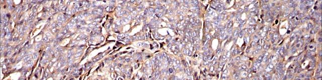
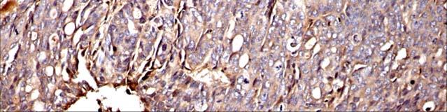
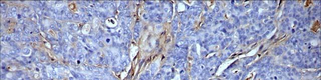
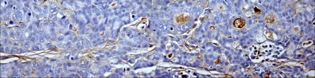
5 Tumorigenicity Assay CD18/HPAF-siMUC4 and CD18/HPAF-Scr cells were harvested and suspended in a normal saline solution at a concentration of 5 x 10 6 cells/ml. Cell viability was examined by trypan blue exclusion assay and single-cell suspensions of >90% viability were used for the injections. Immunodeficient mice were purchased from the Animal Production Area of the National Cancer Institute-Frederick Cancer Research and Development Center (Frederick, MD). The mice were treated in accordance with the Institutional Animal Care and Use Committee (IACUC) guidelines. The orthotopic implantation was performed as previously described (11). All mice were sacrificed on day 21 after implantation, and the presence of metastatic lesions in different organs was determined. Pancreatic tumors were excised, weighed, and their dimensions were measured. Immunohistochemistry The immunohistochemical analysis was performed as previously described (52). Paraffin embedded sections of orthotopic tumors developed by injecting CD18/HPAF-Scr and CD18/HPAF-siMUC4 cells in mice were stained for MUC4 with the specific monoclonal antibody followed by horseradish peroxidase conjugated secondary antibody (ImmPress Reagent Kit, Vector Laboratories) Slides were developed with DAB (Vector Laboratories) and counterstained with hematoxylin. The two panels show the representative images at x100 magnification.
6 Supplementary Figure legends Figure S1. Real-time RT-PCR analysis of MUC4 expression in CD18/HPAF, CD18/HPAF-Scr and CD18/HPAF-siMUC4 cell lines. The level of MUC4 transcript was normalized with that of GAPDH. Bars represents the normalized fold difference in MUC4 transcript expression levels (mean ± SE; n = 3, * P < 0.05). Figure S2. Effect of MUC4 down-regulation on tumorigenicity. Beige nude mice were injected orthotopically with 5X10 5 cells from CD18/HPAF-Scr and CD18/HPAFsiMUC4 cell lines.the mice were sacrificed on day 21 post-injection. Tumors were resected, weighed and their dimensions measured. The mean tumor weight and volume were significantly higher in mice injected with the CD18/HPAF-Scr cells compared to those injected with the CD18/HPAF-siMUC4 cells (weight: vs mg, p=0.02; volume: vs cm 3, p<0.001). Figure S3. Immunohistochemical analysis of primary tumors developed in immunodeficient mice by orthotopic implantation. Paraffin-embedded sections of orthotopic tumors developed by injecting CD18/HPAF-Scr and CD18/HPAF-siMUC4 cells in mice were stained for MUC4 with the specific monoclonal antibody followed by horseradish peroxidase-conjugated secondary antibody. MUC4 expression was decreased in tumor sections derived from mice injected with the CD18/HPAF-siMUC4 cells compared to those from the CD18/HPAF-Scr cells. The panel shows the representative images (x100).
7 Supplementary Table S1. Incidence of metastases of pancreatic tumors developed by orthotopic implantation of CD18/HPAF-Scr and CD18/HPAF-siMUC4 cells in immunodeficient mice. Cell line n Lymph node Liver Lung Spleen CD18/HPAF-Scr 16 16/16 (100%) 6/16 (38%) 3/16 (19%) 3/16 (19%) CD18/HPAF-siMUC4 14 4/14 (26%) 0/14 (0%) 1/14 (7%) 1/14 (7%)