Medical Cytogenetics. Jin Fan, Zhejiang University School of Medicine
|
|
|
- Derek Holland
- 5 years ago
- Views:
Transcription
1 Medical Cytogenetics Jin Fan, Zhejiang University School of Medicine

2 1. Chromatin and chromosome Chromatin in nucleus Euchromatin: Slightly and evenly stained, non- or low-repetitive DNA regions Heterochromatin: Darkly and unevenly stained, highly repetitive DNA regions

3 Chromatin composition Double helix Nucleosome fiber Solenoid Interphase nucleus Chromatin is the basic components in the cell nucleus Composed of DNA, histones and non-histone proteins


4 From Chromatin to Chromosome Different mode of chromatin in metaphase Resulted from highly compaction of chromatin: Cell Cycle

5 Chromosome Preparation Cell culture: Peripheral blood: PHA stimulating Fibroblast from varous cells Bone marrow for leukemia Amniotic fluid cell for fetal diagnosis Colchicine arresting metaphase Harvest a great number of metaphases Hypotonic treatment Chromosome spread preparation Identify each chromosome


6 Telomere Short Arm Chromatids, two after S-phase Centromere Long arm and short arm Telomere Long Arm Chromatid scaffold
7 2. Chromosome identification 1. Morphology of chromosome Length RL: Relative length, Ch L / total L of a haploid set Position of centromere AI: Arm index, Long arm L( q ) / Short arm L ( p ) CI: Centromere index, q / Ch L
8 Metacentric Ch Submetacentric Ch Acrocentric Ch Short Arm Centromere Long Arm Metacentric Ch. CI: 1/2~5/8 Submetacentric Ch. CI: 5/8~7/8 Acrocentric Ch. CI: >7/8
9 International System for Human Cytogenetic Nomenclature ISCN, Denver 1. Rl: from large to small Chromosome length: from long to short 2. CI: from small to large Centromere position: from low to high 3. Variable heterochromatic region: 1qh, 9qh, 16qh, Yq Satellite and satellite arm of acrocentric chromosomes
10 M Subm M Subm A B group Subm C group acroc D M E group F M G acroc Sex Ch. 46, XY Karyotyping, 7 Groups

11 2. Banding of chromosome Q bands: Caspersson (1970) Quinacrine mustard Fluorescence microscopy Bright and dim bands

12 G Bands Giemsa bands: Trypsin digestion Giemsa staining Permanent Dark and light bands
13 R Banding Reversed bands: Heated, KOH Giemsa staining Permanent Dark and light bands reversed to G bands
14 C Bands Heated, KOH Giemsa staining Heterochromatin in the centromeres, long arm of the Y and 1qh, 9qh and 16qh
15 Nomenclature of human chromosome Sister chromatids Zone Short Arm, p Band Centromere heterochromatin Long Arm, q Ch arm zone band Telomere Zones and bands of the chromosme
16 High-resolution bands Chromosomes in different stages of phases of cell cycle More detailed analysis
17 Fluorescence In Situ Hybridization (FISH) Ch. +Probe Co- (denature) (anneal) Using DNA probe labeled with a certain marker Hybridizing with DNA in chromosomes and nuclei on slides Probes hybridized with the fragment in chromosome are detected by signals from the labeled markers

18 Rapid mapping of genes and sequences in chromosome Detecting small fragment in interphase. Detecting cryptic rearrangements or small deletions Banding could not be detected < 4Mb

19 Fragile Sites 46, Y, fra(x)(q27-28) Non-staining gaps that occasionally observer at charcteristic sites on several chromosomes Depend on growth conditions Heritable variants
")


20 Comparative Genomic Hybridization (CGH) Lable test DNA with green Lable normal DNA with red 1:1 mixed Hybrized onto male chromosomal preparation


21 CGH Compare the intensity of two fluorochromes along the chromosome set. Detection of duplication or deletion of chromosomal segment.
22 Array CGH,aCGH

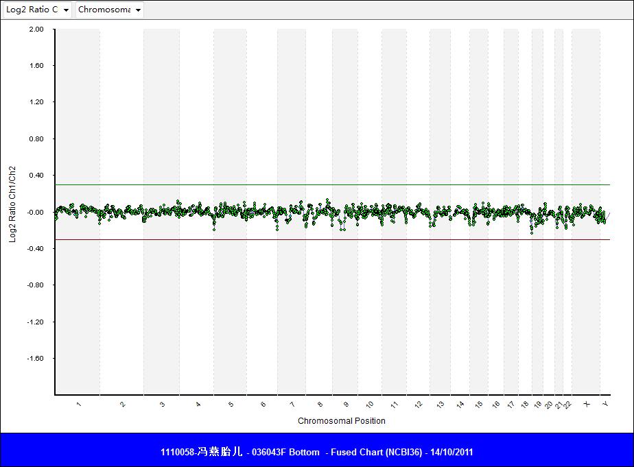
23 Euploid 46,XX 46,XY
24 arr (12)(pter-qter) 1, -12 arr (9)(q12-qter) 3, (15)(q12-qter) 1, der t(12q;15q)
25 3. Chromosome abnormalities (1). Numerical chromosomal abnormalities Heteroploidy: A chromosome complement with chromosome number other than 46 Euploidy: A chromosome complement with an exact multiple of the haploid chromosome number Aneuploidy: A chromosome complement with chromosome number other than an multiple of the haploid chromosome number
26 A. Euploidy: Monoploidy and Polyploidy 1N: Monoploidy 23X or 23Y: in parthenogenesis 3N: Triploidy 69, XXX or 69, XXY in partial mole, aborted fetus and liveborn who does not survive long 4N: Tetraploid 92, XXXX or 92, XXYY in aborted fetus
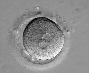
27 Triploidy

28 Triploidy 13-green, 21-red


29 Imprinting: The expression of the phenotype depends on whether the gene or genome inherited from the father or mother. parthenogenesis Pat. diploidy Mat. diploidy Triploidy over-growth of trophoblast over-growth of inner mass cell Two sperm fertilized with a normal oocyte Complete mole Teratomas Partial mole
30 (2). Aneuploidy Loss or gain of chromosomes (not multiple of a haploidy) Monosomy 2n-1: one instead of a pair of homologous chromosomes Trisomy 2n+1: three instead of a pair of homologous chromosomes
 or sister chromatids (mitosis or meiosis")
31 Trisomy Monosomy Resulted from nondisjunction of the homologous chromosomes (meiosis I) or sister chromatids (mitosis or meiosis II).

32 a. Monosomy Almost all monosomy for an entire chromosome is lethal Turner s syndrome: 45,X, the only monosomy can be born and survive 45,X

33 Typical Turner s syndrome Short stature Gonadal dysgenesis: steak gonads Unusual faces, webbed neck, low posterior hairline, broad chest with widely spaced nipples

34 b. Trisomy Trisomy 21, Down s syndrome 47, XY, +21