T-cell response. Taken from NIAID: s.aspx
|
|
|
- Prosper Houston
- 5 years ago
- Views:
Transcription

1 T-cell receptor
2 T-cell response 1. Macrophage or dendritic cell digest antigen bacteria, virus 2. Fragments of Ag bind to major histo-compatiblity (MHC) proteins in macrophage. 3. MHC I-Ag fragment expressed on surface of macrophage 4. T-cell receptor (TCR) protein on the surface of T-cell binds to MHC I-Ag fragments. 5. Binding activates T-cell Cell division à more Ag-specific T-cells Some T-cells bind to bacteria, virus Some T-cells signal to produce more Ag-specific T-cells

3 T-cell response Taken from NIAID: s.aspx

4 T-cell response

5 T-cell receptor
6 Antibodies - Immunoglobulins
7 Immunoglobulins Have to recognize a multitude of different antigens Have to be recognized by the host Bind to phagocytes, T-cells Recognized as self
8 Immunoglobulins
9 Antibody or Immunoglobulin types Immunoglobulin type IgM IgG IgE subtypes IgG1, IgG2a, IgG2b, IgG3, IgG4 function Initial immune response Major Ig for permanent immune response; most research Ab s are this type Mediates allergic reactions IgA, IgD Specialized immune response Taken from:
10 IgM
11 Constant and Variable regions
12 Immunoglobulin structure Antibodies are bivalent two binding sites Heavy (HC) and light chains (LC) separate proteins from separate genes HC and LC are connected by disulphide bonds
13 Immunoglobulin structure Constant region - Similar/same sequence in all Ig s of same type and host species - Recognized by host cells Variable region - Unique sequence for each Ig - Recognizes antigen/epitope
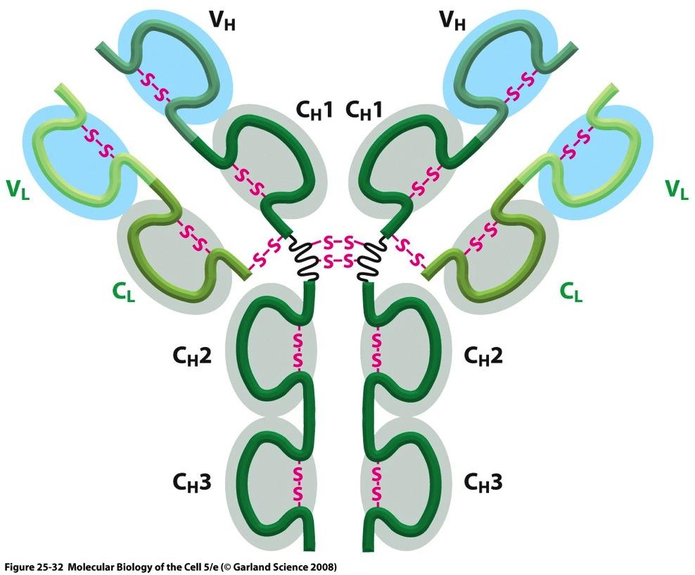
14 Immunoglobulin domains

15 Antibody fragments Fab - Antigen binding Fc fragment - Binds to Fc receptor on cells Digest w/papain

16 Antibody fragments F(ab ) 2 - Antigen binding pfc Digest w/pepsin

17 Selection of heavy and light chains during B-cell development
18 DNA rearrangement produces antibody diversity

19 Immune response
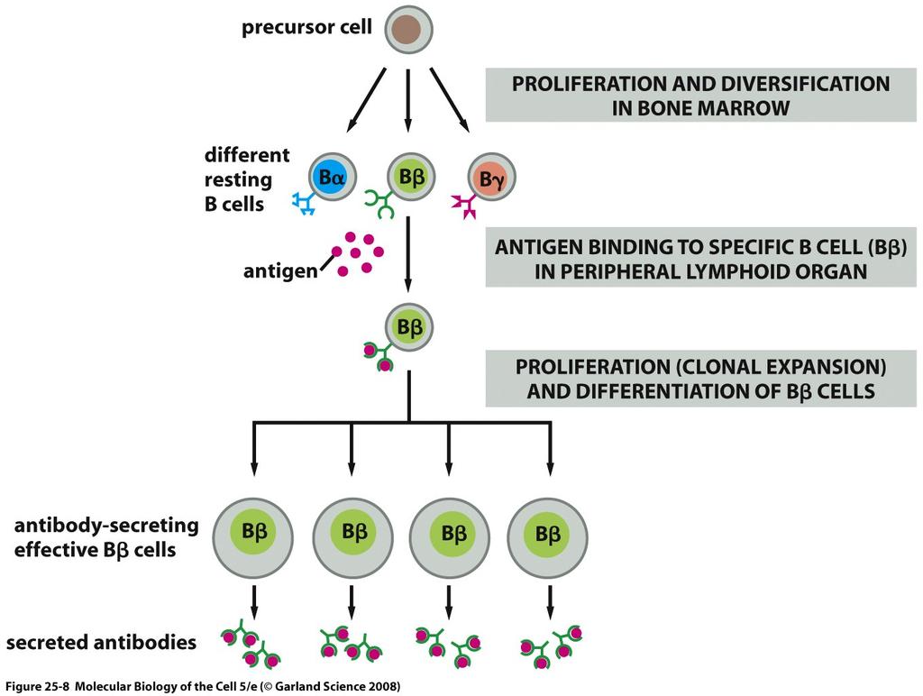

20 Activation of B-cell Memory!
21 Clonal B cells activation activation Monoclonal Monoclonal Polyclonal Monoclonal
22 Polyclonal antibodies Each different B cell generates unique antibody Each antibody may recognize unique antigenic determinant Serum from animal will contain multiple Abs that may recognize multiple determinants antigen


23 Monoclonal antibodies B cell from animal injected with antigen A makes anti-a antibody but does not divide forever. Tumor cell from cell culture divides indefinitely but does not make antibody. FUSE ANTIBOD-SECRETING B CELL WITH TUMOR CELL Hybrid cells called hybridoma cells Hybrid cell makes anti-a antibody and divides indefinitely. Grow hybridoma cells in culture Isolate Ab from media
24 Monoclonal antibodies One type of Ab from a cloned B-cell hybridoma mab, coded by one gene. Use purified mab or culture media from hybridoma cells. Infinite supply of Ab. antigen
25 Polyclonal vs. Monoclonal Abs polyclonal Serum may recognize multiple determinants on single antigen Easier to produce than mabs multiple Abs in sera can give stronger signal in assay Supply limited to lifetime of animal monoclonal Ab recognizes single determinant Extra steps beyond polyclonal Can select high affinity Ab unlimited supply of Ab

26 Western blotting
27 Protein detection Is protein of interest in sample? Compare levels of protein in multiple samples - Size: MW on gel - Activity of protein: assay activity - Specific detection à Ab reactivity Western blot, ELISA
28 Western blot Identify protein by MW and reactivity with Ab. Use antibody to detect protein separated on SDS-PAGE. Make replicate of gel on 2D membrane to allow for contact with Ab.
29 Western blot 1. Run SDS-PAGE 2. Transfer proteins from gel to membrane/paper 3. Bind primary (1 o ) antibodies to proteins on surface of membrane. 4. Bind detecting secondary (2 o ) antibodies to primary antibodies on membrane. 5. Run detection reaction to see where antigen is located.
 GEL transfer (+) MEMBRANE SDS")
30 Protein transferred from gel to membrane Protein is coated with SDS in gel Most of SDS lost during transfer (-) GEL transfer (+) MEMBRANE SDS
31 Transfer membranes Nitrocellulose - Binds protein through electrostatic and hydrophobic interactions. - Binds proteins and nucleic acids - Highly flammable fun! Polyvinylidene Fluoride (PVDF) - Binds through hydrophobic interactions - Stronger than nitrocellulose, less likely to tear
32 Nitrocellulose

33 Nitrocellulose Typical specifications: Mean pore size = 0.2 µm Thickness = mm Image taken without permission from 3 µm


34 Proteins can go through membrane SDS transfer too long MEMBRANE proteins MEMBRANE
35 Primary antibody 1 o Ab Ag membrane
36 Bind enzyme-linked secondary Ab horseradish peroxidase (HRP) 2 o Ab 1 o Ab Ag membrane


37 Add substrate substrate Product + light horseradish peroxidase (HRP) Ag Enzyme converts substrate to product and light membrane

38 Blocking the membrane Non-specific interactions allow Ab to bind to membrane and other proteins 1 o Ab Ag membrane
39 Blocking the membrane Non-specific interactions allow Ab to bind to membrane and other proteins 1 o Ab Ag membrane
40 Ponceau S staining Visualize transferred proteins Determine efficiency of transfer Determine if correct amount of protein was loaded.
41 Ponceau S staining Binds to (+) charged and hydrophobic groups on proteins. Binding is done at low ph to enhance (+) charge on proteins. Binding is reversed (destained) by raising the ph wash in PBS at ph 7.2 Staining procedure does not affect Ab binding if all Ponceau S is removed. Sensitivity ~100 ng (0.1 ug) protein
42 Ponceau S staining procedure 1. Stain in 0.1% Ponceau S in 1% acetic acid 2. Destain membrane with 1% acetic acid 3. Take picture 4. Destain proteins with PBS
43 Blocking the membrane What interactions bind proteins to membrane? Blocking agents - Protein - Lipid - Detergents: Tween-20 Non-fat Milk (casein, low lipid)
44 Non-fat dry milk Major protein is casein - Mixture of four phosphoproteins - Amphipathic protein, forms micelles Small amounts of lipids Combine milk with Tween-20, non-denaturing detergent to form blocking solution.
45 Matching the Ab to the assay Where is the epitope? exposed on surface buried inside protein (need to denature) Denatured, partial denatured antigen Western blot Native antigen ELISA, immunoprecipitation, immunofluorescence, immunocytochemistry
46 Choosing primary Ab Must react with Ag in a partially denatured form with high affinity. Must react with Ag form species being studied. Must not react significantly with other proteins. Need enzyme-linked 2 o Ab that recognizes 1 o Ab.
47 Will 1 o Ab react with Ag? Will it react with species of interest? Does it work for Western blots? - What info can you get? - What is the epitope? - Is the epitope conserved in species of interest?
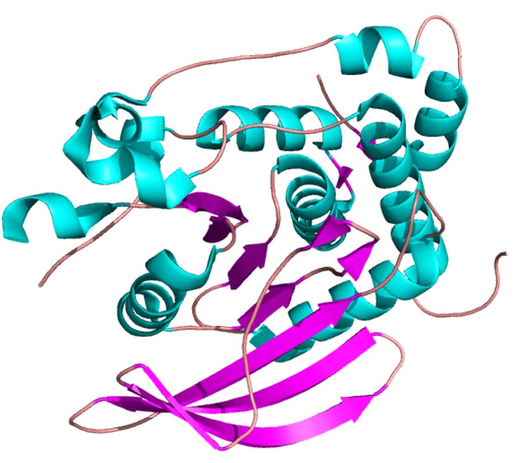
48 FG6: Ab to PTP1B Mouse monoclonal raised against human PTP1B Epitope is somewhere in the catalytic domain aa Works on Western blot, ELISA, IP Reactive with: human, rat, guinea pig, birds, mouse (weak) Where is our PTP1B from?
49 PTP1B catalytic phosphatase domain ER FG6 epitope?
50 Proteolysis of PTP1B in E. coli BL21 stain of E. coli deficient in Deficient in proteases Lon and OmpT E. coli contains other proteases. Recombinant protein partially degraded by E. coli proteases Single cut of one or more peptide bonds Proteases cut at specific sequences or amino acids
51
52 Proteolysis of PTP1B catalytic phosphatase domain 1 321
53 What is interacting? Proteins interact with membrane Proteins interact with proteins Antibodies Specific interactions Antibody Antigen Non-specific interactions Protein with membrane Want all proteins to bind equally well Why? Protein with other proteins

54 Types of interactions - proteins Ionic charge interactions Hydrogen bond Hydrophobic interactions
55 How does protein bind to membrane? Ionic charge interactions Hydrogen bond Hydrophobic interactions proteins unfolded Nitrocellulose
56 How do proteins bind to proteins? Specific interactions antibody-antigen Non-specific interactions Non-Specific Specific
57 Antibody-antigen binding Antigen binding is an equilibrium reaction low [Ab] Ab + Ag Ab-Ag (low) high [Ab] Ab + Ag Ab-Ag (high) [Ab-Ag] [Ab]
58 Antibody non-specific binding Non-specific binding is an equilibrium reaction low [Ab] Ab + Protein/membrane high [Ab] Ab + Protein/membrane very high [Ab] Ab + Protein/membrane Ab-Protein/membrane Ab-Protein/membrane Ab-Protein/membrane
59 How do we ensure specific interactions? ph ( ) salt (NaCl, mm) Amount of antibody Ab + Ag Ab-Ag reduce non-specific interactions

60 How do we limit non-specific interactions? Block hydrophobic interactions Tween-20 Proteins in milk casein - hydrophobic Block electrostatic interactions NaCl ions Proteins in milk Amount of antibody Ab + Protein/membrane Ab-Protein/membrane


61 Chemiluminescence Enzyme-substrate reaction gives off light energy. Detect light with overlaid x-ray film Side view x-ray film light membrane

62 Chemiluminescence 200 kda 116 kda 45 kda