Apolipoprotein Modulation of Aβ Deposition in a Transgenic Mouse Model of Alzheimer s Disease. Ronald B. DeMattos
|
|
|
- Holly Rodgers
- 5 years ago
- Views:
Transcription
1 Apolipoprotein Modulation of Aβ Deposition in a Transgenic Mouse Model of Alzheimer s Disease Ronald B. DeMattos Neuroscience Discovery Research at Eli Lilly
 Mouse")


2 APP V717F (PDAPP) Mouse Model of AD Human Cortex Transgenic Mouse **Aβ Immunostaining**

3 Human APP V717F Tissue Expression Liver Spleen Kidney Lung Sm. Intest. Heart Muscle Skin Cortex Hippocampus CSF 196 kda 125 kda 89 kda 6E10 Western Blot

4 Isolation of Murine CSF from Cisterna Magna
5 Soluble Aβ Total Concentrations in Human and Transgenic Mice Cerebrospinal fluid Plasma (Aβ Total ) (Aβ Total ) Human 14.9 ng/ml pg/ml 2 PDAPP 3 month old 19.4 ng/ml pg/ml 3 PDAPP 9 month old 14.1 ng/ml pg/ml 3 Tg month old ~56.0 ng/ml 17* ~19,000 pg/ml 17* *The Aβ Total levels shown were derived by the addition of the Aβ 40 and Aβ 42 values reported. 1. Matsubara E, Ghiso J, Frangione B, et al. Ann. Neurol. 1999;45: Lannfelt L, Basun H, Vigo-Pelfrey C, et al. Neuroscience Letters 1995;199: DeMattos RB, Bales KR, Parsadanian M, et al. J. Neurochem. 2002;81: Kawarabayashi T, Younkin LH, Saido TC, et al. J. Neurosci. 2001;21:
6 34 kda glycoprotein Apolipoprotein E produced in high levels in liver and brain synthesized by glia in brain (predominantly astrocytes) present in lipoprotein particles in the CNS which are HDL-like exists in 3 common isoforms in humans E2 cys cys E3 cys arg E4 arg arg
7 ApoE and Alzheimer s Disease 1. Epsilon 4 allele of Apolipoprotein E is a genetic risk factor for Alzheimer s disease and cerebral amyloid angiopathy Strittmatter et. al.proc Natl Acad Sci U S A Mar 1;90(5): Saunders et. al. Neurology Aug;43(8): Corder et. al. Science Aug 13;261(5123):921-3 Rebck et al. Neuron Oct;11(4): Increased levels of cortical and vascular Aβ in AD patients with an E4 isoform Schmechel et. al. Proc Natl Acad Sci U S A Oct 15;90(20): In vitro findings demonstrate numerous apoe isoform specific interactions with Aβ as well as CNS cells Strittmatter et. al.proc Natl Acad Sci U S A Mar 1;90(5): LaDu et. al. J Biol Chem Sep 23;269(38): Puttfarcken et. al. J Neurochem Feb;68(2):760-9 DeMattos et. al. J Biol Chem Feb 13;273(7):

8 Murine apoe facilitates fibrillar Aβ (amyloid) formation and toxicity PDAPP, apoe +/+ PDAPP, apoe -/- Aβ stain thioflavine-s stain for fibrillar Aβ One year of age Bales et al., Nat. Genet., 1997; Holtzman et al, PNAS, 2000
9 Effects of human apoe isoforms on Aβ-related pathology in APP transgenic mice Transgenic Breeding Scheme PDAPP +/-, mapoe -/- (Games et al, 1995; Bales et al., 1997) GFAP-ApoE2, E3 or E4 +/- mapoe -/- (Sun et al, 1998) 1. PDAPP +/- mapoe +/+ 2. PDAPP +/- mapoe -/- 3. PDAPP +/- mapoe -/-, ApoE2 +/- 4. PDAPP +/- mapoe -/-, ApoE3 +/- 5. PDAPP +/- mapoe -/-, ApoE4 +/-
10 Hippocampal Aβ levels are suppressed in PDAPP mice expressing human apoe isoforms apoe3 > apoe4 Log Aβ Total x10-5 (pg/gm tissue) 4 mapoe ApoE (-/-) ApoE4 ApoE age (months) Holtzman et al., JCI, 1999 Holtzman et al., PNAS, 2000 Fagan et al., Neurob. Dis. 2002
11 ApoE Hypothesis Hypothesis: Human apoe isoforms influence Aβ deposition in vivo via the following mechanisms: 1. Human apoe isoforms facilitate clearance of the beta amyloid peptide from the brain. (Endocytic and/or chaperone to the plasma compartment) 2. At a critical beta-amyloid concentration, apoe will induce fibril formation.
12 ApoE/Aβ Therapeutics Main Question: Do we want to increase or decrease expression of human apoe in brain with regard to Aβ deposition and related pathology? * Is more human apoe good or bad? - Isoform differences? - Time differences?
13 Animals Analyzed (All animals are Homozygous for the PDAPP transgene) Mouse apoe Knock-out (eko) Heterozygous for GFAP-apoE3 transgene (E3+/-) Homozygous for GFAP-apoE3 transgene (E3+/+)
14 ApoE KO and human ApoE Transgenic mice were analyzed at multiple ages Analysis 3 Months 6 Months 12 Months 15 Months -CSF and Plasma -½ Brain for Biochemistry (hippocampus and cortex) -½ Brain for Histochemistry
15 Sample Analysis CSF Plasma Hippocampus Cortex Fix Brain CSF Aβ40 CSF Aβ42 Plasma Aβ40 Plasma Aβ42 Soluble Aβ40 Soluble Aβ42 Soluble AβTotal Soluble Aβ40 Soluble Aβ42 Soluble AβTotal Pan Aβ Thio-S Silver Soluble Oligo Aβ Soluble Oligo Aβ Insoluble Aβ40 Insoluble Aβ40 Insoluble Aβ42 Insoluble Aβ42 *Serial extraction procedure: carbonate soluble and then guanidine extraction (insoluble)
16 Hippocampal Aβ Load in Month Old PDAPP Mice: apoe3 decreases Aβ deposition in a dose-dependent fashion 30 % Aβ Load E3+/+ E3+/- eko p < (ANOVA)
17 Frequency Distribution of Hippocampal Aβ load in PDAPP,ApoE3+/+ Expressing Mice Percent of Animals % Aβ Load Total N =
18 Distribution of Hippocampal Aβ Load in ApoE3+/- and mapoe KO Mice Percent of Animals Percent of Animals Percent Aβ Load Percent Aβ Load Total N = 37 Total N = 35
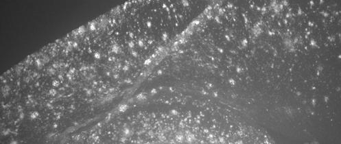
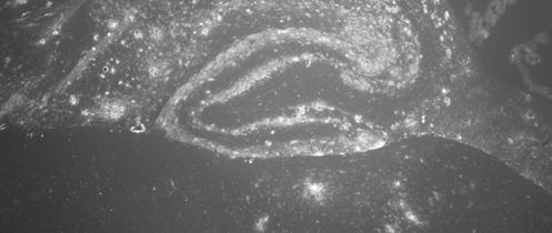
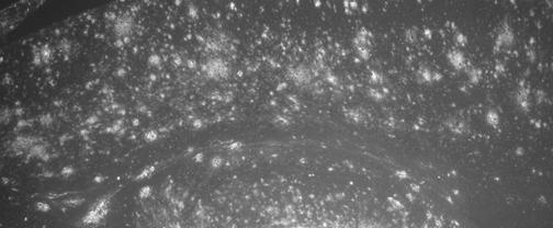
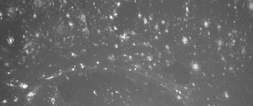
19 ApoE3 decreases percent Aβ load in a dosedependent fashion in PDAPP mice at 12 to 15 months of age ApoE3+/+ ApoE3+/- mapoe KO Aβ Immunostaining
20 Are there differences in Aβ metabolism prior to deposition in PDAPP mice expressing varying levels of apoe3 that may underlie the variation in the amount of Aβ deposition seen at later ages?
21 Analysis of soluble Aβ in young (3 to 6 month old) transgenic animals CSF Aβ40 CSF Aβ Aβ40 (pg/ml) Aβ42 (pg/ml) E3+/+ E3+/- eko 0 E3+/+ E3+/- eko p = p = (ANOVA) (ANOVA)
22 Analysis of soluble Aβ in young (3 to 6 month old) transgenic animals Plasma Aβ40 Plasma Aβ Aβ40 (pg/ml) Aβ42 (pg/ml) E3+/+ E3+/- eko 0 E3+/+ E3+/- eko
23 6MC MW MPJ+/ Abeta40NumberofvaluesMinimum25%PercentileMedian75%PercentileMaximumMeanStd.DeviationStd.Eror12MPEJ-/ M MC MW MPJ+/ Abeta40NumberofvaluesMinimum25%PercentileMedian75%PercentileMaximumMeanStd.DeviationStd.Eror12MPEJ-/ M MC MW MPJ+/ Abeta42NumberofvaluesMinimum25%PercentileMedian75%PercentileMaximumMeanStd.DeviationStd.Eror12MPEJ-/ MC Analysis of soluble Aβ in young 3 to 6 month old PDAPP mice: ApoE3 decreases the CSF:plasma Aβ42 ratio CSF:Plasma Aβ40 CSF:Plasma Aβ Aβ40 Ratio Aβ42 Ratio E3+/+ E3+/- eko 0 E3+/+ E3+/- eko p = (ANOVA)
24 Analysis of carbonate soluble hippocampal Aβ in 3 6 month old PDAPP mice: ApoE3 decreases Aβ Soluble Aβ40 Soluble Aβ Aβ40 (ng/mg) Aβ42 (ng/mg) E3+/+ E3+/- eko 0.0 E3+/+ E3+/- eko p = p = (ANOVA) (ANOVA)
25 0.0138*Yes ValueDataSet-Bq DataSet-CPvalueP>0.05P<0.05P>0.05 ParameterTukey'sMultipleComparisonTest3M40INSvs3MC40INS3M40INSvs3MW40INS3MC40INSvs3MW40INS MeanDif TableAnalyzedDataTable-1One-wayanalysisofvariancePvaluePvaluesumaryAremeansignif.diferent?(P<0.05)NumberofgroupsFRsquared ValueDataSet-Bq DataSet-CPvalueP>0.05P<0.01P<0.01 P<0.01*Yes MeanDif TableAnalyzedDataTable-1One-wayanalysisofvariancePvaluePvaluesumaryAremeansignif.diferent?(P<0.05)NumberofgroupsFRsquared ParameterMultipleComparisonTest3M40Svs3MC40S3M40Svs3Mw40S3MC40Svs3Mw40S nsNo TableAnalyzedDataTable-1One-wayanalysisofvariancePvaluePvaluesumaryAremeansignif.diferent?(P<0.05)NumberofgroupsFRsquared nsNo TableAnalyzedDataTable-1One-wayanalysisofvariancePvaluePvaluesumaryAremeansignif.diferent?(P<0.05)NumberofgroupsFRsquared P<0.01*Yes ValueDataSet-Bq DataSet-CPvalueP>0.05P<0.01P<0.01 TableAnalyzedDataTable-1One-wayanalysisofvariancePvaluePvaluesumaryAremeansignif.diferent?(P<0.05)NumberofgroupsFRsquared ParameterTukey'sMultipleComparisonTest3M42INSvs3MC42INS3M42INSvs3MW42INS3MC42INSvs3MW42INS MeanDif Analysis of carbonate insoluble hippocampal Aβ in 3 6 month old PDAPP mice: ApoE3 decreases Aβ Insoluble Aβ40 Insoluble Aβ Aβ40 (ng/mg) Aβ42 (ng/mg) E3+/+ E3+/- eko 0.0 E3+/+ E3+/- eko p = p = (ANOVA) (ANOVA)
26 Summary 1. In order to understand the mechanisms underlying the effects of human apoe on Aβ metabolism and AD risk, it is critical to assess the Aβ levels in different compartments both prior to and after amyloid deposition. 2. We identified an apoe3 gene dose effect for Aβ deposition: E3-/- > E3+/- > E3+/+ 3. The gene dose effect can be identified for soluble and insoluble Aβ concentrations in multiple CNS compartments in young pre-depositing PDAPP mice. 4. These results support the hypothesis that apoe3 facilitates CNS Aβ clearance.
27 Question: How can we therapeutically increase the expression of ApoE? Goal: To pharmacologically increase apoe expression/secretion in CNS by astrocytes.
28 LXR/RXR Heterodimer Regulates ApoE Expression in CCF-STTG1 Cells Medium Cell Lysate DMSO 9-cis-RA 22-R-CHL T (nm) Liang, Y. et al J. Neurochem 88: , 2004
29 In Vivo Regulation of ApoE in Hippocampus Western Blot Analysis _ T (mg/kg) Apo E 1.8 GFA P Hippocampus * * ** Apo E/GFAP T (mg/kg)
30 Does Acute Increases in ApoE Levels Modify CNS Aβ Metabolism? (7 Day Acute apoe3 TR-PDAPP +/- Study) Mice: 3 month apoe3 +/+ -PDAPP +/- Treatment: (7 day treatment): 12 Vehicle 12 T mg/kg Live Phase: - Oral dosing daily - CSF and Plasma isolated - Hippocampus, cortex, and cerebellum from both hemispheres were dissected Sample Analysis: - Samples processed all at same time - CSF and plasma ELISA s: Aβ 40, Aβ 42, and Aβ Total - Tissue samples: serial extraction (PBS, carbonate, and guan.) - Tissue ELISA s: Aβ 40 and Aβ 42 - Human apoe levels via ELISA
31 7 Day Acute ApoE3 TR-PDAPP +/- Study Carbonate Extraction Cortex Aβ40 (pg/mg tissue) * Aβ42 (pg/mg tissue) ** 0.0 Con Ctx Carb 42 LY Ctx Carb 42 Control T- 50mg/kg Control T-50mg/kg Con Ctx Carb 40 LY Ctx Carb 40 p = p = Carbonate Extraction Hippocampus Aβ40 (pg/mg tissue) Aβ42 (pg/mg tissue) ** 0 Con Hip Carb 42 LY Hip Carb 42 Control T- 50mg/kg Control T-50mg/kg Con Hip Carb 40 LY Hip Carb p =
32 7 Day Acute ApoE3 TR-PDAPP +/- Study Guan Extraction Cortex Aβ40 (pg/mg tissue) Aβ42 (pg/mg tissue) ** 0 Con Ctx Guan 42 LY Ctx Guan 42 Control T- LY 50mg/kg Control T-50mg/kg Con Ctx Guan 40 LY Ctx Guan p = Guan Extraction Hippocampus Aβ40 (pg/mg tissue) Aβ42 (pg/mg tissue) * 0.0 Con Hip Guan 42 LY Hip Guan 42 Control T- LY 50mg/kg Control T-50mg/kg p = Con Hip Guan 40 LY Hip Guan 40 0

33 In-vitro Assay: Removal of Aβ by Exogenous Astrocytes Incubation Chamber Cell suspension of 5x10 5 adult astrocytes/ml Incubate 24 hours at 37 o C poly-l-lysinecoated coverslip Phase-contrast imaging and Quantitative IHC for Abeta, GFAP and DAPI stained nuclei Frozen PDAPP mouse brain section Koistinaho et al. Nature Medicine 2004; 10 (7):


34 ApoE-Dependent Degradation of Aβ in Brain Slices by Astrocytes medium only adult WT adult apoe -/- neonatal WT Koistinaho et al. Nature Medicine 2004; 10 (7):
35 ApoE-Dependent Degradation of Aβ in Brain Slices by Astrocytes ImmunoHistochemistry ELISA 30 % Aβ burden medium only *** adult WT adult neonatal apoe -/- WT Aβ x-42 ng/section medium only *** adult WT adult apoe -/- Koistinaho et al. Nature Medicine 2004; 10 (7):
36 ApoE-Dependent Degradation of Aβ by Astrocytes Control + WT Astrocytes α-apoe + WT Astrocytes * * % Aβ burden α-apoe, no cells medium, WT cells IgG, WT cells α-apoe, WT cells Koistinaho et al. Nature Medicine 2004; 10 (7):


37 RAP Blocks Astrocyte-Mediated Aβ Degradation % Control *** Medium only adult WT RAP adult WT # Receptor Associated Protein *39 kda intracellular chaperone which assists with protein folding *When added exogenously effective (200nM-1mM) antagonist of known LDLR receptors Koistinaho et al. Nature Medicine 2004; 10 (7):

38 Multicellular Astrocyte Aggregates Form in Response to Aβ Deposits In Vitro Koistinaho et al. Nature Medicine 2004; 10 (7):

39 HOHO HOHO HOHO HOHO Cerebral Spinal Fluid Aβ Binding Proteins Brain HO HO O C O C HO HO O C O C Plaque Interstitial Fluid Aβ HO HO O C O C Plasma HO HO O C O C
40 Acknowledgments Eli Lilly Steven Paul Kelly Bales JC Dodart Patrick May Bruce Gitter Guoqing Cao Peggy Racke Matt Bryan Cindy Delong Washington University David Holtzman Maia Parsadanian Mark O Dell Jennie Taylor John Cirrito John Fryer Rich Hartman Bob Brendza
41 Replacement of ApoE3 Particles Restores Aβ Degradation ** 60 *** Burden (% Control) M edium WT ApoE KO ApoE3 (1ug/m l) ApoE3 (10ug/ml)