Non-standard base pairs Non-standard base pairs play critical roles in the varied structures observed in DNA and RNA.
|
|
|
- Gervais Kennedy
- 5 years ago
- Views:
Transcription
1 DNA ORIENTATION


2 Non-standard base pairs Non-standard base pairs play critical roles in the varied structures observed in DNA and RNA.

3 Non-standard base pairs Wobble and mismatched base pairs still use the Watson-Crick edges for hydrogen bonding. Wobble base pairing is seen in mismatches between G-T and G-U base pairs incorporated into DNA and DNA:RNA complexes and play essential roles in the fidelity of DNA replication and transcription. Such mismatches can lead to genome mutations if not accurately detected and corrected by the proof reading activity of DNA polymerase during replication, or post-replicative repair systems. Studies suggest that G-T and A+-C are the most frequent mismatches that cause point mutations in cells.

4 Non-standard base pairs Hoogsteen base pairs take advantage of the Hoogsteen edge of a purine base, which is orthogonal to and, thus, can be accessed without disrupting the Watson-Crick base pairing edge. Consequently, Hoogsteen interactions allow the assembly of multi-stranded DNA complexes, including triplet helixes and G-quadruplexes.
5 Solvent effects As with any biological molecule, solvent interactions directly influence DNA structure and function. Base pairing and stacking are in part stabilized by the hydrophobic effect. Solvent (water and salts) induces base pairs to stack and defines the effective charge of the phosphoribose backbone. Even base pairing is affected by solvent interactions. Numerous hydrogen bond donor and acceptor groups of the backbone and bases must be hydrated. In a G-T wobble base pair, for example, the number of hydrogen bonds between the bases is reduced by one; however, bridging water molecules help to compensate for this loss.
6 Solvent effects Similarly, there are well-defined waters lining the minor groove of B-DNA duplexes (the so-called spine of hydration ) that exchange slowly with the bulk and, therefore, are considered to be integral parts of DNA. In order to minimize the opposing repulsion between the phosphates of the DNA strands, cations help to mitigate the negative charges of the phosphoribose backbone
7 The phosphoribose backbone helps to define the conformation of DNA via the conformation of the deoxyribose sugar The conformation of any polymer is defined by the rotations about each freely rotating chemical bond We can define three categories of bonds: 1) those of the phosphodiester holding two nucleotides together, 2) those within the five-membered ring of the deoxyribose sugar, 3) and the bond holding the nucleotide base to the sugar.
8 Conformations of the deoxyribose sugar
9 Conformations of the deoxyribose sugar the phosphodiester holding two nucleotides together the phosphodiester holding two nucleotides together
10 Conformations of the deoxyribose sugar the five-membered ring of the deoxyribose sugar

11 Conformations of the deoxyribose sugar the bond holding the nucleotide base to the sugar
12 Conformations of the deoxyribose sugar Sugars with atoms puckered above the reference plane (on the same side as the base) are in an endo-form while a pucker that places an atom below this plane is in its exo-form The two general classes of sugar conformations commonly seen in DNA are the C2 -endo and C3 -endo puckers


13 Conformations of the deoxyribose sugar The two conformations have profound effects on the overall DNA conformation in that they specify different phosphate- phosphate distances along each strand (~7 Å for C2 -endo and ~6 Å for C3 -endo). Thus, conformations constructed with C3 -endo sugars will require higher concentrations of salts to counter balance the shorter distance between the negatively charged phosphates.
14 Conformations of the glycosidic bond The base of each nucleotide is attached via the glycosidic bond from the N1 nitrogen of pyrimidines or the N9 nitrogen of purines to the C1 -carbon of the deoxyribose sugar.

, with the base essentially lying on top of the")
15 Conformations of the glycosidic bond Rotation about the glycosidic bond defines χ- angles for the anti- and syn-conformations of the bases The anti conformation (+90 χ +180 ), with the base extended away from the sugar, The syn conformation (-90 χ +90 ), with the base essentially lying on top of the sugar ring
16 Conformations of the glycosidic bond The more compact syn-conformation is more susceptible to steric clashes than the extended anti-form. Purines will more readily adopt the compact syn-conformation than pyrimidines, because of reduced steric collisions. PYRIMIDINES PURINES Similarly, the syn conformation is less sterically hindered when the sugar is puckered as C3 -endo than C2 -endo. The interplay between sugar puckers and χ-rotations can have profound effects on the structures of DNA and the sequence dependence for their formation.
17 Helical parameters Base Pair and Base Step Parameters Translational and rotational relationships of bases within each base pair
18 Helical parameters Base Pair and Base Step Parameters Translational and rotational relationsips between two stacked base pairs
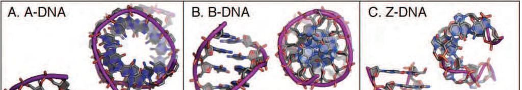

19 Alternative forms of the DNA double helix
20 B-DNA: The Structure of DNA Alternative forms of the DNA double helix B-DNA (Watson-Crick DNA) The predominant form of DNA in vivo is B- DNA, since the inside of a cell is mostly water with a relatively low salt concentration. highly variable and malleable
21 B-DNA: The Structure of DNA Alternative forms of the DNA double helix is a right-handed, antiparallel double-helix in which the Watson-Crick base pairs are stacked directly along and perpendicular to the helical axis, giving rise to major and minor grooves that are similar in depth. The bases are all in the anti-conformation with a majority of deoxyribose sugars in the C2 -endo form, although the sugar puckers are more variable than in many other conformations. The structure is highly variable depending on the sequence and on different environmental conditions.
22 Alternative forms of the DNA double helix A-DNA: is a right-handed antiparallel helical duplex, but is characterized as an underwound structure that is more compact along the helix axis and broader overall across the helix relative to B-DNA. The nucleotide bases, all anti, are shifted by large x displacements towards the minor groove, creating a shallow, wide minor groove and a channel associated with a deep, narrow major groove. The deoxyribose sugars are consistently C3 -endo, which minimizes the potential steric clashes as the sugar is pushed towards the phosphate to accommodate the sliding of the base
23 Alternative forms of the DNA double helix Z-DNA: Z-form DNA is the only characterized lefthanded form of the double-helix. The zig-zagged backbone results from the alternation between syn- and anticonformations, and the respective C3 -endo and C2 -endo sugar puckers. This alternating conformation imposes a sequence preference for alternating purine-pyrimidines, since purines adopt the syn-conformation more readily than do pyrimidines. Thus, the repeating unit is the dinucleotide rather than a single base pair, as in B-DNA.
24 Alternative forms of the DNA double helix Z-DNA: The biological function of Z-DNA has been widely debated and underappreciated; however, several cellular functions for the Z-form are now supported by experimental evidence. Z-DNA was initially characterized as a structure induced by high salt conditions (3 M NaCl). Subsequently, it has been shown that cytosine methylation, and other cations such as spermine and spermidine at millimolar concentrations also stabilize Z-DNA. Most importantly, as a left-handed structure, Z-DNA is the most underwound form of the double-helix and, consequently, serves as a sink for the torsional tension in negatively supercoiled DNA.