Biochemistry and Cell Biology
|
|
|
- Egbert Webster
- 5 years ago
- Views:
Transcription
1 Biochemistry and Cell Biology
2 Monomersare simple molecules that can be linked into chains. (Nucleotides, Amino acids, monosaccharides) Polymersare the long chains of monomers. (DNA, RNA, Cellulose, Protein, Glycogen) Polymers are the most common forms of macromolecules in the body Monomersare hooked together to form polymers through a process called a condensation reaction. These reactions require a H to be removed from one monomer and an OH from another monomer to form water.
3
4
5 Sugars are monomers that consist of ofcarbon atoms, each of which has a hydrogen on one side and an OH group on the other. Polysaccharidesare long chains of sugars that are covalently joined together Starchesare basically super polysaccharides. They are huge polymers of simple sugars. Carbohydratesare a term that refer to polysaccharides, starches, and monosacharides(as long as they re digestible) Nucleic Acids-includes both RNA and DNA, both are made up of nucleotides
 Pentoses (C 5 H 10 O 5 )")

6 Fig. 5-3a Trioses (C 3 H 6 O 3 ) Pentoses (C 5 H 10 O 5 ) Hexoses (C 6 H 12 O 6 ) Glyceraldehyde Ribose Glucose Galactose
 Dehydration reaction in the synthesis of")
7 Fig glycosidic linkage Glucose Glucose (a) Dehydration reaction in the synthesis of maltose Maltose 1 2 glycosidic linkage Glucose Fructose Sucrose (b) Dehydration reaction in the synthesis of sucrose

")
8 Fig. 5-6 Chloroplast Starch Mitochondria Glycogen granules 0.5 µm 1 µm Amylose Glycogen Amylopectin (a) Starch: a plant polysaccharide (b) Glycogen: an animal polysaccharide


9 Fig. 5-7bc (b) Starch: 1 4 linkage of glucose monomers (c) Cellulose: 1 4 linkage of glucose monomers
10 Nucleotides-These are the monomers that are used to make DNA and RNA. Nucleotides all contain 1 sugar, 1 phosphate, and 1 nitrogenous base. In DNA, the sugar is deoxyribose sugar and the potential bases are Adenine, Thymine, Cytosine, and Guanine. DNA is a double stranded complimentary molecule In RNA, the sugar is ribose sugar and the potential bases are Adenine, Uracil, Cytosine, and Guanine. RNA is a single stranded polymer. DNA being double stranded has the ability to act as a template for making a new strand
 Thymine (T, in DNA) Uracil")
 (c) Nucleoside components: nitrogenous")
11 Fig. 5-27c-1 Nitrogenous bases Pyrimidines Cytosine (C) Thymine (T, in DNA) Uracil (U, in RNA) Purines Adenine (A) Guanine (G) (c) Nucleoside components: nitrogenous bases
 (c) Nucleoside components:")
12 Fig. 5-27c-2 Sugars Deoxyribose (in DNA) Ribose (in RNA) (c) Nucleoside components: sugars
 Thymine (T, in DNA) Uracil (U, in RNA) Purines 5C 3C Phosphate group")
 Ribose (in RNA) (c) Nucleoside")
13 Fig C 5 end Nitrogenous bases Pyrimidines 3C Nucleoside Nitrogenous base Cytosine (C) Thymine (T, in DNA) Uracil (U, in RNA) Purines 5C 3C Phosphate group (b) Nucleotide Sugar (pentose) Adenine (A) Guanine (G) 3 end Sugars (a) Polynucleotide, or nucleic acid Deoxyribose (in DNA) Ribose (in RNA) (c) Nucleoside components: sugars
 3' end Old")

14 Fig ' end 3' end Sugar-phosphate backbones Base pair (joined by hydrogen bonding) 3' end Old strands Nucleotide about to be added to a new strand 5' end New strands 3' end 5' end 5' end 3' end
15 RNAis a single stranded polymer and can act as a coding molecule or as an enzyme. This means that RNA was probably the first genetic molecule and the first enzyme. Proteinsare the most common base chemical for enzymes, they are made from Amino acids that are put together in a very specific sequence that is coded for by RNAs that were made from the code in DNA. Proteinshave a Nitrogen and Carbon spine with varying side chains that determine their behavior. Amino Acidsare the monomers of protein
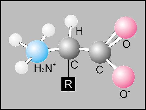
16 Amino Acidsall have the same structure with the exception of the R group. There are two carbons and a nitrogen atom acting as the base of the molecule. R groupsare functional groups that determine amino acid solubility, secondary structure, polarity, and covalent bonding capacity.
17
 Polar Serine (Ser or S) Threonine (Thr or T) Cysteine (Cys or C) Tyrosine (Tyr or Y) Asparagine (Asn or N) Glutamine (Gln or Q) Acidic Electrically charged Basic Aspartic acid (Asp or D)")
18 Fig Nonpolar Glycine (Gly or G) Alanine (Ala or A) Valine (Val or V) Leucine (Leu or L) Isoleucine (Ile or Ι) Methionine (Met or M) Phenylalanine (Phe or F) Trypotphan (Trp or W) Proline (Pro or P) Polar Serine (Ser or S) Threonine (Thr or T) Cysteine (Cys or C) Tyrosine (Tyr or Y) Asparagine (Asn or N) Glutamine (Gln or Q) Acidic Electrically charged Basic Aspartic acid (Asp or D) Glutamic acid (Glu or E) Lysine (Lys or K) Arginine (Arg or R) Histidine (His or H)
 Valine (Val or V) Leucine (Leu or L)")
 Tryptophan (Trp or W)")
19 Fig. 5-17a Nonpolar Glycine (Gly or G) Alanine (Ala or A) Valine (Val or V) Leucine (Leu or L) Isoleucine (Ile or Ι) Methionine (Met or M) Phenylalanine (Phe or F) Tryptophan (Trp or W) Proline (Pro or P)
 Tyrosine (Tyr or Y) Asparagine (Asn or N) Glutamine (Gln")
20 Fig. 5-17b Polar Serine (Ser or S) Threonine (Thr or T) Cysteine (Cys or C) Tyrosine (Tyr or Y) Asparagine (Asn or N) Glutamine (Gln or Q)
 Lysine (Lys or K) Arginine (Arg or R) Histidine")
21 Fig. 5-17c Acidic Electrically charged Basic Aspartic acid (Asp or D) Glutamic acid (Glu or E) Lysine (Lys or K) Arginine (Arg or R) Histidine (His or H)
22 All Eukaryotes have the following organelles and structures. Nucleus Nuclear envelope-a membrane that contains the DNA and the proteins necessary to organize and maintain the DNA Chromatin-DNA and Protein that is found unwound in a cell between divisions. Chromosomes-condensed form of chromatin these are linear and found during mitosis. Nucleolus-area of the nucleus thought to be used to assemble ribosomes. Cytosol Ribosomes-RNA and protein complex that work together to read mrna and build a protein from its code.


23 Fig µm Nuclear envelope: Inner membrane Outer membrane Nucleolus Chromatin Nucleus Nuclear pore Pore complex Surface of nuclear envelope 0.25 µm Ribosome Rough ER 1 µm Close-up of nuclear envelope Pore complexes (TEM) Nuclear lamina (TEM)
24 Organelles are membrane bound, specially designed and tasked parts of cells Endoplasmic Reticulum-network of membrane found in the cell along side the nucleus, makes lipid and protein components of the membrane and materials for export from the cell. Rough ER looks grainy because it has ribosomes embedded in its membrane. Produces membrane bound proteins and proteins for export. Smooth ER is the side of the ER away from the nucleus this is the ER responsible for lipid synthesis and detoxification often refines or modifies products from rough ER

25 Fig Smooth ER Rough ER Nuclear envelope ER lumen Cisternae Ribosomes Transport vesicle Smooth ER Rough ER Transitional ER 200 nm
26 Golgi Apparatus-Acts as a processing center for products from the ER. May modify some chemicals, while just sorting and packaging others for storage or release. Lysosomes are membrane bags of hydrolytic enzymes. Lysosomes keep these dangerous chemicals separate from the rest of the cell s chemicals and concentrated. They bind with food vacuoles to start the digestions of materials that have been consumed. Vacuoles-mean membrane bag and is used to refer to contractile vacuoles that pump out extra water, food vacuoles, and central vacuoles.
 TEM of Golgi")
27 Fig cis face ( receiving side of Golgi apparatus) Cisternae 0.1 µm trans face ( shipping side of Golgi apparatus) TEM of Golgi apparatus
28 Mitochondria-the power house of the cell. This is the organelle that takes glucose or other chemical energy sources and converts them into ATP. Mitochondria have their own circular DNA, a lot of membrane folds, and their own ribosomes. Chloroplasts are the organelles that house chlorophyll allowing them to capture sunlight and convert the energy it carries into the chemical energy (glucose). These also have their own circular DNA, large amounts of internal membrane, and their own ribosomes.


29 Fig Intermembrane space Outer membrane Free ribosomes in the mitochondrial matrix Inner membrane Cristae Matrix 0.1 µm


30 Fig Glucose CYTOSOL Glycolysis No O 2 present: Fermentation Pyruvate O 2 present: Aerobic cellular respiration Ethanol or lactate Acetyl CoA MITOCHONDRION Citric acid cycle

31 Fig. 9-2 Light energy ECOSYSTEM CO 2 + H 2 O Photosynthesis in chloroplasts Cellular respiration in mitochondria Organic molecules + O 2 ATP ATP powers most cellular work Heat energy

32 Fig. 10-3b Chloroplast Stroma Granum Thylakoid Thylakoid space Inner membrane Outer membrane Intermembrane space 1 µm


33 Fig Light Reflected light Chloroplast Absorbed light Granum Transmitted light
34 Cytoskeleton is the structural framework found inside of cells. Microfilaments are the smallest form of cytoskeleton fibers and are made of actin These resist tension very well. These are the fibers that are pulled on in muscles to create a contraction. Microtubules are the largest form of cytoskeleton fibers made of tubulin and they resist compression very well. These provide support to a cell against being crushed and act as a rail along which vacuoles and lysosomes can be transported. They are also the basis for the movement of cilia and flagella

35 Fig Microtubule 0.25 µm Microfilaments

36 Table 6-1b 10 µm Actin subunit 7 nm


37 Table 6-1a 10 µm Column of tubulin dimers 25 nm α β Tubulin dimer
 Motor protein")

38 Fig ATP Vesicle Receptor for motor protein (a) Motor protein (ATP powered) Microtubule of cytoskeleton Microtubule Vesicles 0.25 µm (b)


39 Fig Centrosome Microtubule Centrioles 0.25 µm Longitudinal section of one centriole Microtubules Cross section of the other centriole
40 Fig Centrosome Microtubule Centrioles 0.25 µm Longitudinal section of one centriole Microtubules Cross section of the other centriole