Wound Repair and Regeneration
|
|
|
- Myron Booker
- 5 years ago
- Views:
Transcription


1 Wound Repair and Regeneration Mark A. Carlson, MD Department of Surgery University of Nebraska Medical Center Omaha VA Medical Center Omaha, Nebraska USA UNMC Dept Surgery Research Conference, 3/24/10
")
2 Types of growth (RJ Goss, 1992)

3 Mammalian Healing (Longacre, 1972)

4 Epimorphic regeneration
5 Regeneration vs. conventional healing


6 Paradigm tissue loss response recovery inflammation regeneration death scar with major disability poor healing quality of life scale scar with minor disability good
7 Research 1. Excisional wound model 2. Collagen matrix model (FPCM) 3. Incisional wound model 4. Regeneration plans
8 Excisional Wound Model Effect of Flap
9

 GT after 7 days of splinting, then 1 day of no splint +")
10 Effect of mechanical release on the actin cytoskeleton A B Fig b: phalloidin staining of GT. Exp a. (A) GT after 8 days of splinting. (B) GT after 7 days of splinting, then 1 day of no splint + wound edge release. Images from center of GT, 40x mag.
11 Musculocutaneous Flap
12


13 MCF & apoptosis


14 Wound Edge Release


15
16 Conclusions 1 1. MCF induces GT regression 2. Acute mechanism related to tension 3. Subacute mechanism paracrine?
17 Excisional Wound Model Effect of Splint

18

19 Apoptosis & Wound Splinting splinted desplinted desplinted/released
20 Apoptosis & Wound Splinting


21 BrdU Incorporation & Wound Splinting
22 Conclusions 2 1. Wound splint regulates survival & proliferation 2. Mechanical tension relevant or GT
23 Excisional Wound Model Paracrine Effect
24
25
26 Effect of barrier in MCF model
27 Mediator? Fishing Expedition


28
29 Microarray analysis of MCF model Multiple candidates interferon-gamma


30 interferon-gamma IHC


31 IFNγ KO mice

32 IFNγ KO mice
33 Conclusions 3 1. Subacute MCF mechanism: paracrine (at least) 2. IFNγ: putative paracrine mediator


34
35 Collagen Matrix Model

36 Wound model: fibroblast-populated collagen matrix (FPCM)


37
38 The fibroblast-populated collagen matrix: a model of granulation tissue 1 day detach with spatula 1 day attached/mechanically loaded (isometric tension) granulation tissue dissipation of mechanical load released/mechanically unloaded scar


39

40 FPCM actin cytoskeleton


41 Excisional Wound vs. FPCM
42 Comparison of models (cont d)


43 Is the FPCM a good model? (Comparison of the wound and FPCM)

44 TUNEL PI att att + 1 µm ST Effect of matrix attachment vs. release on fibroblast survival rel

45 FPCM apoptosis (cytospin samples)


46 BrdU incorporation FPCM
47 Conclusions 4 1. FPCM is a reasonable wound model 2. Mechanical tension regulates survival & proliferation in the FPCM


48 FAK and Akt activity in the FPCM

49 FPCM Transfection short RNA phalloidin merge
50 FAK RNAi in primary fibroblasts D units vehicle only vehicle + sirna FAK tub FAK/tub 2 1 FAK/tub ratio post transfection day 0 FAK tubulin Figure A-1: RNA interference of FAK in foreskin fibroblasts. Cells (24-well plates; 10,000 cells per well) were treated with 200 nm of FAK-specific sirna duplex in 0.5% lipid vehicle for 4 hr. Lysates were made on days 1-6 posttransfection, and immunoblotted for total FAK and α-tubulin. Relative FAK expression was calculated from densiometry (D) of the blots (FAK:tubulin band ratio).


51 FAK RNAi in the FPCM TUNEL PI no addition transfection vehicle vehicle + sirna
52 RNAi of FAK in the attached FPCM induces cell death sirna +sirna % apoptotic (TUNEL) day post transfection


53 RNAi of p53 in attached vs. released FPCM (p53 immunohistochemistry) transfection: vehicle only vehicle + sirna p53 FITC PI p53 FITC PI att rel


 TUNEL")


54 RNAi of p53 in attached vs. released FPCM (TUNEL assay) transfection: vehicle only vehicle + sirna TUNEL PI TUNEL PI att rel p53 IHC (% positive) TUNEL (%positive) transfection attached detached attached detached non 3.2 ± ± 4.4* 0.5 ± ± 3.6* vehicle only 7.9 ± ± 7.5* 1.7 ± ± 2.1* RNA duplex 8.4 ± 2.0** 15.0 ± 1.5*,** 5.0 ± 2.6** 4.7 ± 1.9**


55 Exogenous IFNγ in the FPCM
56 Conclusions 5 1. p-fak upregulated by tension in FPCM 2. p53 possibly inhibited by tension 3. FAK and possibly p53 participate in tension-regulated survival
57 Working Hypothesis (and proliferation) (and quiescence)
58 Other FPCM Mediators? Fishing Expedition

59 FPCM microarray design
60 ELISA in the FPCM: IL6 and IL8
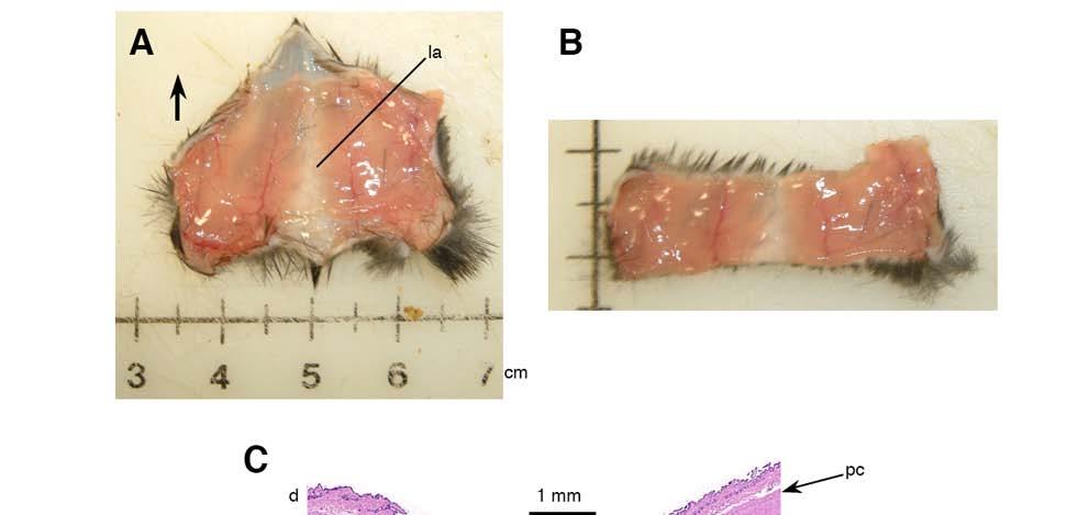
61

62


63 IL6 antibody neutralization
64 Conclusions 6 1. Hundreds of gene products differentially expressed in attached vs. released FPCM 2. IL-6 and IL-8 induced with matrix release 3. Neutralization of IL-6 partially protects against cell cycle inhibition after release
65 The fibroblast-populated collagen matrix: a model of granulation tissue 1 day detach with spatula 1 day IL-6 (quiescence) IL-6 (proliferation) attached/mechanically loaded (isometric tension) granulation tissue dissipation of mechanical load released/mechanically unloaded scar

66 the dark side of research
67 Biologic variability
68 Biologic variability (cont d)
69 Conclusions 7 1. p53 level in FPCM: variable response 2. Source of variability: strain-specific 3. Interpersonal variability in p53 response?
70 Incisional Wound Model


71 Murine incisional model


72
73 peak tensile strength (grams force) 1,500 1,250 WT murine wound disruption strength unwounded, mean ± sd wounded, mean ± sd 1, postwounding day
74 Regeneration Plans
75 Objective: To develop a replacement therapy for dermal tissue (i.e., to engineer a skin equivalent for clinical use)


76 epidermis dermis panniculus carnosus 1 mm epidermis cf fb dermis 5 µm cf = collagen fibril fb = fibroblast normal rat skin 100 µm
77 normal skin structural components epidermis basal lamina dermis blood vessels hair follicles glands cutis (nail, claw, horn) (subcutaneous adipose) (skeletal muscle) functions barrier temperature regulation sensation coloration immune response synthetic specialized


78 dermal replacement: strategy PLA backbone 20 µm 100 µm 20 µm


79 collagen matrix with cells: dermal fibroblasts mesenchymal stem 5 mm neodermis dermal replacement: strategy 50 µm 10 µm
80 basal lamina (type IV collagen, laminin) dermal replacement: strategy neodermis
81 keratinocytes (epidermis) dermal replacement: strategy
82 dermal replacement: strategy finished skin equivalent plug into animal model
 3 2 1 0 goal 0 60 120 180 PWD PWD 204 Additional endpoints Tensile strength Microscopic")
83 1 cm PWD 0 animal model to test skin equivalent (wound contraction assay) PWD 0 4 PWD 7 PWD 14 PWD 28 wound area (cm 2) goal PWD PWD 204 Additional endpoints Tensile strength Microscopic morphology
84 Looks easy, but potential problems: Co-culture conditions Blood supply Infection Inflammation Strength & durability Nerves, glands, hair, etc. Translation from rodents to humans