From light microscopy to multi-photon imaging:
|
|
|
- Drusilla Kennedy
- 5 years ago
- Views:
Transcription
1 From light microscopy to multi-photon imaging: Old and novel approaches for systems biology Frederick Klauschen Program in Systems Immunology and Disease Modeling National Institutes of Health and Institute of Pathology CCM Charité Universitätsmedizin Berlin
2 Overview Systems Biology and Imaging Confocal and Multi-Photon Imaging and modeling applications Bright field microscopy applications Method validation
3 Yet another definition of Systems Biology Integrating information: from single molecules to networks across the scales: from molecules to cell populations with high-throughput methods: Multi-modality data acquisition of spatio-temporal information about multiple molecules in single experiments using advanced data analysis: Large amounts of multidimensional data require standardized and efficient quantitative data analysis methods and modeling&simulation: Design and simulation of realistic models to explain and predict biological behaviour
4 Imaging and Systems Biology Classical view of Systems Biology: Omics -approaches: Genomics, Proteomics, i. e. highthroughput technologies for quantitative measurements of molecular components, used to generate comprehensive (network) representations of biological function. Imaging not considered of particular importance for systems biology! Maybe because conventional, i. e. manual image analysis mostly yields qualitative results! However... imaging methods in combination with quantitative computer-aided image analysis methods highly compatible with needs of systems biology!!!
5 Imaging and Systems Biology Integrating information across the scales: analysis of subcellular structures, single cells and cell populations with high-throughput methods: data acquisition of spatiotemporal information of multiple cells in single experiments using advanced data analysis: From scores to measurements : Computer-aided objective, standardized, quantitative image analysis methods and modeling&simulation: Design and simulation of realistic image-data-based models to explain and predict biological behaviour


6 Microscopy-based experiments spatio-temporal information on biological processes Image Analysis Parameter estimation (dynamics of localization, concentration of proteins, cell geometry model generation) Quantitative results/ conclusions quantitative model validation: direct comparison of simulation and experimental results Modeling and Simulation Simulation results/ conclusions

 Basis for standardized morphology feature reconstruction for realistic modeling & simulation of cellular")
7 Cell-Cell/Cell-Tissue Interaction Image Analysis (confocal/two-photon microscopy data) Consists of four modules: Signal-background separation by adaptive threshold segmentation Single cell/object detection and removal of noise/artefacts Adaptive channel merging Image feature quantification This allows for: Standardized, robust analyses: works for different fluorescent probes, image acquisition parameters, image qualities (z-stack intensity inhomogeneities) Automated, no user intervention necessary (optimization by user possible) Capable of processing large 3-/4-D data sets and detect and quantify even subtle phenomena (differences between experimental groups) Basis for standardized morphology feature reconstruction for realistic modeling & simulation of cellular processes


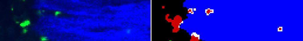
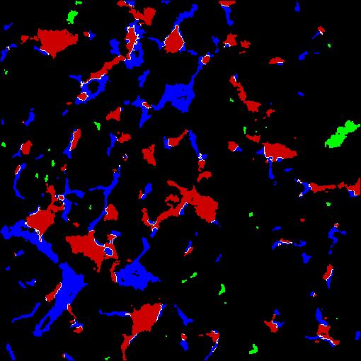
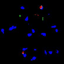
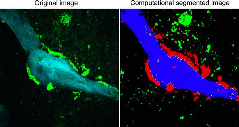
8 bone-osteoclast interaction Applications T-B-cell interaction DC - fiber interaction in LN
 activation inhibits osteoclastogenesis Ishii, Egen, Klauschen et al.")
9 Quantitative analysis of bone homoeostasis RANK activation promotes osteoclast attachment and bone resorption Sphingosine-1-Phosphate-Rec. (S1P 1 ) activation inhibits osteoclastogenesis Ishii, Egen, Klauschen et al., Nature 458, March 2009

10 Relevance of S1P 1 for bone homoeostasis Bone matrix density Bone matrix density Ishii et al., Nature 458, March 2009; Klauschen et al., Nature Prot., 2009.

11 Use of S1P 1 -activation in pathological conditions? S1P 1 -agonist FTY720 reduces effects of estrogendeprivation induced osteoporosis in mice! Ishii et al., Nature 458, March 2009; Klauschen et al., Nature Prot., 2009.

12 Influence of SAP on T-B-cell interaction SAP-mutation: X-linked lymphoproliferative syndrome: - defective humoral immune responses - defective germinal centre formation - reduction in IgG+ memory B-cell numbers SAP: SLAM-associated-protein SLAM: Signaling lymphocyte assoc. molecule André Veillette Nature Reviews Immunology 6, (January 2006)

13 Influence of SAP on T-B-cell interaction SAP is needed for germinal center formation SAP deficiency reduces stability of TB-interaction Qi, Cannons, Klauschen et al., Nature 2008 Oct 9

14 SAP influences synapse size of interacting B and T cells (unpublished data)

15 From descriptive and correlative models to the understanding of underlying mechanisms using realistic computational models quantitative analysis of interface area allows for correlation of experimental condition with biological response use spatial information as basis for mechanistic systems analyses of cellular processes

16 Construction of a spatial cell model from fluorescence microscopy data
17 3D surface reconstruction and volume grid generation Construction of a spatial cell volume model


18 A realistic surface - volume model of T cell - APC interaction Klauschen et al., Nature Protocols, June 2009.


19 Spatial simulation of chemotaxis signalling PI3K Chemotaxis, Xu and Jin, 2006 PTEN Comer and Parent, Cell, 2002 Meier-Schellersheim et al., PLOS CompBiol 2006
20 Spatial simulation of chemotaxis signaling exp -> sim -> exp -> sim -> Klauschen, F. and Meier-Schellersheim, M.., manuscript in preparation
21 Bright- field vs. fluorescence microscopy Bright-field offers high-throughput capability in a single slide! Requires color channel separation and segmentation based on morphology features Max. 1 to 2 simultaneous colors 2-D data Next generation imaging: Multi-color wide-field 3-D confocal microscopy