THE BASICS OF IMMUNOHISTOCHEMISTRY
|
|
|
- Elijah Ira Carpenter
- 5 years ago
- Views:
Transcription
1 THE BASICS OF IMMUNOHISTOCHEMISTRY
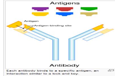
2 Introduction Immunohistochemistry (IHC) identifies specific tissue components by means of a specific antigen/antibody reaction tagged with a visible label. IHC makes it possible to visualize the distribution and localization of specific cellular components within a cell or tissue. IHC utilizes labeled antibodies to localize specific cell and tissue antigens, and is among the most sensitive and specific histochemical techniques.
3 Immunohistochemistry what s good about it? Antibodies bind to antigen in specific manner Gives you a spatial location Can be used to locate particular cells and proteins Can be used to identify cellular events e.g.apoptosis


4 What cellular antigens can we target? Cytoplasmic Nuclear Cell membrane Extracellular matrix

5 Identify replicating cells

6 Locate cells that are signaling

7 Locate apoptotic cells

8 Identify activation states

9 Identify different types of cells in a tissue

10 Examine cytoskeletal structure
11 Important considerations for IHC Antibody selection Fixation Sectioning Antigen Retrieval Blocking Direct method Indirect method Immunoenzyme Fluorescence Multiple labeling Controls
12 IHC steps: Speciment Fixation Antigen retrieval / Permeabilization Blocking endogenous enzymes Blocking aspecific sites Incubation primary Ab Incubation secondary Ab Mounting Controls Microscopy Observation
13 Immunohistochemistry assays may use cells on slides Cells grown, spun into a pellet, frozen or paraffin embedded and sectioned Cells grown as a monolayer or use tissue sections that are frozen or paraffin embedded Sections from tissues contain many different kinds of cells as well as extra-cellular matrix components
14 Fixation Helps to prevent: Degradation Modification Preserves the position of the Ag Provides target for Ab molecules Formaldehyde is the preferred fixative Most of the Ab available are optimized for use with formaldehyde
15 Fixation Aldehyde 10% NBF 4% paraformaldehyde with PBS buffer Frozen LN 2 + Isopentane
16 If the tissue is frozen Advantage: antigens are unaltered Disadvantage: Poor morphology Poor resolution at higher magnification Special storage Cutting difficulty Tissue section on glass slide: Frozen
17 If the tissue is paraffin embedded - Must fix. Remember the paraformaldehyde paradox (12 24 hours) - Process through xylenes and alcohols ruins some antigens - Deparaffinize (remove the infiltrated paraffin wax, by using organic solvents). - The deparaffinized section have to be treated to expose buried antigenic epitopes with either proteases or by heating in low ph citrate buffer (Antigen Retrieval) Best for good architecture Tissue section: Paraffin embedded

18 Slide preparation 5-8 micron tissue sections are cut onto slides Charged slides provide adhesion to tissue sections Deparaffinization Tissue is treated in a series of xylene and alcohol to remove paraffin.
19 Antigen retrieval Enables the partial reversal of formaldehyde induced confirmational change of Ags. Increases the accessibility of the Ab to the Ag. Two methods: Heat Enzyme digestion Choice of Ag retrieval depends on the Ag to be demonstrated. Must determine for each new antibody/antigen target.
20 Antigen retrieval HIER (Heat Induced Epitope Retrieval) Use MW/steamer/pressure cooker ~ 20 minutes, slow cool Citrate 6.0 PIER (Protein Induced Epitope Retrieval) Proteinase K Trypsin Pepsin Pronase, etc. Be careful: Destroys some epitopes Bad for morphology
21 Permabilization: Improving antibody penetration Need this for intracellular (cytoplasmic, nuclear) Ag Detergents most popular Triton-X Tween20 Can t use for membrane proteins Cells grown as a monolayer
22 Blocking Background staining Non-specific immunologic binding usually uniform Endogenous peroxidases Endogenous biotin


23 Non-specific staining Before block After block

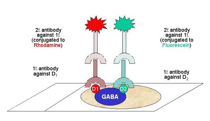
24 Blocking Peroxide Block Blocks endogenous peroxidases 3% H2O2 Protein Block Blocks all non specific sites Reduces background 10% Normal serum or BSA is used

25 Primary Antibodies Two types of Abs Polyclonal Abs Monoclonal Abs

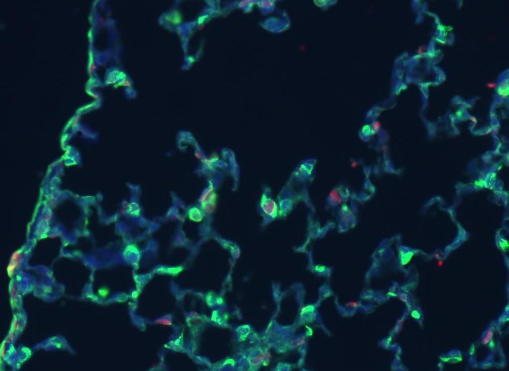
26
27 Monoclonal vs. polyclonal antibody Monoclonal Mouse Tends to be cleaner Very consistent batchto-batch More likely to get false negative results Polyclonal Many different species Tends to have more non-specific reactivity Can have very different affinity batch-to-batch More likely to have success in an unknown application
28 Make sure your antibody is validated for your application!!! IHC-P vs. IF WB, ELISA, IP, etc.
29 Labeling Antibodies: Antibodies are not visible with standard microscopy and must be labeled in a manner that does not interfere with their binding specificity. Common labels include fluorochromes (eg, fluorescein, rhodamine), enzymes demonstrable via enzyme histochemical techniques (eg, peroxidase, alkaline phosphatase), and electron-scattering compounds for use in electron microscopy (eg, ferritin, colloidal gold).


30 Direct methodprimary antibody only Labelled Ab reacts directly with Ag in tissue sections Single step method Short and quick Insensitive due to little signal amplification Goat anti-actin labeled with 594

31 Indirect method primary and secondary antibodies Unlabelled Primary Ab reacts with Ag and the labelled secondary Ab reacts with the primary Ab. Sensitive due to signal amplification Economical as single secondary Ab can be used against many Abs from same species Donkey anti-goat labeled with 488 Goat anti-actin

32 Indirect method primary and secondary antibodies
33 Multiple Immunofluorescence

34 Multiple Labelling of a Tissue Section (FITC + CY3 + DAPI )
35 Enzymatic detection methods Light microscope sufficient for analysis of specimens Resolution of subcellular structures not as good as with fluorescence methods Unlimited shelf life of labelled specimens Substrate reagents often toxic/carcinogenic
36 Enzyme indirect method Ag-Ab conjugates are visualized by the use of a label. Enzymes are used as labels that produce a colored precipitate in the presence of a substrate Labels: Peroxidase Alkaline Phosphatase Substrate: DAB
37 Enzyme indirect method Enzyme labels produce a colored precipitate in the presence of a specific substrate Most widely used label is Peroxidase Produces a dark brown precipitate when Diamino Benzidine (DAB) is added. Alkaline phosphatase is also used and produces either red or blue precipitates.
38 Controls Positive control Best is tissue with known specificity Cells or tissues that are known to contain the specific Ag Detects false negatives due to fixation and processing. Negative control It is to test for the specificity of the antibody involved. Omission of Primary Ab with the same tissue and procedure Useful to detect endogenous biotin and peroxidase activity