Supplemental Information: Phosphorylation of CLIP-170 by Both Plk1 and CK2 Is Involved in the Timely Formation of Kinetochore-microtubule Attachments
|
|
|
- Margery Pitts
- 5 years ago
- Views:
Transcription
1 Supplemental Information: Phosphorylation of CLIP-170 by Both Plk1 and CK2 Is Involved in the Timely Formation of Kinetochore-microtubule Attachments Hongchang Li, X. Shawn Liu, Xiaoming Yang, Yingmin Wang, Yun Wang, Jerrold R. Turner and Xiaoqi Liu Supplemental Materials and Methods: Vector construction and RNAi GFP-CLIP-170 was a kind gift of Dr. Gari G. Borisy. To specifically deplete endogenous CLIP-170 in HeLa cells, plasmid pbs/u6-clip-170 was constructed. The targeting sequence of human CLIP-170 (accession no. NM_ ) was GGAAGAATGATGGCGCTGTT, corresponding to the coding region 752 to 771 relative to the first nucleotide of the start codon. Plasmid pbs/u6-clip-170-1st half (sense strand) was used as a control vector. Plasmid pbs/u6-plk1 was described previously (Liu and Erikson, 2003). The pkd-ck2 construct used to deplete CK2 was obtained from Upstate. The following target sequence was used to design sirna against human Plk1: AAGGGCGGCTTTGCCAAGTGCTT (Liu and Erikson, 2002). Cell culture and transfection HeLa and HEK293T cells were cultured in Dulbecco modified Eagle medium (DMEM) supplemented with 10% (vol/vol) fetal bovine serum, 100 units/ml penicillin, and 100 units/ml streptomycin at 37 C in 8% CO 2. Cells were arrested at mitosis by incubation with 100 ng/ml of nocodazole for 12 h. To synchronize HeLa 46
2 cells at prometaphase, cells were treated with 2.5 mm thymidine for 16 h, released for 8 h, and then treated with thymidine a second time for 16 h, followed by release for 12 h in the presence of 100 ng/ml of nocodazole. sirna was transfected using transmessenger transfection reagent (QIAGEN), whereas plasmid DNA was transfected using GenePORTER (Genlantis) or lipofectamine 2000 (Invitrogen) as described by the manufacturers. For anti-gfp immunofluorescence staining, DNA was transfected using CalPhos Mammalian Transfection Kit (Clontech). Cells were harvested 36 hr after transfection and analyzed by immunoprecipitation and Western blotting. Antibodies The antibodies against CLIP-170 (sc-25613, sc-28325), Plk1 (sc-17783) and CK2α (sc-12738) used in this study were purchased from Santa Cruz Biotechnology. The p150 glued antibody (610474), EB1 antibody (610534), and CK2β antibody (610444) were from BD Transduction Laboratories. The Hec1 antibody (GTX70268), used as a kinetochore marker, and Mad2 antibody (Ab24588), used to detect unattached kinetochores, were from GeneTex and Abcam, respectively. Antibodies against FLAG, GST and tubulin were all obtained from Sigma. Protein purification, mutagenesis and in vitro MT-binding assay Various GST-tagged rat CLIP-170 constructs (accession no. CAB92974) were subcloned into pgex-kg, expressed in E. coli, and purified. Point mutations were generated using the QuickChange Site-Directed Mutagenesis Kit (Stratagene). MT 47
3 pelleting assays were performed using the MT-associated protein spin down assay kit (Cytoskeleton, Inc.). IP, immunoblotting and IF staining Whole cell lysates were incubated with different antibodies indicated in each experiment for 1.5 hr at 4 C, followed by 1 h of incubation with protein A/G-Sepharose beads. Immunocomplexes were resolved by SDS-PAGE, and coimmunoprecipitated proteins were detected by Western blotting using antibodies indicated in the specific experiments. Antibodies against Plk1, CK2α, CK2β, Flag, GST and tubulin were used at 1:1000 dilution, whereas antibodies against ps195 and ps1318 were used at 1:500. IF staining was performed as previously described (Tanenbaum et al., 2006). Antibodies against ps195, ps1318, CLIP-170, p150 glued, Mad2, GFP, and Hec1 were used at 1:100 dilution. Kinase assay Purified recombinant CLIP-170 was incubated with purified Plk1-WT or KM in kinase reaction buffer (50 mm Tris, ph7.5, 10 mm MgCl 2, 2 mm EGTA, 0.5 mm Na 3 VO 4, 100 mm PNPP, 25 mm DTT, 125 μm ATP) supplemented with 10 µci of [γ- 32 P] ATP at 30 C for 30 min. After the reaction mixtures were resolved by SDS-PAGE, the gels were stained with Coomassie brilliant blue, dried, and subjected to autoradiography. Metabolic labeling and phosphopeptide mapping HEK293T cells were transfected with various GFP-CLIP-170 constructs, arrested in 48
4 mitosis by nocodazole treatment (100 ng/ml) for 12 h, and labeled for 3 h with [ 32 P]orthophosphate (ICN) at 1 mci/ml in phosphate-free DMEM. After transfected cells were lysed, the phosphoproteins were immunoprecipitated by GFP antibodies, resolved by SDS-PAGE, stained with Coomassie brilliant blue, and visualized by autoradiography. For phosphopeptide mapping, after lysates were subjected to anti-gfp IP, phosphoproteins were recovered, digested with trypsin or endoproteinase Glu-C, and resolved by 40% alkaline PAGE, followed by autoradiography. Generation of U2OS cells stably expressing GFP-CLIP-170 or -p150 glued constructs One day after U2OS cells were transfected with GFP-CLIP-170 (WT or mutants) or GFP-p150 glued by using MegaTran 1.0, cells were selected with 400 µg/ml of G418 for six weeks. After at least 10 clones per construct were obtained, Western blotting was performed to isolate the cell lines that express various GFP-CLIP-170 or -p150 glued constructs at protein levels close to those of endogenous CLIP-170 or p150 glued. Time-lapse videomicroscopy for live cells U2OS cells stably expressing GFP-CLIP-170 (WT, S195A, S1318A) were used for this purpose. Image acquisition was performed using a Zeiss Axiovert 100TV microscope enclosed in a chamber to maintain CO 2 and temperature for live cells. During filming, cells were maintained in HEPES-buffered DMEM and at 37 C. After treatment with Hoechst 33342, cells were tracked for 10 h with images were acquired at 10-min intervals using SlideBook software. 49
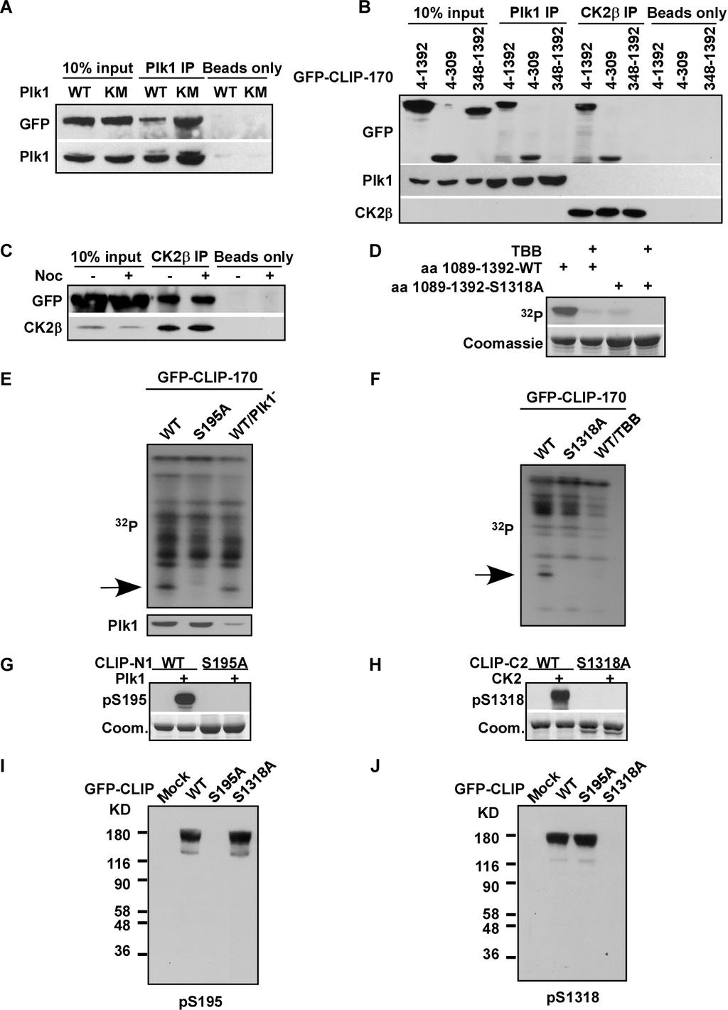
5 Supplemental Figure Legends: Figure S1 CLIP-170 is a Plk1/CK2 interaction partner. (A) Coimmunoprecipitation of Flag-Plk1 and GFP-CLIP-170. HEK293T cells were co-transfected with GFP-CLIP-170 and Flag-Plk1-WT (wild type) or -KM (kinase-defective mutant, K82M), harvested at 24 h posttransfection, and subjected to anti-plk1 IP, followed by Western blotting. 10% of total input is shown in the left two lanes. Protein A/G beads only lanes serve as washing controls (right two lanes). (B) HeLa cells were transfected with GFP-CLIP-170-full length (aa ), CLIP-N (aa 4-309), or CLIP-C (aa ) for 24 h, treated with nocodazole for 12 h, and subjected to anti-plk1 or -CK2β IP, followed by Western blotting. 10% of total input is also shown in the left two lanes. Protein A/G beads only lanes serve as washing controls. (C) Physical association between CK2 and CLIP-170 in HeLa cells. Cells were transfected with GFP-CLIP-170, treated with or without 100 ng/ml nocodazole for 12 h, and subjected to anti-ck2β IP, followed by Western blot analysis. 10% of total input is also shown in the left two lanes. Protein A/G beads only lanes serve as washing controls. (D) CK2 was incubated with CLIP-170-aa (WT or S1318A mutant) in the presence of [γ- 32 P]ATP in the presence or absence of 25 μm TBB. Reaction mixtures were resolved by SDS-PAGE, followed by autoradiography. (E, F) Metabolic labeling and phosphopeptide mapping of GFP-CLIP T cells were transfected with GFP-CLIP-170 (WT, S195A or S1318A), incubated overnight, transfected with dsrna to deplete Plk1 (lane 3 of E), and incubated overnight. After 50
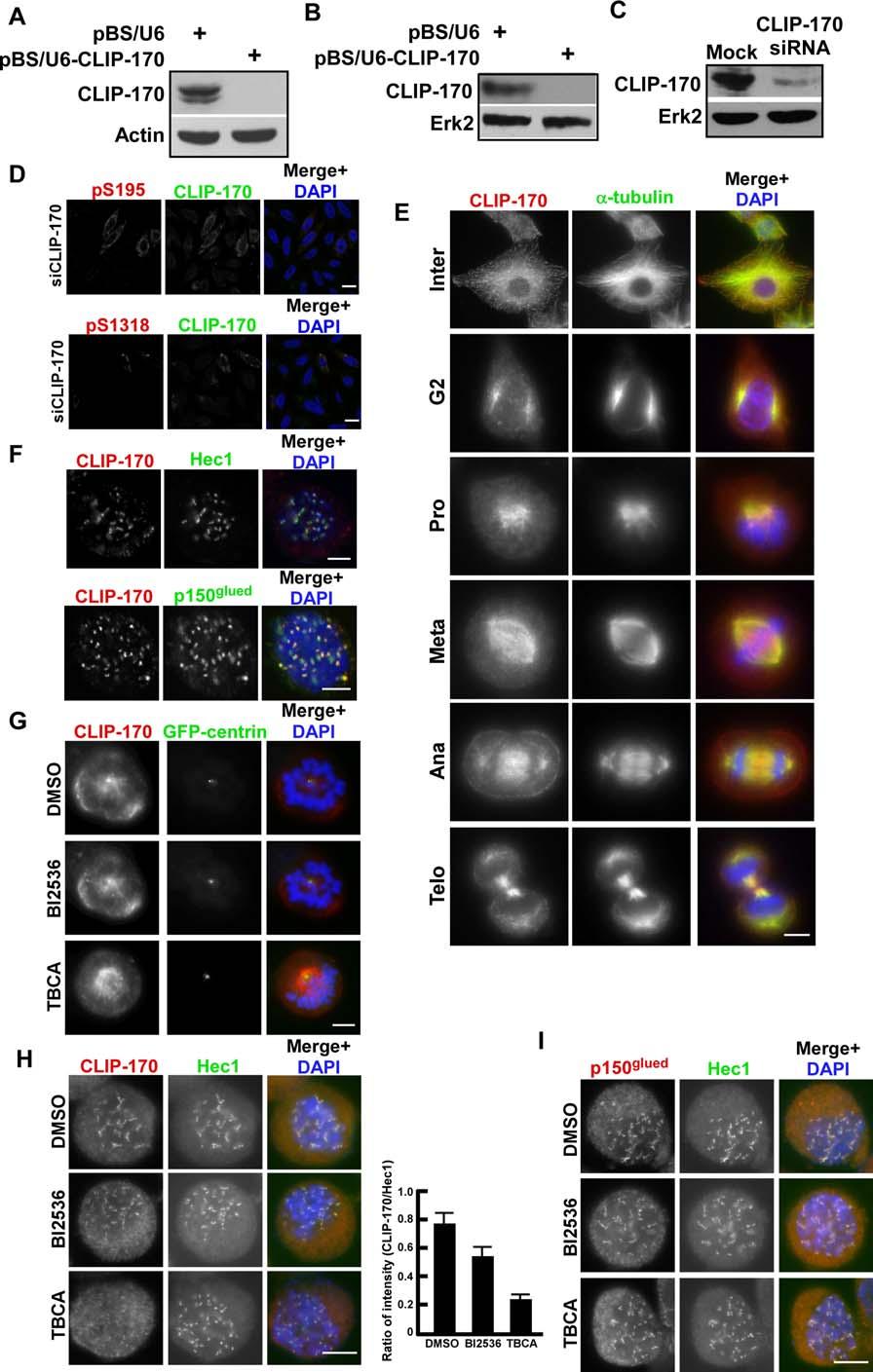
6 cells were metabolically labeled in the presence of TBB (lane 3 of F), lysates were subjected to anti-gfp IP, followed by PAGE. Phosphoproteins were located by autoradiography, excised, digested with trypsin (E) or endoproteinase Glu-C (F), and resolved on an alkaline PAGE. Arrows indicate positions corresponding to peptides containing S195 and S1318, respectively. (G-J). Specificity of ps195 and ps1318 antibodies. (G) Purified Plk1 was incubated with purified GST-CLIP-N1 (WT or S195A) in the presence of unlabeled ATP, followed by an anti-ps195 Western blot. N1, aa (H) CK2 was incubated with purified GST-CLIP-C2 (WT or S1318A) in the presence of unlabeled ATP, followed by an anti-ps1318 Western blot. C2, aa (I, J) HEK 293T cells were transfected with GFP-CLIP-170 constructs (WT, S195A or S1318A), treated with nocodazole, and immunoblotted with antibodies against ps195 (I) or ps1318 (J). Entire gels are shown to indicate specificity of two antibodies. Figure S2 CLIP-170 depletion and its in vivo phosphorylation. (A and B) HeLa cells were co-transfected with pbs/u6-clip-170 and pbabe-puro at a ratio of 7:1. At 1 d after transfection, puromycin was added to select for the transfection-positive cells for 1 d. Harvested cells were immunoblotted using anti-clip-170 antibodies developed in our lab (A) or commercial anti-clip-170 antibodies (B). (C) HeLa cells were depleted of CLIP-170 with sirna and immunoblotted. (D) HeLa cells were depleted of CLIP-170 as in (C) and immunostained. (E) HeLa cells were synchronized with the double thymidine block, released for 10 h to enrich at mitosis, and co-stained with 51
7 antibodies against α-tubulin and CLIP-170. (F) Kinetochore localization of CLIP-170. Nocodazole-treated HeLa cells were co-stained with antibodies against CLIP-170 and Hec1 or p150 glued. (G) HeLa cells stably expressing GFP-centrin were treated with BI2536 or TBCA and immunostained. (H and I) HeLa cells were treated with BI2536 or TBCA for 6 h or 20 h, respectively. Cells were then incubated with nocodazole for additional 2 h and harvested for immunostaining using indicated antibodies. Ratios between the intensities of CLIP-170 and Hec1 staining were quantified by using Image J software. More than 100 kinetochores in 10 cells were taken for this quantification (H, right panel). Bars, 5 µm. Figure S3 The CLIP-170/p150 glued interaction is regulated by CK2-associated phosphorylation at CLIP-S1318. (A) CLIP-170-S1318D has the most dramatic effect in CLIP-170 overexpression-induced bundle formation of GFP-p150 glued. U2OS cells stably expressing GFP-p150 glued were transfected with different forms of myc-clip-170 constructs, co-stained with antibodies against GFP and myc. (B) Quantification results of (A). To quantify, the region with GFP-p150 glued bundles was encircled, and a ratio of fluorescence intensity (GFP versus myc) within the encircled region was calculated. All the imaging parameters were kept constant during data acquisition. At least 100 cells were quantified for each construct. Bar, 5 µm. Figure S4 Phosphorylation at S195/S1318 of CLIP-170 does not affect its microtubule-binding ability. (A) U2OS cells stably expressing various GFP-CLIP-170 constructs were co-stained with antibodies against GFP and α-tubulin. (B) U2OS cells 52
8 stably expressing various GFP-CLIP-170 constructs were co-stained with antibodies against GFP and EB1. (C) Mitotic U2OS cells stably expressing various GFP-CLIP-170 constructs were immunostained. (D) U2OS cells stably expressing various GFP-CLIP-170 constructs were depleted of endogenous CLIP-170 and subjected to anti-clip-170 Western blotting. Positions of endogenous CLIP-170 and GFP-CLIP-170 are labeled on the right. Bars, 5 µm. Figure S5 CLIP-170 phosphorylation does not affect Mad2 stripping from kinetochores. (A) U2OS cells stably expressing GFP-CLIP-170 (WT, S195A or S1318A) were transfected with dsrna to deplete endogenous CLIP-170. Two days post-transfection, cells were processed as indicated, and immunostained with anti-mad2 antibodies. Bars, 5 µm. Supplemental movie legends: Movie S1. A representative movie showing segregation of Hoechst-staining U2OS cells stably expressing CLIP-170-WT, related to Figure 6D. About 40 min is enough for the cell to proceed from the beginning of chromosome condensation to anaphase onset. Movie S2. A representative movie showing segregation of Hoechst-staining U2OS cells stably expressing CLIP-170-S195A, related to Figure 6D. For this cell, one hour is spent from the start of chromosome condensation to anaphase onset. Movie S3. A representative movie showing segregation of Hoechst-staining U2OS cells stably expressing CLIP-170-S1318A, related to Figure 6D. This cell spent 80 53
9 min to proceed to anaphase onset from the beginning of chromosome condensation. Supplemental References: Liu, X., and Erikson, R.L. (2002). Activation of Cdc2/cyclin B and inhibition of centrosome amplification in cells depleted of Plk1 by sirna. Proc Natl Acad Sci U S A 99, Liu, X., and Erikson, R.L. (2003). Polo-like kinase (Plk)1 depletion induces apoptosis in cancer cells. Proc Natl Acad Sci U S A 100, Tanenbaum, M.E., Galjart, N., van Vugt, M.A., and Medema, R.H. (2006). CLIP-170 facilitates the formation of kinetochore-microtubule attachments. Embo J 25,
10
11

12

13

14