Biomedical Optical Imaging Martin Frenz Biomedical Photonics Department Institute of Applied Physics, University of Bern
|
|
|
- Erica Griffin
- 5 years ago
- Views:
Transcription


1 1 Biomedical Optical Imaging Martin Frenz Biomedical Photonics Department Institute of Applied Physics, University of Bern


2 Imaging is one of the most powerful tools in biomedical research. The impact and amount of information contained in visual data is almost impossible to underestimate. A picture is worth ten thousand words A movie is almost priceless 2


3 Seeing inside the body with light 3 Problem: strong scattering of light in biological tissues over the whole spectral range!
4 4 Imaging techniques x-ray techniques: planar (i.e. mammography) computer tomography (1895) (1970s) ultrasound: A/B-mode Doppler (1940) MR techniques: nuclear imaging: magnetic resonant imaging magnetic resonant spectroscopy optical techniques: confocal microscopy optical coherence tomography transillumination techniques fluorescence techniques (1970s) positron emission tomography (1975) single photon emission computer tomography (1986) (1991)
5 Optimum requirements spatial resolution range of λ temporal resolution 1 ms (real time) field of view µm up to cm no ionizing radiation no restrain or anesthesia show structures and function see anywhere in the body low cost and easy to use X-ray nuclear imaging, PET MRI 5
6 6 Imaging techniques x-ray techniques: planar (i.e. mammography) computer tomography (1895) (1970s) ultrasound: A/B-mode Doppler (1940) MR techniques: nuclear imaging: magnetic resonant imaging magnetic resonant spectroscopy optical techniques: confocal microscopy optical coherence tomography transillumination techniques fluorescence techniques (1970s) positron emission tomography (1975) single photon emission computer tomography (1986) (1991)
7 Optical imaging techniques high spatial resolution deep penetration 100'000 OM: confocal microscopy multi-photon microscopy (Markus Kohler) OCT: Optical coherence tomography (Rainer Leitgeb) OA: Optoacoustic ODT: Optical diffusion tomography depth resolution [µm] 10'000 1' OCT OM ODT OA / NIR time of flight tomography frequency modulated tomography '000 10' '000 depth [µm] 7
8 8 Optical detection scatter absorption fluorescence bioluminescence intrinsic scatter of tissue exogenous scatters (gold and silver particles) intrinsic tissue absorption exogenous scatters (dyes, gold and silver particles) tissue auto-fluorescence (NADPH) fluorescence dyes (porphyrin) fluorescence protein-reporter genes (GFP) bioluminescence protein-reporter genes (luciferase)

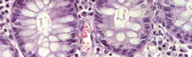
9 9 Autofluorescence Autofluorescence backscattered part of the excitation light white light LFL-München fluorescence image white light and autofluorescence of a flat ptag1-tumor in the bladder after excitation with light at 400 nm. The thickened mucosa (start of a tumor growing) shows a good contrast to the healthy green fluorescent tissue.

10 Photodynamic diagnosis application in urology: bladder carcinoma Fluorescence of Protoporphyrin IX, which accumulates in the tumor after injection of 5- Aminolevulin acid selectively into the tumor 10 excitation: nm detection: > 445 nm Stepp, Kriegmair, Baumgartner red fluorescence blue background
11 Light propagation in tissue - optically inhomogeneous (1 g tissue contains about 10 9 cells) - index of refraction higher than in air (Fresnel reflection) diffusion of light in tissue 3 mm thick slab of female breast tissue 11 strong scattering F.A. Marks, Proc. SPIE 1641, p 227 (1992)
12 Rayleigh- and Mie-scattering reduced scattering coefficient µ s [cm -1 ] µ s =µ s (1-g) NATO ASI Series E: Applied Sciences-Vol.325 Mie scattering: particle size λ (macromolecular structures, membranes) Rayleigh scattering particle size << λ organelles, collagen fibers, cells) 12 optical scattering spectra provide information about size distribution of scatters
13 Optical tissue properties scattering and absorption Beer-Lambert law Id ( ) = (1 R) I exp( µ d) F 0 t µ t = µ a + µ s I 0 = collimated irradiance [W/cm 2 ] R F = Fresnel reflection Mean free path length l path = 1 µ t 13 Doornbos et al. Phys.Med.Biol. 44, p 967 (1999)
14 Tissue absorption diagnostic window 0 14 Hemoglobin and water have relatively low absorption in the near-ir near-ir window enables optical imaging and near-ir spectroscopy
15 Optical imaging 15 absorption coefficient µ -1 a (cm ) Oxygenated blood Deoxygenated blood Melanin 10% Water visible 0.01 UV IR wavelength (nm) angiogenesis (blood concentration) + hypermetabolism (oxygen saturation) optical absorption provides contrast for functional imaging
16 Fluorescence and Raman scattering Fluorescence Raman hν ex excited state 1 excited state 2 hν em hν ex hν s =h(ν ex ν ν ) hν ν hν as =h(ν ex +ν ν ) hν ex hν ν ground state Stokes Anti-Stokes 16 radiative decay from an excited state - coupling to molecular vibrations - each molecule has unique Raman spectrum - typical Raman cross-section σ ~10-29 cm 2 molecule -1 sr -1 (very weak signal) Fluorescence or Raman scattering provides biochemical information because they are related to molecular conformation

17 17 provides higher contrast than conventional optical microscopy image is blurred due to fluorescence from out-of-focus regions bleaching of the chromophore Fluorescence microscopy Arc Lamp EPI-Illumination Excitation Diaphragm Excitation Filter Ocular Dichroic Filter Objective objective Emission Filter

18 Confocal microscopy Laser 500µm Excitation Pinhole Excitation Filter PMT ive 18 Emission Filter Emission Pinhole 3D imaging The fluorescence emission that occurs above and below the focal plane is not confocal with the pinhole aperture. bleaching of the dye excitation in the UV region
 r t")

19 19 Two-photon vs. one-photon fluorescence Fluorescence I(,) r t Fluorescence I 2 (,) r t Single-photon excitation Two-photon excitation linear nonlinear λ em shorter than λ ex less scattering low probability event
 imaging limited to a small")
20 20 Quantitative two-photon microscopy Theileria infected macrophages near-infrared radiation enhances the penetration depth reduces image deterioration due to scattering when passing through biological tissue less photo-bleaching and photo-damage regions above and below the excitation light cone are not excited (no background fluorescence) imaging limited to a small depth
21 21 Targeted contrast agents Clinical diagnostic imaging often relies on different uptake behavior of contrast agents between tumors and the surrounding tissue Non-targeted dyes may accumulate in tumor due to increased vascular density or capillary permeability Some imaging agents specifically target certain receptors, which are overexpressed in malignant cells Examples of targeting ligands for delivery of diagnostic imaging agents include antibodies, hormones, or small peptides.
22 22 Markers - Fluorescent dye (e.g. GFP, ICG) - quantum dots - Bioluminescence (Luciferase) - gold nanoparticles (nano spheres, shells, rods) emission emission absorption fluorescence spectroscopy imaging... opt. spectroscopy confocal OCT photon migration optoacoustics...
 no external light required emission between 400-600 nm molecular imaging high sensitivity but 23 poor spatial resolution due to light scattering C.")
23 Bioluminescence (Luciferase) Bioluminescence: encymes catalyze a bio-chemical process inside the animal that emits light Luciferase emits light when it combines with luciferin, ATP and molecular oxygen (light from firefly) no external light required emission between nm molecular imaging high sensitivity but 23 poor spatial resolution due to light scattering C. Rudolph, LMU
 - broadband excitation - long fluorescence lifetime Q-dots: = 15-20 nm http://probes.invitrogen.")
24 24 Quantum dots biomolecules antibody PSMA biotin - tuneability - no bleaching nm size - high quantum yield ( > 90% ) - broadband excitation - long fluorescence lifetime Q-dots: = nm

 food")


25 Quantum dots multispectral images using different Q-dots LNCaP prostate tumor cells mouse phantom cell culture nm 570 nm 620 nm in-vivo J.R. Mansfield et al, JBO 10, (2005) food autofluorescence C4-2 prostate tumor cells injection of anti-psma antibody coupled to 640 nm Q-dots background = autofluorescence 25 Q-dots PSMA antibody conjugate gold nanorods 2 photon luminescence membrane binding
26 Optical Coherence Tomography (OCT) I o Intensity Spectral distribution λ Source B A Reference mirror Sample λ ο wavelength Detector λ / 2 Detector axial resolution L 26 Mirror Displacement
27 27 Light sources wavelength and bandwidth determine axial resolution ( L): L = 2ln2 π 2 λ λ Typical resolution: SLD Ti:Sapphire laser λ 0 = 1300 nm 800 nm λ = 50 nm 125 nm L = 15 µm 2 µm spatial resolution is given by the spot diameter of the laser
 28 d 2D and 3D images based on interferometric measurement of optical back-reflection or back-scattering from internal tissue mircrostructures z-direction (longitudinal scan)")
28 Optical Coherence Tomography reference mirror L 1 L2 photodiode SLD detection electronics human eye V.J. Srinivasan et al., SPIE Vol. 6079, p (2006) 28 d 2D and 3D images based on interferometric measurement of optical back-reflection or back-scattering from internal tissue mircrostructures z-direction (longitudinal scan) by moving of reference mirror x-y direction (transverse scan) by moving the beam usually implemented with fiber optics Optical transparency of the eye provides unique opportunity for high resolution imaging of the retina
29 29 Diffuse optical tomography measure light that passes through a highly scattering tissue frequency domain: measure amplitude and phase of modulated light absorbing structure light source array of detectors highly scattering medium determine absorption and/or diffusion cross-section
 1 (4 Dt) 3/2 e 2 R µ at 4Dt D = diffusion coefficient [m 2 /s] µ a = absorption coefficient [1/s] R = distance between source and")
30 30 Optical tomography time domain: measure delay of light pulse at detector intensity ps intensity "snake" diffuse time laser pulse highly scattering medium ballistic 1 ns detected laser light PtR (, ) 1 (4 Dt) 3/2 e 2 R µ at 4Dt D = diffusion coefficient [m 2 /s] µ a = absorption coefficient [1/s] R = distance between source and detector


31 31 Optical mammography laser: λ = 780 nm 255 pairs of laser sources and detectors Philips Medical Systems


")
32 Optical mammography x-ray 3D-image-reconstruction laser illumination tumor (1-2 cm) 32 structural changes tumor (increased vascular density) imaging of inhomogenities absorption changes

33 Path of light detector light source skull path of photons reaching detector brain 33 non-invasive and painless continuous measurement i.e. long-term monitoring real-time display


34 Functional brain imaging by near-infrared spectroscopy Neonatology University Hospital Zurich (M. Wolf) 34 Diagnosis of lesions and impending danger is vital for outcome Brain functional disorder - Attention deficit hyperactive disorder (ADHD), epilepsy, psychiatric disorders Research on brain function will - lead to improved understanding of brain and brain development - enhance prevention of diseases and complications - improve treatment
![Video of an activation [HHb] (µm) moving average of 5 points acquisition time 800 ms -1.0-0.5 0.0 0.5 3 1 2 4 B A 8 5 6 7 Franceschini MA et al. Opt.](/docs-images/89/98234370/images/35-1.jpg "Express 2000; 6: 49-57 35 local changes in oxy-and deoxyhemoglobin concentration indicate brain activity quantification possible due to absorption at different wavelengths")
35 Video of an activation [HHb] (µm) moving average of 5 points acquisition time 800 ms B A Franceschini MA et al. Opt. Express 2000; 6: local changes in oxy-and deoxyhemoglobin concentration indicate brain activity quantification possible due to absorption at different wavelengths high temporal resolution

36 Optoacoustic imaging Combines the excellent contrast of optical absorption with the good spatial resolution of ultrasound for deep imaging in the optical diffusive regime. 36


 =Γ µ (x) H(x) H ( x) µ a (")
37 What is optoacoustic imaging? skin surface laser pulse irradiation δ τ p < v tumor p δ v τ < blood vessels illumination heated tissue 37 0 a initial pressure distribution p (x,t = 0) =Γ µ (x) H(x) H ( x) µ a ( x) T( x) = c ρ p


 S(x,t) backward projection")
38 What is optoacoustic imaging? ultrasound propagation and detection array of acoustic sensors reconstruction signals 5 z [mm] x [mm] 38 initial pressure distribution 2 p(x,t) t 2 =Γ t 2 v p(x,t) S(x,t) backward projection FFT - algorithm...
39 39 Absorbers endogenous and exogenous chromophors blood melanin... dyes functional imaging nanoparticles... contrast enhancement goal: strong near-infrared absorption


 Vol")
40 Functional optoacoustic imaging whiskers-stimulation lamda bregma RH LH open skull image 40 left-side right-side increase in vascular blood volume or flow Nature Biotechnology (2003) Vol 21, 803
41 41 Exogenous absorbers antibody nanoparticles IR-laserpulse imaging tumor tissue ultrasoundtransducer


42 42 Selective tumor treatment antibody nanoparticles strong laserpulse therapy T laser thermal spot damage of tumor cells tumor tissue SKBr3 cells + gold conjugate


43 Optoacoustic Imaging 43 Laser 10 Hz, 5 ns Q-Switched Nd:YAG laser with 3ω and OPO ( nm) triggered laser pulse replaces transmitted ultrasound pulse optoacoustic images recorded with US-system (adapted receiver timing)

44 44 Comparison optoacoustic - US vascularization of a finger tip blood vessels US-image bone
45 45 Conclusion Optical photons provide non-ionizing and safe radiation for medical applications. Optical scattering spectra provide information about the size distribution of scatters, such as cell nuclei. Optical absorption provides contrast for functional imaging Optical spectra-based on absorption, fluorescence, bioluminescence or Raman scattering provide biochemical information because they are related to molecular conformation. optical imaging covers a wide range of applications future is multimodal imaging