Mammalian expression and purification system using Strep-tag and/or 6xHistidine-tag
|
|
|
- Bryce Stokes
- 5 years ago
- Views:
Transcription
 551-50672-0 Fax: +49 (0) 551-50672-181 info@iba-go.com www.iba-go.com IBA US Distribution Center 10748 Indianhead Industrial Blvd. St. Louis, MO 63132 Tel.")
1 Mammalian expression and purification system using Strep-tag and/or 6xHistidine-tag Last date of revision May 2005 Version PR IBA Headquarters IBA GmbH Rudolf-Wissell-Str. 28 D Göttingen Germany Tel: +49 (0) Fax: +49 (0) IBA US Distribution Center Indianhead Industrial Blvd. St. Louis, MO Tel IBA-GmbH ( ) Fax Mammalian expression and purification systems using Strep-tag and/or 6xHistidine-tag 1
2 Patents & Licensing IBA patents, licensing and trademarks Strep-tag technology for protein purification and detection is covered by US patent 5,506,121, UK patent and French patent ; the tetracycline promoter based expression system is covered by US patent 5,849,576 and Strep-Tactin is covered by US patent 6,103,493. Further patent applications are pending world-wide. Purchase of reagents related to these technologies from IBA provides a license for non-profit and in-house research use only. Expression or purification or other applications of above mentioned technologies for commercial use require a separate license from IBA. A license may be granted by IBA on a case-by-case basis, and is entirely at IBA's discretion. Please contact IBA for further information on licenses for commercial use. Strep-tag and Strep-Tactin are registered trademarks of IBA GmbH. Other licenses Use of the CMV promoter is covered under U.S. patent nos. 5,168,062 and 5,385,839 owned and licensed by the University of Iowa Research Foundation and may be used for research purposes only. Commercial users must obtain a license to these patents directly from the University of Iowa Research Foundation. Inquiries for commercial use should be directed to: Brenda Akins, University of Iowa Research Foundation (UIRF), 214 Technology Innovation Center, Iowa City, IA The Ni-NTA resin is manufactured by QIAGEN. Hoffmann-La Roche owns patents and patent applications pertaining to the application of Ni-NTA resin (U.S. patent , U.S. patent , EP B1) and to the method of purifying 6xHistidine-tagged proteins using 6xHistidine-tag coding vectors (U.S. patent , U.S. patent , EP B1). All purification of 6xHistidine-tagged proteins by metal affinity chromatography for commercial purposes, and the commercial use of proteins so purified, require a license from Hoffmann-La Roche. Further information about licenses for commercial use is available from QIAGEN GmbH, QIAGEN Strasse 1, D Hilden, Germany. The cartridge design is covered by U.S. patent 4,871,463. Other trademarks and disclaimers Sepharose and FPLC are trademarks of Amersham Pharmacia Biotech Inc.; MacroPrep is a registered trademark of Bio-Rad Laboratories Ltd.; Superflow is a registered trademark of Sterogene Bioseparations Inc., Ni-NTA is a trademark of Qiagen. Registered names, trademarks, etc. used in this document, even when not specifically marked as such, are not to be considered unprotected by law. For research use only 2 Mammalian expression and purification systems using Strep-tag and/or 6xHistidine-tag
3 Content 1 Introduction pexpr-iba vectors Strep-tag /Strep-Tactin system xHistidine-tag in combination with the Strep-tag 6 2 Cloning with pexpr-iba vectors Expression cassettes of pexpr-iba vectors 3 Expression Transient expression in mammalian cells Stable expression in mammalian cells Trouble shooting Expression 13 4 Preparation of soluble cell extracts 4.1 Preparation of soluble extracts after cytoplasmic expression of Strep-tag fusion proteins Preparation of conditioned supernatant after secreted expression of Strep/6xHistidine-tag fusion proteins 15 5 Detection of Strep-tag fusion proteins 5.1 Detection of Strep-tag fusion proteins with Strep-Tactin Alkaline Phosphatase (AP) conjugate Detection of Strep-tag proteins with Strep-Tactin horse radish peroxidase (HRP) conjugates Detection of Strep-tag proteins with the Strep-tag II specific monoclonal antibody Detection of Strep-tag proteins with the Strep-tag II specific monoclonal antibody conjugated to horse radish peroxidase (HRP) 19 6 Purification of Strep-tag fusion proteins 6.1 Purification of Strep-tag fusion proteins using gravity flow columns Purification of Strep-tag fusion proteins using Strep-Tactin Spin Columns Quick purification of Strep-tag fusion proteins using cartridges FPLC purification of Strep-tag fusion proteins using Strep-Tactin Superflow cartridges or Strep-Tactin MacroPrep cartridges HPLC purification of Strep-tag fusion proteins using Strep-Tactin POROS columns Purification of Strep-tag fusion proteins using magnetic beads 6.7 Trouble shooting Strep-tag purification Purification of Strep/6xHistidine-double-tag fusion proteins 7.1 Gravity flow purification of Strep/6xHistidine-tag fusion proteins under native conditions Quick purification of Strep/6xHistidine-tag fusion proteins under native conditions using cartridges Trouble shooting 6xHistidine-tag purification 8 APPENDIX Storage and regeneration of Strep-Tactin resin Storage and regeneration of Ni-NTA resin 38 9 Related products References 41 Mammalian expression and purification systems using Strep-tag and/or 6xHistidine-tag 3
4 1 Introduction 1.1 pexpr-iba vectors pexpr-iba vectors are designed for high-level expression of recombinant proteins in mammalian hosts and for their subsequent purification via Strep-tag and/or 6xHistidinetag. The human cytomegalovirus immediate-early (CMV) promoter provides strong expression in a wide range of mammalian cells. To prolong expression in transfected cells, the vector will replicate in cell lines that are latently infected with SV40 large T antigen (e.g. COS7). In addition, Neomycin resistance gene allows direct selection of stable cell lines. pexpr-iba42 and pexpr-iba44 are designed for secretion of the recombinant protein into the medium. pexpr-iba vectors are available in a multitude of different versions and MCS are compatible with the corresponding pask-iba or ppr-iba vectors for bacterial expression (e.g. pexpr-iba3 pask-iba3; pexpr-iba5 pask-iba5 etc.). Name BM40 signal peptide N-terminal Cleavage C-terminal tag tag pexpr-iba3 no no no Strep-tag pexpr-iba5 no Strep-tag no no pexpr-iba7 no Strep-tag Factor Xa no pexpr-iba13 no Strep-tag Thrombin no pexpr-iba15 no Strep-tag Enterokinase no pexpr-iba42 yes 6xHistidine-tag no Strep-tag pexpr-iba44 yes Strep-tag no 6xHistidine-tag 1.2 Strep-tag /Strep-Tactin system The Strep-tag II is a short peptide (8 amino acids, WSHPQFEK), which binds with high selectivity to Strep-Tactin, an engineered streptavidin. The binding affinity of Strep-tag II to Strep-Tactin (K d = 1 M) is nearly 100 times higher than to streptavidin. This technology allows one-step purification of almost any recombinant protein under physiological conditions, thus preserving its bioactivity. The Strep-tag system can be used to purify functional Strep-tag II proteins from any expression system including baculovirus, mammalian cells, yeast, and bacteria [1,2,3]. After application of the crude extract on a Strep-Tactin column and a short washing step, gentle elution of purified recombinant protein is performed by addition of low concentrations (2.5 mm) desthiobiotin. The Strep-tag/Strep-Tactin interaction is compatible with a variety of reagents (see table 1) making the system attractive for purifying metallo- and membrane proteins, large proteins and protein complexes. Binding capacity ( nmol/ml) depends on the Strep-Tactin matrices and on the fused recombinant protein. Because of its small size, Strep-tag generally does not interfere with the bioactivity of the fusion partner. Thus, removal of the tag becomes superfluous. Comprehensive reviews and scientific publications giving an overview of various Strep-tag applications are listed at 4 Mammalian expression and purification systems using Strep-tag and/or 6xHistidine-tag
5 Reagent Concentration Reducing Agents DTT -mercaptoethanol 50 mm 50 mm Non-Ionic Detergents C 8 E 4 Octyltetraoxyethylene 0.88 % C 10 E 5 ; Decylpentaoxyethylene 0.12 % C 10 E % C 12 E % C 12 E 9 ; Dodecyl nonaoxyethylene (Thesit) % DM; Decyl-ß-D-maltoside 0.35 % LM; N-dodecyl- -D-maltoside % NG; N-nonyl- -D-glucopyranoside 0.2 % OG; N-octyl- -D-glucopyranoside 2.34 % TX; Triton X % Tween 20 2 % Ionic Detergents N-lauryl-sarcosine 2 % 8-HESO;N-octyl-2-hydroxy-ethylsulfoxide 1,32 % SDS; Sodium-N-dodecyl sulfate 0.1 % Zwitter-Ionic Detergents CHAPS 0.1 % DDAO; N-decyl-N,N-dimethylamine-N-oxide % LDAO; N-dodecyl-N,N-dimethylamine-N-oxide 0.13 % Others Ammonium sulfate (NH 4 ) 2 SO 4 CaCl 2 EDTA Urea Table 1. Reagents compatible with the Strep-tag/Strep-Tactin interaction* * Note: These reagents have been successfully tested for the purification of e.g. GAPDH-Streptag with concentrations up to those mentioned. For most reagents higher concentrations may be possible, though. However, since binding depends on the sterical accessibility of Strep-tag in the context of the particular protein the maximal concentration may be different for certain proteins. 2 M 1 M 50 mm Ethanol 10 % Guanidine Glycerol 25 % Imidazole MgCl 2 NaCl 1 M 250 mm 1 M 5 M 1 M Mammalian expression and purification systems using Strep-tag and/or 6xHistidine-tag 5
6 1.3 6xHistidine-tag in combination with the Strep-tag The 6xHistidine-tag Ni-NTA interaction is based on the selectivity and high affinity of Ni- NTA (nickel-nitrilotriacetic acid) resin for proteins containing an affinity tag of six consecutive histidine residues [4,5]. In addition to the Strep-tag, we have included the 6xHistidine-tag in some vectors creating double tag proteins. The 6xHistidine-tag serves for concentrating the recombinant protein and for removing biotin, which may be present in the medium. Since biotin binds to Strep-Tactin in competition to the Strep-tag, biotin in the medium would hamper direct purification of the recombinant protein via Strep-tag. Reagent -mercaptoethanol CaCl 2 Concentration 20 mm 5 mm CHAPS 1 % Ethanol 20 % Glycerol 50 % Guanidine HCl MgCl 2 NaCl 6 M 4 M 2 M Triton X % Tween 20 2 % Urea 8 M Imidazole (reduces binding of contaminating proteins) Up to 20 mm Table 2. Reagents compatible with 6xHistidine-tag/Ni-NTA interaction successfully used in concentrations up to those given. The Strep/6xHistidine double-tag system was further developed to guarantee purification of full-length recombinant proteins at high purity under standardized conditions which is especially useful for high-throughput attempts where extensive protein characterization is not possible. Recombinant proteins that carry 6xHistidine-tag at the N-terminus and Streptag II at the C-terminus (or vice versa) are efficiently expressed in E. coli, yeast, insect, or mammalian cells. After cell lysis and clearing of the lysate, such recombinant proteins may be initially purified using IMAC (Immobilized metal ion affinity chromatography) based on the 6xHistidine-tag/-Ni-NTA interaction. After elution from the Ni-NTA matrix with imidazole, the recombinant protein (which also carries the Strep-tag II epitope) is loaded directly onto a Strep-Tactin matrix. No buffer exchange is required. After a short washing step, the recombinant protein is eluted from the Strep-Tactin matrix using desthiobiotin. Biotin may also be used which enables protein preparations of higher concentrations but renders the column inactive thus preventing its re-use. 2 Cloning with pexpr-iba vectors Cloning of an arbitrary gene into pexpr-iba expression vectors The pexpr-iba vectors multiple cloning sites include many standard unique restriction sites like EcoRI or BamHI for the introduction of foreign genes after PCR. However, the reading 6 Mammalian expression and purification systems using Strep-tag and/or 6xHistidine-tag
7 frame of the corresponding vector has to be considered if such restriction sites are planned to be used. Using standard unique restriction sites, additional polylinker derived amino acids are appended at the respective end of the recombinant protein. To avoid the fusion of such polylinker derived amino acids pexpr-iba vectors offer a general cloning strategy via Type IIS restriction enzymes, BsaI or Eco31I (NEB, MBI Fermentas). They allow the precise fusion of the structural gene with the vector encoded functional elements (Strep-tag II and, depending on the vector, BM40-signal sequence, protease cleavage site, 6xHistidine-tag, start codon, or stop codon). To accomplish this it is necessary to adapt the structural gene at both ends of the coding region via PCR (s. cloning scheme in the IBA catalogue or at The essential primer sequences to introduce the BsaI restriction site into the PCR fragment for the cloning with a defined pexpr-iba vector can be easily determined with our Primer D Signer Software which is free of charge and can be downloaded at our web site. In order to avoid the incorporation of base substitutions, PCR should be performed with a proof reading DNA polymerase (e.g. Pfu, MBI Fermentas) using phosphorothioate (PTO) protected primers. The pexpr-iba vectors are compatible with the pask-iba vectors having the same number (e.g. pask-iba3 with pexpr-iba3; pask-iba5 with pexpr-iba5; etc.) which means that one PCR-fragment can be cloned simultaneously into pask-iba and pexpr-iba via the BsaI cloning strategy. Also transferring genes from pask-iba to pexpr-iba and vice versa is simple due to compatible restriction sites. PCR with Pfu DNA polymerase Standard PCR assay; hot-start; PTO protected primers Mix the following reagents in a 500 µl reaction tube: final concentration: dntp (10 mm each) 1 µl 200 µm Forward primer (10 µm) 2,5 µl 500 nm Reverse primer (10 µm) 2,5 µl 500 nm 10x buffer (supplier) 5 µl Template DNA X µl 20 to 200 pg/µl (plasmid DNA) 0,1 to 1 ng/µl (cdna library) H 2 O ad 50 µl Overlay the sample with 50 µl mineral oil or use a cycler with heated lid and heat the sample at 94 C for 3 min. Add 1 µl Pfu DNA polymerase (2,5 u/µl) and start temperature cycling. Anneal and denature for 30 sec or 1 min. Since the rate of synthesis of Pfu is significantly slower than that of Taq, the duration of the DNA synthesis step should be doubled when using pfu in comparison to protocols referring to the use of Taq polymerase (further information can be obtained from the manufacturer Stratagene). The annealing temperature depends on the primer melting temperatures which can be derived by adding the single base melting temperatures of consecutive bases using 4 C for each GC pairing and 2 C for each AT pairing (and 1 C for each GT pairing). Primers should have a theoretical melting temperature between 60 C and 70 C (this will be achieved automatically if the Primer D Signer Software is used). PCR annealing should be performed at 55 C. Mammalian expression and purification systems using Strep-tag and/or 6xHistidine-tag 7
8 If plasmid DNA with an already cloned gene is used as a template, 15 to 20 cycles are usually sufficient, while 30 to 40 cycles are recommended for cdna libraries as a template. Generally, the number of cycles should be kept as low as possible in order to minimize the possibility of the incorporation of base substitutions. A final 60 C incubation should be performed for 5 min in order to obtain full length products. Samples are stored at 4 C until agarose gel electrophoresis. Essential parameters for optimization are the annealing temperature, the duration of synthesis and the template concentration. Cloning of the PCR product via Type IIS restriction enzymes, BsaI or Eco31I First, the PCR product should be purified. The purification step is recommended to create optimal buffer conditions for effective cleavage of the PCR product. If PCR produced a single product, cleaning can be performed using a spin kit (e.g. Biometra order-no B) without prior separation on an agarose gel. Otherwise, a preparative agarose gel is essential for purification. If a spin kit is used and the DNA fragment is eluted in H 2 O, BsaI restriction can be performed immediately without any precipitation step. The pexpr-iba vectors can be digested with the isoschizomers BsaI or Eco31I. However, both enzymes show different cutting efficiencies in dependence of the DNA source (vector DNA or PCR fragment) and the incubation time. Therefore we performed a comparison of BsaI vs. Eco31I and determined the cloning efficiency by determing the resulting colonies after transformation of the ligation reaction into E. coli DH5alpha cells. As a result, we recommend to use BsaI for 1 hour or Eco31I for 16 hours for the cleavage of both the PCR fragment and the vector. PCR fragment BsaI Eco31I BsaI pask-iba3 Eco31I 1 h 16 h 1 h 16 h 1 h h h h no PCR fragment (control) Table 3. Determination the cloning efficiency of a PCR fragment into pask-iba3 using BsaI or EcoRI. The vector pask-iba3 has been digested by BsaI and Eco31I for 1 or 16 hours, respectively (see columns). To reduce background the linerized vector was dephosphorylated using shrimp alkaline phosphatase. The DNA has been purified via an agarose gel and was ligated to PCR fragments which have been digested in the same way (see rows). After overnight incubation at 16 C the ligation reaction was transformed into DH5alpha cells and plated onto LB/ampicillin plates. The resulting colonies were determined and are indicated in bold. Protocol For restriction digest of the PCR fragment add 5 µl 10x BsaI (or Eco31I) restriction buffer to the spin eluate, respectively. Add H 2 O and restriction enzyme to 50 µl, using 10 to 20 units of the enzyme per µg DNA. Overlay with mineral oil or use a Thermo Cycler with heated lid and incubate at 50 C with BsaI for 1 h (or at 37 C with Eco31I for 16 h). 8 Mammalian expression and purification systems using Strep-tag and/or 6xHistidine-tag
9 For restriction digest of the vector incubate 1-2 µg vector DNA with 10 to 20 units BsaI at 50 C for 1 hour (or Eco31I at 37 C for 16 hours). To reduce background after ligation which may result from re-ligated vector, dephosphorylate linerized vector DNA with phosphatase (e.g. shrimp alkaline phosphatase from USB) according to the manufacturers recommendations. After restriction, the desired vector fragment is purified using a preparative agarose gel with subsequent spin purification whereas the PCR fragment may be purified using the spin kit without prior agarose gel separation. 10 % of the eluates are applied on an analytical agarose gel together with a DNA standard for quantification. Finally the fragments are ligated in a typical assay: Protocol: 100 ng digested vector fragment Digested PCR fragment in 3 times molar excess Buffer for ligation 1 unit T4 DNA ligase H 2 O ad 20 µl Incubate overnight at 16 C and store the sample at 4 C until transformation. Simultaneously, perform the same ligation assay without the addition of PCR fragment for quantifying background reactions. After transformation and screening for a putative correct clone by DNA mini preparation (Biometra order no B) and subsequent restriction analysis, proceed to DNA sequencing. The sequencing primers are also suitable for cycle sequencing. Sequencing primers for pexpr-iba vectors (order-no ): Forward: Reverse: 5 -GAGAACCCACTGCTTACTGGC-3 5 -TAGAAGGCACAGTCGAGG-3 Mammalian expression and purification systems using Strep-tag and/or 6xHistidine-tag 9


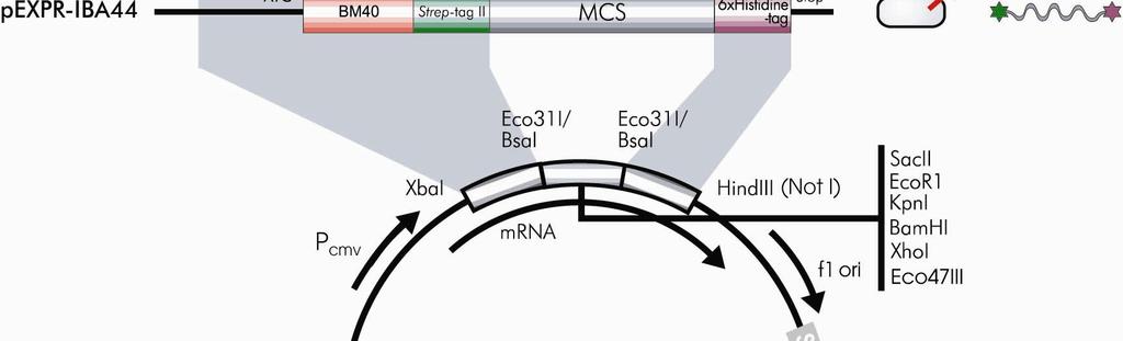
10 2.1 Expression cassettes of pexpr-iba vectors Overview of pexpr-iba vectors Sequences flanking the XbaI/HindIII restriction sites CAAT TATA forward primer GCAAATGGGCGGTAGGCGTGTACGGTGGGAGGTCTATATAAGCAGAGCTCTCTGGCTAACTAGAGAACCCACTGCTTACTGGCTTATCGAAATTA XbaI HindIII NotI reverse primer ATACGACTCACTATAGGGTCTAGA EC* AAGCTTGCGGCCGCAGATCTAGCTTAAGTTTAAACCGCTGATCAGCCTCGACTGTGCCTTCTAGTT *EC = expression cassette 10 Mammalian expression and purification systems using Strep-tag and/or 6xHistidine-tag
11 2.1.2 Multiple Cloning Sites pexpr-iba3 Eco31I PshAI PshAI Eco31I XbaI BsaI SacII EcoRI KpnI BamHI XhoI BsaI Eco47III HindIII TCTAGACCCACaatgGGAGACCGCGGTCCCGAATTCGAGCTCGGTACCCGGGGATCCCTCGAGGTCGACCTGCAGGGGGACCATGGTCTCAgcgcTTGGAGCCACCCGCAGTTCGAAAAATAATAAGCTT MetGlyAspArgGlyProGluPheGluLeuGlyThrArgGlySerLeuGluValAspLeuGlnGlyAspHisGlyLeuSerAlaTrpSerHisProGlnPheGluLysEnd Link Strep-tag II pexpr-iba5 Eco31I PshAI PshAI Eco31I XbaI NheI BsaI SacII EcoRI KpnI BamHI XhoI BsaI EcoRV HindIII TCTAGACCCACCATGGCTAGCTGGAGCCACCCGCAGTTCGAAAAAGgcgcCGAGACCGCGGTCCCGAATTCGAGCTCGGTACCCGGGGATCCCTCGAGGTCGACCTGCAGGGGGACCATGGTCTCTgataTCTAACTAAGCTT MetAlaSerTrpSerHisProGlnPheGluLysGlyAlaGluThrAlaValProAsnSerSerSerValProGlyAspProSerArgSerThrCysArgGlyThrMetValSerAspIleEnd Link Strep-tag II Link ArgProArgSerArgIleArgAlaArgTyrProGlyIleProArgGlyArgProAlaGlyGlyProTrpSerLeuIleSerAsnEnd AspArgGlyProGluPheGluLeuGlyThrArgGlySerLeuGluValAspLeuGlnGlyAspHisGlyLeuEnd pexpr-iba7 Eco31I PshAI PshAI Eco31I XbaI NheI BsaI SacII EcoRI KpnI BamHI XhoI BsaI EcoRV HindIII TCTAGACCCACCATGGCTAGCTGGAGCCACCCGCAGTTCGAAAAAATCGAAGGgcgcCGAGACCGCGGTCCCGAATTCGAGCTCGGTACCCGGGGATCCCTCGAGGTCGACCTGCAGGGGGACCATGGTCTCTgataTCTAACTAAGCTT MetAlaSerTrpSerHisProGlnPheGluLysIleGluGlyArgArgAspArgGlyProGluPheGluLeuGlyThrArgGlySerLeuGluValAspLeuGlnGlyAspHisGlyLeuEnd Link Strep-tag II Factor Xa GluThrAlaValProAsnSerSerSerValProGlyAspProSerArgSerThrCysArgGlyThrMetValSerAspIleEnd ArgProArgSerArgIleArgAlaArgTyrProGlyIleProArgGlyArgProAlaGlyGlyProTrpSerLeuIleSerAsnEnd pexpr-iba13 Eco31I PshAI PshAI Eco31I XbaI NheI BsaI SacII EcoRI KpnI BamHI XhoI BsaI EcoRV HindIII TCTAGACCCACCATGGCTAGCTGGAGCCACCCGCAGTTCGAAAAATCTGGTGGTGGTGGTGGTCTGGTTCCGCGTGGctccCGAGACCGCGGTCCCGAATTCGAGCTCGGTACCCGGGGATCCCTCGAGGTCGACCTGCAGGGGGACCATGGTCTCTgataTCTAACTAAGCTT MetAlaSerTrpSerHisProGlnPheGluLysSerGlyGlyGlyGlyGlyLeuValProArgGlySerArgAspArgGlyProGluPheGluLeuGlyThrArgGlySerLeuGluValAspLeuGlnGlyAspHisGlyLeuEnd Link Strep-tag II Spacer Thrombin GluThrAlaValProAsnSerSerSerValProGlyAspProSerArgSerThrCysArgGlyThrMetValSerAspIleEnd ArgProArgSerArgIleArgAlaArgTyrProGlyIleProArgGlyArgProAlaGlyGlyProTrpSerLeuIleSerAsnEnd pexpr-iba15 Eco31I PshAI PshAI Eco31I XbaI NheI BsaI SacII EcoRI KpnI BamHI XhoI BsaI EcoRV HindIII TCTAGACCCACCATGGCTAGCTGGAGCCACCCGCAGTTCGAAAAAGGCGCCGACGACGACGACAAGGGctccCGAGACCGCGGTCCCGAATTCGAGCTCGGTACCCGGGGATCCCTCGAGGTCGACCTGCAGGGGGACCATGGTCTCTgataTCTAACTAAGCTT MetAlaSerTrpSerHisProGlnPheGluLysGlyAlaAspAspAspAspLysGlySerArgAspArgGlyProGluPheGluLeuGlyThrArgGlySerLeuGluValAspLeuGlnGlyAspHisGlyLeuEnd Link Strep-tag II Enterokinase Link GluThrAlaValProAsnSerSerSerValProGlyAspProSerArgSerThrCysArgGlyThrMetValSerAspIleEnd ArgProArgSerArgIleArgAlaArgTyrProGlyIleProArgGlyArgProAlaGlyGlyProTrpSerLeuIleSerAsnEnd pexpr-iba42 Eco31I PshAI PshAI Eco31I XbaI NheI BsaI SacII EcoRI KpnI BamHI XhoI BsaI Eco47III HindIII TCTAGACCCACCATGAGG./.GCTCTGGCAGCTAGCAGAGGATCGCATCACCATCACCATCACGgcgcCGAGACCGCGGTCCCGAATTCGAGCTCGGTACCCGGGGATCCCTCGAGGTCGACCTGCAGGGGGACCATGGTCTCAgcgcTTGGAGCCACCCGCAGTTCGAAAAATAATAAGCTT MetArg./.AlaLeuAlaAlaSerArgGlySerHisHisHisHisHisHisGlyAlaGluThrAlaValProAsnSerSerSerValProGlyAspPrLeuGluValAspLeuGlnGlyAspHisGlyLeuSerAlaTrpSerHisProGlnPheGluLysEnd BM40 Link 6xHis-tag Link Link Strep-tag II pexpr-iba44 Eco31I PshAI PshAI Eco31I XbaI NheI BsaI SacII EcoRI KpnI BamHI XhoI BsaI HindIII TCTAGACCCACCATGAGG./.GCTCTGGCAGCTAGCTGGAGCCACCCGCAGTTCGAAAAAGgcgcCGAGACCGCGGTCCCGAATTCGAGCTCGGTACCCGGGGATCCCTCGAGGTCGACCTGCAGGGGGACCATGGTCTCAggccTGAGAGGATCGCATCACCATCACCATCACTAATAAGCTT MetArg./.AlaLeuAlaAlaSerTrpSerHisProGlnPheGluLysGlyAlaGluThrAlaValProAsnSerSerSerValProGlyAspProSerArgSerThrCysArgGlyThrMetValSerGlyLeuArgGlySerHisHisHisHisHisHisEnd BM40 Link Strep-tag II Link Link 6xHis-tag Please note: Restriction sites in bold cut twice and are useful for precise and oriented insertion of the recombinant gene by one cleavage reaction only. All the regions denoted with link carry a restriction site which may be useful for cutting out the recombinant gene for e.g. subcloning into pask-iba vectors for bacterial expression. The restriction enzyme cleavage site is not mentioned at the link region when such enzyme has at least one further site in the vector. Mammalian expression and purification systems using Strep-tag and/or 6xHistidine-tag 11
12 3 Expression pexpr-iba vectors are designed for high-level stable as well as non-replicative transient expression in most mammalian cells. 3.1 Transient expression in mammalian cells When the nucleotide sequence is confirmed, prepare enough DNA for the mammalian cell transfection to be carried out. The efficiency of transfection will vary depending on a number of parameters like: DNA purity host cell line used transfection method Choose a method that will give the highest efficiency for the chosen cell line. Protocols for transfection can be found in [6]. An alternative method is presented below (3.1.1 page 12). Depending on cell type and nature of the protein, expression levels of µg protein can be achieved per million cells (when low expression levels or low transfection efficiency are expected, cell numbers should be increased accordingly), usually sufficient for detection with a specific antibody in total lysates (see chapter 5, page 16). Once expression is verified, transfection of approximately 10 9 cells in general allows expression and purification of protein from mammalian cells in the milligram range. Some proteins however, can not be expressed at high levels 1. Cell lines latently infected with SV40 or expressing SV40 large T antigen like COS-1 and COS-7 cells allow episomal replication and thus longer expression periods. Material and important notes To estimate the number of plates of cells to be transfected, a few common examples are given below. Cell line Cell density (cells per cm 2 ) Required for 10 9 cells COS-7 3 x x 500 cm 2 plates CHO-K1 1,25 x x 500 cm 2 plates HEK-293 2,3 x x 500 cm 2 plates Transfection A large number of methods have been establishes for transient transfection. They differ in efficiency, cost and time requirements depending on the cell type used [6]. Among these, magnet assisted transfection (MATra) is a new, easy-to-handle and highly efficient method to transfect cells in culture. Using this new technique nucleic acids, such as plasmid DNA, 1 In some cases proteins can not be expressed at high levels, especially when they are toxic for the cells. Please refer to section 3.3 on page 13 for hints. 12 Mammalian expression and purification systems using Strep-tag and/or 6xHistidine-tag
13 oligonucleotides or sirna, are in a first step associated with magnetic particles. Exploiting magnetic force the full nucleic acid dose is then rapidly drawn towards and delivered into the target cells leading to efficient transfection. Further Information on MATra is available at Stable expression in mammalian cells pexpr-iba vectors contain a neomycin resistance gene to allow for selection of clones that have integrated the pexpr-iba DNA stably into their genome. Neomycin concentrations required need to be determined for each cell line (in advance). Determining expression levels in addition to Neomycin resistance is highly recommended. Depending on cell type and nature of the protein expression levels of µg protein can be achieved per million cells, usually sufficient for detection with a specific antibody in total lysates (see chapter 5, page 16). Some proteins however, can not be expressed at high levels. Please refer to "troubleshooting - expression" in the next section. 3.3 Trouble shooting Expression Possible causes for low expression levels and suggested solutions Problem Possible Cause Comments and Suggestions Low expression Sequence error, mutation Verify sequence and reading frame levels or no expression Protein is toxic Some proteins are inhibiting cell growth or induce apoptosis. In some cases signaling-inactive forms can be expressed at high levels. Protein lacks co-factors Try different cell types, when available cell types related to those endogenously expressing the protein in vivo. Known co-factors can be added to lysis buffer (e.g. metal ions). Protein is secreted Signal sequence present Remove all signal sequences from the coding region. Mammalian expression and purification systems using Strep-tag and/or 6xHistidine-tag 13
14 4 Preparation of soluble cell extracts 4.1 Preparation of soluble extracts after cytoplasmic expression of Strep-tag fusion proteins Material and important notes In general, addition of protease inhibitors is recommended. These are available against specific enzymes like trypsin or pepsin inhibitors or in cocktails. If activity of enzymes needs to be preserved specific inhibitors may be required like phosphatase inhibitors for kinases and other signaling molecules. Reducing agents may be added to prevent disulfide cross-linking. Lysis buffers need to be tested for any expressed protein. Strep-tag:Strep-Tactin binding is compatible with many reagents and detergents (see Table 1 on page 5). Buffers: Buffer W: (washing buffer): 100 mm Tris Cl, 150 mm NaCl, 1 mm EDTA, ph 8 It is recommended to work without EDTA when metalloproteins have been expressed. 5x SDS-PAGE sample buffer: 0.25 M Tris Cl, ph 8.0; 25% glycerol; 7.5% SDS, 0.25 mg/ml bromophenolblue; 12.5% v/v mercaptoethanol 1. Chill buffer W at 4 C. 2. Thaw the cell pellet for 15 minutes on ice and resuspend the cells in buffer W at 4 ml per 10 9 cells. 3. Lyse cells by repeated freeze/thaw cycles. Freeze cells in liquid nitrogen and thaw in a 37 C water bath. Repeat cycle five times. 4. Shear DNA by passing the lysate through a 18-gauge needle four times. (Optional) If the lysate is very viscous, add RNase A (10 µg/ml) and DNase I (5 µg/ml) and incubate on ice for min instead. 5. Centrifuge lysate at 3,000 x g for 15 minutes at 4 C to pellet the cellular debris. A certain proportion of the cellular protein, including the Strep-tag protein, may remain insoluble and will be located in the pellet. More complete recovery of the tagged protein needs optimization. E.g. mild detergents (Table 1) or higher ionic strength (e.g. 500 mm NaCl) may help. 6. Add 5 µl 5x SDS-PAGE sample buffer to 20 µl supernatant and store at -20 C for SDS-PAGE analysis. 7. Proceed to protocols for Strep-tag protein purification under native conditions (see protocol 6 page 21 ). 14 Mammalian expression and purification systems using Strep-tag and/or 6xHistidine-tag
15 4.2 Preparation of conditioned supernatant after secreted expression of Strep/6xHistidine-tag fusion proteins In addition to the Strep-tag, we have introduced the 6xHistidine-tag in some vectors creating double tag proteins. In a first purification step the 6xHistidine-tag serves for concentrating the recombinant protein out of the medium. As a secondary effect any biotin, which may be present in the medium, is removed. Since biotin binds to Strep-Tactin in competition to the Strep-tag, biotin in the medium would hamper direct purification of the recombinant protein via Strep-tag. Material and important notes In general, supernatants of transfected cells can be directly loaded on Ni-NTA cartridges after addition of 1M Tris ph 8, 3M NaCl, 100mM imidazol to a final concentration of 100 mm Tris ph 8, 300 mm NaCl, 10mM imidazol to shift the ph and adjusting ionic strength and after clearing the supernatant by centrifugation (see 7.2, page 33) Very dilute supernatants may need to be concentrated by ultrafiltration or ammonium sulfate precipitation before loading, especially when they are intended to be purified using gravity flow columns. This step can also be used to change the buffer to a ph of 8 required for the purification (see protocols 7 on page 30) For adherent cells: 1. Pipet supernatant off the cells and centrifuge at 3,000 x g for 15 minutes at 4 C to remove the cellular debris. Proceed to step 2. For suspension cells: 1. Centrifuge cell suspension at 750 x g for 10 minutes at 4 C to remove cells and cellular debris. 2. For SDS-PAGE analysis: Take 2 ml of the supernatant and concentrate 100-fold. Add 5 µl 5x SDS-PAGE sample buffer to 20 µl supernatant and store at -20 C until analysis. 3. Adjust ph (and ionic strength) of supernatant to purification conditions, i.e. by addition of 1/10th volume of 1M Tris/HCl, 3M NaCl, 100mM imidazol ph 8. Dilute samples should be concentrated prior to column purification by ultrafiltration. During this step the buffer change could be performed by adding the 10-fold volume of Ni-NTA Lysis Buffer (50 mm NaH 2 PO 4, 300 mm NaCl, 10 mm imidazol, ph 8) to the concentrate. 4. Proceed to protocols for Strep/6xHistidine-tagged protein purification under native conditions (see protocols 7 on page 30). If the target protein concentration is low, it is recommended to use a Ni-NTA cartridge of appropriate capacity connected to a FPLC workstation because large volumes are difficult to handle via syringe operated cartridges or gravity flow columns described under the above mentioned protocols. The second purification via the Strep-tag can then be performed via gravity flow or syringe operated cartridges by transferring the eluate of the Ni-NTA cartridge onto a Strep-Tactin cartridge or gravity flow column as described in the respective protocols. Mammalian expression and purification systems using Strep-tag and/or 6xHistidine-tag 15
16 5 Detection of Strep-tag fusion proteins 5.1 Detection of Strep-tag fusion proteins with Strep-Tactin Alkaline Phosphatase (AP) conjugate Material and important notes PBS buffer: 4 mm KH 2 PO 4, 16 mm Na 2 HPO 4, 115 mm NaCl, ph 7.4 PBS blocking buffer: PBS buffer with 3 % BSA and 0.5 % v/v Tween 20 PBS-Tween buffer: PBS buffer with 0.1 % v/v Tween 20 Reaction buffer: 100 mm NaCl, 5 mm MgCl 2, 100 mm Tris Cl, ph 8.8 NTB solution: 7.5 % w/v nitrotetrazolium blue in 70 % v/v dimethylformamid BCIP solution: 5 % w/v 5-bromo-4-chloro-3-indolyl-phosphate in dimethylformamid Strep-tag protein ladder can be used as positive control (cat. no ). For blocking biotinylated proteins use Biotin Blocking Buffer (cat. no ). 1. Perform SDS-PAGE and electrotransfer of the protein to an appropriate membrane. We recommend to use a nitrocellulose membrane. 2. Block the membrane with 20 ml PBS-blocking buffer. Incubate 1 h (room temperature; with gentle shaking) or overnight in the refrigerator (4 C). Do not use milk powder for blocking, because milk is one of the richest sources of biotin! 3. Wash 3 times with 20 ml PBS-Tween buffer (each step: 5 minutes, room temperature, gentle shaking). 4. After the last washing step, add 10 ml PBS-Tween buffer to the membrane. 5. Optional: Before detection of Strep-tag proteins (step 6) add 20 l Biotin Blocking Buffer (10 minutes, room temperature, gentle shaking). Endogenous biotinylated proteins are specifically blocked. 6. Add 2.5 l Strep-Tactin alkaline phosphatase conjugate (1:4000). Incubate 60 minutes at room temperature, gentle shaking. 7. Wash 2 times with PBS-Tween buffer (each step: 1 minute, room temperature, gentle shaking). 16 Mammalian expression and purification systems using Strep-tag and/or 6xHistidine-tag
17 8. Wash 2 times with PBS-buffer (each step: 1 minute, room temperature, gentle shaking). 9. Transfer membrane in 20 ml reaction buffer and add 10 µl NBT solution and 60 µl BCIP solution. 10. Proceed with the chromogenic reaction under shaking until optimal signal:background ratio is achieved. 11. Stop reaction by washing several times with distilled H 2 O. 12. Air dry the membrane and store it in the dark. 5.2 Detection of Strep-tag proteins with Strep-Tactin horse radish peroxidase (HRP) conjugates Material and important notes PBS buffer: 4 mm KH 2 PO 4, 16 mm Na 2 HPO 4, 115 mm NaCl, ph 7.4 PBS-blocking buffer: PBS buffer with 3 % BSA and 0.5 % v/v Tween 20 PBS-Tween buffer: PBS buffer with 0.1 % v/v Tween 20 Chloronaphtol solution: 3 % w/v 4-chloro-1-naphtol in methanol H 2 O 2 solution: 30 % v/v H 2 O 2 Strep-tag protein ladder can be used as positive control (cat. no ). For blocking biotinylated proteins use Biotin Blocking Buffer (cat. no ). 1. After SDS-PAGE and electrotransfer of the protein to an appropriate membrane block the membrane with 20 ml PBS-blocking buffer. Incubate: 1 h (room temperature; with gentle shaking) or overnight (4 C). Do not use milk powder for blocking, because milk is one of the richest sources of biotin! We recommend to use a nitrocellulose membrane. 2. Wash 3 times with 20 ml PBS-Tween buffer (each step: 5 minutes, room temperature, gentle shaking). 3. After the last washing step, add 10 ml PBS-Tween buffer to the membrane. 4. Optional: Before detection of Strep-tag proteins (step 5), add 20 l Biotin Blocking Buffer (10 minutes, room temperature, gentle shaking). Endogenous biotinylated proteins are specifically blocked. 5. Add 2.5 l Strep-Tactin horse horse radish peroxidase conjugate (1:4000). Incubate 60 minutes at room temperature, gentle shaking. Mammalian expression and purification systems using Strep-tag and/or 6xHistidine-tag 17
18 6. Wash 2 times with PBS-Tween buffer (each step: 1 minute, room temperature, gentle shaking). 7. Wash 2 times with PBS-buffer (each step: 1 minute, room temperature, gentle shaking). 8. Transfer membrane in 20 ml PBS buffer, add 200 l chloronaphtol solution and 20 l H solution. 9. Perform the chromogenic reaction under shaking. 10. Stop reaction by washing several times with distilled H 2 O. 11. Air dry the membrane and store it in the dark. 5.3 Detection of Strep-tag proteins with the Strep-tag II specific monoclonal antibody Material and important notes Use Strep-tag II purified monoclonal antibody solution (e.g.100 g/ml in PBS buffer) or crude cell culture supernatant solution PBS buffer: 4 mm KH 2 PO 4, 16 mm Na 2 HPO 4, 115 mm NaCl, ph 7.4 PBS blocking buffer: PBS buffer with 3 % BSA and 0.5 % v/v Tween 20 PBS-Tween buffer: PBS buffer with 0.1 % v/v Tween 20 Chloronaphtol solution: 3 % w/v 4-chloro-1-naphtol in methanol H 2 O 2 solution: 30 % v/v H 2 O 2 Rabbit anti-mouse antibodies (e.g. DAKO cat. no. P0161) Strep-tag protein ladder can be used as positive control (cat. no ). 1. After SDS-PAGE and electrotransfer of the protein to an appropriate membrane. We recommend to use a nitrocellulose membrane. 2. Block the membrane with 20 ml PBS-blocking buffer. Incubate 1 h (room temperature; with gentle shaking) or overnight (4 C). 3. Wash 3 times with 20 ml PBS-Tween buffer (each step: 5 minutes, room temperature, gentle shaking). 4. After the last washing step, add 10 ml PBS-Tween buffer to the membrane. 5. Add the specific monoclonal antibody to the solution to a final concentration of 0.2 g/ml. 18 Mammalian expression and purification systems using Strep-tag and/or 6xHistidine-tag
19 (e.g. 2 l of a specific monoclonal antibody solution (1 mg/ml) for 10 ml reaction volume) 6. Incubate 60 minutes at room temperature, gentle shaking. 7. Wash 3 times with PBS-Tween buffer (each step: 5 minute, room temperature, gentle shaking). 8. After last washing step, add 10 ml PBS-Tween buffer to the membrane. 9. Add 10 l rabbit anti mouse antibodies (e.g. DAKO cat. No. P0161) and incubate under shaking for 45 minutes 10. Wash 2 times with PBS-Tween buffer (each step: 1 minute, room temperature, gentle shaking). 11. Wash 2 times with PBS buffer (each step: 1 minute, room temperature, gentle shaking). 12. Transfer membrane in 20 ml PBS buffer, add 200 l chloronaphtol solution and 20 l H solution. 13. Perform the chromogenic reaction under shaking. 14. Stop reaction by washing several times with distilled H 2 O. 15. Air dry the membrane and store it in the dark. 5.4 Detection of Strep-tag proteins with the Strep-tag II specific monoclonal antibody conjugated to horse radish peroxidase (HRP) Material and important notes Use Strep-tag II purified monoclonal antibody solution at 1:4000 PBS buffer: 4 mm KH 2 PO 4, 16 mm Na 2 HPO 4, 115 mm NaCl, ph 7.4 PBS blocking buffer: PBS buffer with 3 % BSA and 0.5 % v/v Tween 20 PBS-Tween buffer: PBS buffer with 0.1 % v/v Tween 20 Chloronaphtol solution: 3 % w/v 4-chloro-1-naphtol in methanol H 2 O 2 solution: 30 % v/v H 2 O 2 Strep-tag protein ladder can be used as positive control (cat. no ). 1. After SDS-PAGE and electrotransfer of the protein to an appropriate membrane. We recommend to use a nitrocellulose membrane. Mammalian expression and purification systems using Strep-tag and/or 6xHistidine-tag 19
20 2. Block the membrane with 20 ml PBS-blocking buffer. Incubate 1 h (room temperature; with gentle shaking) or overnight (4 C). 3. Wash 3 times with 20 ml PBS-Tween buffer (each step: 5 minutes, room temperature, gentle shaking). 4. After the last washing step, add 10 ml PBS-Tween buffer to the membrane. 5. Add the specific monoclonal HRP conjugate at 1: Incubate 60 minutes at room temperature, gentle shaking. 7. Wash 2 times with PBS-Tween buffer (each step: 5 minute, room temperature, gentle shaking). 8. Wash 2 times with PBS buffer (each step: 1 minute, room temperature, gentle shaking). 9. Transfer membrane in 20 ml PBS buffer, add 200 l chloronaphtol solution and 20 l H solution. 10. Perform the chromogenic reaction under shaking. 11. Stop reaction by washing several times with distilled H 2 O. 12. Air dry the membrane and store it in the dark. 20 Mammalian expression and purification systems using Strep-tag and/or 6xHistidine-tag
21 6 Purification of Strep-tag fusion proteins 6.1 Purification of Strep-tag fusion proteins using gravity flow columns Material and important notes CV = column bed volume Strep-Tactin Sepharose, Superflow and MacroPrep can be used for gravity flow purification Binding capacity of each matrix is nmol recombinant protein per ml bed volume (100 nmol correspond to 2 mg of a 20 kda protein) Buffer W (washing buffer): 100 mm Tris Cl, 150 mm NaCl, 1 mm EDTA, ph 8 Buffer E (elution buffer): 100 mm Tris Cl, 150 mm NaCl, 1 mm EDTA, 2.5 mm desthiobiotin, ph 8 The composition of the lysis, wash and elution buffers can be modified to suit the particular application, e.g. by adding 0.1% Tween, 5-10 mm -mercaptoethanol, or 1 mm PMSF, or increasing NaCl or glycerol concentrations. The ph should not be lower than 7.5, though. For more information see Table 1 on page 5. Generally, it is recommended to perform chromatography at 4 C. Dependent on the individual equipment this is not always possible and chromatography has to be performed at room temperature. If columns are stored at 4 C and transferred to room temperature air bubbles may form since cold storage buffer is able to take up more gas than buffers at ambient temperature. Therefore, it is recommended to equilibrate the columns immediately after exposure to higher temperatures with buffer that is equilibrated at such temperatures. Column bed volume (CV) Protein extract volume* Washing buffer volume Elution buffer volume 0.2 ml ml 5 x 0.2 ml 6 x 0.1 ml 1 ml ml 5 x 1 ml 6 x 0.5 ml 5 ml ml 5 x 5 ml 6 x 2.5 ml 10 ml ml 5 x 10 ml 6 x 5 ml Table 4: Recommended buffer volumes for chromatography on Strep-Tactin columns *Adjust protein extract volume according to binding capacity of the column and apply the extract as concentrated as possible in the recommended volume range. 1. Equilibrate the Strep-Tactin column with 2 CVs Buffer W. Storage buffer is removed prior to equilibration. The column cannot run dry under gravity flow. Use buffer without EDTA for metalloproteins. Mammalian expression and purification systems using Strep-tag and/or 6xHistidine-tag 21
22 2. Centrifuge soluble extracts (14,000 rpm, 5 minutes, 4 C, microfuge). Insoluble aggregates which may have formed after storage may clog the column and thus have to be removed. 3. Add supernatant of soluble extracts to the column. The volume of the supernatants should be in the range of between 0.5 and 10 CVs (see Table 4 on page 21). Extracts of large volumes with the recombinant protein at low concentration may lead to reduced yields and should be concentrated prior to chromatography. Concentrated cell extracts are preferred; if quantification is possible, apply cell extract containing between 50 and 100 nmol recombinant Strep-tag II fusion protein per 1 ml CV. 4. Wash the column 5 times with 1 CV Buffer W, after the cell extract has completely entered the column. Collect the eluate in fractions having a size of 1 CV. Apply 2 µl of the first washing fraction and 20 l of each subsequent fraction to an analytical SDS-PAGE. 5. Add 6 times 0.5 CVs Buffer E and collect the eluate in 0.5 CV fractions. 20 l samples of each fraction can be used for SDS-PAGE analysis. The purified Strep-tag II fusion protein usually elutes in the 2 nd to 5 th fraction. Desthiobiotin and EDTA can be removed, if necessary, via dialysis or gel chromatography. 6.2 Purification of Strep-tag fusion proteins using Strep-Tactin Spin Columns Material and important notes The spin column matrix binds up to 4 nmol recombinant Strep-tag fusion protein (corresponding to 150 µg of a 37 kda protein (GAPDH-Strep-tag)) Buffer W (washing buffer): 100 mm Tris Cl, 150 mm NaCl, 1 mm EDTA, ph 8 Buffer BE (elution buffer): 100 mm Tris Cl, 150 mm NaCl, 1 mm EDTA, 2 mm D-biotin, ph 8 The composition of the lysis, wash and elution buffers can be modified to suit the particular application, e.g. by adding a mild detergent, a reducing reagent, protease inhibitors, glycerol or by modifying the ionic strength. The ph should not be lower than 7.5, though. For more information see Table 1 on page 5 Generally, it is recommended to perform protein purification at 4 C 22 Mammalian expression and purification systems using Strep-tag and/or 6xHistidine-tag
23 1. Centrifuge soluble extracts (13,000 rpm, 5 minutes, 4 C, microfuge). Insoluble aggregates which may have formed after storage may clog the column and thus have to be removed. 2. Equilibrate the Strep-Tactin Spin Column with 2x 500 µl Buffer W. Centrifuge at each step for 30 seconds at 700 x g (approx rpm). Discard the flow-through. This rehydrates the dried Strep-Tactin resin for the subsequent use. The spin column should be loaded with the soluble extracts containing Strep-tag proteins within 20 min, otherwise the capacity of the column might decrease. Use buffer without EDTA for metalloproteins. 3. Load up to 500 µl supernatant of soluble extracts onto the pre-equilibrated Strep-Tactin Spin Column. Centrifuge for 30 seconds at 700 x g (approx rpm). Collect the flow-through. Apply 2 µl to an analytical SDS-PAGE. Lysates with the recombinant protein at low concentration may lead to reduced yields and should be concentrated prior to chromatography. If quantification is possible, apply a volume of lysate containing between 3 and 5 nmol recombinant Strep-tag II fusion protein. For very concentrated cell lysates, it may be necessary to extend the centrifugation time to 3-4 minutes. 4. Wash the column 4 times with 100 µl Buffer W. Centrifuge at each step for 30 seconds at 13,000 rpm. Collect the flow-through. Apply 2 µl of the first washing fraction and 20 l of each subsequent fraction to an analytical SDS-PAGE. 5. Place the spin column into a fresh 1.5 ml reaction tube and choose one of the following procedures for elution: a. For maximum protein yield: Elute the recombinant protein by adding 3 times 150µl Buffer BE (Biotin-Elution-Buffer). At each step: First, centrifuge for 30 seconds at 700 x g (approx rpm) and finish with 15 seconds at maximum speed. Pool the eluates. b. For maximum protein concentration: Elute the protein with 50µl Buffer BE (Biotin-Elution-Buffer). First, centrifuge for 30 seconds at 700 x g (approx rpm) and finish with 15 seconds at maximum speed. Transfer the eluate from the collection tube onto the spin column and repeat the centrifugation step as above to maximize the yield. Mammalian expression and purification systems using Strep-tag and/or 6xHistidine-tag 23
24 6.3 Quick purification of Strep-tag fusion proteins using cartridges Material and important notes Cartridges filled with 1 ml or 5 ml Strep-Tactin Superflow or MacroPrep are available and can be run under some pressure using syringes or FPLC or HPLC workstations. Binding capacity of each matrix is nmol recombinant protein per ml bed volume (100 nmol of a 20 kda protein correspond to 2 mg) Buffer W (washing buffer): 100 mm Tris Cl, 150 mm NaCl, 1 mm EDTA, ph 8 Buffer E (elution buffer): 100 mm Tris Cl, 150 mm NaCl, 1 mm EDTA, 2.5 mm desthiobiotin, ph 8 The composition of lysis, wash and elution buffers can be modified to suit the particular application, e.g. by adding 0.1% Tween, 5-10 mm -ME, or 1 mm PMSF, or increasing NaCl or glycerol concentrations. The ph should not be lower than 7.5, though. For more information see Table 1 on page 5. Generally, it is recommended to perform chromatography at 4 C. Depending on the individual equipment this is not always possible and chromatography has to be performed at room temperature. If cartridges are stored at 4 C and transferred to room temperature air bubbles may form since cold storage buffer is able to take up more gas than buffers at ambient temperature. Therefore, it is recommended to equilibrate the cartridges immediately after exposure to higher temperatures with buffer that is equilibrated at such temperatures. Cartridges may be connected in series (maximally 3 cartridges) to enlarge capacity Protocol for running a 1 ml cartridge with a syringe. 1. Fill the cartridge inlet with Buffer W. 2. Connect a 10 ml syringe filled with Buffer W. Avoid the inclusion of air bubbles. 3. Inject 2 ml Buffer W with a flow rate of 30 drops/min to equilibrate the cartridge 4. Centrifuge the soluble extracts (14,000 rpm, 5 minutes, 4 C, microfuge) to remove aggregates that may have formed during storage. Insoluble aggregates which may clog the cartridge shall be removed. 5. Fill a syringe with the appropriate amount of the soluble extracts. 6. Remove the 10 ml syringe used for equilibration. 7. Fill the cartridge inlet with Buffer W and connect the cartridge with the soluble extracts. 8. Apply the soluble extracts with a flow rate of 20 drops/min. 24 Mammalian expression and purification systems using Strep-tag and/or 6xHistidine-tag