Directed differentiation of human embryonic stem cells
|
|
|
- Debra George
- 5 years ago
- Views:
Transcription


1 Directed differentiation of human embryonic stem cells TECAN Symposium 2008 Biologics - From Benchtop to Production Wednesday 17 September 2008 Andrew Elefanty Embryonic Stem Cell Differentiation Laboratory, MISCL
2 ES cells as a system to dissect development hes ES cells derived from inner cell mass of preimplantation blastocyst Functionally similar, patient specific cells generated by reprogramming adult or fetal cells: Somatic cell nuclear transfer (SCNT) - oocyte cytoplasm reprograms somatic cell Induced pluripotential stem cell (ips) reprogramming is initiated by genes and/or growth factors introduced into adult cells
3 ES cells can differentiate into all the cell types in the body blood cells differentiation liver heart muscle ES cell line pancreas nerve In vitro differentiation of embryonic stem cells provides an avenue to study early development generate tissue specific stem cells and mature end cells to study disease to screen drugs/therapeutic agents for cell therapies
4 Directed differentiation of ES cells hes Recapitulate embryonic development in vitro to derive lineage precursors Mesendoderm Ectoderm Mesoderm Endoderm
5 Factors that play key roles in embryogenesis also direct differentiation of ES cells hes BMP/Activin/Wnt/FGF Mesendoderm FGF/noggin BMP/Wnt Activin Ectoderm Mesoderm Endoderm
6 Elements that facilitate directed differentiation of HESCs in vitro large, uniform populations of undifferentiated HESCs robust, reproducible differentiation protocol incorporating a serum free defined medium permissive for evaluation of effects of growth factors availability of genetically modified ES cell lines to monitor differentiation or transplantation
")
7 Maintenance and expansion of HESCs T25-T75 'Cut and paste' for stock maintenance and transfer to bulk culture Enzymatic passage to establish bulk cultures Enzymatic passage for expansion for use in FACS analysis, differentiation, genetic manipulation Elefanty/Stanley Laboratory Enzymatic passage for short term maintenance (15-25 passages) T75-T150


8 Enzymatically expanded HESCs express stem cell markers HES 3 d0 Tra 1 60 SSEA 4 E Cadherin control CD 9 Elizabeth Ng, Robyn Mayberry & Amanda Bruce
9 Elements that facilitate directed differentiation of HESCs in vitro large, uniform populations of undifferentiated HESCs robust, reproducible differentiation protocol incorporating a serum free defined medium permissive for evaluation of effects of growth factors availability of genetically modified ES cell lines to monitor differentiation or transplantation


10 Spin EB differentiation of HESCs in APEL, a serum free culture medium APEL Medium a 'neutral' medium that permits evaluation of effects of GFs animal product free, containing only recombinant human proteins reproducible, quantifiable outcomes HESCs Ng et al Curr Protoc Stem Cell Biol, 2008; Ng et al Nat Protocols 2008


11 Reproducible EB formation in spin EBs generated in APEL medium EB size relates to cell input number Reproducible differentiation with different d0 d1 d2 d3 d4 media batches #2 #3 #4 Elizabeth Ng
12 Directed differentiation of ES cells to hematopoietic mesoderm hes Mesendoderm Ectoderm Hematopoietic Mesoderm Endoderm


13 BMP4 directs differentiation of ES cells to hematopoietic mesoderm hes BMP4 induces MIXL1 BMP4 induces hematopoietic mesoderm genes Mesendoderm Hematopoietic Mesoderm Marjorie Pick

14 BMP4 and VEGF are required for efficient formation of CD34+ cells and hematopoietic CFCs Hematopoietic Mesoderm Hematopoietic Progenitors Hematopoietic CFCs Marjorie Pick


15 Hematopoietic blast colony CFC are present transiently during differentiation BMP/VEGF/SCF SF methyl cellulose + VEGF/SCF/IL3/IL6/Epo Elizabeth Ng
16 Elements that facilitate directed differentiation of HESCs in vitro large, uniform populations of undifferentiated HESCs robust, reproducible differentiation protocol incorporating a serum free defined medium permissive for evaluation of effects of growth factors availability of genetically modified ES cell lines to monitor differentiation or transplantation
17 Genetic tags Provide reagents for quantifying the effects of specific growth factors. Provide simply visual assay during the course of ES cell differentiation- allowing the association between different cell types to be observed in real time. Provide a means to isolate and analyse viable cells at specific differentiation stages in vitro and in transplantation assays in vivo
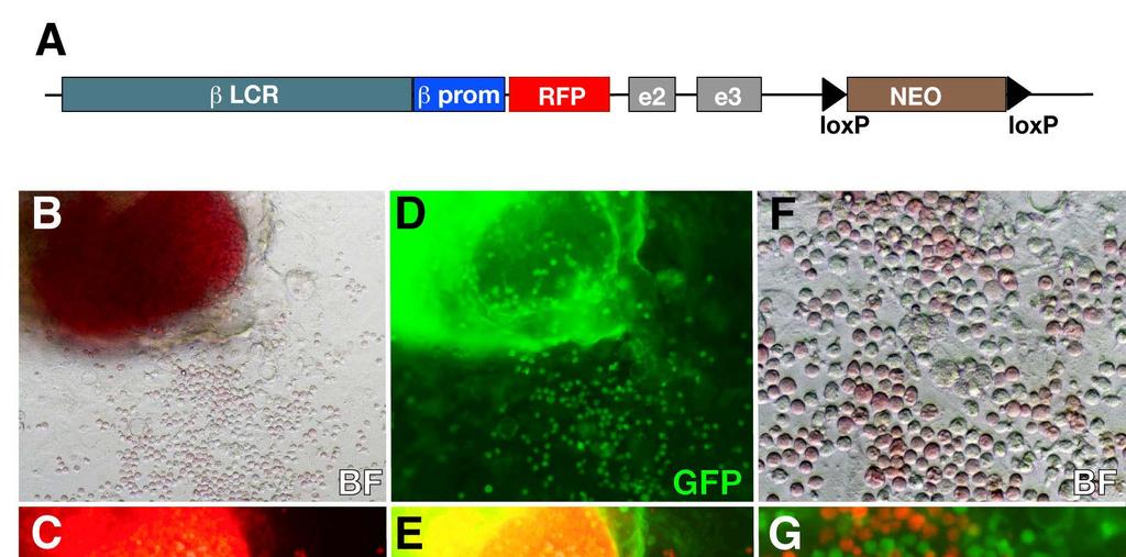
18 Generation of ErythRED HESC line


19 Time course of RFP expression in differentiating ErythRED HESCs RFP GFP CD34 CD45 RFP RFP + cells are GFP dim Most RFP + cells are CD34 - CD45 - Tanya Hatzistavrou


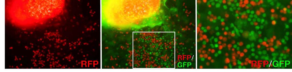
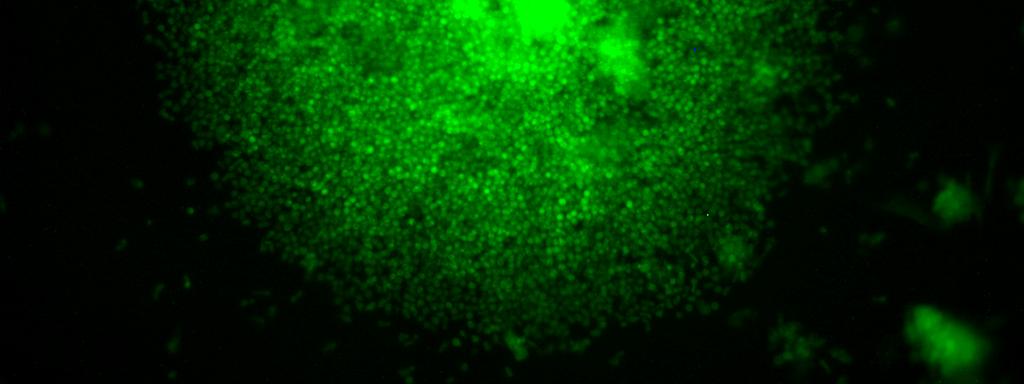
20 Characterisation of d14 sorted EB populations from ErythRED cells GFP dims during erythroid differentiation Tanya Hatzistavrou


21 Expression of erythroid genes is upregulated in RFP+ ErythRED cells * * * * Tanya Hatzistavrou

22 RFP expression is confined to erythroid cells in ErythRED hematopoietic colonies Tanya Hatzistavrou


23 Transplantation of d7 ErythRED EBs under the kidney capsule of immunodeficient mice d7 EBs Grafts harvested d7 d28 BMP/VEGF/SCF No teratomas RFP+ cells in 11/12 grafts at d28 Tanya Hatzistavrou


24 Donor RFP + ErythRED cells distinguished from host erythroid cells in kidney capsule grafts Tanya Hatzistavrou & Sue Micallef


25 Precursors of mesoderm and endoderm pass through the primitive streak during gastrulation BLOOD Inner Cell Mass Primitive endoderm Epiblast Embryonic ectoderm Primitive Streak Extraembryonic and Embryonic mesoderm Definitive endoderm ES cells Primitive Streak MIXL1 MIXL1 is expressed in the Primitive Streak


26 Targeting GFP to MIXL1 locus in HESC lines Clone Validation Excision of selectable marker Single cell cloning Karyotypic analysis Expression of ES cell markers Formation of teratomas Sequencing of genomic junctions Southern Blotting (exclude multiple integration) Richard Davis


27 Gene expression in differentiating MIXL1 MIXL1 GFP/wt cells Richard Davis


28 Correlation between GFP and MIXL1 protein expression in differentiating MIXL1 GFP/w HESCs GFP/w HESCs Richard Davis & Elizabeth Ng


29 Hematopoietic CFCs are enriched in the GFP + PDGFRa + fraction of differentiating d4 MIXL1 GFP/w EBs Richard Davis & Elizabeth Ng


30 Application of robotics to HESC differentiation Stage 2: Spin EB differentiation Enzymatic passage for expansion for use in FACS analysis, differentiation, genetic manipulation Stage 1: HESC maintenance Spin EB set up Growth factor addition and removal Harvesting EBs for further culture or analysis Stage 3: High content image analysis


31 Reporter targeted HESC lines for high content screening analysis


32 Reporter targeted HESC lines for high content screening analysis

33 Reporter targeted HESC lines for high content screening analysis fluorescent signal specificity
34 Summary Enzymatic HESC expansion and passaging enables generation of sufficient cell numbers for differentiation, analysis and genetic manipulation Differentiation in serum free animal product free medium (APEL) using a robust multiwell format (spin EB) enables unbiased assessment of growth factors influencing differentiation Lineage specific fluorescent reporter cells enable objective assessment of differentiation, in vitro and in vivo Protocols lend themselves to application of robotics and high content screening assays


35 Embryonic Stem Cell Differentiation Laboratory, MISCL Maggie Tanya Michael Lloyd Andrew Sue Mei Ed Alex Marjorie Karin Anna Claire Richard Amanda Aude Lisa Elizabeth Xueling Sue Steve Robyn Kathy


36 Research supported by: The Australian Stem Cell Centre Juvenile Diabetes Research Foundation National Health and Medical Research Council