Cellular phenotyping and application of cytometry for diagnostics purposes Part I. dr n. med. Karolina Bukowska-Straková
|
|
|
- Darlene Porter
- 5 years ago
- Views:
Transcription

1 Cellular phenotyping and application of cytometry for diagnostics purposes Part I dr n. med. Karolina Bukowska-Straková
2 Flow cytometry - wikipedia definition ;-) Flow cytometry (abbreviated: FCM) is a technique for counting and examining microscopic particles, such as cells and chromosomes, by suspending them in a stream of fluid and passing them by an electronic detection apparatus. It allows simultaneous multiparametric analysis of the physical and/or chemical characteristics of up to thousands of particles per second. Flow cytometry is routinely used in the diagnosis of health disorders, especially blood cancers, but has many other applications in both research and clinical practice. A common variation is to physically sort particles based on their properties, so as to purify populations of interest.

3

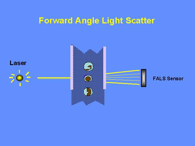
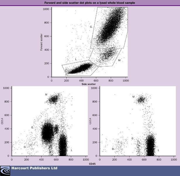
4 Forward scatter - FSC
5

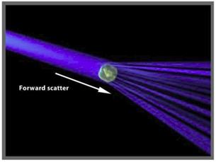
6 Forward scatter - FSC
7 Forward scatter - FSC

8 Forward scatter - FSC
9

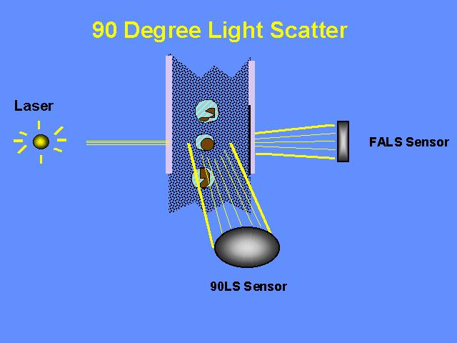
10 Side scatter - SSC

11 Side scatter - SSC
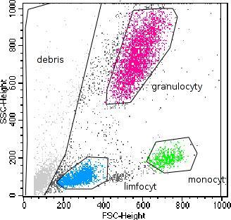
12 FSC versus SSC
13 Monoclonal antibodies conjugated with fluorochrome
14 Monoclonal antibodies (mabs or moabs)

15 Monoclonal antibodies (mabs or moabs): - made by identical immune cells that are all clones of a unique parent cell - monospecific monovalent affinity bind to the same epitope. José M. Casasnovas, Mykol Larvie and Thilo StehleThe EMBO Journal (1999) 18,
16 Monoclonal antibodies (mabs or moabs): The same: isotype, allotype, idiotype AllotypeIndividual differences in amino acid sequence (polymorphisms) Idiotype differences in Ag recognition Isotype different heavy chains: µ, δ, γ, ε, α or light chains: κ, λ
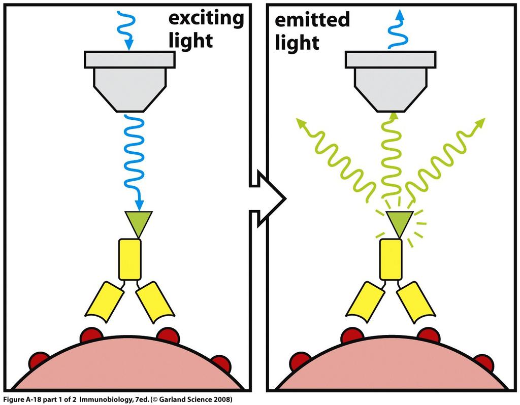
17 Fluorochrome excitation

18 Flow cytometry
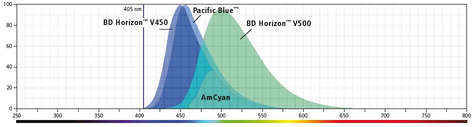
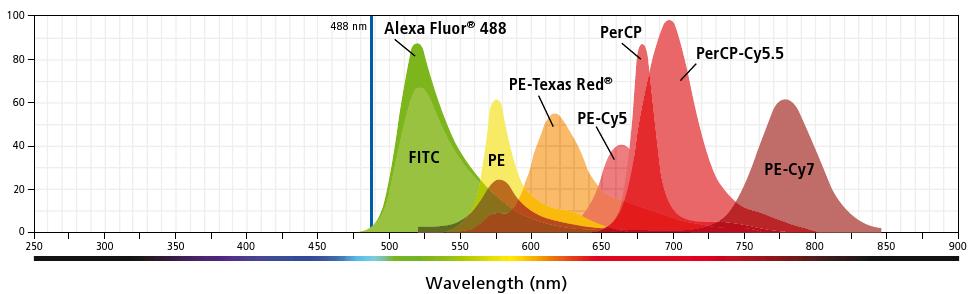
19 Lasers / fluorochroms
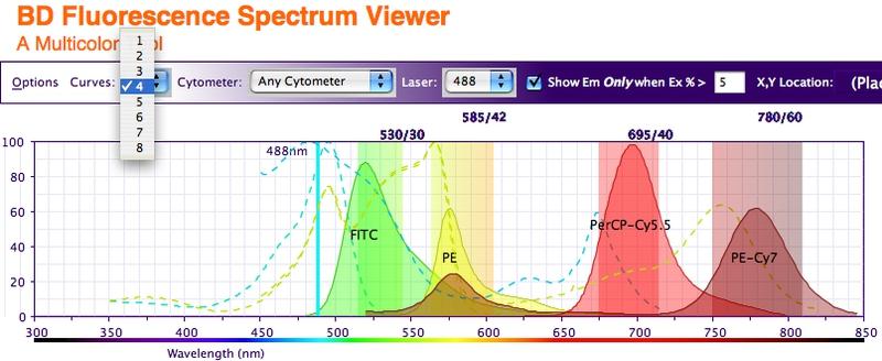
20 Fluorochrome choose
21 Mean fluorescence intensity
22
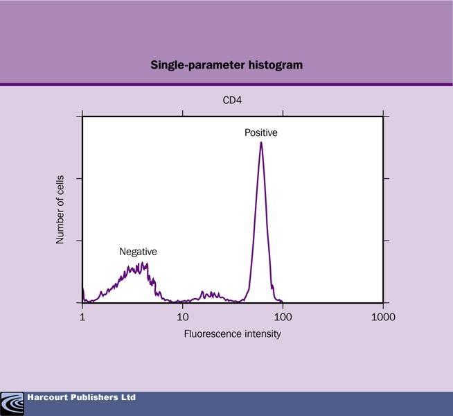
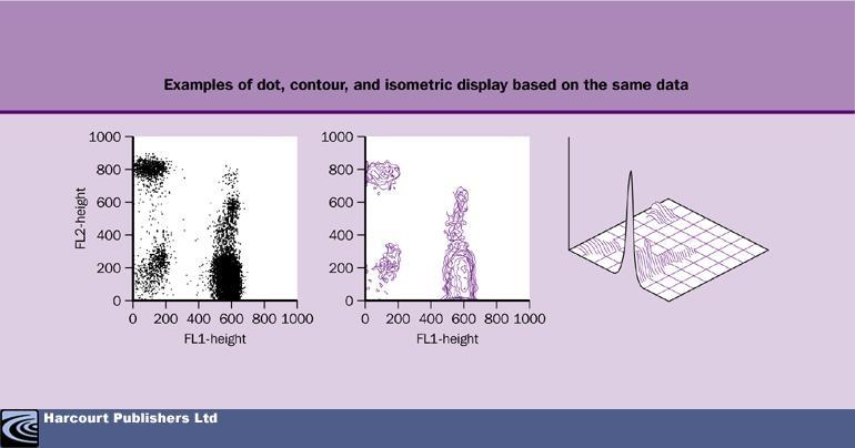
23 Data presentation
24

25 Our instrument LSR II 10 colors
26 Immunophenotyping of leukocytes Flow cytometric immunophenotyping with (monoclonal) antibodies allows the recognition of leukocyte subsets. The CD nomenclature has created clarity in the field of membrane bound leukocyte antigens (but not for intracellular markers) These CD antibodies recognise many different types of markers (lineage specific, differentiation stage specific, etc) and thereby allow the recognition of many different (immature and mature) leukocyte subsets. Usage of multiparameter analyses allow the dissection of differentiation and maturation pathways as well as the detecion of specific proteins (see: Van Lochem et al. Cytometry 2004;60B:1-13).
27 Classical leukocyte markers Immature markers T-cell markers CD34: precursor marker CD1: common thymocyte marker TdT: CD2: pan-t-cell marker terminal deoxnucleotidyl transferase CD117: myeloid precursor marker CD4: helper T-cell marker HLA-DR: precursor cells (and APC cell) CD8: cytotoxic T-cell marker CD3: mature T-cell marker B-cell markers TCR: T-cell receptor CD10: immature B-cell marker CD19: pan-b-cell marker Myeloid markers CD20: mature B-cell marker CD13 and CD33: pan-myeloid SmIg: membrane bound Ig CD14: monocytic marker CyIg: cytoplasmic Ig CD15: granulocytic marker
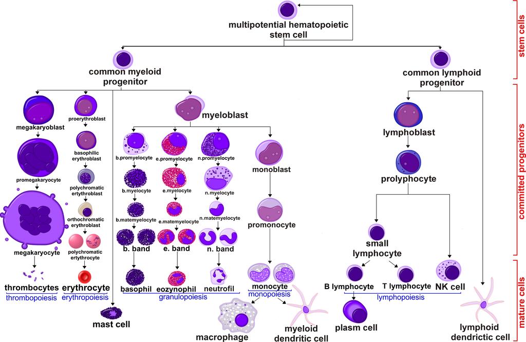
28 Hematopoiesis
29 Physiological balance of hematopoiesis Its regulation depends on: cytokines, celluler interactions, transcription and metabolic factors
30 Hematopoietic cells may be missing or do not function properly Immunodeficiencies
31 Flow cytometry in primary immunodeficiency Flow cytometric immunphenotyping and functional studies of blood / bone marrow / other material Targeted analysis of immune cells: 1) are all cells present in normal frequencies; 2) do they have all relevant proteins expressed; 3) is their function normal?


32 Primary immunodeficiencies (PID) DNA repair defects Defects in innate immunity Congenital defects in fagocyte number or/and function Other well defined immunodeficiency syndromes Diseases of immune dysregulaton Complement deficiencies Alternative complement pathway defects Regulatoty proteins of complement Periodic fever syndromes Predominantly antibody deficiencies Combined T and B-cell defects

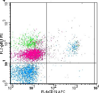
33 Peripheral blood lymphocyte subpopulations


34 Enumeration of lymphocyte subsets in PB healthy control

35 Enumeration of lymphocyte subsets in PB SCID T-B+NK- (common γ chain)


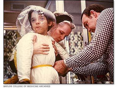
36 David Vetter bubble boy
37 Enumeration of lymphocyte subsets in PB SCID T-B-NK- (ADA deficiency)
38 Enumeration of lymphocyte subsets in PB XLA patient

39 Proliferation assay Lymphocytes Culture with Ag/ mitogen CSFE, 10 min, 37C wash free CSFE culture Signal from unstimulated cells

40 Proliferation assay Lymphocytes CSFE, 10 min, 37C wash free CSFE Culture with Ag/ mitogen
41
42

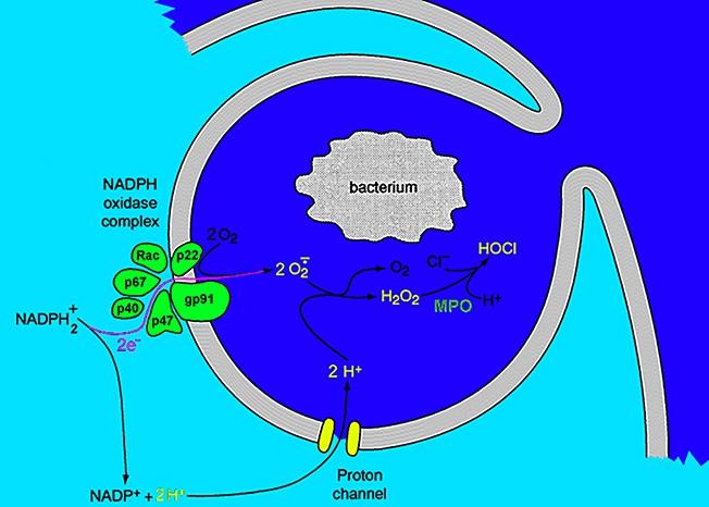
43 Chronic granulomatous disease NADPH oxidase Medical Immunology :4
44 CGD anti-gp91phox Healthy X-linked CGD

45 Reactive oxygen species FCM measurement (superoxide anion) +

46 Reactive oxygen species FC measurement (hydrogen peroxide)
47 Hematopoietic cells may undergo malignant transformation into an abnormally proliferating leukemic cells Leukemia Lymphoma
48 Leukemias and Lymphomas Leukemias (acute or chronic) and lymphomas can be regarded as malignant counterparts of normal hematopoietic cells in different maturation stages.
49 L BAL T - AL L
50 ALL -Acute lymphoblastic leukemia B-ALL from B lineage T-ALL from T lineage AML - Acute myeloid leukemia M0 M1 M2 M3 M4 minimally differentiated acute myeloblastic leukemia acute myeloblastic leukemia, without maturation acute myeloblastic leukemia, with granulocytic maturation promyelocytic, or acute promyelocytic leukemia (APL) acute myelomonocytic leukemia M4eo myelomonocytic together with bone marrow eosinophilia M5 (M5a) acute monoblastic leukemia (M5b) acute monocytic leukemia M6 (M6a) acute erythroid leukemias, including erythroleukemia (M6b) very rare pure erythroid leukemia M7 acute megakaryoblastic leukemia
51 Flow cytometry in hematology Immunophenotyping plays an important role in establishing the diagnosis and classifying hematopoietic malignancies and is a basic investigation, which precisely defines the lineage and stage of differentiation of malignantly transformed hematopoietic cells.

52 CD45 expression PB vs normal BM
53 Pro-B-cell CD34 TdT CD22 Pre-B-I-cell (Pre-pre-B-cell) CD34 TdT CD10bright CD19 CD22 CD45dim Pre-B-II-cell (Pre-B-cell) Immature B-cell (tr. Pre-B-cell) Mature B-cell Plasma cell (TdT) CD10 CD19 CD20-/dim CD22 CD45 CyIgµ CD10dim CD19 CD20 CD22 CD45bright SmIgµ CD19 CD20 CD22bright CD45bright SmIgµ CD19 CD45 Cy/SmIg
 Mature B-cell Plasma cell (TdT) CD10 CD19 CD20-/dim CD22 CD45 CyIgµ CD10dim CD19 CD20 CD22 CD45bright SmIgµ CD19 CD20 CD22bright CD45bright SmIgµ")
54 Pro-B-cell CD34 TdT CD22 Pre-B-I-cell (Pre-pre-B-cell) CD34 TdT CD10bright CD19 CD22 CD45dim Pre-B-II-cell (Pre-B-cell) Immature B-cell (tr. Pre-B-cell) Mature B-cell Plasma cell (TdT) CD10 CD19 CD20-/dim CD22 CD45 CyIgµ CD10dim CD19 CD20 CD22 CD45bright SmIgµ CD19 CD20 CD22bright CD45bright SmIgµ CD19 CD45 Cy/SmIg


55 Pro-B-cell Pre-B-I-cell (Pre-pre-B-cell) Pre-B-II-cell (Pre-B-cell) Immature B-cell (tr. Pre-B-cell) Mature B-cell Plasma cell Physiological shift in B-cell compartment Normal B-cell development Age-related shifts in B-cell compartment of elderly individuals Regenerating B cell precursor in bone marrow after cytotoxic treatment
56 1. AUL 2. prob ALL 3. call 4. preb ALL 5. Trans. preb 6. B-CLL 7. HCL 8. BL, 1. DLBL, MALT 9. LPL 10. MM 3. Lymphoid differentiation Immature T-ALL 12. c T-ALL 13. Mature T-ALL 14. T-LGL 15. NK-LGL

57 Myeloid differentiation A. Plesa, 2008


58 Myelo/monoblast CD34 CD117 HLA-DR CD13 CD33 Pro-monocyte Monocyte Macrophage HLA-DR CD13high CD33high CD11b CD15dim HLA-DR CD13high CD33high CD11b CD15dim CD14 HLA-DR CD13 CD33 CD11b CD14


59 Myelo/monoblast CD34 CD117 HLA-DR CD13 CD33 Promyelocyte Myelocyte Metamyelocyte Neutrophil CD13dim CD33dim CD15 CD11b CD13 CD33dim CD15 CD11b CD16 CD13high CD33 CD15 CD11bhigh CD16high CD117 CD13high CD33high CD15
60 Myeloid differentiation 0. AML-M0 1. AML-M1 2. AML-M2 3. AML-M3 4. AML-M4 5. AML-M5 6. AML-M6 7. AML-M
61 Thank you for your attention =)