Post-translational modification
|
|
|
- Dwayne Glenn
- 5 years ago
- Views:
Transcription
1 Protein expression Western blotting, is a widely used and accepted technique to detect levels of protein expression in a cell or tissue extract. This technique measures protein levels in a biological sample through antibody binding to a specific protein of interest. The precise binding that occurs between an antibody and its target protein epitope allows detection of highly specific amino acid sequences within a protein. 1
2 Post-translational modification Antibodies can also detect specific posttranslational modifications (PTM) of a protein. Phospho-specific antibodies have been used to identify components of specific signaling pathways and to study changes in phosphorylation events in various biological contexts. Antibodies specific to other PTMs have been developed recently, allowing researchers to monitor changes in acetylation, methylation, and ubiquitination status of a protein. 2
3 Primary antibody validation A poor primary antibody can result in dirty, uninterpretable or misleading results. To be confident that an antibody is specific to the target of interest, the antibody specificity should be validated. It takes more than just seeing a band at the expected molecular weight to validate antibody specificity. Antibodies should undergo a stringent validation procedure using a number of different approaches to ensure that the antibody detects the target accurately. 3
4 Be in Control of Your Western Blot An important consideration in any experiment is the inclusion of appropriate controls. 4
5 Primary antibody specificity validation includes: Specificity testing on cell and tissue extracts with documented protein expression levels not just on recombinant protein; Specificity confirmation through the use of sirna transfection or knockout cell lines; Specificity testing of antibodies directed against a post-translational modification by treatment of cell lines with growth factors, chemical activators, or inhibitors, which induce or inhibit target expression. Phospho-specificity testing of phospho-antibodies by phosphatase treatment; 5
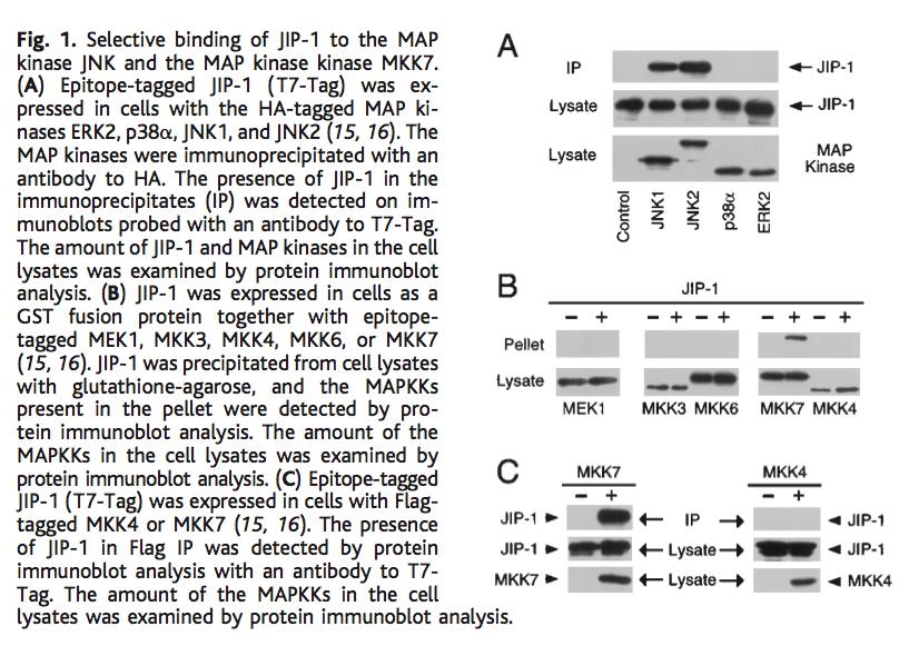
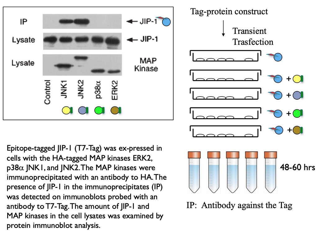
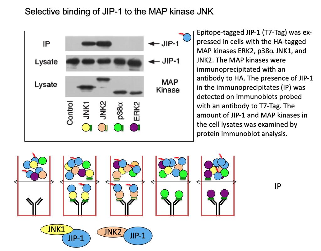
 (Thr202/Tyr204) antibodies are evaluated using negative (U0126-treated) and positive (TPA-treated) Jurkat lysate, as well as")
6 Positive and negative controls ensure confidence that your antibody is detecting a specific signal. mab anti-p-erk1/2 Provider 1 Provider 2 Phospho-p44/42 MAPK (Erk1/2) (Thr202/Tyr204) antibodies are evaluated using negative (U0126-treated) and positive (TPA-treated) Jurkat lysate, as well as purified recombinant tyrosine-phosphorylated proteins. TPA: small molecule drug that activates the signal transduction enzyme protein kinase C (PKC) and ERK phosphorylation 6
 and positive (TPA-treated) Jurkat lysate, as")
7 Positive and negative controls ensure confidence that your antibody is detecting a specific signal. mab anti-p-tyrosine Provider 3 Provider 4 Phospho-tyrosine antibodies are evaluated using negative (U0126-treated) and positive (TPA-treated) Jurkat lysate, as well as purified recombinant tyrosine-phosphorylated proteins. 7
8 Loading controls are used to: Ensure equal loading of a gel Ensure Integrity of the sample Quantitatively compare samples 8

9 Ensure equal loading of a gel Proteins that express well across many cell lines and tissues, such as β-actin, α-tubulin, and GAPDH * are often used as loading controls in experiments designed to compare total protein levels across multiple samples. ( * ) Glyceraldehyde 3-phosphate dehydrogenase 9

10 Ensure equal loading of a gel Cellular fractionation markers should be used when you are preparing nuclear and cytoplasmic extracts to confirm that the lysate was prepared appropriately. Histone H3 is a nuclear protein and Lamin A is a nuclear membrane protein; both of these offer strong nuclear fraction markers. Cytoplasmic proteins like MEK1/2, GAPDH and cytoskeletal proteins like β-actin and α/β-tubulin are common cytoplasm markers. DOI: /mBio Decreased nuclear association of VP16-GFP and HCF-1 in the absence of lamin A/C. (Left) Lmna+/+and Lmna / MEFs were infected with the HSV-1 DG1 virus at an MOI of 50 in the presence of cycloheximide (100 µg/ml), harvested at 3 hpi, and fractionated into cytoplasmic and nuclear fractions. Cytoplasmic and nuclear fractions were loaded at a 1:2 ratio onto an SDS-polyacrylamide gel, and the proteins were resolved in the gel. VP16-GFP and HCF-1 were detected by Western blotting with GFP- and HCF-1-specific antibodies, respectively. GAPDH and lamin B1 were detected as fractionation and loading controls

11 Ensure equal loading of a gel Blot with antibodies against the protein of interest is used as control for the loading control of enzymatic modification of the protein, such as phosphorylation. 11


12 Western blot analysis of EGF Receptor Blot 1 Blot 2 Cell extracts using EGF Cell extracts using Phospho- receptor mab EGF receptor (Tyr1068) mab 12

13 Western blotting Dot blotting 13
14 IP and co-ip Immunoprecipitation and co-immunoprecipitation 14
15 Immunoprecipitation IP is an affinity purification technique widely used to enrich for a protein of interest for further experiments such as WB and Co-IP techniques but also mass spectrometry or structure-function studies. 15
16 IP flowchart 16
 to investigate its identity, structure, expression, or activation or modification state")
17 Immunoprecipitation: affinity purification technique Used to isolate a single protein (the target antigen of the antibody) to investigate its identity, structure, expression, or activation or modification state 17
18 Co-IP versus IP Co-IP is conducted in essentially the same manner as IP, except that antigen precipitated by the antibody is used to co-ip its binding partner(s) or associated protein complex from the lysate. The assumption that is usually made when associated proteins are co-ip is that these proteins are related to the function of the target antigen at the cellular level. This is only an assumption, however, that is subject to further verification. 18
19 Co-IP flowchart 19
. In these methods, the goal is to study the interactors or associated cellular components that are bound to the primary antigen.")
20 Co-IP Co-IP: used to study the interactions of the primary antigen protein with other proteins or nuclei acids (chromatin IP also called ChIP). In these methods, the goal is to study the interactors or associated cellular components that are bound to the primary antigen. 20
21 21
22 Example of scientific questions: Is there a protein-protein association between receptor and signal molecule after treatment? T=0? T=1, T=5, T=10, T=30 min? Hypothesis n.1? Hypothesis n.2 22
23 Hypothesis n.1 Hypothesis n.2 1) Treatment with Ab anti-receptor Y Y 2) Immunoprecipitation Y Y 3)Dissociation with SDS- PAGE loading buffer Surnatant Y Y Pellet 23
24 IP versus WB 24

25 IP versus WB 25


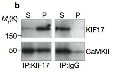
26 Surnatant Pellet a-gfr a-grb2 IP:GFR IP: IgG S P 26

27 Pioneer experiment 27
28 28
29 Tagged-proteins 29
30 Tagged-proteins 30
31 When are Tag-proteins used? Ab against endogenous protein not available to distinguish recombinant constructs from endogenous proteins 31
32 32
33 33
34 34
35 35