Structure of IgG and IgM
|
|
|
- Joy Oliver
- 5 years ago
- Views:
Transcription
1

2 Structure of IgG and IgM Fig. 5-1 A,B

3 Crystal Structure of Secreted IgG Fig. 5-1 C

4 Structure of an Ig Domain Fig. 5-2
5 Proteolytic Fragments of IgG (1) Fig. 5-3A
6 Proteolytic Fragments of IgG (2) Fig. 5-3B
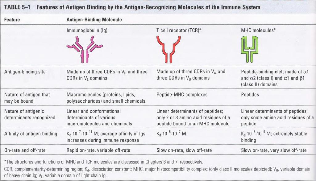
7 Human antibody isotypes Tab. 5-2

8 Examples of Ig Superfamily Proteins Fig. 5-4
9 Ig Light Chain Hypervariable Regions Fig. 5-5

10 Binding of an Antigen by an Antibody Fig. 5-6 A,B

11 Antigen and Antibody Binding Surfaces Fig. 5-6 C

12 Flexibility of Antibody Molecules Fig. 5-7

13 Membrane and Secreted Forms of IgG Fig. 5-8
14 Identification of phenotypic markers unique to particular cell types. The basis for the modern classification of lymphocytes and other leukocytes is the recognition of individual cell populations by specific monoclonal antibodies. These antibodies have been used to define clusters of differentiation (CD) markers for various cell types Immunodiagnosis. The diagnosis of many infectious and systemic diseases relies on the detection of particular antigens or antibodies in the circulation or in tissues by use of monoclonal antibodies in immunoassays Tumor detection. Tumor-specific monoclonal antibodies are used for detection of tumors by imaging techniques and by staining tissues with labeled antibodies.
15 Therapy. Advances in medical research have led to the identification of cells and molecules that are involved in the pathogenesis of many diseases. Monoclonal antibodies, because of their exquisite specificity, provide a means of targeting these cells and molecules. A number of monoclonal antibodies are used therapeutically today. Some examples include antibodies against the cytokine tumor necrosis factor (TNF) used to treat rheumatoid arthritis and other inflammatory diseases, antibodies against CD20 for the treatment of B cell leukemias and for depleting B cells in certain autoimmune disorders, antibodies against the type 2 epidermal growth factor receptor to target breast cancer cells, antibodies against vascular endothelial growth factor (a cytokine that promotes angiogenesis) in patients with colon cancer, and so on
16 Tumor detection. Tumor-specific monoclonal antibodies are used for detection of tumors by imaging techniques and by staining tissues with labeled antibodies. Functional analysis of cell surface and secreted molecules. In biologic research, monoclonal antibodies that bind to cell surface molecules and either stimulate or inhibit particular cellular functions are invaluable tools for defining the functions of surface molecules, including receptors for antigens. Monoclonal antibodies are also widely used to purify selected cell populations from complex mixtures to facilitate the study of the properties and functions of these cells.
17 Generation of Monoclonal Antibodies (1) Fig. 5-9

18 Generation of Monoclonal Antibodies (2) Fig. 5-9

19 Ig Expression During B Cell Maturation Fig. 5-10

20 FcRn Prolongs Half-Life of IgG Molecules Fig. 5-11
21 Types of Antigenic Determinants (1) Fig A,B
22 Types of Antigenic Determinants (2) Fig C

23 Valency and Avidity of Antibodies Fig. 5-13
24 Antigen-Antibody Complexes Fig. 5-14

25 Changes in Ig During Humoral Responses Fig. 5-15