Morphological Investigations - Different Microscopic Techniques (Semicrystalline Polymers)
|
|
|
- Lenard Mitchell
- 5 years ago
- Views:
Transcription


1 Morphological Investigations - Different Microscopic Techniques (Semicrystalline Polymers) Method SEM TEM AFM Typical Sample Preparation Evaporation Surface Etching Ultramicrotomy Selective Staining no special, but very flat surfaces are necessary Typical Results HDPE/EOC blends 1 µm

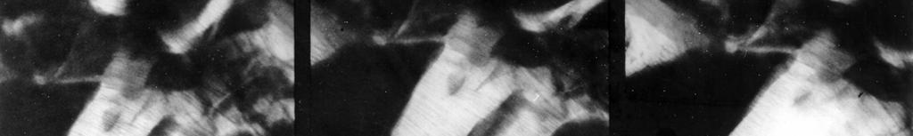
2 Results Morphology: SEM! 500nm " 500nm
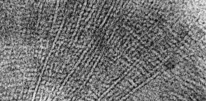
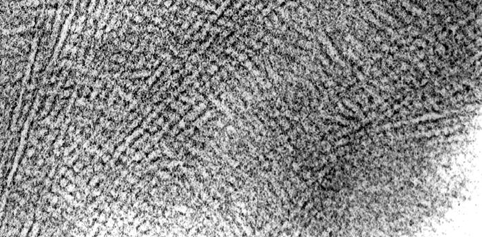


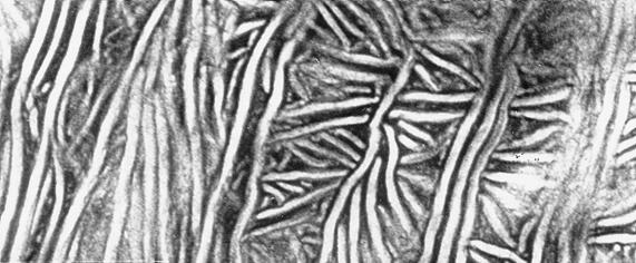
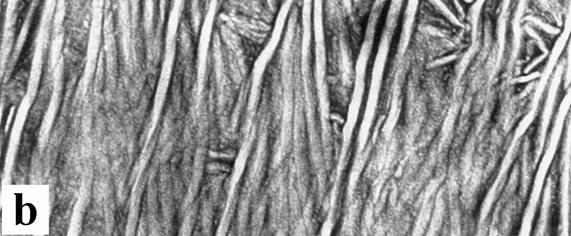
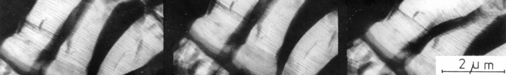
3 Results Morphology: TEM 50 nm 50 nm! "
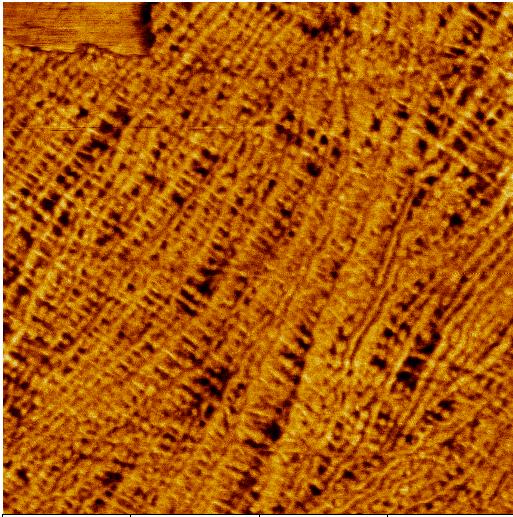

4 Results Morphology: AFM 0! 1 #m 0 " 1 #m
5 II. Sample Preparation Surfaces Cryo Ultramicrotomy Fixation and Staining of Polymers
6 EM investigation of morphology 1. Usual techniques special surfaces brittle fracture surfaces, smooth and selectively etched surfaces bulk polymer (ultra-) thin sections by (cryo-) ultramicrotomy, after fixation and staining 2. Special techniques Irradiation effects $- or electron irradiation at T % RT fixation and contrast enhancement Straining effects Straining of (semi-) thin sections contrast enhancement replication TEM coating SEM ESEM, AFM TEM HVEM AFM
7 Bulk Polymeric Material preparation of surfaces preparation of thin sections surfaces of fibres, foils; freely crystallized from melt or solution polished or sectioned surfaces (by microtomy, polishing) fracture surfaces (fracture at low temperatures,...) fixation, hardening: chemical (cross-linking...) physical (cooling, cross-linking...) mechanical (embedding) effects selective etching (chemical, physical etching) selective staining: selective chemical reactions, physical effects structured surfaces (cryo-) ultramikrotomy replica (one-stage, two-stage) evaporating, conductive layer ultra-thin sections semi-thin sections TEM SEM SFM TEM SFM HVEM

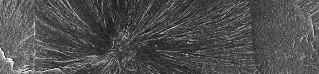

8 Surface of!ipp after permanganic etching, SEM


9 Usual brittle fracture PP / CaSO 4 - Composite Soft matrix fracture


10 Cryo-Ultramicrotome UCT (Fa. Leica)


11 Principle C specimen A specimen arm knife B knife holder specimen holder A - specimen cut with controlled speed B - retraction C - advance of specimen arm determines the specimen thickness specimen holder Specimen Knife specimen arm

 thin")


12 Cutting a) sample thin section water sample knife thin section b) thin section cutting water sample water knife c) sample thin section knife knife water retraction
13 Important Staining Agents for Polymers Polymer Polyolefins Polyamide ClSA / OsO 4 Formalin / OsO 4 Staining agent RuO 4 ClSA / Uranyl acetate PTA / OsO 4 Polyacrylate RuO 4 Hydrazine / OsO 4 ClSA / OsO 4 Polystyrol, Styrol copolymere Polyuretane Polyvinylchlorid RuO 4 RuO 4 OsO 4 ClSA / OsO 4 RuO 4 ClSA / OsO 4 OsO 4 - Osmium tetroxide RuO 4 - Ruthenium tetroxide Cl SA - Chlorosulfonic acid PTA - Phosphotungstic acid
14 Image - Processing - Improvement of contrast - Quantitative measuring of structural details


15 Image Processing - Optimization of gray value distribution Lookup table


16 Image Processing - Filter Operation TEM micrograph of HDPE Low pass High pass

17 PE: Lamellae and laser-light diffraction pattern


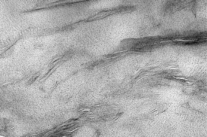
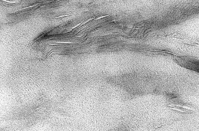
18 Image Processing - Phase Determination TEM image of an ultra thin section 0.5 µm & 48 % & 33 % & 19 %
19 III. Micromechanical Testing of Polymer Properties
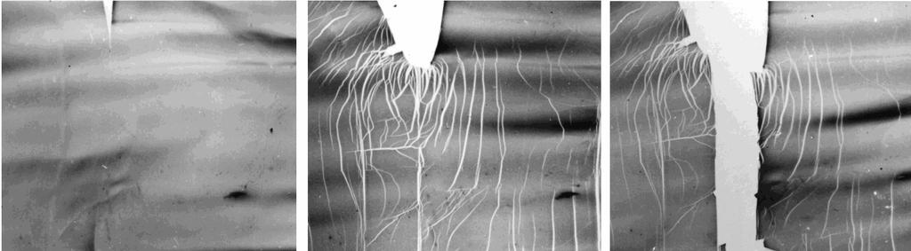
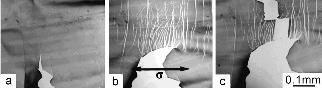
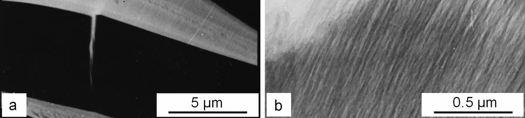
20 Micromechanical Mechanisms mechanical loading scale nm processes stretching of chain segments, reptation movements, chain scission Polymer molecular structure! morphology µm mm microvoid formation microyielding crazing shear band formation micro flow crack initation & propagation fracture
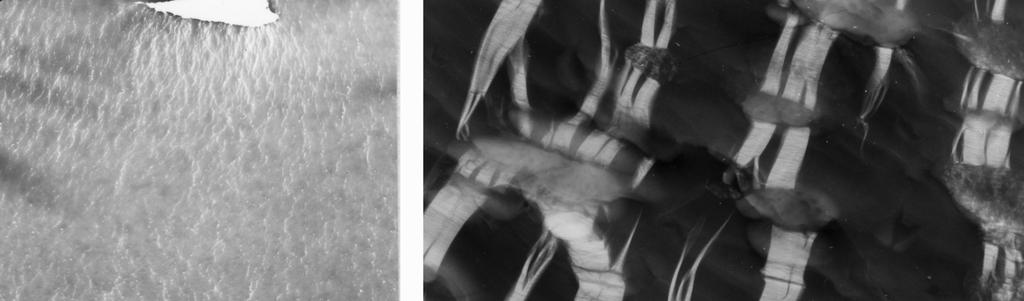
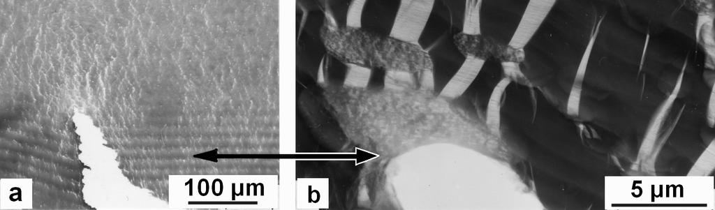
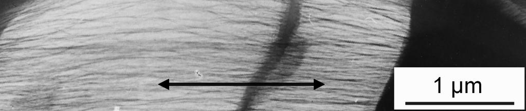
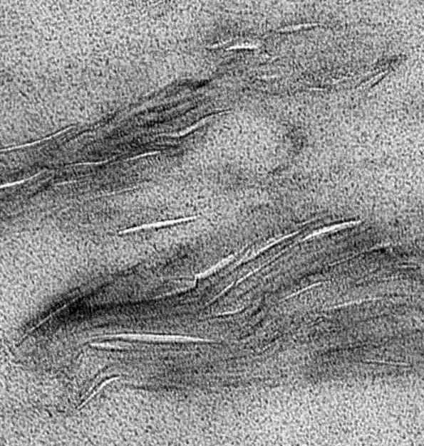
21 Investigation of micromechanical processes using EM bulk polymers SEM TEM (replica) TEM - ultra-thin section - replica SEM ESEM AFM SEM ESEM AFM HVTEM TEM 1. fracture surfaces - tension test - impact test - broken pieces 2. surfaces of deformed (loaded) bulk samples 3. in situ deformation of thin samples


22 Tensile Test under Optical Microscope
23 Tensile Test of HDPE using Miniaturised Specimens under Optical Microscope 25,00 20,00 ( (MPa) 15,00 10,00 5,00 0,00 0% 100% 200% 300% 400% 500% ' 1 mm
")

 Temperature: Room Temperature")
")
24 Tensile stages for micromechanical in situ investigations Scanning Electron Microscope Tensile Stage B156 (Oxford Instruments) Temperature: -180 C C Thickness: 0.5 µm mm Scanning Electron Microscope and Atomic Force Microscope Tensile Module 1000 N (Kammrath &Weiss) Temperature: Room Temperature Thickness: 10 µm... 5 mm High Voltage Electron Microscope Tensile Stage (Jeol) Temperature: Room Temperature Thickness: 100 nm... 5 µm Transmission Electron Microscope Straining Holder Model 671 (Gatan) Temperature: -180 C C Thickness: 100 nm µm


25 Micromechanical in situ Investigations Different Microscopic Techniques (Semicrystalline Polymers) Method SEM TEM AFM Requirements no evaporation, no etching no staining very flat surfaces Techniques Typical Results HDPE/ Copolymer Blends
26 Examples
27 Stiff, very brittle Brittle (PS, SAN, PMMA) Semiductile (PC, PVC) Ductile, tough (PE, PP)
28 In situ deformation of PS in HVTEM
29 a) Pre-craze b) Fibrillated craze c) Laser-light diffraction pattern

")
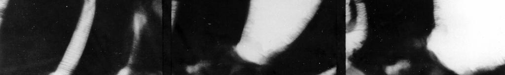
30 Craze rupture in PS (HVTEM)
31 Rubber-toughened Polymer (HIPS)
32 Rubber-toughened Polymer (HIPS)


33 Rubber-toughened Polymer (HIPS)


34 Rubber-toughened Polymer (HIPS) In-situ crack propagation in HIPS


 1")
35 PE Composite Dependence of void size and fibril thickness on particle diameter in filled HDPE: a) 1 µm b) 3 µm c) 8 µm 10 µm
36 Deformation structures of particle-filled PP (10 wt.% Al(OH) 3 )
37 Micro- and Nanocomposites
38 Effect of Nanoparticle Influence of of Interface 100 nm 1 µm 10 nm


39 HVEM Deformation structures of particle-filled PE (10 wt.% SiO 2 ) TEM (RuO 4 stained)
40 PP-Clay-Nanocomposite Deformation structure 200 nm 200 nm
41 RESULT of EM: Better Correlations Polymerization Molecular (chemical) structure Processing Supermolecular structures (Morphology) Loading conditions Micromechanical processes of deformation and fracture Mechanical properties
42 RESULT of EM: Better Properties Stiffness Future now Toughness
43
44 Table of Contents Introduction I Techniques of Electron Microscopy 1. Overview About Techniques 2. Transmission Electron Microscopy Methodical and Instrumental Fundamentals 3. Transmission Electron Microscopy Conventional and Special Investigations of Polymers 4. Scanning Electron Microscopy 5. Atomic Force Microscopy 6. In situ Microscopy 7. Image Processing and Image Analysis
45 II Preparation Techniques 8. Problems of Electron Microscopy of Polymers 9. Preparation of Surfaces 10. Preparation of Thin Sections 11. Special Preparation Techniques 12. Preparation for (in situ) Deformation Tests 13. Contrast Enhancement
46 III Main Groups of Polymer 14. Structural Hierarchy of Polymers 15. Amorphous Polymers 16. Semicrystalline Polymers 17. Polymer Blends 18. High-impact Rubber modified Polymers 19. Block Copolymers 20. Rubbers and Elastomers 21. Polymer Composites 22. Polymer Nanocomposites 23. Biological and Biomedical Polymers 24. Special Processing Forms Index