Structural study of Caenorhabditis elegans
|
|
|
- Roy Arnold
- 5 years ago
- Views:
Transcription


1 aterial Life Science Application Note Structural study of Caenorhabditis elegans related instruments Leica E ICE, Leica E AFS2 edical Industrial anufacturing Natural Resources
2 2 Structural study of Caenorhabditis elegans ATERIALS AND ETHODS Wildtype L4 stage C. elegans (N2 strain) were placed in the 100 µm deep side of Lecithin-coated (see detailed protocol*) type A 3 mm Cu/Au carriers (Leica) with extracellular filler containing 1% (w/v) Agarose type IX and 2% (w/v) Bovine Serum Albumin in bacteria medium (see preparation details**) and sandwiched with the flat side of Lecithin-coated type B 3 mm Cu/Au carriers (Leica). Samples were frozen in a high-pressure freezer (E ICE, Leica). After freezing sandwiches were opened in liquid nitrogen and the type A carriers were transferred into flow-through capsules (plastic capsules D5 x H15 mm, Leica) to the automated freeze-substitution device (AFS2, Leica) set at 90 C. Flow-through capsules with samples were placed into 2.0 ml Sarstedt tubes containing 800 µl precooled 2% (w/v) Osmium tetroxide in anhydrous ethanol (see modified Karreman protocol***). The rubber rings from the lids of these Sarstedt tubes have been removed. Samples were kept at 90 C for 48 h in this solution, next washed 4x 30 min at 90 C with 500 µl precooled 2% (w/v) Osmium tetroxide in anhydrous Acetone and finally left for 46 hat 90 C in 2.0 ml Sarstedt tubes containing 500 µl precooled 2% (w/v) Osmium tetroxide in anhydrous Acetone. Washing steps were carried out by transferring the flow-through capsules with samples to 2.0 ml Sarstedt tubes containing 500 µl precooled 2% (w/v) Osmium tetroxide in anhydrous Acetone. Next samples were gradually warmed from 90 C to 60 C (increment of 2 C/h); kept at 60 C for 8 h before warmed up to 30 C (increment of 2 C/h). At 30 C samples were kept for 8 h and finally were placed on ice for 1 h followed by 1 h at room temperature. Samples were washed 3x 5 min with anhydrous Acetone by transferring the flow-through capsules with samples to 1.5 ml Eppendorf tubes containing 500 µl anhydrous Acetone. A volume of 800 µl freeze-substitution medium was chosen to have enough capacity to replace the water in the sample for Acetone. For all next steps a volume of 500 µl was used to prevent loss of the sample out of the flow-through capsule by overflow. After the last wash with anhydrous Acetone samples were infiltrated with Epon resin (see detailed protocol****). Infiltration with Epon resin/ Acetone mixtures was perfor med at room temperature by the following steps: Epon : Acetone = 1 : 2 for 2 h (33% (v/v) Epon) Epon : Acetone = 1 : 1 for 18 h (50% (v/v) Epon) Epon : Acetone = 2 : 1 for 8 h (66% (v/v) Epon) Epon : Acetone = 3 : 1 for 18 h (75% (v/v) Epon) pure Epon for 8 h (100% Epon) pure Epon for 18 h (100% Epon) pure Epon for 6 h (100% Epon) Finally, individual C. elegans were taken out of the type A carriers and placed separately in the cavities of an embedding mould (Aclar scientific). The cavities were filled with pure EPON and polymerized for 72 h at 65 C. Approximately 70 nm thick sections were cut on an ultramicrotome (Ultracut UCT, Leica) and collected on Carbon coated-formvar-50 mesh hexagonal copper grids. The sections were contrasted 6 min with 7% (w/v) Uranyl Acetate in 70% ethanol and 2 min with Reynolds Lead Citrate. icrographs were acquired on a Tecnai 12 electron microscope (FEI) operated at 100 kv and spotsize 3. Electron micrographs (Binning1, 2048x2048 pixels) were collected with a TIETZ camera (TIETZ TVIPS TemCam F214) at 15,000x magnification (pixel size 0.375nm). Courtesy: Elly van Donselaar 1, artin Harterink 1, Karin Vocking 1 and Rob esman 2 1 Universiteit Utrecht, The Netherlands, 2 Radboud Universiteit Nijmegen, The Netherlands * Lecithin coating of carriers: Prepare a solution of 2% (w/v) Lecithin (L-α-Phosphatidylcholine from egg yolk, Fluka-61755) in Chloroform. Dip the type A carriers one by one in the Lecithin solution and lay the carriers with the 100 µm deep side up on filter paper. Dip also the type B carriers in the Lecithin solution and lay the carriers with the flat side up on filter paper. The carriers will be covered with a thin layer of Lecithin after the Chloroform has evaporated.
 BSA (Bovine Serum Albumin, fraction V, Sigma-Aldrich A-9647) in milliq water and mix gently.")
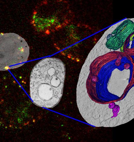
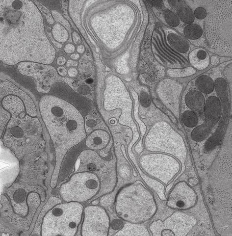
3 LNT Application Note - CAENORHABDITIS ELEGANS 3 ** C.elegans extracellular filler: For 2 ml: 20 mg Agarose, type IX (Ultra-low gellingpoint Agarose, Sigma-Aldrich A-5030) in 1.6 ml bacteria medium, dissolve at 60 C. Let the solution cool down to 37 C, add 400 µl 10% (w/v) BSA (Bovine Serum Albumin, fraction V, Sigma-Aldrich A-9647) in milliq water and mix gently. *** Procedure described in One Protocol Fits All atthia Karreman et al.: Lights will guide you, Sample Preparation and Applications of integrated Laser and Electron icroscopy, published by Uitgeverij BOXPress, s-hertogenbosch, The Netherlands, ISBN: **** Epon resin: For approximately 50 ml pure Epon add together: gram Epoxy embedding medium (Fluka) gram Araldite Hardener 964 (Fluka) gram Epoxy embedding medium hardener NA (Fluka) ix well for at least 10 min and add: ml Epoxy embedding medium accelerator DP 30 (Fluka) ix well for at least another 10 min RESULTS Fig. 1 Caenorahbditis elegans C. elegans processed as described was sectioned perpendicular just behind the head at the position of the kidneys. Overview of a section reveals the subcellular organization of cell of interest taken from the lateral side of C. elegans.


4 4 G LD G ER Fig. 2: Higher magnification of the selected area in Fig. 1 LD-Lipid Droplet, -itochondria, ER-Endoplasmatic reticulum, G-Golgi TEC Fig. 3: Higher magnification of the selected area in Fig. 1 TEC-Tubular Excretory Cell, -itochondria


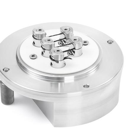
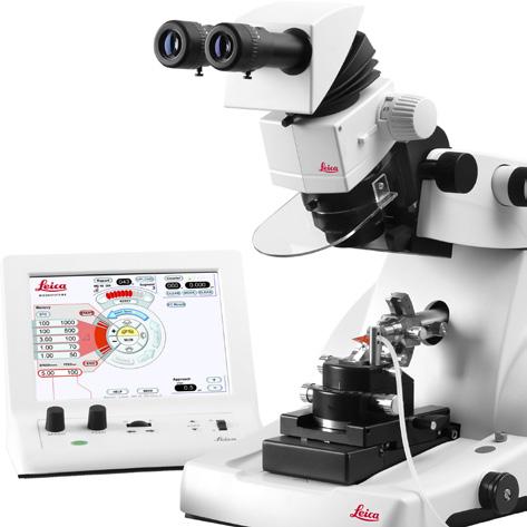
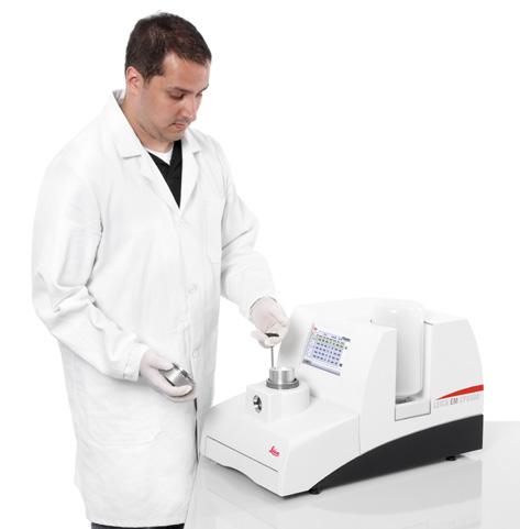
5 RELATED PRODUCTS Leica E ICE Leica ikrosysteme GmbH Vienna, Austria T F Leica E AFS2 CONNECT WITH US! Leica E ICE Application Note CAENORHABDITIS ELEGANS 10/2015 Copyright by Leica ikrosysteme GmbH, Vienna, Austria, Subject to modifications. LEICA and the Leica Logo are registered trademarks of Leica icrosystems IR GmbH.