CHAPTER 1. Aspects of the Three-Dimensional Intracellular Organization of Mesocarp Cells as Revealed by Scanning Electron Microscopy
|
|
|
- Kelley Terry
- 5 years ago
- Views:
Transcription
1 CHAPTER 1 Aspects of the Three-Dimensional Intracellular Organization of Mesocarp Cells as Revealed by Scanning Electron Microscopy Reprinted from Protoplasma by permission of Springer-Verlag Publishers, Inc., New York. Platt-Aloia, K. A Ultrastructure of Mature and Ripening Avocado (Persea americana Mill.) Fruit Mesocarp; Scanning, Transmission and Freeze Fracture Electron Microscopy. PhD. Dissertation. University of California, Riverside. 113 pages. Chapter 1. Aspects of the Three-Dimensional Intracellular Organization of Mesocarp Cells as Revealed by Scanning Electron Microscopy 8
2 Aspects of the Three-Dimensional Intracellular Organization of Mesocarp Cells as Revealed by Scanning Electron Microscopy K. A. Platt-Aloia and W. W. Thomson Department of Botany and Plant Sciences University of California, Riverside, CA 92521, U.S.A. SUMMARY The osmium-ligand binding technique and scanning electron microscopy have been applied to the study of the three-dimensional organization of mesocarp cells of a mature avocado fruit. Using this approach the mitochondria of the cells appear as elongated, branching structures and the endoplasmic reticulum consists of a complex of tubular strands, vesiculated strands and lamellar sheets. Associations of the endoplasmic reticulum with other organelles are also apparent. It is suggested that this approach provides a valuable means to assess the structural transitions in cell organization that occur during development or with functional changes. Keywords: microscopy Endoplasmic Reticulum; Osmium ligand binding; Scanning electron INTRODUCTION Ultrastructural studies on the endoplasmic reticulum (ER) have revealed that this membrane system may have a variety of forms: tubular (Jones and Fawcett 1966, Porter 1961), cisternal (Morre et al. 1971), and fenestrated (Morre and Mollenhauer 1974) as well as forms intermediate to these. Additionally, there are several reports of associations of the ER with cellular organelles such as chloroplasts (Whatley 1971), microbodies, mitochondria (Morre et al. 1971), nuclei, vacuoles, and the plasmalemma (see Morre and Mollenhauer 1974). In the virtual two-dimensional views obtained by transmission electron microscopy (TEM), the three-dimensional organizations of the ER and the extent of the interrelationship of this membrane system with other organelles is often missed. The scanning electron microscope (SEM) provides a means for studying the threedimensional organization of tissues with a high level of resolution. However, most biological tissues are poor electrical conductors and in the SEM this results in an uneven discharge of secondary electrons from the surface of the specimen which distorts the image and greatly reduces resolution. The necessary conductivity can be Correspondence and Reprints: Department of Botany and Plant Sciences, University of California, Riverside, California 92521, USA 9
3 imparted to the sample by several means. The most common method is vacuum evaporation or cathode sputtering of a conductive metal onto the surface to be examined (Boyde and Wood 1969). Although metal coating has proved valuable for many studies, this method is not without disadvantages. Common problems are uneven or incomplete coating, especially of highly irregular surfaces, heat generation during coating which may cause tissue damage, and the masking of structural detail by the layer of metal. Because of these limitations, alternative techniques have been explored which increase the conductivity of biological tissues sufficiently to eliminate discharge, and, at the same time, allow full utilization of the capabilities of the SEM. One alternative procedure takes advantage of the conductivity of osmium and the binding capabilities of osmium tetroxide. This technique is based on earlier work by Seligman and co-workers (Hanker et al. 1966, Seligman et al. 1966) in which they cross-linked tissue-bound osmium with the ligand, thiocarbohydrazide (TCH). The technique involves exposing sections of OsO 4 -treated tissue to TCH, which binds to the osmium in the tissue and then to additional OsO 4 which binds to the unreacted sites on the bound TCH. This procedure results in an increased contrast of osmiophilic components of cells such as membranes and lipid droplets (Seligman et al. 1966). Subsequently, Kelley et al. (1973) and Malick and Wilson (1975) applied this technique to the preparation of material for the SEM. They found that the additional osmium provided adequate conductivity for successful examination of tissue surfaces without coating them with a metal in an evaporator. This method has also been applied to the study of subcellular organelles in the SEM (Woods and Ledbetter 1976). However, in this case, the tissue was dehydrated, embedded in epoxy resin, frozen, broken, and the surface epoxy removed with acetone. Organelles were visible by this technique, but the epoxy limited the depth of field and obscured important details. In the present study we have applied the ligand binding technique to the study of subcellular organelles in avocado fruits. We have found that by further increasing the exposure of the tissue to an additional layer(s) of TCH and osmium (OTOTO), and eliminating the use of epoxys by critical point drying the tissue that not only is the surface of the specimen electrically conductive and suitable for direct examination in the SEM but internal cells and cell organelles can be observed and studied. In this paper we describe some aspects of the three-dimensional organization of organelles in mesocarp cells of avocado fruits after being prepared by this procedure and studied with the SEM. MATERIALS AND METHODS Three different preparative protocols were performed for this study; one for SEM (OTOTO) and two for TEM (OTO and "conventional"). For all three procedures, the initial fixation was the same. Mesocarp tissue samples were taken from mature avocado fruits (Persea americana var. Hass) using a #2 cork borer. The plug of tissue was placed immediately into 1% glutaraldehyde in 50 mm cacodylate buffer (ph 7.2) and cut into smaller pieces (1 x 2 mm for TEM and 2 x 4 mm for SEM). These were then transferred into fresh fixative for 2-3 hours. After a brief rinse in buffer, the material was post-fixed overnight in 1% OsO 4 in 50 mm cacodylate buffer, ph 7.2. Samples for SEM and those for the OTO, TEM preparation, were washed 5-6 times in distilled water 10
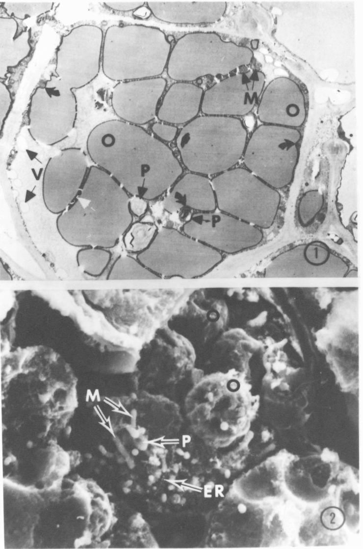
4 over a 20 minute period and then treated with freshly made, filtered, 1% thiocarbohydrazide (TCH) for minutes with frequent agitation (Kelley et al. 1973). The tissue was again washed and then treated overnight with 1% OsO 4 in distilled water. At this point, the samples for TEM (OTO) were removed, dehydrated in acetone and embedded in epoxy resin (Spurr 1969). The above procedure (rinsing, 1% TCH for minutes, rinsing, and OsO 4, overnight) was repeated the next day for the tissues for SEM. Samples were then dehydrated in acetone and critical point dried according to the method of Anderson (1951) using acetone as the transition fluid, with a Tuisimas, CO 2 critical point dryer. After drying, pieces of tissue were broken in half by the use of two pairs of forceps and mounted on aluminum stubs with a conductive paint containing colloidal silver. They were oriented so that the freshly broken surface, relatively free from contamination, was exposed. Samples were stored in a dessicator until viewing with a Joelco JSM-U3 scanning electron microscope at 10 or 20 KV. After the first overnight OsO 4 fixation, samples for TEM ("conventional") were dehydrated in acetone and embedded in Spurr's resin (Spurr 1969). Sections were stained with aqueous Uranyl acetate for 1 hour and with lead citrate (Reynolds 1963) for 1-2 minutes. Sections from the TEM (OTO) samples were studied without post stain. All sections were examined and photographed on a Philips 400 electron microscope. RESULTS Preparation of avocado tissue for transmission electron microscopy by the OTO method resulted in reasonably good preservation of subcellular detail, and sufficient contrast such that post staining of sections was not necessary (Fig. 1). However, some cytoplasmic components and organelles are highly osmiophilic and over-staining sometimes occurs (Fig. 1, white arrows). At higher magnification, some of the overstained membranes are diffuse in appearance, and some components, such as grana, become extremely electron dense, apparently because of excess deposits of osmium (Fig. 3). Nevertheless, this en bloc preparation of material by the OTO technique for TEM serves to illustrate the quality of preservation of subcellular structure for correlation with tissues studied in the SEM. The use of the OTOTO preparation procedure and critical point drying followed by breaking the tissue to expose internal structures for study by SEM gave striking and interesting results. Although positive identification of specific organelles is often difficult, it is possible by careful study and comparison with transmission electron micrographs (compare Figs. 1 and 2). At low magnification, in both SEM and TEM micrographs, the large oil droplets can be easily recognized (Figs. 1 and 2; O). Their identification in the SEM is based on their size, frequency of occurrence in the cells, as well as by the unique surface indentations (Fig. 2, black arrows) which are also observable in thin section images (Fig. 1, black curved arrows). Reconstructions from micrographs of serial thin sections (unpublished observations) and individual thin sections such as Fig. 4, indicate that the branched, often elongated, worm-like structures observed in SEM micro-graphs are mitochondria (Fig. 2; M). In thin sections the plastids vary somewhat in outline but are considerably larger than the mitochondria (Fig. 1; P). This difference in size permits their tentative identification in scanning electron micrographs (Figs. 2, 6; P). We have identified the many smooth surfaced, balloon-like structures in scanning electron micrographs as probable vesicles or vacuoles of different sizes (Fig. 5, 6; V). 11
5 This identification is based on the contour of the vacuoles in thin sections as well as the wide range of sizes that are observed (Fig. 1, V). At higher magnification in scanning electron micrographs, increased detail is apparent and three-dimensional interrelationships of various organelles are revealed. One of the striking features in these cells is the variation in the endoplasmic reticulum. It varies from sheet-like (Fig. 6; ER) to apparent tubular strands (Figs. 2, 5). Often the ER is closely associated with other structures such as plastids (Fig. 6; ER,) and oil droplets (Fig. 6, ER2). Many of the tubular strands of ER are vesiculated and have the appearance of a string of different sized beads (Fig. 5; ER). Individual ballooning of the strands is apparent and in some instances apparent continuities between the sheet-like and tubular forms can be seen (Fig. 6; arrow). DISCUSSION In the present study using scanning electron microscopy we have observed, in the same cell, three different structural forms of endoplasmic reticulum: tubular strands, vesicular strands and lamellar sheets. Our observations in the SEM also substantiate the TEM serial section studies which show mitochondria to be long, branching structures. In the virtual two-dimensional images provided by transmission electron microscopy, many such important structural features of cell organelles can be missed. Also, the three-dimensional interrelationships of different organelles are not always apparent or are observed only infrequently in TEM. Previously, only by extensive serial section reconstruction could these important three-dimensional aspects of cell organization be studied. The osmium-ligand binding technique, as we have applied it, used in conjunction with scanning electron microscopy, provides an avenue for the assessment of these facets of cell and organelle structure. Also, we suggest that the use of this procedure provides a valuable tool for gaining insight into the structural transitions occurring during developmental and/or functional changes in different plant systems. ACKNOWLEDGEMENTS This study was supported in part by a National Science Foundation grant BMS to W. W. Thomson, and a PHS grant 5-S07 RR REFERENCES Anderson, T. F., 1951: Techniques for the preservation of three-dimensional structure in preparing specimens for the electron microscope. Trans. N.Y. Acad. Sci. 13, Boyde, A., Wood, C., 1969: Preparation of animal tissues for surface-scanning electron microscopy. J. Microscopy 90, Hanker, J. S., Deb, C., Wasserkrug, H. L., Seligman, A. M., 1966: Staining tissue for light and electron microscopy by bridging metals with multi-dentate ligands. Science 152, Jones, A. L., Fawcett, D. W., 1966: Hypertrophy of the agranular endoplasmic reticulum 12
6 in hamster liver induced by phenobarital (with a review of the function of this organelle in liver). J. Histochem. Cytochem. 14, Kelley, R. 0., Dekkar, R. A. F., Bluemink, J. G., 1973: Ligand-mediated osmium binding: its application in coating biological specimens for scanning electron microscopy. J. Ultrastruct. Res. 45, Malick, L. E., Wilson, R. B., 1975: Modified Thiocarbohydrazide procedure for scanning electron microscopy: Routine use for normal, pathological, or experimental tissues. Stain Tech. 50, Morre, D. J., Merritt, W. D., Lembi, C. A., 1971: Connections between mitochondria and endoplasmic reticulum in rat liver and onion stem. Protoplasma 73, Morre, D. J., Mollenhauer, H. H., 1974: The endomembrane concept: a functional integration of endoplasmic reticulum and Golgi apparatus. In: Dynamic aspects of plant ultrastructure (Robards, A. W., ed.), pp London: McGraw Hill. Porter, K. R., 1961: The ground substance in the cell. In: The cell. (Brächet, J., Mirsky, A. E., eds.), pp New York, Academic Press. Reynolds, E. S., 1963: The use of lead citrate at high ph as an electron-opaque stain in electron microscopy. J. Cell Biol. 17, Seligman, A. M., Wasserkrug, H. L., Hanker, J. S., 1966: A new staining method (OTO) for enhancing contrast of lipid containing membranes and droplets in osmium tetroxide fixed tissue with osmiophilic thiocarbohydrazide (TCH). J. Cell Biol. _30, Spurr, A. R., 1969: A low viscosity epoxy resin embedding medium for electron microscopy. J. Ultrastruc. Res. 26, Whatley, J. M., 1971: The chloroplasts of Equisetum telmateia Erhr.; A possible developmental sequence. New Phytol. 70, Woods, P. S., Ledbetter, M. C., 1976: Cell organelles at uncoated cryofractured surfaces as viewed with the scanning electron microscope. J. Cell Sci. 21,
7 FIGURES AND FIGURE LEGENDS Fig. 1. Transmission electron micrograph of a thin section of a mature, avocado prepared by the OTO technique. The majority of the cell is occupied by oil droplets (0) which show increased electron density at their periphery, probably due to the osmiophilic nature of lipids. The oil droplets frequently have indentations which are often occupied by organelles (black curved arrows). Some regions of the cytoplasm are excessively osmiophilic (white arrows). Plastids (P) are irregularly shaped and larger than mitochondria (M) which appear either spherical or elongate. V = vesicle X 2000 Fig. 2 Scanning electron micrograph of a cell on the freshly broken surface of avocado fruit tissue prepared by the osmium-ligand binding technique. Oil droplets (O) can be identified by their comparable size, number, and shape when compared to thin sections as in Fig. 1. Black arrows indicate depressions or indentations in the oil droplets. Plastids (P) are irregularly shaped and of larger diameter than mitochondria (M) which are long and of a more consistent diameter. Microbodies and small vesicles (*) are, however, not discernable. Endoplasmic reticulum (ER) of the vesiculating-strand type can be seen. X 2,500 14
8 15
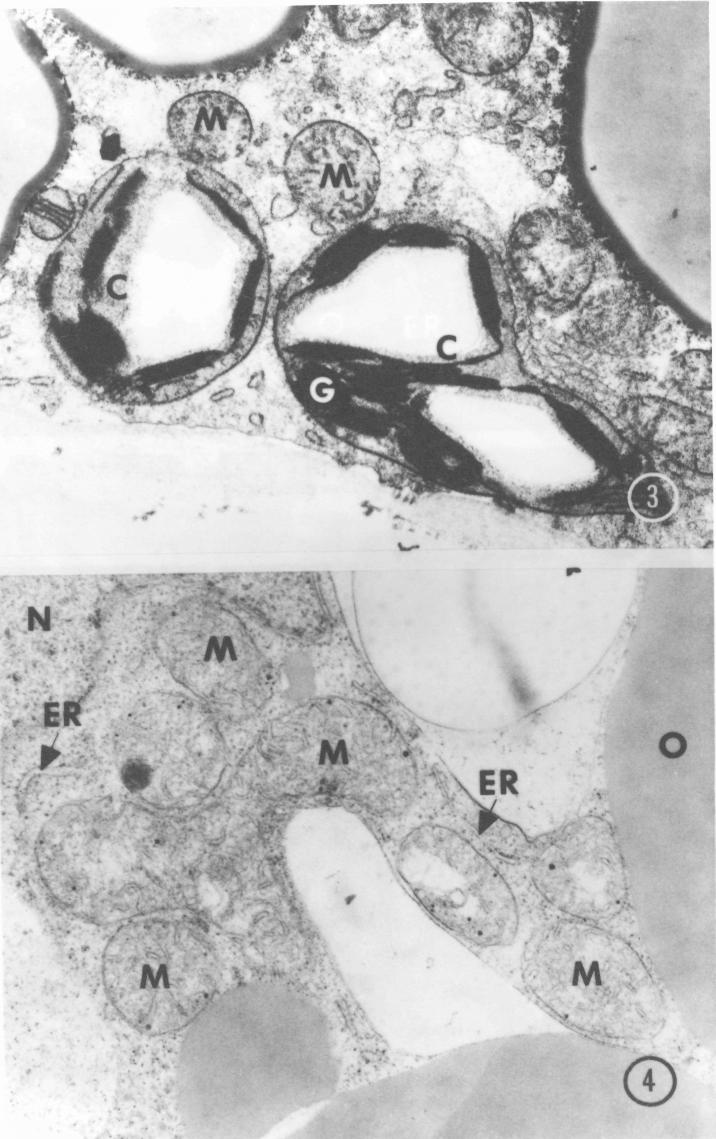
9 Fig. 3. Transmission electron micrograph of a portion of a mesophyl cell of a mature avocado prepared by the OTO method. The membranes of the chloroplasts (C) and mitochondria (M) show good contrast but are diffuse and granular in appearance. Grana (6) are extremely electron dense. X 15,000 Fig. 4. Transmission electron micrograph of what appears to be several mitochondria (M) in a mesophyl cell of an avocado. Actually, serial sections were taken of this cell and all seven "mitochondria" are part of a single, elongate branching mitochondrion. ER = endoplasmic reticulum, 0 = oil, N = nucleus. X 25,400 16
10 17
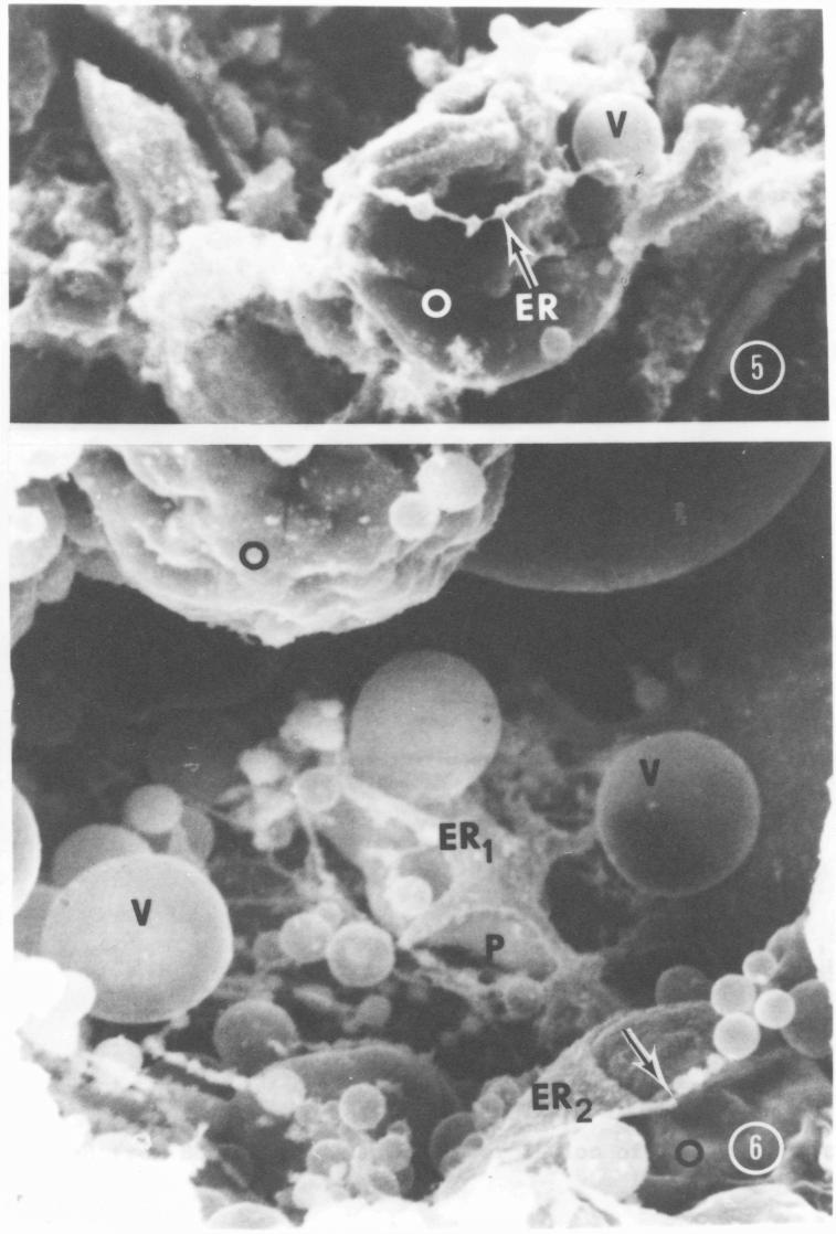
11 Fig. 5. Portion of a cell of an avocado fruit showing a vesiculating strand of ER (arrow) winding around an oil droplet (0). V = vesicle X 4,600 Fig. 6. Scanning electron micrograph of avocado fruit subcellular organelles. A stellarshaped lamellar sheet of endoplasmic reticulum (ER-.) is in an apparently close association with an underlying plastid (P). Another sheet (ER2) can be seen ensheathing an oil droplet (0). Tubular strands of ER can also be seen in various degrees of vesiculation and in apparent continuity with the sheet forms (arrow). V = vesicle X 8,000 18
12 19
COPYRIGHTED MATERIAL. Tissue Preparation and Microscopy. General Concepts. Chemical Fixation CHAPTER 1
 CHAPTER 1 Tissue Preparation and Microscopy General Concepts I. Biological tissues must undergo a series of treatments to be observed with light and electron microscopes. The process begins by stabilization
Chapter. Methods for and. Analysis of Plant Cell Tissue Ultrastructure. Contents. 1.1 Introduction. John E. Mayfield and William V.
 In: Dashek, William V., ed. Methods in plant biochemistry and molecular biology. Boca Raton, FL: CRC Press: pp. 3-11. Chapter 1. 1997. Chapter Methods for and Analysis of Plant Cell Tissue Ultrastructure
BIOLOGICAL SAMPLE PREPARATION FOR TEM OBSERVATION. TEM Seminar Nov 16, 2017 Astari Dwiranti, Ph.D
 BIOLOGICAL SAMPLE PREPARATION FOR TEM OBSERVATION TEM Seminar Nov 16, 2017 Astari Dwiranti, Ph.D Why do we need EM for biological samples? (O'Connor and Adams, 2010) Why do we need EM for biological samples?
Investigation of cellular uptake mechanisms by correlative TEM and SIM
 Collaborative Research Center (SFB 1278) Investigation of cellular uptake mechanisms by correlative TEM and SIM Rainer Heintzmann, Fengjiao Ma, Institute of Physical Chemistry Stephanie Höppener, Martin
THE USE OF LEAD CITRATE AT HIGH ph AS AN ELECTRON-OPAQUE STAIN IN ELECTRON MICROSCOPY
 THE USE OF LEAD CITRATE AT HIGH ph AS AN ELECTRON-OPAQUE STAIN IN ELECTRON MICROSCOPY EDWARD S. REYNOLDS. From the Department of Anatomy, Harvard Medical School, Boston Aqueous solutions of lead salts
A rapid method for electron microscopic examination of blood cells
 Journal of Clinical Pathology, 1979, 32, 162-1 67 A rapid method for electron microscopic examination of blood cells ANTONIO COPPOLA From the Department ofpathology, College of Medicine, Downstate Medical
Preparation of tissues for study
 Preparation of tissues for study HISTOLOGY : It is the branch of science which deals with the microscopic study of normal tissue HISTOPATHOLOGY : It is the branch of science which deals with the microscopic
A Comparison of Techniques Useful for Preparing Nematodes for Scanning Electron Microscopy 1
 Journal of Nematology 18(4):479-487. 1986. The Society of Nematologists 1986. A Comparison of Techniques Useful for Preparing Nematodes for Scanning Electron Microscopy 1 J. D. EISENBACK ~ Abstract: Second-stage
Transmission Electron Microscopic Study of Antibiotic Action on Klebsiella pneumoniae Biofilm
 ANTIMICROBIAL AGENTS AND CHEMOTHERAPY, Aug. 2002, p. 2679 2683 Vol. 46, No. 8 0066-4804/02/$04.00 0 DOI: 10.1128/AAC.46.8.2679 2683.2002 Copyright 2002, American Society for Microbiology. All Rights Reserved.
SONOMA STATE UNIVERSITY DEPARTMENT OF BIOLOGY BIOLOGY 344: CELL BIOLOGY Fall 2013
 SONOMA STATE UNIVERSITY DEPARTMENT OF BIOLOGY BIOLOGY 344: CELL BIOLOGY Fall 2013 Instructor Murali C. Pillai, PhD Office 214 Darwin Hall Telephone (707) 664-2981 E-mail pillai@sonoma.edu Website www.sonoma.edu/users/p/pillai
 -------------------------------------- Biology Name Summary Sbeet Cell Structure Lab Class Date --------------------~ ---------- A. Cork Cells: Wet mount I Higb Power 1. Is it easier to see individual
Technical Overview Cross-linking fixatives: What they are, what they do, and why we use them
 Technical Overview Cross-linking fixatives: What they are, what they do, and why we use them Focus on: Formaldehyde, Glutaraldehyde, and Osmium tetroxide M. Kuwajima/Kristen Harris Lab EM processing and
Cell Structure and Function
 Cell Structure and Function Dead White Men Who Discovered (and were made of) Cells: Anton Van Leeuwenhoek Robert Hooke Where the Magic Happened Schleiden Cell Theory All plants are made of cells Schwann
PREPARATION OF HISTOLOGICAL SPECIMENS
 PREPARATION OF HISTOLOGICAL SPECIMENS Histo-techniques Preparation of tissue for microscopic examination Series of processes Ultimate aim to make tissue visible as it is Pathology Vs Anatomy Steps vary
Experiments on fixation for electron microscopy 2. Alkalized osmium tetroxide. By S. K. MALHOTRA
 28? Experiments on fixation for electron microscopy 2. Alkalized osmium tetroxide By S. K. MALHOTRA (From the Cytological Laboratory, Department of Zoology, University Museum, Oxford) With 6 plates (figs.
Supplementary Figure legends. Supplementary Methods
 Supplementary Methods Transmission electron microscopy. For transmission electron microscopy (TEM), the cell populations were rinsed with 0.1 Sorensen s buffer (ph 7.5), fixed in 2.5% glutaraldehyde for
A Study of Potassium Permanganate 'Fixation' for Electron Microscopy
 241 A Study of Potassium Permanganate 'Fixation' for Electron Microscopy By S. BRADBURY AND G. A. MEEK (From the Department of Human Anatomy, South Parks Road, Oxford) With five plates (figs, i, 2, 4,
Activity 2.1. Activity 2.2. Looking at animal cells. Looking at plant cells
 Activity 2.1 Looking at animal cells Skills C1, C2 a source of animal cells, for example some macerated liver or scrapings from the lining of the trachea from a set of sheep or other lungs (obtainable
Examination of Virus-Infected Cultured Cells
 APPLIED MICROBIOLOGY, OCt. 1970, p. 633-637 Copyright 1970 American Society for Microbiology Examination of Virus-Infected Cultured Cells by Scanning Electron Microscopy CATHERINE ARANYI, JAMES FENTERS,
From Eye to Insight. Leica EM ACE. Coater Family
 From Eye to Insight Leica EM ACE Coater Family EM ACE COATERS A Coater Family to cover all your needs. Developed in cooperation with leading scientists, the EM ACE coaters cover all the requirements for
Name Date Block LAB: Exploring Plant & Animal Cells
 Name Date Block LAB: Exploring Plant & Animal Cells Background Information: One of the first scientists to look at cells under a microscope was an English scientist by the name of Robert Hooke. He viewed
Visualization of the real microarchitecture of glycoprotein matrices with scanning electron microscopy
 Visualization of the real microarchitecture of glycoprotein matrices with scanning electron microscopy G. Familiari *, M. Relucenti, A. Familiari, E. Battaglione, G. Franchitto, and R. Heyn Laboratory
Electron Microscopy (EM) Grid
 Grid") Anirban Som 25-01-14 Instrumental technique presentation Electron Microscopy (EM) Grid What I will talk about Some basic topics about EM grid Home-made grid preparation Grid cleaning Carbon coating and
Anirban Som 25-01-14 Instrumental technique presentation Electron Microscopy (EM) Grid What I will talk about Some basic topics about EM grid Home-made grid preparation Grid cleaning Carbon coating and
Reinforcement. Cells and Life CHAPTER 1 LESSON 1
 Reinforcement Cells and Life LESSON 1 Directions: In numbers 1 through 4 below, a code letter has been substituted for each letter of the alphabet. To find out what the sentence says, use the following
nucleolus nucleus number proteins ribosomes type
 Name Use with textbook pages 123 129 Inside the nucleus Cloze Activity Section 41 Vocabulary 23 46 chromosomes DNA genes genetic molecule nucleolus nucleus number proteins ribosomes type Use the terms
Specimen Preparation Techniques for Scanning Electron Microscopy of Developing Peanut Pegs' H. E. Pattee', S. C. Mohapatra3 and E. K.
 Peanut Science (1983) 10:93-97 Specimen Preparation Techniques for Scanning Electron Microscopy of Developing Peanut Pegs' H. E. Pattee', S. C. Mohapatra3 and E. K. Agnello3 ABSTRACT This study evaluated
High pressure freezing
 High pressure freezing Introduction Technique Freeze-substitution Andres Käch Center for Microscopy and Image Analysis, University of Zurich andres.kaech@zmb.uzh.ch, www.zmb.uzh.ch Introduction Plunge
Cells and Tissues. Overview CELLS
 Cells and Tissues WIll The basic unit of structure and function in the human body is the cell. Each of a cell's parts, or organelles, as well as the entire cell, is organized to perform a specific function.
Resolution of Microscopes Visible light is nm Dry lens(0.5na), green(530nm light)=0.65µm=650nm for oil lens (1.4NA) UV light (300nm) = 0.13µm f
, green(530nm light)=0.65µm=650nm for oil lens (1.4NA) UV light (300nm) = 0.13µm f") Microscopes and Microscopy MCB 380 Good information sources: Alberts-Molecular Biology of the Cell http://micro.magnet.fsu.edu/primer/ http://www.microscopyu.com/ Approaches to Problems in Cell Biology
Microscopes and Microscopy MCB 380 Good information sources: Alberts-Molecular Biology of the Cell http://micro.magnet.fsu.edu/primer/ http://www.microscopyu.com/ Approaches to Problems in Cell Biology
Visualizing Cells Molecular Biology of the Cell - Chapter 9
 Visualizing Cells Molecular Biology of the Cell - Chapter 9 Resolution, Detection Magnification Interaction of Light with matter: Absorbtion, Refraction, Reflection, Fluorescence Light Microscopy Absorbtion
On colouring epon-embedded tissue sections with Sudan black B or Nile blue A for light microscopy
 109 On colouring epon-embedded tissue sections with Sudan black B or Nile blue A for light microscopy By S. M. McGEE-RUSSELL and N. B. SMALE (From the Electron Microscopy Laboratory, Virus Research Unit,
QX-102 Applications Manual
 QX-102 Applications Manual QuantomiX Ltd. 2005. All rights reserved. UQX003 Issue 2.1 November 2005 QX-102 Applications Manual, Issue 2.1 2 3 QX-102 Applications Manual, Issue 2.1 This publication is the
Supplement Figure 1. Plin5 Plin2 Plin1. KDEL-DSRed. Plin-YFP. Merge
 Supplement Figure 1 Plin5 Plin2 Plin1 KDEL-DSRed Plin-YFP Merge Supplement Figure 2 A. Plin5-Ab MitoTracker Merge AML12 B. Plin5-YFP Cytochrome c-cfp merge Supplement Figure 3 Ad.GFP Ad.Plin5 Supplement
666 THE JOURNAL OF CELL BIOLOGY' VOLUME 71, 1976" pages
 ph-dependent BINDING OF IMMUNOGLOBULINS TO INTESTINAL CELLS OF THE NEONATAL RAT RICHARD RODEWALD. From the Department of Biology, University of Virginia, Charlottesville, Virginia 22901. Neonatal rats
University of Ulm. Nadja El-Mecharrafie Grade 10. Division: Electron Microscopy
 University of Ulm Nadja El-Mecharrafie Grade 10 February 20-24th 2006 University of Ulm Division: Electron Microscopy Universität Ulm Zentrale Einrichtung Elektronenmikroskopie Materialwissenschaftliche
NANOGOLD PRODUCT INFORMATION
 NANOGOLD 95 Horse Block Road, Yaphank NY 11980-9710 Tel: (877) 447-6266 (Toll-Free in US) or (631) 205-9490 Fax: (631) 205-9493 Tech Support: (631) 205-9492 tech@nanoprobes.com www.nanoprobes.com PRODUCT
Histological preparation of embryonic and adult zebrafish eyes
 Histological preparation of embryonic and adult zebrafish eyes Richard J. Nuckels 1 and Jeffrey M. Gross 1,2,3 1 Section of Molecular Cell and Developmental Biology 2 Institute of Cell and Molecular Biology
High Resolution, Low Voltage Scanning Electron Microscopy of Uncoated Yeast Cells Fixed by the Freeze-Substitution Method
 /. Electron Microsc, Vol. 37, No. 1, 17-30, 1988 High Resolution, Low Voltage Scanning Electron Microscopy of Uncoated Yeast Cells Fixed by the Freeze-Substitution Method Masako OSUMI,* Misuzu BABA,* Nobuko
Samples Conductive and Observations on the Surface Morphology of Human Erythrocytes and Ehrlich Ascites Cells*
 Proc. Nat. Acad. Sci. USA Vol. 70, No. 12, Part I, pp. 3599-3603, December 1973 A Wet Chemical Method for Rendering Scanning Electron Microscopy Samples Conductive and Observations on the Surface Morphology
SEM Immunocytochemistry for Cells & Materials
 SEM Immunocytochemistry for Cells & Materials R. Geoff Richards AO Research Institute, AO Foundation, Davos, Switzerland. Immunohistochemistry Immunocytochemistry can be performed on a biological specimen,
CHAPTER 17 FROM GENE TO PROTEIN. Section C: The Synthesis of Protein
 CHAPTER 17 FROM GENE TO PROTEIN Section C: The Synthesis of Protein 1. Translation is the RNA-directed synthesis of a polypeptide: a closer look 2. Signal peptides target some eukaryotic polypeptides to
Baraa Ayed AL-Odat. Israa Ayed. Heba kalbouneh
 1 Baraa Ayed AL-Odat Israa Ayed Heba kalbouneh Introduction: "lecture #1" The name " histology " is derived from the Greek words: "histo" means a tissue and "logos" means the study of. So, Histology mean
Scanning and transmission electron microscopic observations of the topographic anatomy of dendritic lesions in the rabbit cornea
 Scanning and transmission electron microscopic observations of the topographic anatomy of dendritic lesions in the rabbit cornea William H. Spencer and Thomas L. Hayes Scanning electron microscopic observations
Specimen Preparation Technique for a Microstructure Analysis Using the Focused Ion Beam Process
 Specimen Preparation Technique for a Microstructure Analysis Using the Focused Ion Beam Process by Kozue Yabusaki * and Hirokazu Sasaki * In recent years the FIB technique has been widely used for specimen
The principles and practice of electron microscopy
 The principles and practice of electron microscopy Second Edition Ian M. Watt CAMBRIDGE UNIVERSITY PRESS Contents Preface tofirstedition page ix Preface to second edition xi 1 Microscopy with light and
COMPLEX OF EUGLENA GRACILIS
 Published Online: 1 February, 1965 Supp Info: http://doi.org/10.1083/jcb.24.2.253 Downloaded from jcb.rupress.org on May 12, 2018 THE ULTRASTRUCTURE OF THE PELLICLE COMPLEX OF EUGLENA GRACILIS JOACHIM
Chapter 1. A Preview of the Cell. Lectures by Kathleen Fitzpatrick Simon Fraser University Pearson Education, Inc.
 Chapter 1 A Preview of the Cell Lectures by Kathleen Fitzpatrick Simon Fraser University The Cell Theory: A Brief History Robert Hooke (1665) observed compartments in cork, under a microscope, and first
Cell Lab. Problem: How do bacterial, animal and plant cells differ?
 Cell Lab Problem: How do bacterial, animal and plant cells differ? Objective: Create a list of characteristics and criteria to identify bacteria, animal and plant cells. Materials: Light microscope Microviewer
Methods of Culturing Microorganisms. Chapter 3. Five Basic Techniques of Culturing Bacteria. Topics
 Chapter 3 Topics Methods of Culturing Microorganisms Microscope (History, Types, Definitions) Staining (Gram s) Methods of Culturing Microorganisms Five basic techniques of culturing Media Microbial growth
THE PATTERNED ORGANIZATION OF THICK AND THIN MICROFILAMENTS IN THE CONTRACTING PSEUDOPOD OF DIFFLUGIA
 y. Ceii sd. 13,727-739 (1973) 727 Printed in Great Britain THE PATTERNED ORGANIZATION OF THICK AND THIN MICROFILAMENTS IN THE CONTRACTING PSEUDOPOD OF DIFFLUGIA B. S. ECKERT* AND S. M. MCGEE-RUSSELL Department
Specimen configuration
 APPLICATIONNOTE Model 1040 NanoMill TEM specimen preparation system Specimen configuration Preparing focused ion beam (FIB) milled specimens for submission to Fischione Instruments. The Model 1040 NanoMill
LAMININ. For Immunohistochemical Demonstration of Laminin in Paraffin-embedded and Frozen Human Tissue Sections Stock No. IMMH-7
 LAMININ For Immunohistochemical Demonstration of Laminin in Paraffin-embedded and Frozen Human Tissue Sections Stock No. IMMH-7 TABLE OF CONTENTS BACKGROUND AND PRINCIPLE... 4 REAGENTS AND EQUIPMENT PROVIDED...
Manufactured by. Zyagen Barnes Canyon Road San Diego, CA 92121, USA
 Alkaline Phosphatase Immunohistochemistry Detection kits For detection of mouse, rabbit, goat, rat, sheep, chicken, guinea pig, and human primary antibodies Size: 500 Tests Catalog #: AK-011, Mouse Kit
BIO 315 Lab Exam I. Section #: Name:
 Section #: Name: Also provide this information on the computer grid sheet given to you. (Section # in special code box) BIO 315 Lab Exam I 1. In labeling the parts of a standard compound light microscope
The Nucleus and DNA Replication
 OpenStax-CNX module: m46073 1 The Nucleus and DNA Replication OpenStax College This work is produced by OpenStax-CNX and licensed under the Creative Commons Attribution License 3.0 By the end of this section,
Improved Monitoring of P. aeruginosa on Agar Plates
 Electronic Supplementary Material (ESI) for Analytical Methods. This journal is The Royal Society of Chemistry 2015 Improved Monitoring of P. aeruginosa on Agar Plates SUPPLEMENTAL INFORMATION T. A. Webster,
Visualization of the intact endoplasmic reticulum by immunofluorescence with antibodies to the major ER glycoprotein, endoplasmin
 Visualization of the intact endoplasmic reticulum by immunofluorescence with antibodies to the major ER glycoprotein, endoplasmin G. L. E. KOCH, D. R. J. MACER and M. J. SMITH Medical Research Council
Complete the table by giving the letter labelling the organelle that matches the function. Function of organelle
 Q1.The diagram shows a eukaryotic cell. (a) Complete the table by giving the letter labelling the organelle that matches the function. Function of organelle Letter Protein synthesis Modifies protein (for
A Level. A Level Biology. Cells, Microscopes, Cell Cycle and Immunity Questions. AQA, OCR, Edexcel. Name: Total Marks: Page 1
 AQA, OCR, Edexcel A Level A Level Biology Cells, Microscopes, Cell Cycle and Immunity Questions Name: Total Marks: Page 1 Q1.The diagram shows a eukaryotic cell. (a) Complete the table by giving the letter
Epigenetics, Environment and Human Health
 Epigenetics, Environment and Human Health A. Karim Ahmed National Council for Science and the Environment Washington, DC May, 2015 Epigenetics A New Biological Paradigm A Question about Cells: All cells
Answers to the multiple choice questions are at the bottom of the last page of this document.
 Review for Unit Test #2: Cell Parts, Functions and Protein Synthesis, Answers Answers to the multiple choice questions are at the bottom of the last page of this document. 1. Know all of the material on
Electron Microscope Unit The University of Hong Kong SAMPLE PREPARATION TECHNIQUES ON ELECTRON MICROSCOPY
 Electron Microscope Unit The University of Hong Kong SAMPLE PREPARATION TECHNIQUES ON ELECTRON MICROSCOPY (A) Processing of fresh specimen for TEM study 1 Page a) Manual processing procedure for soft tissue
IV. Observation of the Internal Structural Arrangement
 THE MYOSIN FILAMENT IV. Observation of the Internal Structural Arrangement FRANK A. PEPE and BARBARA DRUCKER From the Department of Anatomy, The School of Medicine, University of Pennsylvania, Philadelphia,
For labelling sub-cellular organelles in tissue sections, cell cultures and cell free experiments using our proprietary Red fluorescence probe
 ab112127 CytoPainter F-actin Staining Kit - Red Fluorescence Instructions for Use For labelling sub-cellular organelles in tissue sections, cell cultures and cell free experiments using our proprietary
Supplementary Figure 1. Photographs of the Suaeda glauca (S. glauca) Bunge at different stages of metal ion absorption. (a) Photographs of S.
 Bunge at different stages of metal ion absorption. (a) Photographs of S.") 1 2 3 4 5 6 7 Supplementary Figure 1. Photographs of the Suaeda glauca (S. glauca) Bunge at different stages of metal ion absorption. (a) Photographs of S. glauca after absorption of tin salt. (b) Photographs
1 2 3 4 5 6 7 Supplementary Figure 1. Photographs of the Suaeda glauca (S. glauca) Bunge at different stages of metal ion absorption. (a) Photographs of S. glauca after absorption of tin salt. (b) Photographs
The Preservation and Investigation of Plant Mitochondria
 The Preservation and Investigation of Plant Mitochondria By J. CHAYEN AND URSULA J. MILES (From the Wheatstone Physics Laboratory, King's College, London, W.C. 2) SUMMARY A method for making squash preparations
KCC Path-Core Page 1 of 5
 Instructions for Sample preparation for Paraffin embedding PLEASE NOTE: There is no one-size-fits-all method of tissue preparation for all experimental designs. Before harvesting tissue, you need to assess
Methodology for Immunohistochemistry. Learning Objectives:
 Proteomics Methodology for Immunohistochemistry Methodology for Immunohistochemistry A staining process for identifying the proteins location in cells, tissues by using antigen-antibody property. Immuno
CytoPainter Golgi Staining Kit Green Fluorescence
 ab139483 CytoPainter Golgi Staining Kit Green Fluorescence Instructions for Use Designed for the detection of Golgi bodies by microscopy This product is for research use only and is not intended for diagnostic
MICRO-CT ANALYSIS. Related instrument: Leica EM CPD300 MATERIAL RESEARCH LIFE SCIENCE RESEARCH INDUSTRIAL MEDICAL MANU RESEARCH FACTURING
 MATERIAL RESEARCH LIFE SCIENCE RESEARCH Application Booklet MICRO-CT ANALYSIS Related instrument: Leica EM CPD300 INDUSTRIAL MEDICAL MANU RESEARCH FACTURING NATURAL RESOURCES 2. 3 X-RAY MICRO-COMPUTED
Cleaning samples. Options available. » Do not use organic. » Never, never, use squeeze. » Use detergents instead e.g. » Carbon Dioxide snow
 Sample Preparation Cleaning samples» Do not use organic solvents as these are always contaminated, even when fresh electronic grade» Never, never, use squeeze or spray bottles» Use detergents instead e.g.
Electron microscopy technology of reticulocytes after sorting with
 Electron microscopy technology of reticulocytes after sorting with magnetic beads The Cell Analysis Center Scientific Bulletin Part 2 For efficient analysis of cells, sorting of the target cells is crucial.
Immunohistochemistry: Basics and Methods
 Immunohistochemistry: Basics and Methods Bearbeitet von Igor B Buchwalow, Werner Böcker 1st Edition. 2010. Buch. x, 153 S. Hardcover ISBN 978 3 642 04608 7 Format (B x L): 15,5 x 23,5 cm Gewicht: 445 g
!! PLEASE READ BEFORE USE!!
 In situ Proximity Ligation Assay protocols!! PLEASE READ BEFORE USE!! The test protocol is a guideline, user need to determine their optimal experimental condition for best performance. The following protocol
Supplemental Information. Site-Specific Cryo-focused Ion Beam Sample Preparation Guided by 3D. Correlative Microscopy
 Biophysical Journal, Volume 110 Supplemental Information Site-Specific Cryo-focused Ion Beam Sample Preparation Guided by 3D Correlative Microscopy Jan Arnold, Julia Mahamid, Vladan Lucic, Alex de Marco,
ab CytoPainter ER Staining Kit Red Fluorescence
 ab139482 CytoPainter ER Staining Kit Red Fluorescence Instructions for Use Designed to detect Human endoplasmic reticulum by microscopy. This product is for research use only and is not intended for diagnostic
Total Test Questions: 66 Levels: Grades Units of Credit: 1.0 STANDARD 2. Demonstrate appropriate use of personal protective devices.
 DESCRIPTION Biotechnology is designed to create an awareness of career possibilities in the field of biotechnology. Students are introduced to diagnostic and therapeutic laboratory procedures that support
A STUDY OF THE DIFFERENTIATION PRODUCTS OF THE HAIR FOLLICLE
 A STUDY OF THE DIFFERENTIATION PRODUCTS OF THE HAIR FOLLICLE CELLS WITH THE ELECTRON MICROSCOPE* PAUL F. PARAKKAL, Pn.D. AND A. GEDEON MATOLTSY, M.D. Investigations with the light microscope have shown
CORRELATION BETWEEN FIBER LENGTH, ULTRASTRUCTURE, AND THE LENGTH-TENSION RELATIONSHIP OF MAMMALIAN SMOOTH MUSCLE
 CORRELATION BETWEEN FIBER LENGTH, ULTRASTRUCTURE, AND THE LENGTH-TENSION RELATIONSHIP OF MAMMALIAN SMOOTH MUSCLE PETER H. COOKE and FREDRIC S. FAY From the Department of Muscle Research, Boston Biomedical
ferruginea. At the present time four species of
 ELECTRON MICROSCOPY OF GALLIONELLA FERRUGINEA' A. E. VATTER AND R. S. WOLFE Electron Microscope Laboratory and the Department of Bacteriology, University of Illinois, For over 80 years prior to the work
THE EFFECT OF SOL-GEL TECHNIQUE ON THE ALUMINIUM SiCp COMPOSITE
 Jurnal Mekanikal June 2005, No. 19, 11 21 THE EFFECT OF SOL-GEL TECHNIQUE ON THE ALUMINIUM SiCp COMPOSITE Jamaliah Idris [1] and N.J. Nee [2] [1] Assoc. Prof. [2] Undergraduate student Faculty of Mechanical
Application Note Investigating Structure-property Relationships in a Carbon-fiber Composite
 Application Note Investigating Structure-property Relationships in a Carbon-fiber Composite ZEISS Correlative Microscopy Investigating Structure-property Relationships in a Carbon-fiber Composite ZEISS
Supporting Information
 Copyright WILEY-VCH Verlag GmbH & Co. KGaA, 69469 Weinheim, Germany, 2013. Supporting Information for Adv. Funct. Mater., DOI: 10.1002/adfm.201302405 Self-Assembly Mechanism of Spiky Magnetoplasmonic Supraparticles
DNA isolation from tissue DNA isolation from eukaryotic cells (max. 5 x 10⁶ cells) DNA isolation from paraffin embedded tissue
 DNA isolation from paraffin embedded tissue") INDEX KIT COMPONENTS 3 STORAGE AND STABILITY 3 BINDING CAPACITY 3 INTRODUCTION 3 IMPORTANT NOTES 4 EUROGOLD TISSUE DNA MINI KIT PROTOCOLS 5 A. DNA isolation from tissue 5 B. DNA isolation from eukaryotic
INDEX KIT COMPONENTS 3 STORAGE AND STABILITY 3 BINDING CAPACITY 3 INTRODUCTION 3 IMPORTANT NOTES 4 EUROGOLD TISSUE DNA MINI KIT PROTOCOLS 5 A. DNA isolation from tissue 5 B. DNA isolation from eukaryotic
Comparing the Quality of Fixation for Gel-based Formalin (Formagel) versus Traditional Liquid-Based Formalin for Immunohistochemistry
 versus Traditional Liquid-Based Formalin for Immunohistochemistry") Comparing the Quality of Fixation for Gel-based Formalin (Formagel) versus Traditional Liquid-Based Formalin for Immunohistochemistry Brian H. Le, M.D., Reading Hospital Reviewed by Michael R. LaFrinere,
Comparing the Quality of Fixation for Gel-based Formalin (Formagel) versus Traditional Liquid-Based Formalin for Immunohistochemistry Brian H. Le, M.D., Reading Hospital Reviewed by Michael R. LaFrinere,
Immunohistochemistry: Basics and Methods
 Immunohistochemistry: Basics and Methods Igor B. Buchwalow l Werner Böcker Immunohistochemistry: Basics and Methods Prof. Dr. Igor B. Buchwalow Prof. Dr. Werner Böcker Gerhard-Domagk-Institut für Pathologie
Chapter 2 Methods of Collecting and Studying Microinsects
 Chapter 2 Methods of Collecting and Studying Microinsects 2.1 Introduction Microinsects are one of the least thoroughly studied groups of insects and quite objective reasons are largely associated with
Microstructure and Vacuum Leak Characteristics of SiC coating Layer by Three Different Deposition Methods
 Microstructure and Vacuum Leak Characteristics of SiC coating Layer by Three Different Deposition Methods Y. Kim Professor, Department of Materials Science and Engineering, College of Engineering, Kyonggi
An XPS and Atomic Force Microscopy Study of the Micro-Wetting Behavior of Water on Pure Chromium* 1
 Materials Transactions, Vol. 44, No. 3 (2003) pp. 389 to 395 #2003 The Japan Institute of Metals An XPS and Atomic Force Microscopy Study of the Micro-Wetting Behavior of Water on Pure Chromium* 1 Rongguang
test 7 3. What is the main function of a vacuole in a cell?
 test 7 Name: Date: 1. ase your answer(s) to the following question(s) on the diagram below and on your knowledge of biology. The diagram represents a model cell setup. The locations of three different
General Education Learning Outcomes
 BOROUGH OF MANHATTAN COMMUNITY COLLEGE City University of New York Department of Science Title of Course: Cell Biology Class hours 3 BIO Section: 260 Lab hours 3 Semester Spring 2018 Credits 4 Schedule:
Electron Microscopic Study of a Slime Layer
 JOURNAL OF BACTERIOLOGY, July 1969, p. 316-325 Copyright @ 1969 American Society for Microbiology Vol. 99, No. I Printed in U.S.A. Electron Microscopic Study of a Slime Layer H. C. JONES, I. L. ROTH, AND
Light Sensitization of DNA Nanostructures via Incorporation of Photo-Cleavable Spacers
 Electronic Supplementary Material (ESI) for Chemical Communications. This journal is The Royal Society of Chemistry 2016 Light Sensitization of DNA Nanostructures via Incorporation of Photo-Cleavable Spacers
Drying Cellulose Nanocrystal Suspensions
 Drying Cellulose Nanocrystal Suspensions Abstract. Drying cellulose nanocrystals (CNCs) while maintaining their nanoscale dimensions is a major challenge for uses which require a dry form of the material.
Leveraging the Precision of Electroforming over Alternative Processes When Developing Nano-scale Structures
 VOLUME 4 - ELECTROFORMING Leveraging the Precision of over Alternative Processes When Developing Nano-scale Structures Electrical and mechanical component and subsystem designers generally have five techniques
A STRAIN GAUGE TECHNIQUE FOR THE DYNAMIC MEASUREMENT OF ICE*
 A STRAIN GAUGE TECHNIQUE FOR THE DYNAMIC MEASUREMENT OF ICE* Don B. Clark? Introduction N THE study of ice properties exists a need for a measuring technique I that will provide automatic recording of
Materials and Methods Materials Required for Fixing, Embedding and Sectioning. OCT embedding matrix (Thermo Scientific, LAMB/OCT)
") Page 1 Introduction Tissue freezing and sectioning is a rapid method of generating tissue samples (cryosections) for histological analysis, and obviates the need for wax embedding. The method is popular
Page 1 Introduction Tissue freezing and sectioning is a rapid method of generating tissue samples (cryosections) for histological analysis, and obviates the need for wax embedding. The method is popular
Relationship between Microstructure and Vacuum Leak Characteristics of SiC Coating Layer
 , pp.47-51 http://dx.doi.org/10.14257/astl.2015.117.11 Relationship between Microstructure and Vacuum Leak Characteristics of SiC Coating Layer Yootaek Kim 1 and Junwon Choi 2 1 Dept. of Materials Engineering,
Hon. Rinenart and Winston, inc. 383 Madison Avenue, New York DNA AND CHROMOSOMES CELL STRUCTURE AND FUNCTION INTERACTING SYSTEMS IN
 CELLS AND ORGANELLES Alex B Novikoff, Albert Einstein College of Medicine, Yeshiva University, and Eric Holtzman, Columbia University Providing the most modern approach to the study of cell structure yet
ApoTrack Cytochrome c Apoptosis ICC Antibody
 ab110417 ApoTrack Cytochrome c Apoptosis ICC Antibody Instructions for Use For the Immunocytochemistry analysis of cytochrome c and a mitochondrial marker (Complex Vα) in apoptotic cells and nonapoptotic