2016 Catalog Products & Services
|
|
|
- Noel Hawkins
- 6 years ago
- Views:
Transcription


1 2016 Catalog Products & Services
2

3 Dear Valued Customer, Agilent is committed to the fight against cancer. Leveraging our tremendous strength within pathology, genomics and companion diagnostics enables us to serve you, our pathology customers, with a full breadth of workflow solutions for routine diagnostics. At the same time, we are in a unique position to accelerate the adoption of new groundbreaking technologies from a research into a clinical setting. These new solutions further address critical issues in bringing robust and timely diagnosis to patients. We want to be your dedicated partner that can offer a broad product portfolio of products and the promise of exciting new technologies, with an ever-increasing ability to provide you with trusted answers that positively affect patient diagnosis and ultimately patient treatment. We are committed to meet your lab s needs, both today and tomorrow. It is this ability to drive innovation and implement game-changing technologies into a diagnostic setting which makes us truly unique. And as we continually develop new and compliant solutions, collaborating with key pathology labs, our pharma partners and leading academic institutions from around the world, we will work together with you to continue to develop technologies which will advance the diagnosis and treatment of cancer. In this year s catalog, we are pleased to present several new products, including 15 new FLEX Ready-to-Use antibodies for Dako Omnis, new SureFISH* probes and an IQFISH Panel for Lung Cancer. In addition, we are working closely with several pharma partners to very soon bring you the most important tests in recent cancer treatment history for PD- L1 testing. These products, currently only available in the United States, are the most recent example of our leadership in the diagnostics space, as the first company providing FDA-approved tests for PD-L1. We hope you enjoy reading and using the new catalog. We are here for you and your laboratory, and will continue to do our best to be first choice as a laboratory partner in clinical research and diagnostics, so that together we can provide patients with trusted answers. Sincerely, Christian Sauber Vice President and General Manager Pathology Division * SureFISH probes are manufactured by Agilent Technologies, Inc.
4 Table of Contents New in New Products 10 Presentation of Dako Solutions Dako Lab Control Solutions 14 Experience a new level of lab control and insight Advanced Staining Solutions 18 Introduction 20 Dako Omnis Solution for IHC and ISH 21 Dako Omnis 23 IHC Ancillaries and Accessories 24 ISH Ancillaries and Accessories 25 pharmdx Kit 26 Primary Antibodies (FLEX Ready-to-Use) 33 Negative Controls (FLEX Ready-to-Use) 34 Visualization Systems (EnVision FLEX) 35 Autostainer Link Solution for IHC 36 Autostainer Link Ancillaries and Accessories 38 PT Link, Instrument and Accessories 39 pharmdx Kits 40 Primary Antibodies (FLEX Ready-to-Use) 53 Negative Controls FLEX Ready-to-Use 53 Visualization Systems (EnVision FLEX) 54 Optional Reagents (EnVision FLEX) 54 Doublestaining System (EnVision DuoFLEX) 55 Autostainer Plus for IHC 55 Ancillaries and Accessories 56 pharmdx Kits (Dako Autostainer) 57 Primary Antibodies (FLEX Ready-to-Use) 60 Negative Controls (FLEX Ready-to-Use) 60 Visualization Systems (EnVision FLEX) 61 Optional Reagents (EnVision FLEX) 62 Visualization Systems 63 Antibodies and Controls 64 Overview of FLEX Ready-to-Use Antibodies 74 Primary Antibodies 119 Antibody Cocktails 121 Multipurpose Antibodies 123 Secondary Antibodies 125 Control Reagents 126 Visualization Systems 127 Overview of Dako Visualization Systems 128 Overview of EnVision FLEX and FLEX+ Visualization Systems 130 EnVision FLEX Systems 132 EnVision FLEX Single Reagents 134 EnVision DuoFLEX System 135 EnVision Systems 136 Other Visualization Systems 137 Ancillaries for IHC 137 Chromogenic Substrates 137 Blocking Reagents, Buffers, Diluents 138 Counterstains 139 Mounting Media 139 Proteolytic Enzymes 139 Dako Pen, Slides and Pascal Quality Strips 140 Labeling Systems 140 Label Printers 141 Slide Labels pharmdx Solution 145 Introduction 147 c-kit pharmdx Kits 148 EGFR pharmdx Kits 150 ER/PR pharmdx Kits 152 HercepTest Kits 156 HER2 pharmdx Kits 160 TOP2A IQFISH pharmdx Kit Molecular Pathology 163 Introduction 164 Hybridizer Instrument 165 CISH and FISH Kits 165 Overview of Dako FISH and CISH Kits 166 Dako DuoCISH Kit 167 HER2 pharmdx Kits 172 Telomere PNA FISH Kits 173 FISH Probes 173 Split Signal FISH DNA Probes 180 FISH DNA/PNA Probe Mix 181 IQFISH Panel for Lung Cancer 183 SureFISH ALK/RET/ROS1 Probes 184 Labeled Probes and Detection Systems 184 Human Papillomavirus DNA Probes 185 GenPoint Amplified Signal Detection System 2
5 Table of Contents (continued) 186 Standard Nucleic Acid Detection System 186 PNA Probes and Detection System 187 ISH Ancillaries and Accessories H&E Solution 190 Introduction 191 Dako CoverStainer 192 Coverslipper for Glass Slides 192 Ancillaries and Accessories Special Stains Solution 196 Introduction 197 Artisan Link Pro Special Staining System 199 Artisan Accessories 200 Artisan Link and Link Pro Special Stains 202 Artisan Special Stains Dako Acadmy 208 Introduction 209 Training 214 Literature 215 e-learning 216 Events Service and Support 218 Introduction 219 Deployment Services 220 Instrument Services 221 Application and Technical Support 222 Instrument Service Agreements General Product Information 224 Monoclonal Antibodies 224 Polyclonal Antibodies 225 Biotinylated Antibodies 225 Alkaline Phosphatase-Conjugated Antibodies 226 Peroxidase-Conjugated Antibodies 227 Fluorescein-Conjugated Antibodies for Tissue Staining 228 Rhodamine-Conjugated Antibodies for Tissue Staining Indexes 230 Alphabetical Index 251 Synonym List 254 Antibody Clone Index 256 Product Code System 257 Product Code Index Distribution 276 Dako Sales Offices and Distributors Flow Cytometry and Specific Proteins Reagent Partnership Division provides Dako s clinical diagnostic products within the area of flow cytometry and specific proteins. The Division focuses on two business areas: Retail sales of IVD-approved products within the areas of flow cytometry and specific proteins, including a broad range of assays for turbidimetry OEM bulk sales and assay development of antibody solutions and kits with special expertise in assay development and validation for turbidimetric platforms To acquire a product catalog for Flow Cytometry and/or Specific Proteins, please contact rpsupport@agilent.com or visit our homepage 3
6
7 New in 2016 New Products 7 Presentation of Dako Solutions 10
8 6

 60 tests, 12 ml 27 75 GA500 Rb a Hu Alpha-1-Fetoprotein, Ready-to-Use (Dako Omnis) 60 tests, 12 ml 27 77 GA702 Mo a Hu Beta-Catenin, Clone β-catenin-1, Ready-to-Use (Dako Omnis) 60 tests, 12")
9 New Products pharmdx Solution We are working closely with several pharma partners to very soon bring you the most important tests in recent cancer treatment history for PD-L1 testing. These products, currently only available in the United States, are the most recent example of our leadership in the diagnostics space, as the first company providing FDA-approved tests for PD-L1. Advanced Staining Solutions Dako Omnis Solution for IHC and ISH FLEX Ready-to-Use Antibodies for Dako Omnis Page Code Product Package Size GA505 Rb a Hu Alpha-1-Antitrypsin, Ready-to-Use (Dako Omnis) 60 tests, 12 ml GA500 Rb a Hu Alpha-1-Fetoprotein, Ready-to-Use (Dako Omnis) 60 tests, 12 ml GA702 Mo a Hu Beta-Catenin, Clone β-catenin-1, Ready-to-Use (Dako Omnis) 60 tests, 12 ml GA515 Rb a Hu Calcitonin, Ready-to-Use (Dako Omnis) 60 tests, 12 ml GA054 Mo a Hu Caldesmon, Clone h-cd, Ready-to-Use (Dako Omnis) 60 tests, 12 ml GA623 Mo a Hu CD8, Clone C8/114B, Ready-to-Use (Dako Omnis) 60 tests, 12 ml GA781 Mo a Hu CD23, Clone DAK-CD23, Ready-to-Use (Dako Omnis) 60 tests, 12 ml GA508 Rb a Hu Chorionic Gonadotropin, Ready-to-Use (Dako Omnis) 60 tests, 12 ml GA083 Rb a Hu Cyclin D1, Clone EP12, Ready-to-Use (Dako Omnis) 60 tests, 12 ml GA659 Rb a Hu ERG, Clone EP111, Ready-to-Use (Dako Omnis) 60 tests, 12 ml GA510 Rb a Hu IgA, Ready-to-Use (Dako Omnis) 60 tests, 12 ml GA513 Rb a Hu IgM, Ready-to-Use (Dako Omnis) 60 tests, 12 ml GA074 Mo a Hu Mammaglobin, Clone 304-1A5, Ready-to-Use (Dako Omnis) 60 tests, 12 ml GA607 Mo a Hu Neurofilament Protein, Clone 2F11, Ready-to-Use (Dako Omnis) 60 tests, 12 ml GA075 Mo a Hu Renal Cell Carcinoma Marker, Clone SPM314, Ready-to-Use (Dako Omnis) 60 tests, 12 ml ISH Ancillaries and Accessories for Dako Omnis Page Code Product Package Size 24 GC206 Dako Omnis Vial with Mixing Ball 2 ml 7
 The slide rack for Dako")
10 New Products (continued) Advanced Staining Solutions (continued) Autostainer Link Solution for IHC PT Link Page Code Product Package Size 38 PT200 PT Link Instrument 1 unit PT Link Accessories Page Code Product Package Size 38 PT202 Replacement Tank for PT200 1 unit 38 PT203 Spare Tank Cover for PT200 1 unit H&E Solution Dako CoverStainer Slide Rack (Code CS119) The slide rack for Dako CoverStainer has a unique design which minimizes reagent carryover, extending reagent longevity and enabling consistent staining results. At the same time, the Dako CoverStainer slide rack gives you full visibility of your slides which will help you reduce the time spent sorting them. 8
11 New Products (continued) Molecular Pathology IQFISH Panel for Lung Cancer Page Code Product Package Size 181 G ALK IQFISH Break-Apart Probe 20 tests 181 G ALK IQFISH Break-Apart Probe, 6 packs 6 x 20 tests 181 G MET IQFISH Probe with CEP7 20 tests 181 G MET IQFISH Probe with CEP7, 6 packs 6 x 20 tests 182 G RET IQFISH Break-Apart Probe 20 tests 182 G RET IQFISH Break-Apart Probe, 6 packs 6 x 20 tests 182 G ROS1 IQFISH Break-Apart Probe 20 tests 182 G ROS1 IQFISH Break-Apart Probe, 6 packs 6 x 20 tests SureFISH* Probes Page Code Product Package Size 183 G ALK BA P5 5 µl 183 G ALK BA P20 20 µl 183 G ALK BA P20 x 6 6 x 20 µl 183 G ALK BA P µl 183 G RET BA P5 5 µl 183 G RET BA P20 20 µl 183 G RET BA P20 X 6 6 x 20 µl 183 G RET BA P µl 183 G ROS BA P5 5 µl 183 G ROS BA P20 20 µl 183 G ROS BA P20 X 6 6 x 20 µl 183 G ROS BA P µl ISH Accessories Page Code Product Package Size 188 G9415A IQFISH Fast Hybridization Buffer µl 188 G9416A IQFISH Fast Hybridization Buffer 200 x 6 6 x 200 µl 188 G9414A IQFISH Fast Hybridization Buffer µl * SureFISH probes are manufactured by Agilent Technologies, Inc. 9


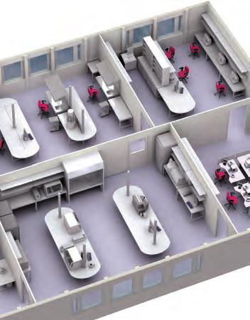
12 Get the full picture with Dako Solutions FISH Test request in LIS Dako Omnis Instrument connectivity DakoLink Omnis workstation DakoLink server IHC Molecular Pathology CISH 10


13 Supported by excellent service and support for your laboratory Dako CoverStainer H&E DakoLink workstation Artisan Link Pro Special Stains Companion Diagnostics pharmdx Autostainer Link IHC 11
14
15 Dako Lab Control Solutions Experience a new level of lab control and insight 14


16 Processing Grossing Embedding Accessioning Sectioning Slide Review Case Assembly True Positive ID IHC and ISH H&E and Special Stains 14


 adds sample creation and tracking capabilities, from accessioning to archiving.")
 and even connect between multiple locations, providing access from your lab to")
17 Dako Lab Control Solutions Experience a new level of lab control and insight The Dako Lab Control solutions consist of staining management, sample tracking and connectivity software that is both flexible and scalable to meet the needs of each individual lab. Either as separate modules or combined, DakoLink and True Positive ID enable your lab to: Minimize errors to improve patient safety Improve efficiency by reducing hands-on time Provide a full electronic audit trail to support quality and regulatory needs The DakoLink and the DakoLink Omnis staining management software connect all Dako staining instruments and allow you to share information across functions, create customized reports based on information captured and easily manage all instruments, slides, reagents and protocols. DakoLink True Positive ID (TPID) adds sample creation and tracking capabilities, from accessioning to archiving. By registering every action for all case parts throughout all of the lab processes, TPID increases patient safety by reducing the risk of transcription errors and misplaced samples. Dako connectivity for total lab control DakoLink and TPID can integrate with your Laboratory Information System (LIS) and even connect between multiple locations, providing access from your lab to anywhere on your network. DakoLink has the ability to read LIS barcodes or create its own unique 2D barcode, ensuring every slide is uniquely identified. With flexible connectivity capabilities, unique identification, work lists and reports, TPID and DakoLink work together to give you total control of your lab. DakoLink 15
18
19 Advanced Staining Solutions Introduction 18 Dako Omnis Solution for IHC and ISH 20 Autostainer Link Solution for IHC 35 Autostainer Plus for IHC 55 Antibodies and Controls 63 Antibody Cocktails 119 Multipurpose Antibodies 121 Secondary Antibodies 123 Control Reagents 125 Visualization Systems 126 Ancillaries for IHC 137 Labeling Systems 140

20 Introduction to the Advanced Staining Solutions Advanced Staining Solutions Introduction We listened. We responded. Our commitment to advancing pathology begins with something very simple listening. By listening carefully to pathologists and lab personnel around the world, we learned that there is growing pressure to: Manage increasing slide volumes with limited personnel and financial resources Process slides faster, to minimize time to diagnosis Cope with fluctuations in workload without sacrificing turnaround time Improve quality control of processes and secure consistency in quality Increase the traceability of patient samples to enable accreditation Find and retain well-trained, qualified staff With almost 50 years of dialogue with our customers, We have helped drive scientific advancement and certainty in cancer diagnostics. We remain committed to delivering novel solutions and innovative technologies which support you to meet the challenges of today and tomorrow. One supplier. Two choices. With the addition of Dako Omnis, we can now deliver a unique and flexible combination of comprehensive advanced staining solutions. These solutions can be used independently or together, to help you meet the individual needs of your lab, without compromising on quality and consistency results. Speak to your local representative to assess which solution addresses the needs of your lab now and in the future. Dako Omnis. Developed by the lab for the lab. Developed together with pathologists, lab managers and lab technicians from around the world, with the needs of the pathology lab very much in focus. Dako Omnis builds on our reputation for delivering quality reagents and staining solutions that bring certainty to cancer diagnostics. Dako Omnis provides: A true automated, walk-away solution High throughput and overnight capacity Same-day IHC and ISH provides complete patient case management Unparalleled onboard capacity of temperature-controlled reagents Increased productivity with limited setup and minimal maintenance time Dako Omnis delivers what pathologists, lab managers and technicians are asking for in terms of time, choice and better patient care. Dako Omnis is a generation ahead in IHC and ISH. Parallel or batch loading, the choice is yours. With a high throughput and full automation, this is a true walk-away solution. A controlled onboard environment facilitates unattended overnight processing of patient cases. Autostainer Link 48 is a compact, bench-top, open system that delivers the flexibility required in a research and clinical environment. Adaptable to your individual setup, and helps to maximize productivity by the decoupled pre-treatment and the ability to run either large batches of up to 48 IHC slides, or mini batches. 18


21 The Dako FLEX RTU solution Ensures optimal staining results, slide after slide Using validated protocols and optimized reagents reduces the risk of false negative or false positive results. A robust, specific and sensitive IHC assay is critical for providing staining results that support an accurate patient diagnosis. The most important element in the qualification of the staining results of clinical samples is accurate selection and staining of control tissue. Dako protocols are the result of a comprehensive study of numerous different tissue types, tissue thicknesses, protocol step durations, target retrieval methods, antibody dilutions, and pre-analytical variations. The Dako optimized protocol ensures that every test performance is highly robust, accurate and consistent, compensating for variations in preanalytical parameters to provide increased certainty slide after slide. Dako FLEX RTU is developed in collaboration with pathology experts to ensure optimal staining results. The solution consists of: Pre-diluted antibody - FLEX RTU antibody Visualization system - EnVision FLEX/FLEX+ Optimized and validated protocol - the recipe for consistent high-quality results Introduction Advanced Staining Solutions Find our range of FLEX RTU antibodies for all of our advanced staining platforms at FLEX RTU antibodies EnVision FLEX visualization system Validated protocols Pre-diluted and carefully selected for specificity and sensitivity Proven concept since 2007 Excellent antibody quality - confirmed by various EQA schemes Broad portfolio of IVD antibodies Visualization system with low complexity and very high sensitivity Visibility of antigen in high and low expressing tissue Kit configuration provides Flexibility Consistency Simplicity Automated protocols developed for Dako solutions Optimized, validated protocols developed in cooperation with leading pathology experts Robust protocols compensate for pre-analytical variations Plug and play on Dako advanced staining platforms 19

22 Dako Omnis Solution for IHC and ISH Advanced Staining Solutions Dako Omnis Solution for IHC and ISH Dako Omnis meets the challenges of the modern pathology lab. It accommodates an increasing number of diverse, advanced staining methods in an increasingly unpredictable working day. Dako Omnis achieves this by automating any advanced staining method using a simple interface with little hands-on time. Lab staff can deliver consistent IHC and ISH results with minimal training. Continuous sample loading allows prioritized patient cases to stream seamlessly into an ongoing workflow. With turnaround times of less than four hours for FISH slides, they are ready within the same time frame as IHC slides. Dako Omnis delivers consistent results in IHC and ISH, regardless of operator experience. The system logs operator actions and built-in controls reduce possible human errors. Dako Omnis gives more time Process 165 IHC slides in a typical workday, including setting up overnight runs Handle the workload with fewer instruments thanks to an unparalleled capacity Enable faster diagnosis of whole patient cases with same-day IHC and ISH results Minimize hands-on time with automation designed for the clinical laboratory Free up lab techs for other tasks thanks to accurate run-time information Dako Omnis allows greater choice Choose continuous loading to match patient cases, or load in batches to utilize full through-put capacity Absorb peaks in workload by processing up to 60 slides in unattended overnight runs Eliminate operator waiting time by loading slides and/or reagents anytime, also during runs, while keeping an optimal throughput because runs continue uninterrupted during the loading Ensure transparency by enabling staff to monitor the slide flow from their workstations Dako Omnis enables better patient care Get results with ease and greater certainty thanks to the FLEX Readyto-Use reagents and optimized protocols Increase lab quality and staffing options because Dako Omnis minimizes the risk of human error Facilitate lab accreditation and improve patient safety by automatic tracking and reporting Apply patient case workflow while optimizing capacity utilization thanks to the 5-slide racks Achieve consistent staining conditions with temperature-controlled and humidity-controlled staining chambers alongside temperaturecontrolled reagent positions 20

23 Dako Omnis Dako Omnis C GI100 Advanced staining system 1 unit IHC and ISH automated on the same platform, coupled with fully optimized and validated protocols, enables a fast turnaround time of patient cases. It supports your lab to deliver consistent quality and optimal results day after day and slide after slide for increased certainty. Dako Omnis provides: Automated IHC and ISH, from deparaffinization to counterstaining Parallel or batch processing Flexible loading, virtually zero waiting time to add slides or reagents Up to 60 slides processed simultaneously Capacity for 60 temperature-controlled reagents on board Limited setup and little maintenance High throughput, including possibility for overnight run Full traceability of patient cases through onboard and workstation software Intuitive user interface and individual user log in LAN seats that display information where needed, including information from the LIS Dynamic Gap staining technology that helps ensure consistent, highquality staining results with very low variation between slides, instruments and days. IHC and ISH automated on the same platform Dako Omnis meets head-on the challenges in the modern pathology lab to accommodate an increasing number of diverse, advanced staining methods in an increasingly unpredictable working day. Dako Omnis achieves this by automating any advanced staining method using the simplest of user interfaces with little hands-on time, so new users can start producing results in minutes. Compensate for increasing fluctuations in workflow Process 60 IHC slides completely unattended overnight (or 45 IHC plus 15 ISH). You decide if the slides should be ready as soon as possible or at the start of the next working day. Batch or continuous flow, the choice is yours. Continuous sample loading allows prioritized patient cases to stream seamlessly into an ongoing workflow. Full flexibility for the unpredictable lab environment. Monitor and control your staining workflow Monitor the progress of your run at a glance. Clear visual alerts notify you when user interactions are necessary. Dako Omnis connects to your LIS system. Share, monitor and track slides wherever you are. Manage increasing slide volumes with limited resources Load your IHC or ISH slides when convenient and the system informs you which reagents are needed. Once slides are loaded, you are free to perform other tasks. Just load and walk away. With minimal hands-on time, daily setup takes just 15 minutes and little daily maintenance is required. Less time for preparation, faster processing. Dako Omnis Solution for IHC and ISH Advanced Staining Solutions 21
24 Dako Omnis (continued) Hardware Specifications Advanced Staining Solutions Dako Omnis Solution for IHC and ISH Turn-around time IHC: 2 hours 30 minutes ISH: 3 hours 40 minutes Throughput 165 slides can be loaded in a typical workday (including preparation for an overnight run) Slide capacity 60 slides for IHC or ISH (up to 15 ISH slides) Reagent capacity 60 reagent vials Bulk fluid capacity 8 x 3.5 L bottle and 4 x 7 L bottle Waste capacity 4 x 7 L bottle (non-hazardous) 1 x 7 L bottle (low-hazardous, below limit values) Dimensions 57.1" W x 31.2" D x 60.4" H (145 cm W x 79.3 cm D x cm H) Weight 1,323 lbs (600 kg) fully loaded Voltage 120/ VAC Power consumption 1200 W Features Processes Fully automated and simultaneous IHC and/or ISH Deparaffinization, staining and counterstaining with parallel processing Operation Continuous or batch workflow 5-slot racks to optimize capacity utilization and patient-case management Reagents and slides can be loaded anytime, also during runs Easy-to-use software interface and ready-to-use reagents Built-in controls to reduce possible human errors Staining conditions Temperature controlled onboard reagent storage Dynamic Gap staining technology Temperature and humidity controlled processing environment FLEX RTU reagents and protocols for optimal staining results Connectivity and control LAN seats for setup and monitoring from anywhere Full integration with Laboratory Information Systems 1D and 2D barcodes Data logging, reporting and access rights for traceability and accreditation Learn more about Dako Omnis by visiting 22
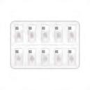
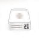
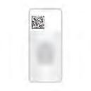
25 IHC Ancillaries and Accessories (Dako Omnis) Clearify TM GC L Clearify TM is used onboard Dako Omnis to remove paraffin from tissue sections for both IHC and ISH staining in a two-phase dewaxing procedure. DAB+ Substrate Chromogen System (Dako Omnis) C GV825 Onboard mixing 150 tests EnVision FLEX DAB+ Substrate Chromogen System (Dako Omnis) is intended for use in immunohistochemistry together with Dako Omnis. The working solution is prepared onboard by the Dako Omnis instrument. It is a high sensitivity DAB system suitable for use in combination with the EnVision FLEX visualization system (Codes GV800/GV823). Upon oxidation, DAB forms a brown endproduct at the site of the target antigen. The reagent is intended for use on formalin-fixed, paraffin-embedded tissue sections. Hematoxylin (Dako Omnis) C GC808 Ready-to-use 8 x 22.5 ml, 600 tests Intended for use in immunohistochemistry together with Dako Omnis. The reagent is recommended for counterstaining on formalin-fixed, paraffinembedded tissue sections providing a clear blue, nuclear staining. IHC Microscope Slides, FLEX C K8020 Coated glass slides 5 x 100 slides Coated microscope slides for adhesion of formalin-fixed, paraffin-embedded tissue sections for use in immunohistochemistry with Dako EnVision FLEX visualization systems. FLEX IHC Microscope Slides are compatible with, but not limited to, the following Dako instruments: Dako Omnis, Autostainer Link, Dako Autostainer/Autostainer Plus and PT Link. Mixing Strip, for Dako Omnis GC well mixing strip 25 strips Dako Omnis Mixing Strip is intended for mixing of the chromogen working solution during staining onboard Dako Omnis. Dako Omnis Mixing Strip has ten wells designed to hold chromogen for five slides with minimal dead volume. Wells are covered with a lid to limit spill of reagent during disposal of strips. Dako Omnis Mixing Strip can stand unsupported on a table. Arrows indicate correct insertion on Dako Omnis. Dako Omnis Mixing Strip is single use only and used strips are classified as hazardous waste due to chromogen residuals. Reagent Vial, Small/Large, for Dako Omnis GC201 Small vial 25 x 2 ml GC202 Large vial 25 x 30 ml Reagent vials designed to allow the use of a user-defined reagent on Dako Omnis. Each single-use bottle is labeled with positive identification technology. User-fillable reagent vial may be filled to a maximum fill volume of approximately 2 ml/30 ml, respectively. The vial closure contains a septum to reduce evaporation of reagent during onboard use in Dako Omnis reagent storage. Slide Rack, for Dako Omnis GC101 Slide racks holding 5 slides each 6 racks Dako Omnis Slide Rack is designed for use on Dako Omnis. The Slide Rack holds the slides with samples to be processed on Dako Omnis.Each Slide Rack can carry up to five slides. Each slide is placed in a positioning groove and fixated by a spring. Dako Omnis is validated with FLEX IHC Microscope Slides and Superfrost Plus Slides. Dako does not recommend the use of other slide types. Dako Omnis Slide Rack is classified as non-hazardous waste, and Slide Rack parts comply with incineration or parts may be dismantled for recycling. Slide Rack Color Clips, for Dako Omnis GC104 Blue 25 clips GC105 Green 25 clips GC106 Gray 25 clips GC103 Red 25 clips The colored clips are attached to the slide rack for visual identification of individual racks. Each Dako Omnis Slide Rack can hold two Dako Omnis Slide Rack Color Clips and the colors available are: blue, green, gray and red. Dako Omnis Slide Racks are supplied with black color clips as default. Sulfuric Acid, 0.3 M, for Dako Omnis GC x 22.5 ml Sulfuric Acid, 0.3 M is a generic cleaning agent used to remove residue (primarily protein) from various surfaces. It is used on Dako Omnis to automatically clean the Liquid Handling Tip after pipetting of reagents with high protein content, specifically primary antibodies. Wash Buffer (20x) (Dako Omnis) C GC807 Concentrate 20 x 175 ml, 1700 tests Wash Buffer 20x (Dako Omnis) is intended for use in immunohistochemistry. The product is used as wash buffer for immunohistochemical staining procedures onboard Dako Omnis. Dako Omnis Solution for IHC and ISH Advanced Staining Solutions 23
 is intended for mounting of formalin-fixed, paraffin-embedded (FFPE) tissue sections after FISH staining performed onboard the Dako Omnis instrument.")
26 ISH Ancillaries and Accessories (Dako Omnis) Advanced Staining Solutions Dako Omnis Solution for IHC and ISH Fluorescence Mounting Medium (Dako Omnis) C GM304 Ready-to-use 20 tests, 0.8 ml Fluorescence Mounting Medium (Dako Omnis) is intended for mounting of formalin-fixed, paraffin-embedded (FFPE) tissue sections after FISH staining performed onboard the Dako Omnis instrument. The mounting medium also contains 500 µg/l DAPI for improved nuclei staining. ISH Cleaning Solution (Dako Omnis) C GC207 Ready-to-use NEW 100 tests, 10 ml ISH Cleaning Solution (Dako Omnis) is an accessory to the Dako Omnis instrument. It is used for cleaning the pipette tip between dispenses of in situ hybridization probes. Washing with ISH Cleaning Solution dissolves ISH probe, allowing remaining probe to be effectively washed away with water. The product is provided in a ready-to-use vial for the Dako Omnis instrument. ISH Ethanol Solution, 96% (Dako Omnis) C GM300 Ready-to-use 20 tests, 14 ml ISH Ethanol Solution, 96% (Dako Omnis) is intended for use in automated in situ hybridization assays together with the Dako Omnis instrument on formalin-fixed, paraffin-embedded (FFPE) tissue sections. The solution is used in the wash step after target retrieval. The product is provided in a ready-to-use vial for the Dako Omnis instrument. ISH Lid, for Dako Omnis GC102 5 lids Dako Omnis ISH Lid is intended for use in FISH procedures. Each Dako Omnis ISH Lid holds five slides and has five built-in Cover Glasses and one Humidity Pad. The Cover Glasses serve to distribute probe buffer across the staining area and to reduce buffer evaporation. The Humidity Pad with deionized water added serves to increase the humidity inside Dako Omnis ISH Lid to further reduce evaporation. Dako Omnis ISH Lid also provides insulation to maintain proper denaturation temperature. Dako Omnis ISH Lid is single use only and is classified as non-hazardous waste. ISH Pepsin (Dako Omnis) C GM302 Ready-to-use 20 tests, 7 ml ISH Pepsin (Dako Omnis) is intended for use in automated in situ hybridization assays together with the Dako Omnis instrument on formalin-fixed, paraffinembedded (FFPE) tissue sections. The solution is used in the digestion step. The product is provided in a ready-to-use vial for the Dako Omnis instrument. ISH Pre-Treatment Solution (20x) (Dako Omnis) C GM301 Concentrate 175 ml, 20x concentrated ISH Pre-Treatment Solution (20x) (Dako Omnis) is intended for use in automated in situ hybridization assays together with the Dako Omnis instrument on formalin-fixed, paraffin-embedded (FFPE) tissue sections. The solution is used in the pre-treatment step. An inert green color is added to the buffer for easy identification and user friendliness. The volume is tailored for dilution in one Dako Omnis bulk bottle. ISH Stringent Wash Buffer (20x) (Dako Omnis) C GM303 Concentrate 175 ml, 20x concentrated ISH Stringent Wash Buffer (20x) (Dako Omnis) is intended for use in automated in situ hybridization assays together with the Dako Omnis instrument on formalin-fixed, paraffin-embedded (FFPE) tissue sections. The solution is used in the post-hybridization step. An inert yellow color is added to the buffer for easy identification and user friendliness. The volume is tailored for dilution in one Dako Omnis bulk bottle. Mixing Device, for Dako Omnis GC116 1 unit Dako Omnis Mixing Device is an accessory to the Dako Omnis instrument. It is designed specifically to support the fluorescence in situ hybridization (FISH) and the chromogenic in situ hybridization (CISH) procedures. The Dako IQISH buffer is extremely viscous, and during storage the reagent phase separates. Hence the Dako IQISH reagents require a particular preparatory processing to thaw and unify the content. Some Dako ISH reagents are therefore provided in dedicated ISH reagent vials containing a mixing ball, and the Dako Omnis Mixing Device is designed to fit together with these ISH reagent vials. Dako Omnis Mixing Device contains a magnet that enables the mixing ball to move up and down (110 cycles) inside the vial after 40 minutes thawing of the ISH reagent; thus ensuring a homogenous probe mix prior to application on the Dako Omnis instrument. Vial with Mixing Ball, 2 ml, for Dako Omnis GC vials NEW 2 ml Dako Omnis Vial with Mixing Ball, 2 ml has been designed as an accessory for Dako Omnis and Dako Omnis Mixing Device and is intended for use in ISH procedures using user-provided FISH probes diluted in ethylene carbonatebased hybridization buffer (IQFISH). Dako Omnis Vial with Mixing Ball, 2 ml includes a mixing ball that is used by Dako Omnis Mixing Device to mix the IQFISH hybridization buffer with the user-provided probe. Each package contains 25 vials, 25 caps and 25 mixing balls. 24
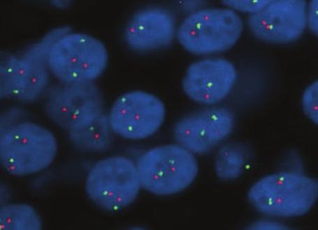
27 pharmdx Kit (Dako Omnis) HER2 IQFISH pharmdx (Dako Omnis) C GM333 Ready-to-use 20 tests, 1.6 ml HER2 IQFISH pharmdx (Dako Omnis) is the hybridization probe for the automated direct fluorescence in situ hybridization (FISH) assay onboard Dako Omnis instruments. It consists of a HER2 and CEN-17 probe mix in IQISH hybridization buffer and is provided in a ready-to-use vial for the Dako Omnis instrument. The IQISH hybridization buffer is non-toxic and allows genomic DNA probe hybridization to be performed in just 75 minutes on the Dako Omnis instrument. The short hybridization time results in a turnaround time of less than 4 hours for a complete FISH staining from deparaffinization to mounting. HER2 IQFISH pharmdx (Dako Omnis) is indicated in adjunction to HercepTest in the assessment of patients for whom Herceptin TM (trastuzumab) treatment is being considered. For breast cancer patients, results from HER2 IQFISH pharmdx (Dako Omnis) are intended for use as an adjunct to the clinicopathologic information currently used for estimating prognosis in stage II, node-positive breast cancer patients. Accessory reagents to be used together with HER2 IQFISH pharmdx (Dako Omnis): HER2 IQFISH pharmdx (Dako Omnis) is, together with accessory reagent devices, designed to quantitatively determine HER2 gene amplification in formalin-fixed, paraffin-embedded (FFPE) breast cancer tissue specimens and FFPE specimens from patients with adenocarcinoma of the stomach including gastroesophageal junction. Gene amplification is determined from the ratio between the number of signals from the hybridization of the HER2 gene probe (red signals) and the number of signals from the hybridization of the CEN-17 reference chromosome 17 probe (green signals). Breast carcinoma (FFPE) stained with HER2 IQFISH pharmdx (Dako Omnis), Code GM333. Tumor cells show HER2 gene amplification. Product Name Dako Omnis ISH Lid Dako Omnis Mixing Device Fluorescence Mounting Medium (Dako Omnis) ISH Ethanol Solution, 96% (Dako Omnis) ISH Pepsin (Dako Omnis) ISH Pre-Treatment Solution (20x) (Dako Omnis) ISH Stringent Wash Buffer (20x) (Dako Omnis) ISH Cleaning Solution (Dako Omnis) Code GC102 GC116 GM304 GM300 GM302 GM301 GM303 GC207 Breast carcinoma (FFPE) stained with HER2 IQFISH pharmdx (Dako Omnis), Code GM333. Cells show HER2 gene nonamplification. Dako Omnis Solution for IHC and ISH Advanced Staining Solutions Go to page 143 to read about all our pharmdx products. Product Name 25

28 Primary Antibodies (FLEX Ready-to-Use) (Dako Omnis) For Dako Omnis, we offer a dedicated series of high-quality, pre-diluted, ready-to-use (RTU) primary antibodies. FLEX Ready-to-Use antibodies are pre-diluted primary antibodies specifically developed for automated use while maintaining the highquality staining performance for which Dako antibodies is known. Each FLEX RTU antibody is accompanied by a validated protocol that is optimized to absorb variations related to pre-analytical factors. This enables a reliable staining performance in various tissue types containing both high and low-expression structures. The antibody specificity and protocol have both been evaluated and approved by external pathology experts. Advanced Staining Solutions Dako Omnis Solution for IHC and ISH Optimized FLEX protocols Excellent staining results Dako Omnis Antibodies with high specificity and sensitivity Key Features Optimized staining performance of both high and low-expression structures Dako Omnis and the dynamic gap staining technology provide consistent and uniform staining with excellent morphology Crisp and clear staining with no background Optimal laboratory efficiency with RTU antibodies on Dako Omnis The GA-Series FLEX Ready-to-Use Primary Antibodies listed in this section are packaged in Dako Omnis vials for use on Dako Omnis instruments, and can be used only with the EnVision FLEX system for Dako Omnis. 26
 stained with FLEX Anti-Alpha-1-Antitrypsin, Code GA505.")
 stained with FLEX Anti-BCL6, Code GA625.")
 stained with FLEX Anti-AMACR, Code GA060.")
 stained with FLEX Anti-Amyloid A, Code GA605.")
29 Primary Antibodies (FLEX Ready-to-Use) (Dako Omnis) (continued) Polyclonal Rabbit Anti-Human Alpha-1-Antitrypsin C GA505 NEW 60 tests, 12 ml BCL6 Protein C GA625 Clone PG-B6p 60 tests, 12 ml A1AT-deficient liver (FFPE) stained with FLEX Anti-Alpha-1-Antitrypsin, Code GA505. Polyclonal Rabbit Anti-Human Alpha-1-Fetoprotein C GA500 NEW 60 tests, 12 ml Embryonal carcinoma (FFPE) stained with FLEX Anti-Alpha-1-Fetoprotein, Code GA500. Monoclonal Rabbit Anti-Human AMACR C GA060 Clone 13H4 60 tests, 12 ml Follicular lymphoma (FFPE) stained with FLEX Anti-BCL6, Code GA625. Beta-Catenin C GA702 Clone b-catenin-1 NEW 60 tests, 12 ml Colon adenocarcinoma (FFPE) stained with FLEX Anti-Beta-Catenin, Code GA702. CA 125 C GA701 Clone M11 60 tests, 12 ml Dako Omnis Solution for IHC and ISH Advanced Staining Solutions Prostate adenocarcinoma (FFPE) stained with FLEX Anti-AMACR, Code GA060. Amyloid A C GA605 Clone mc1 60 tests, 12 ml Ovarian carcinoma (FFPE) stained with FLEX Anti-CA 125, Code GA701. Polyclonal Rabbit Anti-Human Calcitonin C GA515 NEW 60 tests, 12 ml Kidney with amyloidosis (FFPE) stained with FLEX Anti-Amyloid A, Code GA605. B-Cell-Specific Activator Protein C GA650 Clone DAK-Pax5 60 tests, 12 ml Thyroid medullary carcinoma (FFPE) stained with FLEX Anti-Calcitonin, Code GA515. Caldesmon C GA054 Clone h-cd NEW 60 tests, 12 ml Lymph node (FFPE) stained with FLEX Anti- BSAP, Code GA650. Leiomyosarcoma (FFPE) stained with FLEX Anti-Caldesmon, Code GA
 stained with FLEX Anti-CEA, Code GA622.")
 stained with FLEX Anti-CD3, Code GA503. CD7 C GA643 Clone CBC.")
30 Primary Antibodies (FLEX Ready-to-Use) (Dako Omnis) (continued) Carcinoembryonic Antigen (CEA) C GA622 Clone II-7 60 tests, 12 ml CD8 C GA623 Clone C8/144B NEW 60 tests, 12 ml Advanced Staining Solutions Dako Omnis Solution for IHC and ISH Colon adenocarcinoma (FFPE) stained with FLEX Anti-CEA, Code GA622. Polyclonal Rabbit Anti-Human Carcinoembryonic Antigen (CEA) C GA tests, 12 ml CD2 C GA651 Clone AB75 60 tests, 12 ml Precursor T-lymphoblastic lymphoma (FFPE) stained with FLEX Anti-CD2, Code GA651. Polyclonal Rabbit Anti-Human CD3 C GA tests, 12 ml T-cell lymphoma (FFPE) stained with FLEX Anti-CD3, Code GA503. CD7 C GA643 Clone CBC tests, 12 ml Lymphoma (FFPE) stained with FLEX Anti- CD7 Code GA643. Angioimmunoblastic T-cell lymphoma (FFPE) stained with FLEX Anti-CD8, Code GA623. CD10 C GA648 Clone 56C6 60 tests, 12 ml Lymphoma (FFPE) stained with FLEX Anti- CD10, Code GA648. CD15 C GA062 Clone Carb-3 60 tests, 12 ml Hodgkin's Lymphoma (FFPE) stained with FLEX Anti-CD15, Code GA062. CD20cy C GA604 Clone L26 60 tests, 12 ml B-cell chronic lymphocytic leukemia/small lymphocytic lymphoma (FFPE) stained with FLEX Anti-CD20cy, Code GA604. CD23 C GA781 Clone DAK-CD23 NEW 60 tests, 12 ml Chronic lymphocytic leukemia/small lymphocytic lymphoma (FFPE) stained with FLEX Anti-CD23, Code GA
31 Primary Antibodies (FLEX Ready-to-Use) (Dako Omnis) (continued) CD31, Endothelial Cell C GA610 Clone JC70A 60 tests, 12 ml CD68 C GA613 Clone PG-M1 60 tests, 12 ml Angiosarcoma (FFPE) stained with FLEX Anti-CD31, Code GA610. CD34 Class II C GA632 Clone QBEnd tests, 12 ml Angiosarcoma (FFPE) stained with FLEX Anti-CD34, Code GA632. CD43 C GA636 Clone DF-T1 60 tests, 12 ml Tonsil (FFPE) stained with FLEX Anti-CD68, Code GA613. CD79a C GA621 Clone JCB tests, 12 ml Plasmacytoma (FFPE) stained with FLEX Anti-CD79a, Code GA621. CD138 C GA642 Clone MI15 60 tests, 12 ml Dako Omnis Solution for IHC and ISH Advanced Staining Solutions Tonsil (FFPE) stained with FLEX Anti-CD43, Code GA636. CD45, Leucocyte Common Antigen C GA751 Clones 2B11 + PD7/26 60 tests, 12 ml High grade myeloma (FFPE) stained with FLEX Anti-CD138, Code GA642. CD246, ALK Protein C GA641 Clone ALK1 60 tests, 12 ml Tonsil (FFPE) stained with FLEX Anti-CD45, Code GA751. CD68 C GA609 Clone KP1 60 tests, 12 ml Anaplastic large cell lymphoma (FFPE) stained with FLEX Anti-CD246, ALK Protein, Code GA641. CDX2 C GA080 Clone DAK-CDX2 60 tests, 12 ml Tonsil (FFPE) stained with FLEX Anti-CD68, Code GA609. Colon adenocarcinoma (FFPE) stained with FLEX Anti-CDX2, Code GA
32 Primary Antibodies (FLEX Ready-to-Use) (Dako Omnis) (continued) Polyclonal Rabbit Anti-Human Chorionic Gonadotropin (hcg) C GA508 NEW 60 tests, 12 ml Cytokeratin 18 C GA618 Clone DC tests, 12 ml Advanced Staining Solutions Dako Omnis Solution for IHC and ISH Placenta (FFPE) stained with FLEX Anti- Human Chorionic Gonadotropin, Code GA508. Monoclonal Rabbit Anti-Human Cyclin D1 C GA083 Clone EP12 NEW 60 tests, 12 ml Mantle cell lymphoma (FFPE) stained with FLEX Anti-Cyclin D1, Code GA083. Cytokeratin C GA053 Clone AE1/AE3 60 tests, 12 ml Adenocarcinoma (FFPE) stained with FLEX Anti-Cytokeratin, Code GA053. Cytokeratin 5/6 C GA780 Clone D5/16 B4 60 tests, 12 ml Cytokeratin 7 C GA619 Clone OV-TL 12/30 60 tests, 12 ml Ductal carcinoma (FFPE) stained with FLEX Anti-Cytokeratin 7, Code GA619. Renal clear cell carcinoma stained with FLEX Anti-Cytokeratin 18, Code GA618. Cytokeratin 19 C GA615 Clone RCK tests, 12 ml Thyroid papillary carcinoma (FFPE) stained with FLEX Anti-Cytokeratin 19, Code GA615. Cytokeratin 20 C GA777 Clone K s tests, 12 ml Merkel cell carcinoma (FFPE) stained with FLEX Anti-Cytokeratin 20, Code GA777. Cytokeratin, High Molecular Weight C GA051 Clone 34bE12 60 tests, 12 ml Prostate (FFPE) stained with FLEX Anti- Cytokeratin HMW, Code GA051. Monoclonal Mouse Anti- Cytomegalovirus C GA752 Clones CCH2 + DDG9 60 tests, 12 ml Lung tissue (FFPE) stained with FLEX Anti- Cytomegalovirus, Code GA
33 Primary Antibodies (FLEX Ready-to-Use) (Dako Omnis) (continued) E-Cadherin C GA059 Clone NCH tests, 12 ml Gross Cystic Disease Fluid Protein-15 C GA077 Clone 23A3 60 tests, 12 ml Poorly differentiated ductal carcinoma (FFPE) stained with FLEX Anti-E-Cadherin, Code GA059. Epithelial Antigen C GA637 Clone Ber-EP4 60 tests, 12 ml Adenocarcinoma (FFPE) stained with FLEX Anti-Epithelial Antigen, Code GA637. Monoclonal Rabbit Anti-Human ERG (Ets-Related Gene) C GA659 Clone EP111 NEW 60 tests, 12 ml Breast hyperplasia (FFPE) stained with FLEX Anti-GCDFP-15, Code GA077. Polyclonal Rabbit Anti- Helicobacter Pylori C GA tests, 12 ml Gastric mucosa (FFPE) stained with FLEX Anti-Helicobacter Pylori, Code GA523. Hepatocyte C GA624 Clone OCH1E5 60 tests, 12 ml Dako Omnis Solution for IHC and ISH Advanced Staining Solutions Prostate carcinoma (FFPE) stained with FLEX Anti-ERG, Code GA659. Polyclonal Rabbit Anti-Human Gastrin C GA tests, 12 ml Hepatocellular carcinoma (FFPE) stained with FLEX Anti-Hepatocyte, Code GA624. Polyclonal Rabbit Anti- Herpes Simplex Virus Type 1 C GA tests, 12 ml Gastrin-producing tumor (FFPE) stained with FLEX Anti-Gastrin, Code GA519. Polyclonal Rabbit Anti- Glial Fibrillary Acidic Protein C GA tests, 12 ml Brain with herpes simplex virus (FFPE) stained with FLEX Anti-Herpes Simplex Virus Type 1, Code GA521. Polyclonal Rabbit Anti-Human IgA C GA510 NEW 60 tests, 12 ml Glioblastoma (FFPE) stained with FLEX Anti- GFAP, Code GA524. Multiple myeloma (FFPE, bone marrow) stained with FLEX Anti-IgA, Code GA
34 Primary Antibodies (FLEX Ready-to-Use) (Dako Omnis) (continued) Polyclonal Rabbit Anti-Human IgM C GA513 NEW 60 tests, 12 ml Melanosome C GA052 Clone HMB tests, 12 ml Advanced Staining Solutions Dako Omnis Solution for IHC and ISH Mantle cell lymphoma (FFPE) stained with FLEX Anti-IgM, Code GA513. Polyclonal Rabbit Anti-Human Kappa Light Chains C GA tests, 12 ml Tonsil (FFPE) stained with FLEX Anti-Kappa light chains, Code GA506. Ki-67 Antigen C GA626 Clone MIB-1 60 tests, 12 ml High grade lymphoma (FFPE) stained with FLEX Anti-Ki-67, Code GA626. Polyclonal Rabbit Anti-Human Lambda Light Chains C GA tests, 12 ml Tonsil (FFPE) stained with FLEX Anti-Lambda Light Chains, Code GA507. Mammaglobin C GA074 Clone 304-1A5 NEW 60 tests, 12 ml Invasive ductal carcinoma (FFPE) stained with FLEX Anti-Mammaglobin, Code GA074. Melanoma (FFPE) stained with FLEX Anti- Melanosome, Code GA052. MUM1 Protein C GA644 Clone MUM1p 60 tests, 12 ml Diffuse large B-cell lymphoma (FFPE) stained with FLEX Anti-MUM1, Code GA644. Polyclonal Rabbit Anti-Human Myeloperoxidase C GA tests, 12 ml Acute myeloid leukemia (FFPE) stained with FLEX Anti-Myeloperoxidase, Code GA511. Neurofilament Protein C GA607 Clone 2F11 NEW 60 tests, 12 ml Merkel cell tumor (FFPE) stained with FLEX Anti-Neurofilament Protein, Code GA607. Nucleophosmin C GA652 Clone tests, 12 ml Acute myeloid leukemia (AML) (FFPE) stained with FLEX Anti-Nucleophosmin, Code GA
 stained with FLEX Anti-p53, Code GA616.")
35 Primary Antibodies (FLEX Ready-to-Use) (Dako Omnis) (continued) p53 Protein C GA616 Clone DO-7 60 tests, 12 ml Polyclonal Rabbit Anti-Human Thyroglobulin C GA tests, 12 ml Invasive transitional cell carcinoma (FFPE) stained with FLEX Anti-p53, Code GA616. Polyclonal Rabbit Anti-Human Prostate-Specific Antigen (PSA) C GA tests, 12 ml Prostate adenocarcinoma (FFPE) stained with FLEX Anti-Prostate Specific Antigen, Code GA514. Renal Cell Carcinoma Marker C GA075 Clone SPM314 NEW 60 tests, 12 ml Renal clear cell carcinoma (FFPE) stained with FLEX Anti-Renal Cell Carcinoma Marker, Code GA075. Thyroid follicular carcinoma (FFPE) stained with FLEX Anti-Thyroglobulin, Code GA509. Monoclonal Mouse Anti- Vimentin C GA630 Clone V9 60 tests, 12 ml Bladder wall (FFPE) stained with FLEX Anti- Vimentin, Code GA630. Polyclonal Rabbit Anti-Human Von Willebrand Factor C GA tests, 12 ml Angiosarcoma (FFPE) stained with FLEX Anti-Von Willebrand Factor, Code GA527. Dako Omnis Solution for IHC and ISH Advanced Staining Solutions Polyclonal Rabbit Anti- S100 C GA tests, 12 ml Breast carcinoma (FFPE) stained with FLEX Anti-S100, Code GA504. Negative Controls (FLEX Ready-to-Use) (Dako Omnis) Universal Negative Control for GA-Series Mouse Primary Antibodies C GA750 Ready-to-use 120 tests, 24 ml Universal negative control for all FLEX ready-to-use mouse primary antibodies for use on the Dako Omnis instrument. Packaged in vials for Dako Omnis. Universal Negative Control for GA-Series Rabbit Primary Antibodies C GA600 Ready-to-use 120 tests, 24 ml Universal negative control for all FLEX ready-to-use rabbit primary antibodies for use on the Dako Omnis instrument. Packaged in vials for Dako Omnis. 33
36 Visualization Systems (EnVision FLEX) (Dako Omnis) EnVision FLEX Visualization Systems for Dako Omnis EnVision FLEX, the well-known Dako visualization system, has been configured into a dedicated system for Dako Omnis. The highly sensitive polymer-based EnVision FLEX system builds upon simple intelligent chemistry that allows for distinct clear staining. The Dynamic Gap staining technology utilized onboard Dako Omnis, the high-quality primary antibodies and the EnVision FLEX system all come together to provide a robust system that produces stains with excellent morphology and diagnostic certainty. The streamlined kits and optional reagents for Dako Omnis are packaged for your convenience and are easy to order, making the system flexible, versatile and functional. Advanced Staining Solutions Dako Omnis Solution for IHC and ISH Code Code FLEX High ph GV800 or GV823 FLEX Low ph GV800 + GV805 or GV823 + GV805 EnVision FLEX, High ph (Dako Omnis) C GV800 HRP. Rabbit/Mouse. High ph 600 tests EnVision FLEX, High ph is a high-sensitivity visualization system intended for use in immunohistochemistry together with Dako Omnis. The dual link system detects primary mouse and rabbit antibodies and the reaction is visualized by DAB+ Chromogen. The convenience kit includes Peroxidase-Blocking Reagent, EnVision/HRP, DAB+ Chromogen, Substrate Buffer and Target Retrieval Solution, High ph (50x Tris/EDTA buffer, ph 9). EnVision FLEX convenience kits are compatible with all optional EnVision FLEX and FLEX+ reagents for Dako Omnis. EnVision FLEX Mini Kit, High ph (Dako Omnis) C GV823 HRP. Rabbit/Mouse. High ph 150 tests EnVision FLEX Mini Kit, High ph is a high-sensitivity visualization system intended for use in immunohistochemistry together with Dako Omnis. The dual link system detects primary mouse and rabbit antibodies and the reaction is visualized by DAB+ Chromogen. The convenience kit includes Peroxidase- Blocking Reagent, EnVision/HRP, DAB+ Chromogen, Substrate Buffer and Target Retrieval Solution, High ph (50x Tris/EDTA buffer, ph 9). EnVision FLEX convenience kits are compatible with all optional EnVision FLEX and FLEX+ reagents for Dako Omnis. EnVision FLEX Systems FLEX+ High ph GV800 + GV821 (Mouse LINKER) or GV800 + GV809 (Rabbit LINKER) FLEX+ Low ph GV800 + GV805 + GV821 (Mouse LINKER) or GV800 + GV805 + GV809 (Rabbit LINKER) Mouse LINKER (Dako Omnis) C GV821 Ready-to-use 75 tests, 22.5 ml EnVision FLEX+ Mouse LINKER is an optional EnVision FLEX+ reagent and may be used with EnVision FLEX convenience kits (GV800 and GV823) for Dako Omnis to amplify the signal of primary mouse antibodies. Rabbit LINKER (Dako Omnis) C GV809 Ready-to-use 75 tests, 22.5 ml EnVision FLEX+ Rabbit LINKER is an optional EnVision FLEX+ reagent and may be used with EnVision FLEX convenience kits (GV800 and GV823) for Dako Omnis to amplify the signal of primary rabbit antibodies. Target Retrieval Solution, High ph (Dako Omnis) C GV804 Concentrate 3 x 68 ml, 225 tests EnVision FLEX Target Retrieval Solution, High ph (Dako Omnis) is an optional EnVision FLEX reagent containing 50x concentrated Tris/EDTA, ph 9 and is compatible with EnVision FLEX convenience kits for Dako Omnis. The volume is optimized for dilution in Dako Omnis bulk bottles. Target Retrieval Solution, Low ph (Dako Omnis) C GV805 Concentrate 3 x 68 ml, 225 tests EnVision FLEX Target Retrieval Solution, Low ph (Dako Omnis) is an optional EnVision FLEX reagent containing 50x concentrated citrate buffer, ph 6.1 and is compatible with EnVision FLEX convenience kits for Dako Omnis. The volume is optimized for dilution in Dako Omnis bulk bottles. 34

37 Autostainer Link Solution for IHC Automated Link Platforms is the line of instruments with which pathology laboratories will experience an outstanding level of integration that provides high productivity and efficient workflow. The Autostainer Link 48 staining instrument with the latest release of DakoLink software enables improved productivity in a pathology laboratory by staining 48 slides in less than three hours. When processing slides in parallel, using only one Autostainer Link 48 and one PT Link pretreatment module, up to 144 slides can be processed in a regular working day, including setting up an overnight run. With PT Link, pathology laboratories can further maximize productivity by reducing the number of operations needed in the specimen preparation processes of deparaffinization, rehydration and target retrieval. The fact that pre-treatment and staining are decoupled gives high flexibilitly and productivity. The revolutionary DakoLink software and connectivity options will improve workflow and productivity even further by, among other things, completely eliminating re-labeling steps and repetitive test request entries. Autostainer Link 48 Process 48 slides in less than three hours Organize your working day to the minute with precise run-time estimation Achieve high quality, when staining slides with FLEX RTU primary antibodies and EnVision FLEX/FLEX+ visualization optimized for Autostainer Link 48 PT Link Maximize productivity by processing slides in parallel Run deparaffinization, target retrieval and dehydration in one step with the 3-in-1 buffer Have confidence in your pre-treatment process, as it is controlled every second Possibility to track via DakoLink software DakoLink Software Enables a fully integrated pathology solution with Dako instrumentation for Advanced Staining and Histostaining Significant tracking improvements with included slide pre-treatment Full laboratory connectivity by controlling all slides and slide IDs from one workstation Reporting made easy Improved laboratory efficiency Autostainer Link Solution for IHC Advanced Staining Solutions 35


38 Autostainer Link 48 Autostainer Link 48 C AS480 Slide-processing instrument 1 unit Advanced Staining Solutions Autostainer Link Solution for IHC Reliability and innovation come together in Autostainer Link 48. Our trusted immunohistochemistry stainer is united with revolutionary software and connectivity options, delivering an outstanding level of integration that provides high productivity and efficient workflow. Get high quality staining results - on time Process 48 slides in less than three hours. This makes it possible to finalize 96 slides during a regular working day with only one Autostainer Link 48 and one PT Link Gain up to 45 minutes of your run time compared to our previously fastest Autostainer Autostainer Plus Get the most out of your laboratory time by processing slides in parallel using PT Link and the fastest ever Autostainer Link 48 Have the freedom to set up your own standards and a possibility to control these Autostainer Link 48 DakoLink software Autostainer Link 48 ensures optimal staining results and offers a high slide and reagent capacity. Save space and centralize slide programming by connecting up to three instruments and three PT Links to one computer. The DakoLink software has optimized run-time estimation. Confidence secured The Autostainer Link solution Consistent high-quality staining is ensured by validated staining protocols optimized with Dako reagents FLEX ready-to-use primary antibodies and EnVision FLEX/FLEX+ visualization systems Get necessary quality control documentation with DakoLink consolidated reporting. Any kind of customized report is just a few mouse clicks away FLEX Ready-to-Use reagents PT Link 36
 Electrical specifications 120 V: 110/120 V (+/- 10%), 60 Hz (+/- 2 Hz) 220 V: 220/240 V (+/- 10%), 50 Hz (+/- 2 Hz) Current requirements 3 A at 220 V; 6 A at 110 V Normal operating temperature")
39 Autostainer Link 48 (continued) Hardware Specifications Dimensions 35'' W x 26'' D x 27'' H (0.89 m W x 0.66 m D x 0.68 m H) Weight 147 lbs (66.7 kg) Electrical specifications 120 V: 110/120 V (+/- 10%), 60 Hz (+/- 2 Hz) 220 V: 220/240 V (+/- 10%), 50 Hz (+/- 2 Hz) Current requirements 3 A at 220 V; 6 A at 110 V Normal operating temperature C (64-79 F) Total slide capacity 48 slides (US and international sizes) Reagent capacity 42 reagents Bulk fluid capacity 2 x 10 L; slides (at 200 µl dispense volume) Waste capacity 2 x 10 L; slides (at 200 µl dispense volume) Software requirements Windows XP SP3, Windows 7 (32 bit) or higher Ancillaries and Accessories (Autostainer Link) Hematoxylin (Link) C SK308 Ready-to-use 45 ml This product is optimized for use on Autostainer Link Instruments. This histological staining reagent is suitable for visualization of nuclei in tissue sections and cell preparations. This product does not contain alcohol and is suitable for use with all chromogens commonly used in immunohistochemistry applications. IHC Microscope Slides, FLEX C K8020 Coated glass slides 5 x 100 slides Coated microscope slides for adhesion of formalin-fixed, paraffin-embedded tissue sections for use in immunohistochemistry with Dako EnVision FLEX visualization systems. FLEX IHC Microscope Slides are compatible with, but not limited to, the following Dako instruments: Dako Omnis, Autostainer Link, Dako Autostainer/Autostainer Plus and PT Link. Instrument Cleaning Kit (Link) C SK301 Ready-to-use 18 runs The cleaning kit provides enough solution for 18 cleaning procedures for Autostainer Link 48. The easy-to-follow instructions for use can be found in Autostainer Link 48 Basic User Guide. Reagent Bottles, User-Fillable, for Autostainer Link Instruments C SK bottles 5 ml C SK bottles 12 ml C SK bottles 25 ml C SK bottles 50 ml Reagent bottles designed to allow the use of a user-defined reagent on Autostainer Link instruments. Each single-use bottle is labeled with positive identification technology. Autostainer Link Solution for IHC Advanced Staining Solutions 37


40 PT Link, Instrument and Accessories Advanced Staining Solutions Autostainer Link Solution for IHC PT Link, Pre-Treatment Module for Tissue Specimens C PT200 NEW 1 unit PT Link allows the entire pre-treatment process of deparaffinization, rehydration and epitope retrieval to be combined into a well-documented, 3-in-1 specimen preparation procedure. With PT Link, pathology laboratories can maximize productivity by reducing the number of operations needed in the pre-treatment process, while saving time by using the same slide rack from pre-treatment all the way through the immunohistochemical staining. Quality control reports from the pre-treatment process can be printed directly from the user-friendly software, while additional confidence in the procedures come from features such as no-boil option and low-fluid warning at 5 mm below the frosted label area of a slide. Options such as delayed start and preheat mode provide the flexibility that is required to make pre-treatment work in parallel with other processes. DakoLink Software Enables a fully integrated pathology solution with Dako instrumentation for advanced staining and histostaining Significant tracking improvements by implementing slide pre-treatment Full laboratory connectivity by maintaining all slides and slide IDs from one workstation Reporting made easy Improved laboratory efficiency Hardware Specifications Pre-treatment tanks 2 Total slide capacity PT Link Rinse Station C PT109 1 container and lid This container is for the working solution of Dako Wash Buffer (10x), Code S3006, used for the rinse step in the 3-in-1 pre-treatment procedure for deparaffinization, rehydration and epitope retrieval. The container should be used in conjunction with PT Link, Code PT100/PT101/PT200. The container holds two Autostainer slide racks. 48 (each tank holds 24 slides in two Autostainer slide racks) Dimensions 29.0 cm W x 64.7 cm D x 32.0 cm H (11.4" W x 25.5" D x 12.6" H) Weight Electrical specifications Normal operating temperature Temperature range for target retrieval mode Temperature range for preheat mode 23 kg (51 lbs) V, 50 Hz/60 Hz; V, 50 Hz/60 Hz C (59-86 F) C ( F) C ( F) Tank for PT Link PT102 Replacement tank for PT100/PT101 PT202 Replacement tank for PT200 NEW Tank Cover for PT Link PT103 Spare tank cover for PT100/PT101 PT203 Spare tank cover for PT200 NEW 1 unit 1 unit 1 unit 1 unit 38
 a protein and progesterone receptor (PR) protein expression in normal and neoplastic tissues.")


 overexpression in breast")
41 pharmdx Kits (Autostainer Link) ER/PR pharmdx Kit for Automated Link Platforms C SK tests ER/PR pharmdx Kit is a semi-quantitative immunohistochemical kit system to identify estrogen receptor (ER) a protein and progesterone receptor (PR) protein expression in normal and neoplastic tissues. The assay specifically detects the ER a protein as well as the PR protein located in the cell nuclei of ER and PR-expressing cells, respectively. ER/PR pharmdx Kit is indicated as an aid in identifying patients eligible for treatment with anti-hormonal or aromatase inhibitor therapies as well as an aid in the prognosis and management of breast cancer. The kit utilizes a simple two-step staining procedure and is suitable for formalinfixed, paraffin-embedded specimens. The kit provides all the reagents needed to run the ER/PR tests, including control slides to validate each run, and detailed instructions. A scoring guideline is included to facilitate interpretation. Estrogen receptor (FFPE) stained with ER/PR pharmdx Kit. HercepTest for Automated Link Platforms C SK tests HercepTest is a semi-quantitative immunohistochemical assay for determination of HER2 protein (c-erbb-2 oncoprotein) overexpression in breast cancer tissues routinely processed for histological evaluation and formalin-fixed, paraffinembedded cancer tissue from patients with adenocarcinoma of the stomach, including the gastroesophageal junction. HercepTest with the indication adenocarcinoma of the stomach, including the gastroesophageal junction, is not available on selected markets. HercepTest specifically demonstrates overexpression of HER2 protein. HercepTest is indicated as an aid in the assessment of patients for whom Herceptin TM (trastuzumab) treatment is being considered. The kit includes reagents required for the immunohistochemical staining (except wash buffer), control slides representing different expression levels of HER2 protein, and detailed instructions. SK001 has been tailored especially for use on the Autostainer Link instruments. HercepTest TM and Herceptin TM are trademarks of Genentech, Inc. subject to licenses held by Dako Denmark A/S and F. Hoffmann-La Roche Ltd. HercepTest TM is subject to an exclusive trademark license to Dako Denmark A/S. Gastric adenocarcinoma (FFPE) stained with HercepTest, 3+ staining. Autostainer Link Solution for IHC Advanced Staining Solutions Progesterone receptor (FFPE) stained with ER/PR pharmdx Kit. Breast carcinoma (FFPE) stained with HercepTest, 3+ staining. Go to page 143 to read about all our pharmdx products. 39
 antibodies are pre-diluted primary antibodies specifically developed for automated use while maintaining the")
42 Primary Antibodies (FLEX Ready-to-Use) (Autostainer Link) Advanced Staining Solutions Autostainer Link Solution for IHC FLEX Ready-to-Use (RTU) antibodies are pre-diluted primary antibodies specifically developed for automated use while maintaining the highquality staining performance for which Dako antibodies is known. Each FLEX RTU antibody has been developed with focus on delivering a consistent, high-quality staining performance with just one flexible staining protocol. The staining performance of all antibodies has been defined, tested and approved through collaboration with leading international pathologists. For each FLEX RTU antibody, one protocol is recommended to obtain optimal staining results. The quality of the stainings has been reviewed by a group of expert pathologists. In our Atlas of Stains guide book, we present staining images of high and low-expression structures as well as of recommended control tissues. NordiQC Pass Rate Overview (3). In a sample of top antibodies, Dako FLEX RTU antibodies deliver high pass rate. References: 1. Skaland I, Nordhus M, Gudlaugsson E, Klos J, Kjellevold KH, Janssen EA, et al. Evaluation of 5 different labeled polymer immunohistochemical detection systems. Appl Immunohistochem Mol Morphol 2010;18: Atlas of Stains - 4th edition, Dako Order No Test results from FLEX RTU Antibodies Dako FLEX RTU antibody selection together with the easy-to-use Dako EnVision FLEX/FLEX+ Visualization Systems (1) provides: Efficient epitope retrieval High-quality antibodies/clones Optimal antibody dilution Optimal visualization system Unique reference document: Dako Atlas of Stains (2) The IR-Series FLEX Ready-to-Use Primary Antibodies listed in this section are packaged in Universal Reagent Vials for use on Autostainer Link instruments, and can only be used with EnVision FLEX and EnVision FLEX+ Visualization Systems. High-Quality Antibodies Empirical data from the quality assurance organization, NordiQC, published on their Web site (3), shows that applying high-quality antibodies/clones brings staining results to a higher level. Clone quality, combined with a high degree of protocol standardization, delivers lower error rates and higher staining quality. Antibody Name Clone Optimal/Good No. Samples AMACR 13H4 100 % 5 BCL2 Oncoprotein % 14 B-Cell-Specific Activator Protein DAK-Pax5 95 % 21 CD10 56C6 98 % 47 CD15 CARB-3 96 % 49 CD31 JC70A 97 % 34 CD45, Leucocyte Common Antigen 2B11 + PD 7/ % 31 Cytokeratin 18 DC % 15 Cytokeratin 20 Ks % 25 Ki-67 MIB-1 97 % 38 MutL Protein Homolog 1 ES05 92 % 27 Podoplanin D % 15 Progesterone Receptor Pgr % 78 40
 C IR700 Clone HHF35 60 tests, 12 ml Amyloid A C IR605 Clone mc1 60")

 stained with FLEX Anti-Actin (Smooth Muscle), Code IR611/ IS611.")
 stained with FLEX Anti-Amyloid A, Code IR605/")


 stained with")


43 Primary Antibodies (FLEX Ready-to-Use) (Autostainer Link) (continued) Actin (Muscle) C IR700 Clone HHF35 60 tests, 12 ml Amyloid A C IR605 Clone mc1 60 tests, 12 ml Rhabdomyosarcoma (FFPE) stained with FLEX Anti-Actin (Muscle), Code IR700/ IS700. Actin (Smooth Muscle) C IR611 Clone 1A4 60 tests, 12 ml Uterine leiomyoma (FFPE) stained with FLEX Anti-Actin (Smooth Muscle), Code IR611/ IS611. Polyclonal Rabbit Anti-Human Alpha-1-Antitrypsin C IR tests, 12 ml Appendix with amyloidosis (FFPE) stained with FLEX Anti-Amyloid A, Code IR605/ IS605. B-Cell-Specific Activator Protein C IR650 Clone DAK-Pax5 60 tests, 12 ml B-cell chronic lymphatic leukemia (FFPE) stained with FLEX Anti-BSAP, Code IR650/ IS650. BCL2 Oncoprotein C IR614 Clone tests, 12 ml Autostainer Link Solution for IHC Advanced Staining Solutions A1AT deficient liver (FFPE) stained with FLEX Anti-Alpha-1-Antitrypsin, Code IR505/IS505. Polyclonal Rabbit Anti-Human Alpha-1-Fetoprotein C IR tests, 12 ml Follicular lymphoma (FFPE) stained with FLEX Anti-BCL2 Oncoprotein, Code IR614/ IS614. BCL6 Protein C IR625 Clone PG-B6p 60 tests, 12 ml Embryonal carcinoma (FFPE) stained with FLEX Anti-Alpha-1-Fetoprotein, Code IR500/ IS500. Monoclonal Rabbit Anti-Human AMACR C IR060 Clone 13H4 60 tests, 12 ml Follicular lymphoma (FFPE) stained with FLEX Anti-BCL6 Protein, Code IR625/IS625. Beta-Catenin C IR702 Clone b-catenin-1 60 tests, 12 ml Prostate adenocarcinoma (FFPE) stained with FLEX Anti-AMACR, Code IR060/IS060. Colon adenoma (FFPE) stained with FLEX Anti-Beta-Catenin, Code IR702/IS


 stained with")
")
 C IR622 Clone II-7 60 tests, 12 ml Medullary")
 stained with FLEX Anti-CEA, Code IR526/IS526.")


44 Primary Antibodies (FLEX Ready-to-Use) (Autostainer Link) (continued) CA 125 C IR701 Clone M11 60 tests, 12 ml Polyclonal Rabbit Anti-Human Carcinoembryonic Antigen (CEA) C IR tests, 12 ml Advanced Staining Solutions Autostainer Link Solution for IHC Mesothelioma (FFPE) stained with FLEX Anti-CA 125, Code IR701/IS701. Polyclonal Rabbit Anti-Human Calcitonin C IR tests, 12 ml Thyroid medullary carcinoma (FFPE) stained with FLEX Anti-Calcitonin, Code IR515/ IS515. Caldesmon C IR054 Clone h-cd 60 tests, 12 ml Leiomyosarcoma (FFPE) stained with FLEX Anti-Caldesmon, Code IR054/IS054. Calretinin C IR627 Clone DAK-Calret 1 60 tests, 12 ml Granulosa cell tumor (FFPE) stained with FLEX Anti-Calretinin, Code IR627/IS627. Carcinoembryonic Antigen (CEA) C IR622 Clone II-7 60 tests, 12 ml Medullary carcinoma (FFPE) stained with FLEX Anti-CEA, Code IR622/IS622. Secondary adenocarcinoma (FFPE) stained with FLEX Anti-CEA, Code IR526/IS526. CD1a C IR069 Clone tests, 12 ml Thymoma (FFPE) stained with FLEX Anti- CD1a, Code IR069/IS069. CD2 C IR651 Clone AB75 60 tests, 12 ml Precursor T-lymphoblastic lymphoma (FFPE) stained with FLEX Anti-CD2, Code IR651/ IS651. Polyclonal Rabbit Anti-Human CD3 C IR tests, 12 ml Precursor T-lymphoblastic lymphoma (FFPE) stained with FLEX Anti-CD3, Code IR503/ IS503. CD4 C IR649 Clone 4B12 60 tests, 12 ml Anaplastic large cell lymphoma (FFPE) stained wit FLEX Anti-CD4, Code IR649/ IS


 stained with FLEX Anti-CD19, Code IR656/ IS656.")

 stained with")
 stained")


45 Primary Antibodies (FLEX Ready-to-Use) (Autostainer Link) (continued) CD5 C IR082 Clone 4C7 60 tests, 12 ml CD19 C IR656 Clone LE-CD19 60 tests, 12 ml Mantle cell lymphoma (FFPE) stained with FLEX Anti-CD5, Code IR082/IS082. CD7 C IR643 Clone CBC tests, 12 ml Peripheral T-cell lymphoma (FFPE) stained with FLEX Anti-CD7, Code IR643/IS643. CD8 C IR623 Clone C8/144B 60 tests, 12 ml B-cell chronic lymphatic leukemia (FFPE) stained with FLEX Anti-CD19, Code IR656/ IS656. CD20cy C IR604 Clone L26 60 tests, 12 ml Mantle cell lymphoma (FFPE) stained with FLEX Anti-CD20cy, Code IR604/IS604. CD21 C IR608 Clone 1F8 60 tests, 12 ml Autostainer Link Solution for IHC Advanced Staining Solutions Peripheral T-cell lymphoma (FFPE) stained with FLEX Anti-CD8, Code IR623/IS623. CD10 C IR648 Clone 56C6 60 tests, 12 ml Follicular lymphoma (FFPE) stained with FLEX Anti-CD21, Code IR608/IS608. CD23 C IR781 Clone DAK-CD23 60 tests, 12 ml Precursor B-lymphoblastic lymphoma (FFPE) stained with FLEX Anti-CD10, Code IR648/ IS648. CD15 C IR062 Clone Carb-3 60 tests, 12 ml B-cell chronic lymphocytic leukemia/small lymphocytic lymphoma (FFPE) stained with FLEX Anti-CD23, Clone DAK-CD23, Code IR781/IS781. CD30 C IR602 Clone Ber-H2 60 tests, 12 ml Lung adenocarcinoma (FFPE) stained with FLEX Anti-CD15, Code IR062/IS062. Anaplastic large cell lymphoma (FFPE) stained with FLEX Anti-CD30, Code IR602/ IS


")


 (FFPE) stained with FLEX Anti-CD68, Code IR609/IS609.")
 (FFPE) stained with FLEX Anti-CD68, Code IR613/IS613.")
46 Primary Antibodies (FLEX Ready-to-Use) (Autostainer Link) (continued) CD31, Endothelial Cell C IR610 Clone JC70A 60 tests, 12 ml CD57 C IR647 Clone TB01 60 tests, 12 ml Advanced Staining Solutions Autostainer Link Solution for IHC Kaposi sarcoma (FFPE) stained with FLEX Anti-CD31, Code IR610/IS610. CD34 Class II C IR632 Clone QBEnd tests, 12 ml Angiosarcoma (FFPE) stained with FLEX Anti-CD34 Class II, Code IR632/IS632. CD43 C IR636 Clone DF-T1 60 tests, 12 ml Peripheral B-cell lymphoma (FFPE) stained with FLEX Anti-CD43, Code IR636/IS636. CD45, Leucocyte Common Antigen C IR751 Clones 2B11 + PD7/26 60 tests, 12 ml B-cell chronic lymphatic leukemia (FFPE) stained with FLEX Anti-CD45, Code IR751/ IS751. CD56 C IR628 Clone 123C3 60 tests, 12 ml Small cell carcinoma (FFPE) stained with FLEX Anti-CD56, Code IR628/IS628. Hodgkin lymphoma (LP subtype) (FFPE) stained with FLEX Anti-CD57, Code IR647/ IS647. CD68 C IR609 Clone KP1 60 tests, 12 ml Acute myeloid leukemia (FAB Type M1) (FFPE) stained with FLEX Anti-CD68, Code IR609/IS609. CD68 C IR613 Clone PG-M1 60 tests, 12 ml Acute myeloid leukemia (FAB Type M1) (FFPE) stained with FLEX Anti-CD68, Code IR613/IS613. CD79a C IR621 Clone JCB tests, 12 ml Plasmacytoma (FFPE) stained with FLEX Anti-CD79a, Code IR621/IS621. CD99, MIC2 Gene Products, Ewing's Sarcoma Marker C IR057 Clone 12E7 60 tests, 12 ml Granulosa cell tumor (FFPE) stained with FLEX Anti-CD99, Code IR057/IS


 stained with FLEX")


 stained with FLEX Anti- Cytokeratin 7, Code IR619/IS619.")
 stained with FLEX AntihCG, Code IR508/IS508.")
 stained with FLEX Anti-Cytokeratin 8/18, Code IR094.")
47 Primary Antibodies (FLEX Ready-to-Use) (Autostainer Link) (continued) CD138 C IR642 Clone MI15 60 tests, 12 ml Cytokeratin C IR053 Clone AE1/AE3 60 tests, 12 ml Myeloma (FFPE) stained with FLEX Anti- CD138, Code IR642/IS642. CD246, ALK Protein C IR641 Clone ALK1 60 tests, 12 ml Anaplastic large cell lymphoma (FFPE) stained with FLEX Anti-CD246, ALK Protein, Code IR641/IS641. CDX2 C IR080 Clone DAK-CDX2 60 tests, 12 ml Merkel cell tumor (FFPE) stained with FLEX Anti-Cytokeratin, Code IR053/IS053. Cytokeratin 5/6 C IR780 Clone D5/16 B4 60 tests, 12 ml Prostate hyperplasia and carcinoma (FFPE) stained with FLEX Anti-Cytokeratin 5/6, Code IR780/IS780. Cytokeratin 7 C IR619 Clone OV-TL 12/30 60 tests, 12 ml Autostainer Link Solution for IHC Advanced Staining Solutions Carcinoids (FFPE) stained with FLEX Anti- CDX2, Code IR080/IS080. Polyclonal Rabbit Anti-Human Chorionic Gonadotropin (hcg) C IR tests, 12 ml Breast lobular (FFPE) stained with FLEX Anti- Cytokeratin 7, Code IR619/IS619. Monoclonal Rabbit Anti-Human Cytokeratin 8/18 C IR094 Clone EP17/EP30 60 tests, 12 ml Seminoma (FFPE) stained with FLEX AntihCG, Code IR508/IS508. Monoclonal Rabbit Anti-Human Cyclin D1 C IR083 Clone EP12 60 tests, 12 ml Hepatocellular carcinoma (FFPE) stained with FLEX Anti-Cytokeratin 8/18, Code IR094. Monoclonal Mouse Anti- Cytokeratin 17 C IR620 Clone E3 60 tests, 12 ml Mantle cell lymphoma (FFPE) stained with FLEX Anti-Cyclin D1, Code IR083/IS083. Pancreatic adenocarcinoma (FFPE) stained with FLEX Anti-Cytokeratin 17, Code IR620/ IS


 stained with FLEX Anti-Cytokeratin, High Molecular Weight,")

 stained with FLEX Anti-Desmin, Code IR606/IS606.")

 stained with FLEX Anti-Epithelial Antigen, Code IR637/ IS637.")
48 Primary Antibodies (FLEX Ready-to-Use) (Autostainer Link) (continued) Cytokeratin 18 C IR618 Clone DC tests, 12 ml Desmin C IR606 Clone D33 60 tests, 12 ml Advanced Staining Solutions Autostainer Link Solution for IHC Merkel cell carcinoma (FFPE) stained with FLEX Anti-Cytokeratin 18, Code IR618/ IS618. Cytokeratin 19 C IR615 Clone RCK tests, 12 ml Thyroid papillary carcinoma (FFPE) stained with FLEX Anti-Cytokeratin 19, Code IR615/ IS615. Cytokeratin 20 C IR777 Clone K s tests, 12 ml Merkel cell carcinoma (FFPE) stained with FLEX Anti-Cytokeratin 20, Code IR777/ IS777. Cytokeratin, High Molecular Weight C IR051 Clone 34bE12 60 tests, 12 ml Prostate adenocarcinoma (FFPE) stained with FLEX Anti-Cytokeratin, High Molecular Weight, Code IR051/IS051. Monoclonal Mouse Anti- Cytomegalovirus C IR752 Clones CCH2 + DDG9 60 tests, 12 ml Lung tissue (FFPE) stained with FLEX Anti- Cytomegalovirus, Code IR752/IS752. Uterine leiomyoma (FFPE) stained with FLEX Anti-Desmin, Code IR606/IS606. E-Cadherin C IR059 Clone NCH tests, 12 ml Breast ductal carcinoma (FFPE) stained with FLEX Anti-E-Cadherin, Code IR059/IS059. Epithelial Antigen C IR637 Clone Ber-EP4 60 tests, 12 ml Lung adenocarcinoma (FFPE) stained with FLEX Anti-Epithelial Antigen, Code IR637/ IS637. Epithelial Membrane Antigen (EMA) C IR629 Clone E29 60 tests, 12 ml Mesothelioma (FFPE) stained with FLEX Anti-EMA, Code IR629/IS629. Monoclonal Mouse Anti- Epstein-Barr Virus, LMP C IR753 Clones CS tests, 12 ml Burkitt lymphoma (FFPE) stained with FLEX Anti-Epstein-Barr Virus, LMP, Code IR753/ IS


 stained with FLEX Anti-ERG, Code")
")


 stained with FLEX Anti-Helicobacter Pylori, Code")
 stained")

49 Primary Antibodies (FLEX Ready-to-Use) (Autostainer Link) (continued) ERCC1 C IR091 Clone 4F9 60 tests, 12 ml Polyclonal Rabbit Anti- Glial Fibrillary Acidic Protein C IR tests, 12 ml Breast carcinoma (FFPE) stained with FLEX Anti-ERCC1, Code IR091. Monoclonal Rabbit Anti-Human ERG (Ets-Related Gene) C IR659 Clone EP tests, 12 ml Prostate adenocarcinoma (FFPE) stained with FLEX Anti-ERG, Code IR659. Estrogen Receptor a C IR657 Clone 1D5 60 tests, 12 ml Glioblastoma (FFPE) stained with FLEX Anti- GFAP, Code IR524/IS524. Gross Cystic Disease Fluid Protein-15 C IR077 Clone 23A3 60 tests, 12 ml Breast hyperplasia (FFPE) stained with FLEX Anti-GCDFP-15, Code IR077/IS077. Polyclonal Rabbit Anti- Helicobacter Pylori C IR tests, 12 ml Autostainer Link Solution for IHC Advanced Staining Solutions Breast ductal carcinoma (FFPE) stained with FLEX Anti-Estrogen Receptor a, Code IR657/IS657. Monoclonal Rabbit Anti-Human Estrogen Receptor a C IR084 Clone EP1 60 tests, 12 ml Gastric mucosa (FFPE) stained with FLEX Anti-Helicobacter Pylori, Code IR523/IS523. Hepatocyte C IR624 Clone OCH1E5 60 tests, 12 ml Breast tissue (FFPE) stained with FLEX Anti- Estrogen Receptor a, Clone EP1. Polyclonal Rabbit Anti-Human Gastrin C IR tests, 12 ml Hepatocellular carcinoma (FFPE) stained with FLEX Anti-Hepatocyte, Code IR624/ IS624. Polyclonal Rabbit Anti- Herpes Simplex Virus Type 1 C IR tests, 12 ml Gastrin-producing tumor spread to the liver (FFPE) stained with FLEX Anti-Gastrin, Code IR519/IS519. Placenta (FFPE) stained with FLEX Anti- Herpes Simplex Virus Type 1, Code IR521/ IS

")


 stained with")
 stained with FLEX Anti- Insulin, Code IR002/IS002.")
 stained with FLEX Anti-Kappa Light Chains, Code IR506/IS506.")

50 Primary Antibodies (FLEX Ready-to-Use) (Autostainer Link) (continued) Polyclonal Rabbit Anti-Human IgA C IR tests, 12 ml Polyclonal Guinea Pig Anti- Insulin C IR tests, 12 ml Advanced Staining Solutions Autostainer Link Solution for IHC Tonsil (FFPE) stained with FLEX Anti-IgA, Code IR510/IS510. Polyclonal Rabbit Anti-Human IgD C IR tests, 12 ml Splenic marginal zone lymphoma (FFPE) stained with FLEX Anti-IgD, Code IR517/ IS517. Polyclonal Rabbit Anti-Human IgG C IR tests, 12 ml Plasmacytoma IgG subtype (FFPE) stained with FLEX Anti-IgG, Code IR512/IS512. Polyclonal Rabbit Anti-Human IgM C IR tests, 12 ml Chronic lymphocytic leukemia IgM subtype (FFPE) stained with FLEX Anti-IgM, Code IR513/IS513. Inhibin a C IR058 Clone R1 60 tests, 12 ml Granulosa cell tumor (FFPE) stained with FLEX Anti-Inhibin a, Code IR058/IS058. Pancreas (FFPE) stained with FLEX Anti- Insulin, Code IR002/IS002. Polyclonal Rabbit Anti-Human Kappa Light Chains C IR tests, 12 ml Plasmacytoma (FFPE) stained with FLEX Anti-Kappa Light Chains, Code IR506/IS506. Ki-67 Antigen C IR626 Clone MIB-1 60 tests, 12 ml Burkitt lymphoma (FFPE) stained with FLEX Anti-Ki-67 Antigen, Code IR626/IS626. Polyclonal Rabbit Anti-Human Lambda Light Chains C IR tests, 12 ml Plasmacytoma (FFPE) stained with FLEX Anti-Lambda Light Chains, Code IR507/ IS507. Mammaglobin C IR074 Clone 304-1A5 60 tests, 12 ml Breast hyperplasia (FFPE) stained with FLEX Anti-Mammaglobin, Code IR074/IS


 stained with FLEX")
 with loss of MSH2")

 with loss of")
51 Primary Antibodies (FLEX Ready-to-Use) (Autostainer Link) (continued) Mast Cell Tryptase C IR640 Clone AA1 60 tests, 12 ml MUM1 Protein C IR644 Clone MUM1p 60 tests, 12 ml Mastocytosis in the subcutis (FFPE) stained with FLEX Anti-Mast Cell Tryptase, Code IR640/ IS640. Melan-A C IR633 Clone A tests, 12 ml Granulosa cell tumor (FFPE) stained with FLEX Anti-Melan-A, Code IR633/IS633. Melanosome C IR052 Clone HMB tests, 12 ml Classic Hodgkin's lymphoma, (FFPE) stained with FLEX Anti-MUM1 Protein, Code IR644/ IS644. MutL Protein Homolog 1 C IR079 Clone ES05 60 tests, 12 ml Colon adenocarcinoma (FFPE) stained with FLEX Anti-MutL Protein Homolog 1, Code IR079/IS079. MutS Protein Homolog 2 C IR085 Clone FE11 60 tests, 12 ml Autostainer Link Solution for IHC Advanced Staining Solutions Metastatic melanoma (FFPE) stained with FLEX Anti-Melanosome, Code IR052/IS052. MUC2 C IR658 Clone CCP58 60 tests, 12 ml Colon adenocarcinoma (FFPE) with loss of MSH2 protein stained with FLEX Anti-MSH2, Code IR085. Monoclonal Rabbit Anti-Human MutS Protein Homolog 6 C IR086 Clone EP49 60 tests, 12 ml Barrett's esophagus (FPPE) stained with FLEX Anti-MUC2, Code IR658. MUC5AC C IR661 Clone CLH2 60 tests, 12 ml Colon adenocarcinoma (FFPE) with loss of MSH6 protein stained with FLEX Anti-MSH6, Code IR086. Polyclonal Rabbit Anti-Human Myeloperoxidase C IR tests, 12 ml Pancreas adenocarcinoma (FFPE) stained with FLEX Anti-MUC5AC, Clone CLH2, Code IR661. Acute myeloid leukemia (FFPE) stained with FLEX Anti-Myeloperoxidase, Code IR511/ IS


 stained with FLEX Anti-NSE, Code IR612/IS612.")
 stained with FLEX Anti-Nucleophosmin, Code IR652.")
 stained with FLEX Anti- OCT3/4, Code IR092.")

 stained with FLEX Anti-p63 Protein, Code IR662.")
 stained with FLEX Anti- Placental Alkaline Phosphatase, Code")
52 Primary Antibodies (FLEX Ready-to-Use) (Autostainer Link) (continued) Monoclonal Mouse Anti- Myogenin C IR067 Clone F5D 60 tests, 12 ml p53 Protein C IR616 Clone DO-7 60 tests, 12 ml Advanced Staining Solutions Autostainer Link Solution for IHC Wilms' tumor (FFPE) stained with FLEX Anti- Myogenin, Code IR067/IS067. Neurofilament Protein C IR607 Clone 2F11 60 tests, 12 ml Merkel cell carcinoma (FFPE) stained with FLEX Anti-Neurofilament Protein, Code IR607/IS607. Neuron-Specific Enolase (NSE) C IR612 Clone BBS/NC/VI-H14 60 tests, 12 ml Carcinoid tumor (FFPE) stained with FLEX Anti-NSE, Code IR612/IS612. Nucleophosmin C IR652 Clone tests, 12 ml Acute myeloid leukemia (FFPE) stained with FLEX Anti-Nucleophosmin, Code IR652. Octamer-Binding Transcription Factor 3/4 C IR092 Clone N1NK 60 tests, 12 ml Seminoma (FFPE) stained with FLEX Anti- OCT3/4, Code IR092. Transitional cell carcinoma (FFPE) stained with FLEX Anti-p53 Protein, Code IR616/ IS616. p63 Protein C IR662 Clone DAK-p63 60 tests, 12 ml Breast carcinoma in situ (FFPE) stained with FLEX Anti-p63 Protein, Code IR662. Placental Alkaline Phosphatase C IR779 Clone 8A9 60 tests, 12 ml Seminoma (FFPE) stained with FLEX Anti- Placental Alkaline Phosphatase, Code IR779/IS779. Monoclonal Mouse Anti- Pneumocystis Jiroveci C IR635 Clone 3F6 60 tests, 12 ml Lung (FFPE) stained with FLEX Anti- Pneumocystis Jiroveci, Code IR635/IS635. Podoplanin C IR072 Clone D tests, 12 ml Mesothelioma (FFPE) stained with FLEX Anti-Podoplanin, Code IR072/IS

 stained with FLEX")
 C IR514 60 tests, 12 ml Renal cell carcinoma (FFPE)")
 stained with FLEX Anti-S100, Code")

 stained with FLEX")
 stained with FLEX Anti-PSMA, Code")
53 Primary Antibodies (FLEX Ready-to-Use) (Autostainer Link) (continued) Monoclonal Rabbit Anti-Human Postmeiotic Segregation Increased 2 C IR087 Clone EP51 60 tests, 12 ml Renal Cell Carcinoma Marker C IR075 Clone SPM tests, 12 ml Colon adenocarcinoma (FFPE) with loss of PMS2 protein stained with FLEX Anti-PMS2, Code IR087. Progesterone Receptor C IR068 Clone PgR tests, 12 ml Breast ductal carcinoma (FFPE) stained with FLEX Anti-Progesterone Receptor, Code IR068. Polyclonal Rabbit Anti-Human Prostate-Specific Antigen (PSA) C IR tests, 12 ml Renal cell carcinoma (FFPE) stained with FLEX Anti-Renal Cell Carcinoma Marker, Code IR075/IS075. Polyclonal Rabbit Anti- S100 C IR tests, 12 ml Malignant melanoma (FFPE) stained with FLEX Anti-S100, Code IR504/IS504. Smooth Muscle Myosin Heavy Chain C IR066 Clone SMMS-1 60 tests, 12 ml Autostainer Link Solution for IHC Advanced Staining Solutions Prostatic adenocarcinoma (FFPE) stained with FLEX Anti-PSA, Code IR514/IS514. Prostate-Specific Membrane Antigen C IR089 Clone 3E6 60 tests, 12 ml Breast hyperplasia (FFPE) stained with FLEX Anti-Myosin Heavy Chain (Smooth Muscle), Code IR066/IS066. Synaptophysin C IR660 Clone DAK-SYNAP 60 tests, 12 ml Prostate adenocarcinoma (FFPE) stained with FLEX Anti-PSMA, Code IR089. Prostein C IR088 Clone 10E3 60 tests, 12 ml Small intestine carcinoid (FFPE) stained with FLEX Anti-Synaptophysin, Code IR660. Monoclonal Rabbit Anti-Human Terminal Deoxynucleotidyl Transferase (TdT) C IR093 Clone EP tests, 12 ml Prostate adenocarcinoma (FPPE) stained with FLEX Anti-Prostein, Code IR088. Thymoma (FFPE) stained with FLEX Anti- TdT, Code IR


 C IR056 Clone 8G7G3/1 60")

 stained with FLEX Anti- Tyrosinase, Code IR061/IS061.")
 stained with FLEX Anti-Villin, Code IR076/IS076.")

 (FFPE) stained with FLEX Anti-Von Willebrand Factor, Code")
54 Primary Antibodies (FLEX Ready-to-Use) (Autostainer Link) (continued) Polyclonal Rabbit Anti-Human Thyroglobulin C IR tests, 12 ml Monoclonal Mouse Anti- Vimentin C IR630 Clone V9 60 tests, 12 ml Advanced Staining Solutions Autostainer Link Solution for IHC Thyroid follicular carcinoma (FFPE) stained with FLEX Anti-Thyroglobulin, Code IR509/ IS509. Monoclonal Mouse Anti- Thyroid Transcription Factor (TTF-1) C IR056 Clone 8G7G3/1 60 tests, 12 ml Thyroid follicular carcinoma (FFPE) stained with FLEX Anti-TTF-1, Code IR056/IS056. Monoclonal Muose Anti-Human Tyrosinase C IR061 Clone T tests, 12 ml Melanoma (FFPE) stained with FLEX Anti- Tyrosinase, Code IR061/IS061. Monoclonal Mouse Anti- Villin C IR076 Clone 1D2 C3 60 tests, 12 ml Renal cell carcinoma (FFPE) stained with FLEX Anti-Villin, Code IR076/IS076. B-cell chronic lymphocytic leukemia (FFPE) stained with FLEX Anti-Vimentin, Code IR630/ IS630. Polyclonal Rabbit Anti-Human Von Willebrand Factor C IR tests, 12 ml Acute myeloid leukemia (FAB Type M7) (FFPE) stained with FLEX Anti-Von Willebrand Factor, Code IR527/IS527. Wilms' Tumor 1 (WT1) Protein C IR055 Clone 6F-H2 60 tests, 12 ml Serous ovarian adenocarcinoma (FFPE) stained with FLEX Anti-Wilms' Tumor 1 (WT1) Protein, Code IR055/IS055. ZAP-70 C IR653 Clone 2F tests, 12 ml B-cell chronic lymphocytic leukemia (FFPE) stained with FLEX Anti-ZAP-70, Code IR
55 Negative Controls FLEX Ready-to-Use (Autostainer Link) Negative Control for IR-Series Mouse Primary Antibodies C IR750 Ready-to-use 120 tests, 24 ml Universal negative control for all FLEX ready-to-use mouse primary antibodies for use on Automated Link instruments. Packaged in Universal Vial. Negative Control for IR-Series Rabbit Primary Antibodies C IR600 Ready-to-use 120 tests, 24 ml Universal negative control for all FLEX ready-to-use rabbit primary antibodies for use on Automatedr Link instruments. Packaged in Universal Vial. Visualization Systems (EnVision FLEX) (Autostainer Link) EnVision FLEX and FLEX+ visualization systems are one of the building blocks in the FLEX IHC solution. EnVision FLEX and FLEX+ kits have been validated and packaged to ensure the reagents are working optimally together. Furthermore, the EnVision FLEX and FLEX+ protocols are available in the software for Autostainer Link 48, and this concept of having only one protocol for each system adds another level of standardization. When combined with the FLEX RTU antibody series - which are optimized to one of the EnVision FLEX or FLEX+ protocols - an even further level of standardization is achieved. The result is robust and high-quality staining of the tissues. Customer usage has confirmed that EnVision FLEX and FLEX+ easily can be the preferred visualization system, e.g. Skaland et al.* have produced the below results in their laboratory. Sensitivity Ranking No. False Negative Background Staining TAT (minutes) Overall Conclusion EnVision FLEX+ 1 No No 259 Best choice EnVision FLEX 2 No No 224 OK choice EnVision 2 No No 224 OK choice Competitor Product 1 2 (mouse) 3 (rabbit) Sometimes Yes 342 Not the best choice Competitor Product 2 4 Sometimes Yes 308 Not the best choice * Skaland I, Nordhus M, Gudlaugsson E, Klos J, Kjellevold KH, Janssen EA, et al. Evaluation of 5 different labeled polymer immunohistochemical detection systems. Appl Immunohistochem Mol Morphol 2010;18:90-6. Autostainer Link Solution for IHC Advanced Staining Solutions EnVision FLEX, High ph (Link) C K8000 HRP. Rabbit/Mouse. High ph tests EnVision FLEX, High ph is a high-sensitivity visualization system intended for use in immunohistochemistry together with Autostainer Link Instruments. The dual link system detects primary mouse and rabbit antibodies and the reaction is visualized by DAB+ Chromogen. The convenience kit includes Peroxidase- Blocking Reagent, EnVision/HRP, DAB+ Chromogen, Substrate Buffer, Target Retrieval Solution, High ph (50x Tris/EDTA buffer, ph 9), and Wash Buffer (20x). EnVision FLEX convenience kits are compatible with all optional EnVision FLEX and FLEX+ reagents for Autostainer Link Instruments. EnVision FLEX Mini Kit, High ph (Link) C K8023 HRP. Rabbit/Mouse. High ph tests EnVision FLEX Mini Kit, High ph is a high-sensitivity visualization system intended for use in immunohistochemistry together with Autostainer Link Instruments. The dual link system detects primary mouse and rabbit antibodies and the reaction is visualized by DAB+ Chromogen. The convenience kit includes Peroxidase-Blocking Reagent, EnVision/HRP, DAB+ Chromogen, Substrate Buffer, Target Retrieval Solution, High ph (50x Tris/EDTA buffer, ph 9), and Wash Buffer (20x). EnVision FLEX convenience kits are compatible with all optional EnVision FLEX and FLEX+ reagents for Autostainer Link Instruments. EnVision FLEX+, Mouse, High ph (Link) C K8002 HRP. Mouse. High ph tests EnVision FLEX+, Mouse, High ph is a very-high-sensitivity visualization system intended for use in immunohistochemistry together with Autostainer Link Instruments. The EnVision FLEX+ Mouse (LINKER) amplifies the signal of primary mouse antibodies and the reaction is visualized by DAB+ Chromogen. In addition to the EnVision FLEX+ Mouse (LINKER) the convenience kit includes Peroxidase-Blocking Reagent, EnVision/HRP, DAB+ Chromogen, Substrate Buffer, Target Retrieval Solution, High ph (50x Tris/EDTA buffer, ph 9), and Wash Buffer (20x). The EnVision FLEX+ Rabbit (LINKER), Code K8009, is an optional EnVision FLEX reagent that may be used with EnVision FLEX and FLEX+ convenience kits to amplify the signal of primary rabbit antibodies. EnVision FLEX+ convenience kits are compatible with all optional EnVision FLEX and FLEX+ reagents for Autostainer Link Instruments. 53
56 Optional Reagents (EnVision FLEX) (Autostainer Link) Advanced Staining Solutions Autostainer Link Solution for IHC Antibody Diluent C K8006 Diluent tests, 120 ml, EnVision FLEX Antibody Diluent is an optional EnVision FLEX reagent and is recommended for the dilution of Dako concentrated Primary Antibodies. EnVision FLEX Antibody Diluent is compatible with all EnVision FLEX and FLEX+ convenience kits. Hematoxylin (Link) C K8008 Ready-to-use tests, 3 x 45 ml EnVision FLEX Hematoxylin is an optional EnVision FLEX reagent and is recommended for counterstaining. The reagent provides a clear blue, nuclear staining. EnVision FLEX Hematoxylin is compatible with EnVision FLEX and FLEX+ convenience kits. Mouse (LINKER) (Link) C K8021 Ready-to-use tests, 40 ml EnVision FLEX+ Mouse (LINKER) is an optional EnVision FLEX+ reagent and may be used with EnVision FLEX and FLEX+ convenience kits to amplify the signal of primary mouse antibodies. Rabbit (LINKER) (Link) C K8009 Ready-to-use tests, 40 ml EnVision FLEX+ Rabbit (LINKER) is an optional EnVision FLEX+ reagent and may be used with EnVision FLEX and FLEX+ convenience kits to amplify the signal of primary rabbit antibodies. Doublestaining System (EnVision DuoFLEX) (Autostainer Link) EnVision DuoFLEX Doublestain System (Link) C SK110 HRP/AP, Rabbit/Mouse tests, 30 ml EnVision DuoFLEX Doublestain System is intended for use in immunohistochemistry together with Autostainer Link instruments. This system is useful for the simultaneous detection of multiple antigens present in low or high concentrations within one specimen. The visualization is based on peroxidase (HRP) using DAB+ as chromogen and alkaline phosphatase (AP) using Permanent Red as chromogen. EnVision DuoFLEX Doublestain System is biotin-free, thus significantly reducing non-specific staining resulting from endogenous avidin-biotin activity. This visualization system should be used for Dako DuoFLEX Cocktail antibodies. Note: The number of tests is based on the use of 200 µl or 300 µl of reagent per slide. Target Retrieval Solution, High ph C K8004 Concentrate 3 x 30 ml, 50x concentrated EnVision FLEX Target Retrieval Solution, High ph is an optional EnVision FLEX reagent containing 50x concentrated Tris/EDTA, ph 9 and is compatible with all EnVision FLEX and FLEX+ convenience kits for both Autostainer Link Instruments and Dako Autostainer Instruments. One 30 ml bottle, when properly diluted, is enough to fill one PT Link tank. Target Retrieval Solution, Low ph C K8005 Concentrate 3 x 30 ml, 50x concentrated EnVision FLEX Target Retrieval Solution, Low ph is an optional EnVision FLEX reagent containing 50x concentrated citrate buffer, ph 6.1 and is compatible with all EnVision FLEX and FLEX+ convenience kits for both Autostainer Link Instruments and Dako Autostainer Instruments. One 30 ml bottle, when properly diluted, is enough to fill one PT Link tank. Wash Buffer C K8007 Concentrate 1 L, 20x concentrated EnVision FLEX Wash Buffer is an optional EnVision FLEX reagent containing 20x concentrated wash buffer and is compatible with all EnVision FLEX and FLEX+ convenience kits for both Autostainer Link Instruments and Dako Autostainer Instruments. Prostate (FFPE) stained with DuoFLEX Cocktail Anti-AMACR + Anti-Cytokeratin HMW + Anti- Cytokeratin 5/6, Code IC
57 Autostainer Plus for IHC Dako Autostainer Plus Staining System allows for staining procedures and processing of specimens to be performed automatically. The instrument is suited for immunostaining of tissue sections, cytospins and cell smears. The system has four elements: Autostainer Plus Instrument The instrument is no longer available, but is still being supported Autostainer Plus Software The flexible and open software allows for creating and saving staining protocols. It does not provide means of connectivity to Laboratory Information System or Local Area Network Reagents A dedicated line of FLEX ready-to-use reagents gives high quality staining results Slide Labeling System Slide labels can be printed from the Autostainer Plus software Ancillaries and Accessories (Dako Autostainer) Accessory Utensils S3424 Dako Autostainer Reagent Racks, One White/ One Blue S3425 Dako Autostainer Reagent Vials S3704 Dako Autostainer Slide Racks 2 racks 100 vials 4 racks AEC+ Substrate-Chromogen C K3461 Ready-to-use 150 tests, 15 ml C K3469 Ready-to-use 1100 tests, 110 ml AEC+ Substrate-Chromogen is especially useful in applications requiring high sensitivity. It is suitable for use in peroxidase-based immunohistochemical staining methods. AEC (3-amino-9-ethylcarbazole) forms a red end-product at the site of the target antigen. AEC must be used together with aqueous mounting fluids. Clear-It Cleaning Reagent for Dako Autostainer C SL L This reagent is suitable for cleaning the Dako Autostainer sink and reservoir after performing histological stains. DAB+, Liquid C K x 11 ml " Liquid DAB+ is a high sensitivity substrate-chromogen system for use in peroxidase-based immunohistochemical staining methods. DAB (diaminobenzidine) forms a very stable, brown end-product at the site of the target antigen. DAB may be used together with mounting fluids containing organic solvents. DAB-Away, Cleaning Agent C S1967 DAB chromogen removal system 50 tests, 250 ml For cleaning of glassware, parts and surfaces that have come in contact with 3,3'-diaminobenzidine (DAB). Is recommended for routine cleaning of the Dako Autostainer probe and surfaces. Contains materials sufficient for 250 ml of cleaning reagent working solution and 250 ml of decolorizer. Hematoxylin C S ml Developed for use with the Dako Autostainer. IHC Microscope Slides, FLEX C K8020 Coated glass slides 5 x 100 slides Coated microscope slides for adhesion of formalin-fixed, paraffin-embedded tissue sections for use in immunohistochemistry with Dako EnVision FLEX visualization systems. FLEX IHC Microscope Slides are compatible with, but not limited to, the following Dako instruments: Dako Omnis, Autostainer Link, Dako Autostainer/Autostainer Plus and PT Link. Peroxidase-Blocking Solution, Dako REAL C S2023 Ready-to-use 1250 tests Strongly inhibits endogenous peroxidase in frozen and formalin-fixed, paraffinembedded tissue sections and is especially optimized for automated use. Peroxidase and Alkaline Phosphatase Blocking Reagent (Dual Endogenous Enzyme-Blocking Reagent) C S x 11 ml " Suppresses endogenous alkaline phosphatase and peroxidase in cell preparations, frozen tissue sections, and formalin-fixed, paraffin-embedded tissue sections. Proteinase K C S3020 Ready-to-use 10 x 11 ml " Proteinase K is intended for proteolytic epitope retrieval in formalin-fixed, paraffin-embedded tissues prior to immunohistochemical procedures. Proteinase K, Dako REAL C S2019 Concentrate 750 tests, 4 ml, 40x concentrated Makes 160 ml of working solution. Dako REAL Proteinase K is intended for proteolytic epitope retrieval in combination with heat-induced epitope retrieval in batch processing of slides. The working solution is prepared by diluting the concentrate 40 times with Dako REAL Proteinase K Diluent, S2032. Proteinase K Diluent, Dako REAL C S2032 Ready-to-use diluent 250 ml For dilution of Dako REAL Proteinase K, S2019. Proteolytic Enzyme C S3007 Ready-to-use 10 x 11 ml " Proteolytic Enzyme, Ready-to-Use, is intended for the proteolytic digestion of formalin-fixed, paraffin-embedded tissues, cell blocks or cell specimens prior to immunohistochemical (IHC) or in situ hybridization (ISH) procedures. Proteolytic digestion of formalin-fixed tissues improves accessibility of antibodies and DNA probes to target sites within tissues. In IHC, proteolytic digestion exposes certain epitopes which have been masked during fixation. In ISH procedures, accessibility of DNA sequences is enhanced allowing better probe penetration and hybridization. Wash Buffer 10x C S3006 Concentrate 1 L, 10x concentrated Tris-buffered saline solution containing 0.05% Tween 20, ph 7.6. Well-suited for use in manual and automated immunohistochemical staining protocols. Autostainer Plus for IHC Advanced Staining Solutions " Packaged in vials for use with Dako Autostainer instruments 55
58 pharmdx Kits (Dako Autostainer) Advanced Staining Solutions Autostainer Plus for IHC c-kit pharmdx for Dako Autostainer C K tests c-kit pharmdx is a qualitative immunohistochemical kit system for the identification of c-kit (CD117) protein expression in normal and neoplastic tissues. c-kit pharmdx is indicated as an aid in the differential diagnosis of gastrointestinal stromal tumors (GIST). Accurate assessment of c-kit protein expression is now a critical factor in the diagnosis of GIST and is becoming increasingly important in influencing decisions regarding clinical management, including the use of Gleevec /Glivec (imatinib mesylate) for the treatment of patients with confirmed GIST. c-kit pharmdx utilizes a simple two-step staining procedure and is suitable for formalin-fixed, paraffin-embedded specimens. The kit includes ready-to-use primary antibody, negative control reagent, cell line control slides and detailed instructions. The kit has been tailored especially for use on the Dako Autostainer. EGFR pharmdx Kit for Dako Autostainer C K tests EGFR pharmdx Kit is a qualitative immunohistochemical kit system that includes all reagents necessary to identify expression of epidermal growth factor receptor (EGFR) protein on the surface of normal and neoplastic cells. EGFR protein is also called HER1 protein. Through the use of standard methods and reagents, EGFR pharmdx Kit will provide reproducible results from laboratory to laboratory. EGFR is indicated as an aid in identifying colorectal cancer patients eligible for treatment with Erbitux (cetuximab) or Vectibix TM (panitumumab). The kit utilizes a simple two-step staining procedure and is suitable for formalinfixed, paraffin-embedded specimens. Results can be available within 1 day, giving clinicians EGFR expression levels in a quick and reliable manner. The kit has been designed for use on the Dako Autostainer. Go to page 143 to read about all our pharmdx products. ER/PR pharmdx Kit for the Dako Autostainer C K tests ER/PR pharmdx Kit is a semi-quantitative immunohistochemical kit system to identify estrogen receptor (ER) a protein and progesterone receptor (PR) protein expression in normal and neoplastic tissues. The assay specifically detects the ER a protein as well as the PR protein located in the cell nuclei of ER and PR-expressing cells, respectively. ER/PR pharmdx Kit is indicated as an aid in identifying patients eligible for treatment with anti-hormonal or aromatase inhibitor therapies as well as an aid in the prognosis and management of breast cancer. The kit utilizes a simple two-step staining procedure and is suitable for formalinfixed, paraffin-embedded specimens. The kit provides all the reagents needed to run the ER/PR tests, including control slides to validate each run, and detailed instructions. A scoring guideline is included to facilitate interpretation. HercepTest for Dako Autostainer C K tests HercepTest is a semi-quantitative immunohistochemical assay for determination of HER2 protein (c-erbb-2 oncoprotein) overexpression in breast cancer tissues routinely processed for histological evaluation and formalin-fixed, paraffinembedded cancer tissue from patients with adenocarcinoma of the stomach, including the gastroesophageal junction. HercepTest with the indication adenocarcinoma of the stomach, including the gastroesophageal junction, is not available on selected markets. HercepTest specifically demonstrates overexpression of HER2 protein. HercepTest is indicated as an aid in the assessment of patients for whom Herceptin TM (trastuzumab) treatment is being considered. K5207 has been tailored especially for use on the Dako Autostainer. HercepTest TM and Herceptin TM are trademarks of Genentech, Inc. subject to licenses held by Dako Denmark A/S and F. Hoffmann-La Roche Ltd. HercepTest TM is subject to an exclusive trademark license to Dako Denmark A/S. 56
59 Primary Antibodies (FLEX Ready-to-Use) (Dako Autostainer) The IS-Series FLEX Ready-to-Use Primary Antibodies listed in this section are packaged in Dako Autostainer Reagent Vials for convenience, ease of operation, and time savings. The package insert included with each Dako antibody gives a detailed product description. IS-Series Antibodies can only be used with EnVision FLEX and EnVision FLEX+ Visualization Systems. Actin (Muscle) C IS700 Clone HHF35 30 tests, 6 ml Polyclonal Rabbit Anti-Human Carcinoembryonic Antigen (CEA) C IS tests, 6 ml Actin (Smooth Muscle) C IS611 Clone 1A4 30 tests, 6 ml Polyclonal Rabbit Anti-Human Alpha-1-Antitrypsin C IS tests, 6 ml Polyclonal Rabbit Anti-Human Alpha-1-Fetoprotein C IS tests, 6 ml Monoclonal Rabbit Anti-Human AMACR C IS060 Clone 13H4 30 tests, 6 ml Amyloid A C IS605 Clone mc1 30 tests, 6 ml B-Cell-Specific Activator Protein C IS650 Clone DAK-Pax5 30 tests, 6 ml CD1a C IS069 Clone tests, 6 ml CD2 C IS651 Clone AB75 30 tests, 6 ml Polyclonal Rabbit Anti-Human CD3 C IS tests, 6 ml CD4 C IS649 Clone 4B12 30 tests, 6 ml CD5 C IS082 Clone 4C7 30 tests, 6 ml CD7 C IS643 Clone CBC tests, 6 ml Autostainer Plus for IHC Advanced Staining Solutions BCL2 Oncoprotein C IS614 Clone tests, 6 ml BCL6 Protein C IS625 Clone PG-B6p 30 tests, 6 ml Beta-Catenin C IS702 Clone b-catenin-1 30 tests, 6 ml CA 125 C IS701 Clone M11 30 tests, 6 ml Polyclonal Rabbit Anti-Human Calcitonin C IS tests, 6 ml Caldesmon C IS054 Clone h-cd 30 tests, 6 ml Calretinin C IS627 Clone DAK-Calret 1 30 tests, 6 ml Carcinoembryonic Antigen (CEA) C IS622 Clone II-7 30 tests, 6 ml CD8 C IS623 Clone C8/144B 30 tests, 6 ml CD10 C IS648 Clone 56C6 30 tests, 6 ml CD15 C IS062 Clone Carb-3 30 tests, 6 ml CD19 C IS656 Clone LE-CD19 30 tests, 6 ml CD20cy C IS604 Clone L26 30 tests, 6 ml CD21 C IS608 Clone 1F8 30 tests, 6 ml CD23 C IS781 Clone DAK-CD23 30 tests, 6 ml CD30 C IS602 Clone Ber-H2 30 tests, 6 ml 57
60 Primary Antibodies (FLEX Ready-to-Use) (Dako Autostainer) (continued) CD31, Endothelial Cell C IS610 Clone JC70A 30 tests, 6 ml CD34 Class II C IS632 Clone QBEnd tests, 6 ml Cytokeratin 7 C IS619 Clone OV-TL 12/30 30 tests, 6 ml Monoclonal Mouse Anti- Cytokeratin 17 C IS620 Clone E3 30 tests, 6 ml Advanced Staining Solutions Autostainer Plus for IHC CD43 C IS636 Clone DF-T1 30 tests, 6 ml CD45, Leucocyte Common Antigen C IS751 Clones 2B11 + PD7/26 30 tests, 6 ml CD56 C IS628 Clone 123C3 30 tests, 6 ml CD57 C IS647 Clone TB01 30 tests, 6 ml CD68 C IS609 Clone KP1 30 tests, 6 ml CD68 C IS613 Clone PG-M1 30 tests, 6 ml CD79a C IS621 Clone JCB tests, 6 ml CD99, MIC2 Gene Products, Ewing's Sarcoma Marker C IS057 Clone 12E7 30 tests, 6 ml CD138 C IS642 Clone MI15 30 tests, 6 ml CD246, ALK Protein C IS641 Clone ALK1 30 tests, 6 ml CDX2 C IS080 Clone DAK-CDX2 30 tests, 6 ml Polyclonal Rabbit Anti-Human Chorionic Gonadotropin (hcg) C IS tests, 6 ml Monoclonal Rabbit Anti-Human Cyclin D1 C IS083 Clone EP12 30 tests, 6 ml Cytokeratin C IS053 Clone AE1/AE3 30 tests, 6 ml Cytokeratin 5/6 C IS780 Clone D5/16 B4 30 tests, 6 ml Cytokeratin 18 C IS618 Clone DC tests, 6 ml Cytokeratin 19 C IS615 Clone RCK tests, 6 ml Cytokeratin 20 C IS777 Clone K s tests, 6 ml Cytokeratin, High Molecular Weight C IS051 Clone 34ßE12 30 tests, 6 ml Monoclonal Mouse Anti- Cytomegalovirus C IS752 Clones CCH2 + DDG9 30 tests, 6 ml Desmin C IS606 Clone D33 30 tests, 6 ml E-Cadherin C IS059 Clone NCH tests, 6 ml Epithelial Antigen C IS637 Clone Ber-EP4 30 tests, 6 ml Epithelial Membrane Antigen (EMA) C IS629 Clone E29 30 tests, 6 ml Monoclonal Mouse Anti- Epstein-Barr Virus, LMP C IS753 Clones CS tests, 6 ml Estrogen Receptor a C IS657 Clone 1D5 30 tests, 6 ml Monoclonal Rabbit Anti-Human Estrogen Receptor a C IS084 Clone EP1 30 tests, 6 ml Polyclonal Rabbit Anti-Human Gastrin C IS tests, 6 ml Polyclonal Rabbit Anti- Glial Fibrillary Acidic Protein C IS tests, 6 ml Gross Cystic Disease Fluid Protein-15 C IS077 Clone 23A3 30 tests, 6 ml 58
61 Primary Antibodies (FLEX Ready-to-Use) (Dako Autostainer) (continued) Polyclonal Rabbit Anti- Helicobacter Pylori C IS tests, 6 ml Hepatocyte C IS624 Clone OCH1E5 30 tests, 6 ml MutL Protein Homolog 1 C IS079 Clone ES05 30 tests, 6 ml Polyclonal Rabbit Anti-Human Myeloperoxidase C IS tests, 6 ml Polyclonal Rabbit Anti- Herpes Simplex Virus Type 1 C IS tests, 6 ml Polyclonal Rabbit Anti-Human IgA C IS tests, 6 ml Polyclonal Rabbit Anti-Human IgD C IS tests, 6 ml Polyclonal Rabbit Anti-Human IgG C IS tests, 6 ml Polyclonal Rabbit Anti-Human IgM C IS tests, 6 ml Inhibin a C IS058 Clone R1 30 tests, 6 ml Polyclonal Guinea Pig Anti- Insulin C IS tests, 6 ml Monoclonal Mouse Anti- Myogenin C IS067 Clone F5D 30 tests, 6 ml Neurofilament Protein C IS607 Clone 2F11 30 tests, 6 ml Neuron-Specific Enolase (NSE) C IS612 Clone BBS/NC/VI-H14 30 tests, 6 ml p53 Protein C IS616 Clone DO-7 30 tests, 6 ml Placental Alkaline Phosphatase C IS779 Clone 8A9 30 tests, 6 ml Monoclonal Mouse Anti- Pneumocystis Jiroveci C IS635 Clone 3F6 30 tests, 6 ml Podoplanin C IS072 Clone D tests, 6 ml Autostainer Plus for IHC Advanced Staining Solutions Polyclonal Rabbit Anti-Human Kappa Light Chains C IS tests, 6 ml Ki-67 Antigen C IS626 Clone MIB-1 30 tests, 6 ml Polyclonal Rabbit Anti-Human Lambda Light Chains C IS tests, 6 ml Mammaglobin C IS074 Clone 304-1A5 30 tests, 6 ml Mast Cell Tryptase C IS640 Clone AA1 30 tests, 6 ml Melan-A C IS633 Clone A tests, 6 ml Melanosome C IS052 Clone HMB tests, 6 ml MUM1 Protein C IS644 Clone MUM1p 30 tests, 6 ml Progesterone Receptor C IS068 Clone PgR tests, 6 ml Polyclonal Rabbit Anti-Human Prostate-Specific Antigen (PSA) C IS tests, 6 ml Renal Cell Carcinoma Marker C IS075 Clone SPM tests, 6 ml Polyclonal Rabbit Anti- S100 C IS tests, 6 ml Smooth Muscle Myosin Heavy Chain C IS066 Clone SMMS-1 30 tests, 6 ml Polyclonal Rabbit Anti-Human Thyroglobulin C IS tests, 6 ml Monoclonal Mouse Anti- Thyroid Transcription Factor (TTF-1) C IS056 Clone 8G7G3/1 30 tests, 6 ml Tyrosinase C IS061 Clone T tests, 6 ml 59
62 Primary Antibodies (FLEX Ready-to-Use) (Dako Autostainer) (continued) Monoclonal Mouse Anti- Villin C IS076 Clone 1D2 C3 30 tests, 6 ml Monoclonal Mouse Anti- Vimentin C IS630 Clone V9 30 tests, 6 ml Polyclonal Rabbit Anti-Human Von Willebrand Factor C IS tests, 6 ml Wilms' Tumor 1 (WT1) Protein C IS055 Clone 6F-H2 30 tests, 6 ml Advanced Staining Solutions Autostainer Plus for IHC Negative Controls (FLEX Ready-to-Use) (Dako Autostainer) Negative Control for IS-Series Mouse Primary Antibodies C IS750 Ready-to-use 60 tests, 12 ml Universal negative control for all FLEX ready-to-use mouse primary antibodies for use on Dako Autostainer/Autostainer Plus instruments. Packaged in Dako Autostainer Vial. Visualization Systems (EnVision FLEX) (Dako Autostainer) EnVision FLEX, High ph (Dako Autostainer/Autostainer Plus) C K8010 HRP. Rabbit/Mouse. High ph tests EnVision FLEX, High ph is a high-sensitivity visualization system intended for use in immunohistochemistry together with Dako Autostainer Instruments. The dual link system detects primary mouse and rabbit antibodies and the reaction is visualized by DAB+ Chromogen. The convenience kit includes Peroxidase- Blocking Reagent, EnVision/HRP, DAB+ Chromogen, Substrate Buffer, Target Retrieval Solution, High ph (50x Tris/EDTA buffer, ph 9), and Wash Buffer (20x). EnVision FLEX convenience kits are compatible with all optional EnVision FLEX and FLEX+ reagents for Dako Autostainer Instruments. EnVision FLEX Mini Kit, High ph (Dako Autostainer/Autostainer Plus) C K8024 HRP. Rabbit/Mouse. High ph tests EnVision FLEX Mini Kit, High ph is a high-sensitivity visualization system intended for use in immunohistochemistry together with Dako Autostainer Instruments. The dual link system detects primary mouse and rabbit antibodies and the reaction is visualized by DAB+ Chromogen. The convenience kit includes Peroxidase-Blocking Reagent, EnVision/HRP, DAB+ Chromogen, Substrate Buffer, Target Retrieval Solution, High ph (50x Tris/EDTA buffer, ph 9), and Wash Buffer (20x). EnVision FLEX convenience kits are compatible with all optional EnVision FLEX and FLEX+ reagents for Dako Autostainer Instruments. Negative Control for IS-Series Rabbit Primary Antibodies C IS600 Ready-to-use 60 tests, 12 ml Universal negative control for all FLEX ready-to-use rabbit primary antibodies for use on Dako Autostainer/Autostainer Plus instruments. Packaged in Autostainer Reagent Vial. EnVision FLEX+, Mouse, High ph (Dako Autostainer/Autostainer Plus) C K8012 HRP. Mouse. High ph tests EnVision FLEX+, Mouse, High ph is a very-high-sensitivity visualization system intended for use in immunohistochemistry together with Dako Autostainer Instruments. The EnVision FLEX+ Mouse (LINKER) amplifies the signal of primary mouse antibodies and the reaction is visualized by DAB+ Chromogen. In addition to the EnVision FLEX+ Mouse (LINKER) the convenience kit includes Peroxidase-Blocking Reagent, EnVision/HRP, DAB+ Chromogen, Substrate Buffer, Target Retrieval Solution, High ph (50x Tris/EDTA buffer, ph 9), and Wash Buffer (20x). The EnVision FLEX+ Rabbit (LINKER), Code K8019, is an optional EnVision FLEX reagent that may be used with EnVision FLEX and FLEX+ convenience kits to amplify the signal of primary rabbit antibodies. EnVision FLEX+ convenience kits are compatible with all optional EnVision FLEX and FLEX+ reagents for Dako Autostainer Instruments. 60
63 Optional Reagents (EnVision FLEX) (Dako Autostainer) Antibody Diluent C K8006 Diluent tests, 120 ml, EnVision FLEX Antibody Diluent is an optional EnVision FLEX reagent and is recommended for the dilution of Dako concentrated Primary Antibodies. EnVision FLEX Antibody Diluent is compatible with all EnVision FLEX and FLEX+ convenience kits. Hematoxylin (Dako Autostainer/Autostainer Plus) C K8018 Ready-to-use tests, 10 x 13 ml EnVision FLEX Hematoxylin is an optional EnVision FLEX reagent and is recommended for counterstaining. The reagent provides a clear blue, nuclear staining. EnVision FLEX Hematoxylin is compatible with EnVision FLEX and FLEX+ convenience kits. Mouse (LINKER) (Dako Autostainer/Autostainer Plus) C K8022 Ready-to-use tests, 3 x 13 ml EnVision FLEX+ Mouse (LINKER) is an optional EnVision FLEX+ reagent and may be used with EnVision FLEX and FLEX+ convenience kits to amplify the signal of primary mouse antibodies. Rabbit (LINKER) (Dako Autostainer/Autostainer Plus) C K8019 Ready-to-use tests, 3 x 13 ml EnVision FLEX+ Rabbit (LINKER) is an optional EnVision FLEX+ reagent and may be used with EnVision FLEX and FLEX+ convenience kits to amplify the signal of primary rabbit antibodies. Target Retrieval Solution, High ph C K8004 Concentrate 3 x 30 ml, 50x concentrated EnVision FLEX Target Retrieval Solution, High ph is an optional EnVision FLEX reagent containing 50x concentrated Tris/EDTA, ph 9 and is compatible with all EnVision FLEX and FLEX+ convenience kits for both Autostainer Link Instruments and Dako Autostainer Instruments. One 30 ml bottle, when properly diluted, is enough to fill one PT Link tank. Target Retrieval Solution, Low ph C K8005 Concentrate 3 x 30 ml, 50x concentrated EnVision FLEX Target Retrieval Solution, Low ph is an optional EnVision FLEX reagent containing 50x concentrated citrate buffer, ph 6.1 and is compatible with all EnVision FLEX and FLEX+ convenience kits for both Autostainer Link Instruments and Dako Autostainer Instruments. One 30 ml bottle, when properly diluted, is enough to fill one PT Link tank. Wash Buffer C K8007 Concentrate 1 L, 20x concentrated EnVision FLEX Wash Buffer is an optional EnVision FLEX reagent containing 20x concentrated wash buffer and is compatible with all EnVision FLEX and FLEX+ convenience kits for both Autostainer Link Instruments and Dako Autostainer Instruments. Autostainer Plus for IHC Advanced Staining Solutions 61
64 Visualization Systems (Dako Autostainer) Advanced Staining Solutions Autostainer Plus for IHC ADVANCE C K4069 HRP. Rabbit/Mouse 55 tests C K4068 HRP. Rabbit/Mouse 550 tests This ready-to-use, peroxidase-based ADVANCE kit is compatible with suitably diluted rabbit and mouse primary antibodies. The ADVANCE kit is a supersensitive, non-biotin based, immunohistochemical visualization system that is useful for the detection of antigens in low concentrations, for short incubation time or for higher dilution of primary antibodies. ADVANCE is 5 or more times more sensitive than EnVision+ and with approximately the same sensitivity as CSA II. Note: The number of tests for this kit is based on the use of 200 µl of reagent per slide. Dako REAL Detection Systems (LSAB+) C K5005 AP/RED, Rabbit/Mouse 500 tests C K5003 HRP/AEC, Rabbit/Mouse 500 tests C K5001 HRP/DAB+, Rabbit/Mouse 500 tests For use with both rabbit and mouse primary antibodies. The kits contain, in userfriendly dropper bottles, ready-to-use biotinylated link antibody and ready-touse streptavidin conjugated with alkaline phosphatase (K5005) or peroxidase (K5001, K5003). The substrates provided with the kits are: K5005: Five-component naphthol phosphate/fast Red. K5003: One-component, ready-to-use hydrogen peroxide/ aminoethylcarbazole. K5001: Two-component hydrogen peroxide/diaminobenzidine. Dako Autostainer templates exist for all three kit versions. EnVision Detection Systems Peroxidase/DAB, Rabbit/Mouse C K4065 HRP/DAB+, Rabbit/Mouse 150 tests C K5007 HRP/DAB+, Rabbit/Mouse 500 tests For use with both rabbit and mouse primary antibodies. The kit contains, in userfriendly dropper bottles, ready-to-use EnVision reagent. Also included is twocomponent high-sensitivity diaminobenzidine (DAB+) chromogenic substrate system. The EnVision reagent of this kit is a peroxidase-conjugated polymer backbone, which, in addition, also carries secondary antibody molecules directed against rabbit and mouse immunoglobulins. The combination of several peroxidase molecules and several secondary antibody molecules on the same polymer provides a simple, yet sensitive, visualization system. Endogenous biotin will not affect staining results. Other reagent provided with the kit is: Extra DAB for double application of chromogen. Dako Autostainer template exists for Code K5007. EnVision+ Dual Link, Single Reagent C K4061 HRP. Rabbit/Mouse 10 x 11 ml This ready-to-use, peroxidase-conjugated EnVision+ Dual Link reagent is for use when a detection system more sensitive than the LSAB2 kit is needed. It is compatible with suitably diluted rabbit and mouse primary antibodies. The reagent is provided in Dako Autostainer Reagent Vials. EnVision G 2 Doublestain System, Rabbit/Mouse (DAB+/Permanent Red) C K tests EnVision G 2 Doublestain System is a high-sensitivity peroxidase and alkalinephosphatase-based 2nd generation visualization kit. The kit is intended for use in immunohistochemistry for the simultaneous detection of two different antigens within the same specimen, and is compatible with suitably diluted rabbit and mouse primary antibodies. The kit may be used on formalin-fixed, paraffin-embedded tissue sections and fixed cell smears. In addition to the ready-to-use EnVision G 2 reagents packaged in Dako Autostainer Reagent Vials, the kit includes both DAB+ and Permanent Red chromogenic substrate systems. Note: The number of tests for this kit is based on the use of 200 µl of reagent per slide. EnVision G 2 System/AP, Rabbit/Mouse (Permanent Red) C K tests/500 tests EnVision G 2 System/AP is a high-sensitivity alkaline-phosphatase-based 2nd generation visualization kit. The kit is intended for use in immunohistochemistry, and it is compatible with suitably diluted rabbit and mouse primary antibodies. The kit may be used on formalin-fixed, paraffin-embedded tissue sections, frozen sections and fixed cell smears. In addition to the ready-to-use EnVision G 2 reagents packaged in Dako Autostainer Reagent Vials, the kit includes a Permanent Red chromogenic substrate system. The kit may be used in manual procedures or with the Dako Autostainer instruments. Note: The number of tests for this kit is based on the use of 200 µl of reagent per slide. EnVision+ Kits C K4005 HRP. Mouse (AEC+) 1100 tests C K4007 HRP. Mouse (DAB+) 1100 tests C K4009 HRP. Rabbit (AEC+) 1100 tests C K4011 HRP. Rabbit (DAB+) 1100 tests These ready-to-use, peroxidase-based EnVision+ kits are compatible with suitably diluted mouse or rabbit primary antibodies, respectively. In addition to the ready-to-use EnVision+ reagent, the kits include a blocking reagent for endogenous peroxidase, and a high-sensitivity 3-amino-9-ethylcarbazole (AEC+) chromogenic substrate system. Universal LSAB2 Kit/HRP, Rabbit/Mouse C K x 11 ml Link + 10 x 11 ml Streptavidin/HRP This peroxidase-based visualization kit is for use with both rabbit and mouse primary antibodies. The biotinylated link antibody in the kit is produced in goat. Reagents are ready-to-use and are provided in Dako Autostainer Reagent Vials. 62
65 Antibodies and Controls Primary Antibodies Primary monoclonal and polyclonal antibodies, primary antibody conjugates, and prediluted ready-to-use antibody systems are presented here. These products are intended for use on tissue sections and/or cell smears. Products originating from the same primary antibody have been grouped together. For each group, suitable tissue fixation and pre-treatment of sections are indicated. All Dako antibody reagents for immunohistochemistry are in liquid form and contain an antimicrobial agent. A detailed product description, a guideline for dilution, and a recommended staining procedure are given in the package insert included with each vial. Antibody Forms Dako primary antibodies for immunohistochemistry are provided undiluted as well as in ready-to-use forms adapted for different staining procedures and detection kits as described below. Concentrated antibody reagents The polyclonal antibody reagents are provided as whole serum, Ig fractions, F(ab') 2 fragments or affinity-isolated while the monoclonal antibodies are provided as tissue culture supernatants or purified from culture supernatants. In a few cases, monoclonal antibodies have been purified from ascitic fluid. Ready-to-use, for EnVision FLEX (GA, IR and IS-Series) GA-series is designed for Dako Omnis, IR-series is designed for Automated Link Platforms, and the IS-series is designed for Dako Autostainer and Autostainer Plus. For optimal performance these prediluted primary antibodies can only be used with EnVision FLEX and EnVision FLEX+ visualization systems. FLEX Ready-to-Use Universal Negative Controls are available separately for monoclonal mouse (Code GA750, IR750 and IS750) and polyclonal rabbit (Code GA600, IR600 and IS600) primary antibodies. Ready-to-Use, for EnVision DuoFLEX (IC-Series) These ready-to-use antibody cocktails are designed for optimal performance using EnVision DuoFLEX Doublestain System, and will provide a two-color staining reaction on the same tissue section. The IC-series is designed for Automated Link Platforms. Antibody Applications Antigens may resist fixation to a variable degree. Therefore, suitable tissue fixation and, when necessary, antigen retrieval method are listed for each antibody and explained below. Frozen: The appropriately diluted and concentrated antibody is useful for labelling sections of frozen tissue fixed in acetone for 10 minutes. It also works well on cell smears fixed in a 19:19:1 mixture of acetone:methanol:36% fomalin for 90 seconds, or in acetone for 10 minutes. Formalin: The antibody is suitable for labelling formalin-fixed, paraffin-embedded (FFPE) tissue sections. The duration of the formalin fixation should, generally, not exceed hours. Enzyme: Optimal staining results require adequate treatment of deparaffinized, formalin-fixed tissue sections with a proteolytic enzyme before incubation with the antibody. Detailed procedures for the use of Dako proteolytic enzymes are included with the products. HIER: Optimal staining results require heat-induced epitope retrieval (HIER) of deparaffinized, formalin-fixed tissue sections, for example in a microwave oven, a pressure cooker or a water bath, before incubation with the antibody. During heat treatment the sections must be immersed in a suitable buffer. A detailed procedure is available from Dako. Enzyme/HIER: Optimal staining results on deparaffinized, formalinfixed tissue sections require either treatment with a proteolytic enzyme or heating in a suitable buffer before incubation with the antibody. Enzyme + HIER: Optimal staining results on deparaffinized, formalinfixed tissue sections require treatment with a proteolytic enzyme as well as heating in a suitable buffer before incubation with the antibody. Stains All immunohistochemical stains are from formalin-fixed, paraffinembedded (FFPE) tissue sections unless otherwise specified. For more images and high-resolution zoom tool, please visit the Products section on Autostainer Link 48 Installers DakoLink users can download Installers, which will update specific DakoLink database protocols. Go to the and locate the antibody code number/ protocol in question, click the link to download the Installer to a portable device, e.g. a USB flash drive, and transfer the file to the DakoLink Server computer. Read the full installation guide before beginning the update process. Antibodies and Controls Advanced Staining Solutions 63
 antibodies are pre-diluted primary antibodies specifically developed for automated use while maintaining the")
66 Overview of FLEX Ready-to-Use Antibodies Advanced Staining Solutions Antibodies and Controls FLEX Ready-to-Use (RTU) antibodies are pre-diluted primary antibodies specifically developed for automated use while maintaining the highquality staining performance that Dako s antibodies are known for. Each FLEX RTU antibody has been developed with focus on delivering a consistent, high-quality staining performance with just one flexible staining protocol. The staining performance of all antibodies has been defined, tested and approved through collaboration with leading international pathologists. For each FLEX RTU antibody, one protocol is recommended to obtain optimal staining results. The quality of the stainings has been reviewed by a group of expert pathologists. In our Atlas of Stains guide book, we present staining images of high and low-expression structures as well as of recommended control tissues. In the following, the FLEX RTU antibodies are presented in tables according to organ or tissue type. This is done to ease and enhance the selection of antibodies for relevant markers to examine the expression pattern in the different tissue or organ types. INTERPRETATION FLEX Ready-to-Use Atlas of Stains - 4 th Edition A Better Path for Cancer Diagnostics Terms and Conditions for Use of Target-Specific FLEX Ready-to-Use Tables The Target-Specific FLEX Ready-to-Use tables are by no means intended as a replacement of the professional judgment of a certified pathologist. The contents of the tables should not be regarded as indicative of a standard procedure for diagnosis or treatment. The products mentioned in the Target- Specific FLEX Ready-to-Use tables should be used by qualified personnel only. The user of such products should refer to the Package Inserts accompanying the selected Dako product. Dako and its partners cannot be held responsible for the use of products in any other way than described in the Package Inserts, or for conclusions drawn based on results obtained with our products. FLEX RTU Antibodies Deliver a high-quality staining performance reviewed and accepted by leading pathologists Provide a basis for consistency Improve overall laboratory efficiency Increase productivity Content 1. Breast Tissue 2. Endocrine System 3. Gastrointestinal Tract 4. Kidney and Urinary Tract 5. Liver, Biliary System and Exocrine Pancreas 6. Lymphatic Tissue and Bone Marrow 7. Mesothelial Surfaces 8. Nervous System 9. Prostate 10. Reproductive System 11. Respiratory System 12. Skeletal Muscles 13. Skin 14. Soft Tissue and Bones 15. Undifferentiated Tumors For product descriptions, staining images, package inserts and contact information for local Dako representatives, please visit: Dako does not claim or make warranties that the information provided in the Target-Specific FLEX Ready-to-Use tables are valid everywhere, since there are differences in the acceptance of the relevance of various markers between countries and pathologists and in the standard operating procedures used. 64


67 Overview of FLEX Ready-to-Use Antibodies (continued) FLEX RTU Antibodies Breast Tissue Page Anti- Clone 76 BCL2 Oncoprotein Caldesmon h-cd 83 CD31, Endothelial Cell JC70A 83 CD34 Class II QBEnd Cytokeratin 8/18 EP17/EP30 92 E-Cadherin NCH Epithelial Membrane Antigen E29 95 Estrogen Receptor α 1D5 95 Estrogen Receptor α EP1 97 Gross Cystic Disease Fluid Protein-15 23A3 101 Ki-67 Antigen MIB Mammaglobin 304-1A5 57 Muscle Actin HHF Myosin Heavy Chain, Smooth Muscle SMMS p53 Protein DO p63 Protein DAK-p Progesterone Receptor PgR S100 Polyclonal 74 Smooth Muscle Actin 1A4 Breast carcinoma stained with FLEX Anti-Estrogen Receptor a, Code IR084/IS084. Breast ductal carcinoma stained with Anti-Gross Cystic Disease Fluid Protein-15, Code IR077/IS077. Breast ductal carcinoma stained with Anti- Mammaglobin,Code IR074/IS074. Antibodies and Controls Advanced Staining Solutions FLEX RTU Antibodies Endocrine System Page Anti- Clone General neuroendocrine markers 85 CD56 123C3 107 Neuron-Specific Enolase BBS/NC/VI- H S100 Polyclonal 114 Synaptophysin DAK-SYNAP Alimentary tract and pancreas 88 CDX2 DAK-CDX2 96 Gastrin Polyclonal 100 Insulin Polyclonal Gonades 88 Chorionic Gonadotropin Polyclonal 100 Inhibin α R1 103 Melan-A A103 Pituitary glands 88 Chorionic Gonadotropin Polyclonal Thyroid and parathyroid glands 78 Calcitonin Polyclonal 114 Thyroglobulin Polyclonal 115 Thyroid Transcription Factor, TTF-1 8G7G3/1 Gastrointestinal carcinoma stained wit hanti-cdx2, Code IR080/IS080. Insulinoma stained with Anti-Insulin, Code IR002/ IS002. Granulosa cell tumor stained with Anti-Melan A, Code IR633/IS633. Lung small cell carcinoma stained with Anti-Thyroid Transcription Factor, Code IR056/IS


 metastatic to the ovary stained with FLEX Anti-MUC2, Code IR658.")
 with loss of MSH6 protein stained with FLEX Anti-MSH6, Code IR086.")
68 Overview of FLEX Ready-to-Use Antibodies (continued) FLEX RTU Antibodies Gastrointestinal Tract Advanced Staining Solutions Antibodies and Controls Page Anti- Clone 77 Beta-Catenin β-catenin-1 78 Calretinin DAK-Calret 1 84 CD45, Leucocyte Common Antigen 2B11 + PD7/ CDX2 DAK-CDX2 92 Desmin D33 92 E-Cadherin NCH MUC2 CCP MUC5AC CLH2 105 MutL Protein Homolog 1 ES MutS Protein Homolog 2 FE MutS Protein Homolog 6 EP Neurofilament Protein 2F Neuron-Specific Enolase BBS/NC/VI- H p53 Protein DO Podoplanin D Postmeiotic Segregation Increased 2 EP S100 Polyclonal 74 Smooth Muscle Actin 1A4 116 Villin 1D2 C3 117 Vimentin V9 Colon adenocarcinoma stained with Anti-CDX2, Code IR080/IS080. Rhabdomyosarcoma stained with Anti-Desmin, Code IR606/IS606. Colon adenocarcinoma (FFPE) metastatic to the ovary stained with FLEX Anti-MUC2, Code IR658. Colon adenocarcinoma (FFPE) with loss of MSH2 protein stained with FLEX Anti-MSH2, Code IR085. Colon adenocarcinoma (FFPE) with loss of MSH6 protein stained with FLEX Anti-MSH6, Code IR086. Epitheloid mesothelioma stained with Anti- Podoplanin, Code IR072/IS072. Colon adenocarcinoma (FFPE) with loss of PMS2 protein stained with FLEX Anti-PMS2, Code IR


69 Overview of FLEX Ready-to-Use Antibodies (continued) FLEX RTU Antibodies Kidney and Urinary Tract Page Anti- Clone 81 CD10 56C6 94 Epithelial Membrane Antigen E Ki-67 Antigen MIB Melan-A A p53 Protein DO Renal Cell Carcinoma Marker SPM S100 Polyclonal 113 S100 + Tyrosinase + Melan-A Polyclonal + T311 + A Smooth Muscle Actin 1A4 117 Vimentin V9 FLEX RTU Antibodies Liver, Biliary System and Exocrine Pancreas Page Anti- Clone 74 Alpha-1-Antitrypsin Polyclonal 75 Alpha-1-Fetoprotein Polyclonal 77 CA 125 M11 81 CD10 56C6 83 CD31, Endothelial Cell JC/70A 83 CD34 Class II QBEnd CDX2 DAK-CDX2 94 Epithelial Membrane Antigen E29 98 Hepatocyte OCH1E5 101 Ki-67 Antigen MIB p53 Protein DO Progesterone Receptor PgR Smooth Muscle Actin 1A4 Transitional carcinoma of the bladder stained with Anti-p53 Protein, Code IR616/IS616. Renal cell carcinoma stained with Anti-Renal Cell Carcinoma Marker, Code IR075/IS075. A1AT deficient liver stained with Anti-Alpha-1- Antitrypsin, Code IR505/IS505. Hemangiosarcoma stained with Anti-CD31, Endothelial Cell, Code IR610/IS610. Antibodies and Controls Advanced Staining Solutions Angiosarcoma stained with Anti-CD34, Class II, Code IR632/IS632. Hepatocellular carcinoma stained with Anti- Hepatocyte, Code IR624/IS


70 Overview of FLEX Ready-to-Use Antibodies (continued) FLEX RTU Antibodies Lymphatic Tissue and Bone Marrow Advanced Staining Solutions Antibodies and Controls Page Anti- Clone 76 B-Cell-Specific Activator Protein DAK-Pax-5 76 BCL2 Oncoprotein BCL6 Protein PG-B6p 79 CD1a CD2 AB75 79 CD3 Polyclonal 119 CD3 + CD20cy Polyclonal + L26 80 CD4 4B12 80 CD5 4C7 80 CD7 CBC CD8 C8/144B 81 CD10 56C6 81 CD15 Carb-3 82 CD19 LE-CD19 82 CD20cy L26 82 CD21 1F8 82 CD23 DAK-CD23 83 CD30 Ber-H2 83 CD31, Endothelial Cell JC70A 83 CD34 Class II QBEnd CD43 DF-T1 84 CD45, Leucocyte Common Antigen 2B11 + PD7/ CD56 123C3 85 CD57 TB01 85 CD68 KP1 85 CD68 PG-M1 86 CD79α JCB CD138 MI15 87 CD246, ALK Protein ALK1 88 Cyclin D1 EP12 94 Epithelial Membrane Antigen E29 99 IgA Polyclonal 99 IgD Polyclonal 99 IgG Polyclonal 100 IgM Polyclonal 100 Kappa Light Chains Polyclonal 101 Ki-67 Antigen MIB Lambda Light Chains Polyclonal 105 MUM1 Protein MUM1p 106 Myeloperoxidase Polyclonal 108 Nucleophosmin S100 Polyclonal 114 Terminal Deoxynucleotidyl Transferase EP Von Willebrand Factor Polyclonal 118 ZAP-70 2F3.2 Precursor T-cell lymphoblastic lymphoma/leukemia stained with Anti-CD2, Code IR651/IS651. Anaplastic large cell lymphoma stained with Anti- CD4, Code IR649/IS649. Precursor B-cell lymphoblastic lymphoma/leukemia stained with Anti-CD10, Code IR648/IS648. Mantle cell lymphoma stained with Anti-Cyclin D1, Code IR083/IS


71 Overview of FLEX Ready-to-Use Antibodies (continued) FLEX RTU Antibodies Mesothelial Surfaces Page Anti- Clone 77 CA 125 M11 78 Calretinin DAK-Calret 1 78 Carcinoembryonic Antigen II-7 79 Carcinoembryonic Antigen Polyclonal 92 Desmin D33 93 Epithelial Antigen Ber-EP4 94 Epithelial Membrane Antigen E Podoplanin D Smooth Muscle Actin 1A4 117 Vimentin V9 117 Wilms Tumor 1 Protein 6F-H2 Uterine leiomyoma stained with Anti-Desmin, Code IR606/IS606. Mesothelioma stained with Anti-Epithelial Antigen, Code IR637/IS637. Mesothelioma stained with Anti-Podoplanin, Code IR072/IS072. Mesothelioma stained with Anti-Wilms Tumor 1 Protein, Code IR055/IS055. Antibodies and Controls Advanced Staining Solutions FLEX RTU Antibodies Nervous System Page Anti- Clone 84 CD45, Leucocyte Common Antigen 2B11 + PD7/ Cytokeratin AE1/AE3 94 Epithelial Membrane Antigen E29 96 Glial Fibrillary Acidic Protein 6F2 107 Neurofilament Protein 2F S100 Polyclonal 114 Synaptophysin DAK-SYNAP Astrocytoma stained with Anti-Glial Fibrillary Acidic Protein, Code IR524/IS524. Merkel cell carcinoma stained with Anti- Neurofilament Protein, Code IR607/IS607. Schwannoma stained with Anti-S100, Code IR504/IS


 stained with FLEX Anti-ERG, Code IR659.")

72 Overview of FLEX Ready-to-Use Antibodies (continued) FLEX RTU Antibodies Prostate Advanced Staining Solutions Antibodies and Controls Page Anti- Clone 75 AMACR 13H4 119 AMACR + Cytokeratin HMW + Cytokeratin 5/6 13H4 + 34βE12 + D5/16 B4 89 Cytokeratin 5/6 D5/16 B4 94 ERG EP Ki-67 Antigen MIB p53 Protein DO p63 Protein DAK-p Prostate-Specific Antigen Polyclonal 112 Prostate-Specific Membrane Antigen 3E6 112 Prostein 10E3 FLEX RTU Antibodies Reproductive System Page Anti- Clone 75 Alpha-1-Fetoprotein Polyclonal 77 CA 125 M11 78 Calretinin DAK-Calret 1 78 Carcinoembryonic Antigen II-7 79 Carcinoembryonic Antigen Polyclonal 88 CDX2 DAK-CDX2 94 Epithelial Membrane Antigen E29 94 ERCC1 4F9 100 Inhibin α R1 101 Ki-67 Antigen MIB Melan-A A Melanosome HMB Octamer-Binding Transcription Factor 3/4 N1NK 109 p53 Protein DO Placental Alkaline Phosphatase 8A9 111 Progesterone Receptor PgR S100 Polyclonal 74 Smooth Muscle Actin 1A4 114 Synaptophysin DAK-SYNAP 117 Vimentin V9 117 Wilms Tumor 1 Protein 6F-H2 Prostate hyperplasia and prostate carcinoma stained with Anti-Cytokeratin 5/6, Code IR780/IS780. Prostate adenocarcinoma (FFPE) stained with FLEX Anti-ERG, Code IR659. Prostate adenocarcinoma (FPPE) stained with FLEX Anti-Prostein, Code IR088. Yolk sac tumor stained with Anti-Alpha-1- Fetoprotein, Code IR500/IS500. Serous ovarian carcinoma stained with Anti-p53 Protein, Code IR616/IS616. Intratubular germ cell tumor stained with Anti- Placental Alkaline Phosphatase, Code IR779/IS779. Serous ovarian adenocarcinoma stained with Anti- Wilms Tumor 1 Protein, Code IR055/IS


 stained with FLEX Anti- Synaptophysin, Code IR660.")

73 Overview of FLEX Ready-to-Use Antibodies (continued) FLEX RTU Antibodies Respiratory System Page Anti- Clone 78 Calretinin DAK-Calret 1 78 Carcinoembryonic Antigen II-7 79 Carcinoembryonic Antigen Polyclonal 85 CD56 123C3 93 Epithelial Antigen Ber-EP4 94 Epithelial Membrane Antigen E29 94 ERCC1 4F9 101 Ki-67 Antigen MIB p53 Protein DO p63 Protein DAK-p Podoplanin D Pneumocystis Jiroveci 3F6 113 S100 Polyclonal 114 Synaptophysin DAK-SYNAP 115 Thyroid Transcription Factor, TTF-1 8G7G3/1 117 Vimentin V9 FLEX RTU Antibodies Skeletal Muscles Small cell carcinoma of the lung stained with Anti- CD56, Code IR628/IS628. Small cell lung cancer (FFPE) stained with FLEX Anti- Synaptophysin, Code IR660. Lung adenocarcinoma stained with Anti-Epithelial Antigen, Code IR637/IS637. Antibodies and Controls Advanced Staining Solutions Page Anti- Clone 78 Caldesmon h-cd 92 Desmin D33 74 Muscle Actin HHF Myogenin F5D 107 Myosin Heavy Chain, Smooth Muscle SMMS-1 74 Smooth Muscle Actin 1A4 Leiomyosarcoma stained with Anti-Caldesmon, Code IR054/IS054. Leiomyosarcoma stained with Anti-Desmin, Code IR606/IS


74 Overview of FLEX Ready-to-Use Antibodies (continued) FLEX RTU Antibodies Skin Advanced Staining Solutions Antibodies and Controls Page Anti- Clone 84 CD45, Leucocyte Common Antigen 2B11 + PD7/ E-Cadherin NCH Epithelial Membrane Antigen E Melan-A A Melanosome HMB S100 Polyclonal 120 S100 + Tyrosinase + Melan-A Polyclonal + T311 + A Smooth Muscle Actin 1A4 114 Synaptophysin DAK-SYNAP 116 Tyrosinase T311 FLEX RTU Antibodies Soft Tissue and Bones Page Anti- Clone 83 CD31, Endothelial Cell JC70A 83 CD34 Class II QBEnd CD68 KP1 86 CD68 PG-M1 86 CD99, MIC2 Gene Products, Ewing s Sarcoma Marker 12E7 92 Desmin D33 94 Epithelial Membrane Antigen E Ki-67 Antigen MIB Melan-A A Melanosome HMB Muscle Actin HHF Myogenin F5D 107 Myosin Heavy Chain, Smooth Muscle SMMS p53 Protein DO Podoplanin D S100 Polyclonal 74 Smooth Muscle Actin 1A4 114 Synaptophysin DAK-SYNAP 117 Vimentin V9 117 Von Willebrand Factor Polyclonal 117 Wilms Tumor 1 Protein 6F-H2 Melanoma stained with Anti-Melan-A, Code IR633/IR633. Melanoma stained with Anti-Melanosome, Code IR052/IS052. Malignant melanoma stained with Anti-Tyrosinase, Code IR061/IS061. Ewing s sarcoma stained with Anti-CD99, Code IR057/IS057. Rhabdomyosarcoma (FFPE) stained with FLEX Anti- Myogenin, Code IR067/IS


75 Overview of FLEX Ready-to-Use Antibodies (continued) FLEX RTU Antibodies Undifferentiated Tumors Page Anti- Clone 84 CD45, Leucocyte Common Antigen 2B11 + PD7/ Cytokeratin AE1/AE3 113 S100 Polyclonal 117 Vimentin V9 Acute myeloid leukemia stained with Anti-CD45, Code IR751/IS751. Merkel cell tumor stained with Anti-Cytokeratin, Code IR053/IS053. Schmannoma stained with Anti-S100, Code IR504/ IS504. B-cell chronic lympocytic lymphoma stained with Anti-Vimentin, Code IR630/IS630. Antibodies and Controls Advanced Staining Solutions 73
 Clone: HHF35 Isotype: IgG1, kappa Frozen Formalin HIER C M0635 Culture supernatant 1 ml C IR700 RTU 5, FLEX 60 tests, 12 ml 2 C")

76 Primary Antibodies Advanced Staining Solutions Antibodies and Controls Actin (Muscle) Clone: HHF35 Isotype: IgG1, kappa Frozen Formalin HIER C M0635 Culture supernatant 1 ml C IR700 RTU 5, FLEX 60 tests, 12 ml 2 C IS700 RTU 5, FLEX 30 tests, 6 ml " Labels myocardial, skeletal and smooth muscle cells as well as myoepithelial cells. It also reacts with 'myofibroblasts' in the stroma of certain tumors. The antibody is a useful aid for classification of rhabdomyosarcomas, leiomyomas and leiomyosarcomas, and many carcinomas. Leiomyosarcoma (FFPE) stained with FLEX Anti-Actin (Muscle), Code IR700/IS700. Monoclonal Mouse Anti- Actin (Sarcomeric) Clone: Alpha-Sr-1 Isotype: IgM, kappa Frozen Formalin HIER C M0874 Culture supernatant 1 ml Reacts with human sarcomeric actin expressed in striated and cardiac muscle cells. Results aid in the classification of neoplasms derived from these types of cells. Sarcomeric actin from rabbit has been used for immunization. Actin (Smooth Muscle) Clone: 1A4 Isotype: IgG2a, kappa Frozen Formalin HIER C M0851 Culture supernatant 0.2 ml/1 ml C IR611 RTU 5, FLEX 60 tests, 12 ml 2 C IS611 RTU 5, FLEX 30 tests, 6 ml " This antibody labels smooth muscle cells, myofibroblasts and myoepithelial cells. It is a useful aid for classification of leiomyomas, leiomyosarcomas (1), and pleomorphic adenomas (2). References: 1. Rizeq MN, van de Rijn M, Hendrickson MR, Rouse RV. A comparative immunohistochemical study of uterine smooth muscle neoplasms with emphasis on the epithelioid variant. Hum Pathol 1994;25: Brennan PA, Umar T, Zaki GA, Langdon JD, Spedding A, Buckley J, et al. Are myoepithelial cells responsible for the widespread expression of inducible nitric oxide synthase in pleomorphic adenoma? An immunohistochemical study. J Oral Pathol Med 2000;29: Leiomyosarcoma (FFPE) stained with FLEX Anti-Actin (Smooth Muscle), Code IR611/IS611. Monoclonal Mouse Anti- Adrenocorticotropin (ACTH) Clone: 02A3 Isotype: IgG1, kappa Frozen Formalin C M3501 Culture supernatant 1 ml Is specific for the C-terminal sequence of ACTH (ACTH 24-39) and was found not to cross-react with blph by radioimmunoassay. The antibody labels corticotrophs in the adenohypophysis. It may be a useful aid for classification of pituitary adenomas and for classification of primary and metastatic tumors of the pituitary. Monoclonal Rabbit Anti-Human Akt-pS473, Phosphorylation Site Specific Clone: 14-5 Frozen Formalin HIER U M3628 Culture supernatant 1 ml This monoclonal rabbit antibody labels activated Akt protein that is phosphorylated at serine residue 473. Akt, also known as protein kinase B (PKB) or Rac-a, is a serine/threonine protein kinase that functions as an important regulator of various cell processes including apoptosis, proliferation, differentiation and metabolism. Akt is a critical downstream effector of PI3- kinase (PI-3K), which mediates signal transduction initiated by a variety of stimuli including hormones, growth factors and cytokines. PI-3K activates Akt through a second messenger, which results in phosphorylation of Akt at threonine 308 and at serine 473 by upstream protein kinases. Activated Akt phosphorylates a number of protein substrates including BAD, caspase-9, forkhead transcription factors, GSK-3-a-b, CREB and mtor/frap. Polyclonal Rabbit Anti-Human Albumin Formalin C F0117 FITC. Ig fraction 2 ml ALK Protein See: CD246, ALK Protein Polyclonal Rabbit Anti-Human Alpha-1-Antitrypsin Formalin Enzyme C GA505 RTU 5, FLEX NEW 60 tests, 12 ml 8 C IR505 RTU 5, FLEX 60 tests, 12 ml 2 C IS505 RTU 5, FLEX 30 tests, 6 ml " Alpha-1-antitrypsin (A1AT) is a 51 kda glycoprotein mainly synthesized in the liver. It is an acute phase protein and functions as a major inhibitor of serine proteases. A1AT is present in normal liver cells, histiocytes and monocytes, and in a large variety of tumors of both epithelial and mesenchymal differentiation. A1AT-deficient liver (FFPE) stained with FLEX Anti-Alpha-1-Antitrypsin, Code GA505. Dako FLEX RTU Antibodies for Liver/Biliary/Pancreas Testing See our panel of FLEX antibodies at page Packaged in vials for use with Dako Omnis 2 Packaged in vials for use with Autostainer Link instruments " Packaged in vials for use with Dako Autostainer instruments 5 Ready-to-use antibody. To be used with EnVision FLEX or EnVision FLEX+ visualization systems on formalin-fixed, paraffin-embedded tissue sections
 is a 70 kda glycoprotein, synthesized by the cells")

77 Primary Antibodies (continued) Polyclonal Rabbit Anti-Human Alpha-1-Fetoprotein Formalin HIER C A0008 Ig fraction 0.2 ml C GA500 RTU 5, FLEX NEW 60 tests, 12 ml 8 C IR500 RTU 5, FLEX 60 tests, 12 ml 2 C IS500 RTU 5, FLEX 30 tests, 6 ml " Alpha-1-fetoprotein (AFP) is a 70 kda glycoprotein, synthesized by the cells of the embryonic yolk sac, fetal liver and fetal intestinal tract. Expression of AFP has been demonstrated in many hepatocellular carcinomas and in gonadal and extragonadal germ cells tumors, including yolk sac tumors. The antibody is a useful aid for classification of neoplastic liver diseases, yolk sac tumors and mixed germ cell tumors. Embryonal carcinoma (FFPE) stained with FLEX Anti-Alpha-1- Fetoprotein, Code GA500, on Dako Omnis. Monoclonal Rabbit Anti-Human AMACR Clone: 13H4 Frozen Formalin HIER C M3616 Culture supernatant 0.2 ml/1 ml C GA060 RTU 5, FLEX 60 tests, 12 ml 8 C IR060 RTU 5, FLEX 60 tests, 12 ml 2 C IS060 RTU 5, FLEX 30 tests, 6 ml " Recognizes a 382-amino-acid protein, alpha-methylacyl-coa racemase (AMACR), that was identified by cdna library subtraction in conjunction with high throughput microarray screening of prostate adenocarcinomas. AMACR, also known as P504S, is an enzyme that is involved in bile acid biosynthesis and b-oxidation of branched-chain fatty acids. Results aid in the classification of premalignant high-grade prostatic intraepithelial neoplasia (HGPIN) and prostate adenocarcinoma (1). AMACR is present at low or undetectable levels in glandular epithelial cells of normal and benign hyperplastic prostates. Reference: 1. Luo J, Zha S, Gage WR, Dunn TA, Hicks JL, Bennett CJ, et al. Alphamethylacyl-CoA racemase: a new molecular marker for prostate cancer. Cancer Res 2002;62: Amyloid A Clone: mc1 Isotype: IgG2a, kappa Frozen Formalin Enzyme/HIER C M0759 Culture supernatant 1 ml C GA605 RTU 5, FLEX 60 tests, 12 ml 8 C IR605 RTU 5, FLEX 60 tests, 12 ml 2 C IS605 RTU 5, FLEX 30 tests, 6 ml " Amyloid A (AA) is an extracellular deposited insoluble fibrillar protein, highly resistant to proteolytic degradation. Such deposition is common for a group of disease known as amyloidosis. The antibody is a useful aid for the identification and classification of AA-amyloidosis. Kidney with amyloidosis (FFPE) stained with FLEX Anti-Amyloid A, Code GA605, on Dako Omnis. Androgen Receptor Clone: AR441 Isotype: IgG1, kappa Frozen Formalin HIER C M3562 Culture supernatant 1 ml Labels the nuclei of cells known to contain the androgen receptor. In Western blot, the antibody identifies a 110 and 112 kda doublet in extracts of the metastatic prostate cancer cell line, LNCap, and in extracts of cells transfected with the gene for androgen receptor. Benign prostatic hyperplasia (FFPE) stained with Anti-Androgen Receptor, Code M3562. Antibodies and Controls Advanced Staining Solutions Prostate adenocarcinoma (FFPE) stained with FLEX Anti-AMACR, Code GA060, on Dako Omnis. 1 Packaged in vials for use with Dako Omnis 2 Packaged in vials for use with Autostainer Link instruments " Packaged in vials for use with Dako Autostainer instruments 5 Ready-to-use antibody. To be used with EnVision FLEX or EnVision FLEX+ visualization systems on formalin-fixed, paraffin-embedded tissue sections 75

78 Primary Antibodies (continued) Advanced Staining Solutions Antibodies and Controls B-Cell-Specific Activator Protein Clone: DAK-Pax5 Isotype: IgG1, kappa Frozen Formalin HIER C M7307 Culture supernatant 1 ml C GA650 RTU 5, FLEX 60 tests, 12 ml 8 C IR650 RTU 5, FLEX 60 tests, 12 ml 2 C IS650 RTU 5, FLEX 30 tests, 6 ml " B-cell-specific activator protein, BSAP, also known as Pax-5, is a transcription factor expressed in B cells. Antibodies to BSAP may be useful for the identification of pro, pre, and mature B cells and for classification of lymphomas and subclassification of classic Hodgkin's lymphoma and anaplastic large cell lymphoma of the T and null-cell type. Lymph node (FFPE) stained with FLEX Anti-BSAP, Code GA650, on Dako Omnis. BCL2 Oncoprotein Clone: 124 Isotype: IgG1, kappa Frozen Formalin HIER C M0887 Culture supernatant 0.2 ml/1 ml C IR614 RTU 5, FLEX 60 tests, 12 ml 2 C IS614 RTU 5, FLEX 30 tests, 6 ml " Reacts with the BCL2 oncoprotein encoded by a gene involved in the t(14;18) chromosomal translocation. The BCL2 oncoprotein plays a central role in apoptosis. The antibody may be a useful aid for classification of follicular lymphomas and various diffuse lymphoproliferative diseases (1). Reference: 1. Pezzella F, Tse AGD, Cordell JL, Pulford KAF, Gatter KC, Mason DY. Expression of the bcl-2 oncogene protein is not specific for the 14;18 chromosomal translocation. Am J Pathol 1990;137: Follicular lymphoma (FFPE) stained with FLEX Anti-BCL2 Oncoprotein, Code IR614/IS614. BCL6 Protein Clone: PG-B6p Isotype: IgG1, kappa Frozen Formalin HIER C M7211 Culture supernatant 0.2 ml/1 ml C GA625 RTU 5, FLEX 60 tests, 12 ml 8 C IR625 RTU 5, FLEX 60 tests, 12 ml 2 C IS625 RTU 5, FLEX 30 tests, 6 ml " The BCL6 gene encodes a 706 amino acid nuclear protein of the Kruppel-type zinc finger protein. It is rearranged in about 30% of diffuse large B-cell lymphomas, and is expressed predominantly in normal germinal centre B cells and related lymphomas. The antibody is a useful aid for classification of follicular lymphomas, diffuse large B-cell lymphomas, Burkitt's lymphomas, and nodular, lymphocyte-predominance Hodgkin's lymphoma. The BCL6 antibody, together with BCL2 antibody, is also a useful aid in classification of mantle cell lymphomas, and nodular, lymphocyte-predominance Hodgkin's lymphoma. BCL6 protein is not expressed in B-CLL, hairy cell leukemia, mantle cell and marginal-zone derived lymphomas (1, 2). References: 1. Flenghi L, Bigerna B, Fizzotti M, Venturi S, Pasqualucci L, Pileri S, et al. Monoclonal antibodies PG-B6a and PG-B6p recognize, respectively, a highly conserved and a formol-resistant epitope on the human BCL-6 protein amino-terminal region. Am J Pathol 1996;148: Falini B, Bigerna B, Pasqualucci L, Fizzotti M, Martelli MF, Pileri S, et al. Distinctive expression pattern of the BCL-6 protein in nodular lymphocyte predominance Hodgkin's disease. Blood 1996;87: Follicular lymphoma (FFPE) stained with FLEX Anti-BCL6, Code GA625, on Dako Omnis. BCL10 Protein Clone: 151 Isotype: IgG1, kappa Formalin HIER C M7260 Culture supernatant 0.2 ml BCL10 is an apoptotic regulatory molecule identified through its direct involvement in t(1;14) of mucosa-associated lymphoid tissue (MALT) lymphoma. The antibody labels subpopulations of normal B and T cells and is a useful aid for subclassification of MALT lymphomas (1). Reference: 1. Ye H, Dogan A, Karran L, Willis TG, Chen L, Wlodarska I, et al. BCL10 expression in normal and neoplastic lymphoid tissue. Nuclear localization in MALT lymphoma. Am J Pathol 2000;157: Packaged in vials for use with Dako Omnis 2 Packaged in vials for use with Autostainer Link instruments " Packaged in vials for use with Dako Autostainer instruments 5 Ready-to-use antibody. To be used with EnVision FLEX or EnVision FLEX+ visualization systems on formalin-fixed, paraffin-embedded tissue sections

79 Primary Antibodies (continued) Beta-Amyloid Clone: 6F/3D Isotype: IgG1, kappa Formalin HIER C M0872 Culture supernatant 1 ml Labels deposits of beta-amyloid in senile plaques of brain tissue from patients with Alzheimer's disease. Formalin-fixed, paraffin-embedded tissue sections must be treated with formic acid prior to the immunohistochemical staining. C5b-9 (TCC) Clone: ae11 Isotype: IgG2a, kappa Frozen U M0777 Culture supernatant 1 ml The antibody is directed against a neoepitope formed by poly (C9) in the terminal complement complex (TCC). It reacts both with the membrane-bound C5b-9(m) and the fluid-phase (SC5b-9) complexes. Beta-Catenin Clone: b-catenin-1 Isotype: IgG1, kappa Frozen Formalin HIER C M3539 Culture supernatant 1 ml C GA702 RTU 5, FLEX NEW 60 tests, 12 ml 8 C IR702 RTU 5, FLEX 60 tests, 12 ml 2 C IS702 RTU 5, FLEX 30 tests, 6 ml " Beta-catenin is an 88 kda multifunctional protein playing an essential role in cell-cell adhesion by binding to the transmembrane protein, cadherin. Betacatenin is also involved in the regulation of gene expression as a mediator of the Wnt signaling pathway. The expression and intracellular localization of betacatenin is altered in many types of cancers. Colon adenocarcinoma (FFPE) stained with FLEX Anti-Beta- Catenin, Code GA702, on Dako Omnis. Monoclonal Mouse Anti- Bromodeoxyuridine Clone: Bu20a Isotype: IgG1, kappa Frozen Formalin HIER U M0744 Culture supernatant 1 ml Binds to cells which have incorporated bromodeoxyuridine into their DNA during the S-phase of the cell cycle. CA 19-9 Clone: 1116-NS-19-9 Isotype: IgG1, kappa Formalin C M3517 Ascites 1 ml Reacts with sialylated Le a -active pentasaccharide (sialylated lacto-nfucopentaose II) which is enzymatically synthesized by sialylation of type 1 carbohydrate chains. The CA 19-9 antigen has been immunohistochemically demonstrated in ductal epithelium of the breast, kidney, salivary glands and sweat glands. The antibody reacts with epithelium of the lung and the colon, pancreatic acini and ducts, biliary epithelium in the liver and ductal epithelium of the prostate. Results aid in the classification of gastrointestinal carcinomas, including adenocarcinomas of the stomach, intestine, and pancreas. CA 125 Clone: M11 Isotype: IgG1, kappa Formalin HIER C M3520 Ascites 1 ml C GA701 RTU 5, FLEX 60 tests, 12 ml 8 C IR701 RTU 5, FLEX 60 tests, 12 ml 2 C IS701 RTU 5, FLEX 30 tests, 6 ml " Recognizes a mucin-like glycoprotein larger than 200 kda, expressing the CA 125 epitope. The antibody is a useful aid for classification of a variety of tumors, such as some adenocarcinomas of the colon, breast carcinomas, malignant mesothelioma, uterine adenomatoid tumor, lung bronchoalveolar carcinoma, and ovarian endometrioid and serous carcinomas. Results may also aid in the classification of adenocarcinomas (1). Reference: 1. Neal S. Goldstein, MD. Immunophenotypic characterization of 225 prostate adenocarcinomas with intermediate or high Gleason scores. Am J Clin Pathol 2002;117: Antibodies and Controls Advanced Staining Solutions Polyclonal Rabbit Anti-Human C1q Complement Frozen Formalin C F0254 FITC. Ig fraction 2 ml Polyclonal Rabbit Anti-Human C3c Complement Formalin C F0201 FITC. Ig fraction 2 ml The antigen used for immunization is C3c. Thus the antibody reacts both with C3c as well as with the C3c part of native C3 and C3b. There is no reaction with C3d and C3a. Polyclonal Rabbit Anti-Human C4c Complement C F0169 FITC. Ig fraction 2 ml Ovarian carcinoma (FFPE) stained with FLEX Anti-CA 125, Code GA701, on Dako Omnis. 1 Packaged in vials for use with Dako Omnis 2 Packaged in vials for use with Autostainer Link instruments " Packaged in vials for use with Dako Autostainer instruments 5 Ready-to-use antibody. To be used with EnVision FLEX or EnVision FLEX+ visualization systems on formalin-fixed, paraffin-embedded tissue sections 77


80 Primary Antibodies (continued) Advanced Staining Solutions Antibodies and Controls Polyclonal Rabbit Anti-Human Calcitonin Formalin HIER C A0576 Ig fraction 1 ml C GA515 RTU 5, FLEX NEW 60 tests, 12 ml 8 C IR515 RTU 5, FLEX 60 tests, 12 ml 2 C IS515 RTU 5, FLEX 30 tests, 6 ml " Calcitonin is a 32 amino acid peptide hormone, produced in the parafollicular C cells of the thyroid. Calcitonin acts through its receptors, causing osteoclastmediated bone resorption and calcium excretion by the kidney. The antibody is useful for the identification of calcitonin-producing C cells and is a useful aid for classification of medullary thyroid carcinoma. Thyroid medullary carcinoma (FFPE) stained with FLEX Anti- Calcitonin, Code GA515, on Dako Omnis. Caldesmon Clone: h-cd Isotype: IgG1, kappa Frozen Formalin Enzyme + HIER C M3557 Culture supernatant 1 ml C GA054 RTU 5, FLEX NEW 60 tests, 12 ml 8 C IR054 RTU 5, FLEX 60 tests, 12 ml 2 C IS054 RTU 5, FLEX 30 tests, 6 ml " Caldesmon is a smooth muscle-specific protein involved in the regulation of smooth muscle contraction. The antibody recognizes the high molecular mass variant of caldesmon (h-caldesmon) and does not react with the non-muscle variant. Leiomyosarcoma (FFPE) stained with FLEX Anti-Caldesmon, Code GA054, on Dako Omnis. Calponin Clone: CALP Isotype: IgG1, kappa Frozen Formalin Enzyme + HIER C M3556 Culture supernatant 1 ml Calponin is a developmentally regulated protein thought to play a role in the regulation of the thin filament-associated system of smooth muscle contraction. On Western blots, the antibody reacts with a 34 kda protein (calponin) found in tissue extracts from smooth muscle, but not in fibroblast extracts. Fibroadenoma (FFPE) stained with Anti-Calponin, Code M3556. Calretinin Clone: DAK-Calret 1 Isotype: IgG1, kappa Formalin Enzyme/HIER C M7245 Culture supernatant 0.2 ml/1 ml C IR627 RTU 5, FLEX 60 tests, 12 ml 2 C IS627 RTU 5, FLEX 30 tests, 6 ml " Calretinin is a 32 kda member of the superfamily of calcium-binding proteins. It is abundantly expressed in central and peripheral neural tissues, particularly in the retina and in the neurons of the sensory pathways, and calretinin may play an important role in the survival of nerve cells during disturbances in calcium homeostasis. Calretinin is also expressed by mesothelial cells, and the antibody is a useful aid for classification of malignant mesotheliomas of the epithelial type. Mesothelioma (FFPE) stained with FLEX Anti-Calretinin, Code IR627/ IS627. Carcinoembryonic Antigen (CEA) Clone: II-7 Isotype: IgG1, kappa Frozen Formalin HIER C M7072 Culture supernatant 0.2 ml/1 ml C GA622 RTU 5, FLEX 60 tests, 12 ml 8 C IR622 RTU 5, FLEX 60 tests, 12 ml 2 C IS622 RTU 5, FLEX 30 tests, 6 ml " Monoclonal antibodies to CEA have been classified into five essentially noninteracting epitope groups, designated Gold 1 to 5. Dako Anti-CEA, clone II-7, belongs to epitope group Gold 1 and shows a high degree of CEA specificity (1). The antibody is a useful aid for classification of adenocarcinomas, notably in the gastrointestinal tract, including colonic and pancreatic carcinomas. Results also aid in the classification of secretory meningiomas (2) and medullary carcinoma of the thyroid (3). References: 1. Nap M, Hammarström ML, Börmer O, Hammarström S, Wagener C, Handt S, et al. Specificity and affinity of monoclonal antibodies against carcinoembryonic antigen. Cancer Res 1992;52: Probst-Cousin S, Villagran-Lillo R, Lahl R, Bergmann M, Schmid KW, Gullotta F. Secretory meningioma. Clinical, histologic, and immunohistochemical findings in 31 cases. Cancer 1997;79: Uribe M, Fenoglio-Preisler CM, Grimes M, Feind C. Medullary carcinoma of the thyroid gland. Am J Surg Pathol 1985;9: Packaged in vials for use with Dako Omnis 2 Packaged in vials for use with Autostainer Link instruments " Packaged in vials for use with Dako Autostainer instruments 5 Ready-to-use antibody. To be used with EnVision FLEX or EnVision FLEX+ visualization systems on formalin-fixed, paraffin-embedded tissue sections

 and CEACAM6 (non-specific cross-reacting antigen, NCA). Reference: 1.")
81 Primary Antibodies (continued) Polyclonal Rabbit Anti-Human Carcinoembryonic Antigen (CEA) Colon adenocarcinoma (FFPE) stained with FLEX Anti-CEA, Code GA622, on Dako Omnis. Formalin Enzyme C GA526 RTU 5, FLEX 60 tests, 12 ml 8 C IR526 RTU 5, FLEX 60 tests, 12 ml 2 C IS526 RTU 5, FLEX 30 tests, 6 ml " The antibody has been absorbed with blood group antigens A and B, and insolubilized normal human plasma. The antibody shows a strong reaction with CEA and CEA-like proteins, such as CEACAM1 (biliary glycoprotein, BGP1) and CEACAM6 (non-specific cross-reacting antigen, NCA). Reference: 1. Sheahan K, O'Brien MJ, Burke B, Dervan PA, O'Keane JC, Gottlieb LS, et al. Differential reactivities of carcinoembryonic antigen (CEA) and CEA-related monoclonal and polyclonal antibodies in common epithelial malignancies. Am J Clin Pathol 1990;94: CD1a Clone: 010 Isotype: IgG1, kappa Frozen Formalin HIER C M3571 Culture supernatant 1 ml C IR069 RTU 5, FLEX 60 tests, 12 ml 2 C IS069 RTU 5, FLEX 30 tests, 6 ml " CD1a, a member of the CD1 antigen family, is a non-polymorphic MHC class I- related cell surface glycoprotein expressed in association with ß 2 -microglobulin. Langerhans' cells, interdigitating dendritic cells and medullary thymocytes in thymic medulla are labeled by anti-cd1a. This antibody is a useful aid for classification of thymomas and malignancies of T-cell precursors. CD2 Clone: AB75 Isotype: IgG1, kappa Formalin HIER C M7309 Culture supernatant 1 ml C GA651 RTU 5, FLEX 60 tests, 12 ml 8 C IR651 RTU 5, FLEX 60 tests, 12 ml 2 C IS651 RTU 5, FLEX 30 tests, 6 ml " CD2 is a transmembrane glycoprotein that is considered a pan-t-cell antigen expressed on the majority of thymocytes and virtually all peripheral T lymphocytes. The antibody may be a useful aid for classification of peripheral T- cell lymphoma, anaplastic large cell lymphoma and precursor T-cell lymphoma. Precursor T-lymphoblastic lymphoma (FFPE) stained with FLEX Anti-CD2, Code GA651, on Dako Omnis. CD3 Clone: F Isotype: IgG1, kappa Frozen Formalin HIER C M7254 Culture supernatant 0.2 ml/1 ml CD3 is expressed by peripheral T cells, thymocytes, and activated natural killer cells. The antibody is a useful aid for classification of T-cell neoplasms. The antibody recognizes an epitope on the intracytoplasmic portion of the e-chain of CD3. The performance of the F antibody is comparable to Dako Polyclonal Rabbit Anti-Human CD3, Code A0452 (1). Reference: 1. Alibaud L, Llobera R, AI Saati T, March M, Delsol G, Rubin B. A new monoclonal anti-cd3e antibody reactive on paraffin sections. J Histochem Cytochem 2000;48: Antibodies and Controls Advanced Staining Solutions Thymoma (FFPE) stained with FLEX Anti-CD1a, Code IR069/IS069. Tonsil (FFPE) stained with Anti- CD3, Code M Packaged in vials for use with Dako Omnis 2 Packaged in vials for use with Autostainer Link instruments " Packaged in vials for use with Dako Autostainer instruments 5 Ready-to-use antibody. To be used with EnVision FLEX or EnVision FLEX+ visualization systems on formalin-fixed, paraffin-embedded tissue sections 79

82 Primary Antibodies (continued) Advanced Staining Solutions Antibodies and Controls Polyclonal Rabbit Anti-Human CD3 Frozen Formalin HIER C A0452 Affinity isolated 0.2 ml/1 ml C GA503 RTU 5, FLEX 60 tests, 12 ml 8 C IR503 RTU 5, FLEX 60 tests, 12 ml 2 C IS503 RTU 5, FLEX 30 tests, 6 ml " Synthetic peptide from the intracellular part of the e-chain of human CD3 was coupled to bovine thyroglobulin and used for immunization. The antibody is a pan-t cell marker for identification of T cells. It is well-suited for labeling reactive T cells in tissue with lymphoid infiltrates, and for classification of T-cell neoplasms. The antibody shows a stronger labeling intensity than corresponding monoclonal antibodies to CD3, and should, generally, be preferred on formalinfixed, paraffin-embedded tissue sections. T-cell lymphoma (FFPE) stained with FLEX Anti-CD3, Code GA503, on Dako Omnis. CD4 Clone: 4B12 Isotype: IgG1, kappa Frozen Formalin HIER C M7310 Culture supernatant 0.2 ml/1 ml C IR649 RTU 5, FLEX 60 tests, 12 ml 2 C IS649 RTU 5, FLEX 30 tests, 6 ml " CD4 is a transmembrane glycoprotein, expressed on normal thymocytes, T- helper cells, majority of mature peripheral T cells, and a subset of suppressor or cytotoxic T cells. CD4 is not found on immature thymocytes. The antibody is a useful aid for classification of anaplastic large cell lymphomas, unspecified peripheral T-cell lymphomas and mycosis fungoides. Peripheral T-cell lymphoma (FFPE) stained with FLEX Anti-CD4, Code IR649/IS649. CD5 Clone: 4C7 Isotype: IgG1, kappa Formalin HIER C M3641 Culture supernatant 1 ml C IR082 RTU 5, FLEX 60 tests, 12 ml 2 C IS082 RTU 5, FLEX 30 tests, 6 ml " Reacts with CD5 expressed on B and T cells and may be a useful aid for theclassification of B and T-cell malignancies. This includes B-cell chronic lymphoid leukemia (B-CLL), B-cell small lymphocytic lymphoma (B-SLL), mantle cell lymphoma (MCL) and T-cell lymphoma and leukemia. Mantle cell lymphoma (FFPE) stained with FLEX Anti-CD5, Code IR082/IS082. CD7 Clone: CBC.37 Isotype: IgG2b, kappa Frozen Formalin HIER C M7255 Culture supernatant 1 ml C GA643 RTU 5, FLEX 60 tests, 12 ml 8 C IR643 RTU 5, FLEX 60 tests, 12 ml 2 C IS643 RTU 5, FLEX 30 tests, 6 ml " CD7 is expressed by the majority of peripheral blood T cells, NK cells, and all thymocytes. It is one of the earliest surface antigens on T and NK-cell lineages. The antibody is a useful aid for classification of T-cell malignancies (1). Reference: 1. Al Saati T, Alibaud L, Lamant L, Boyes J, March M, Delsol G. A new monoclonal anti-cd7 antibody reactive on paraffin sections. Appl Immunohistochem Mol Morphol 2001;9: Lymphoma (FFPE) stained with FLEX Anti-CD7, Code GA643, on Dako Omnis Packaged in vials for use with Dako Omnis 2 Packaged in vials for use with Autostainer Link instruments " Packaged in vials for use with Dako Autostainer instruments 5 Ready-to-use antibody. To be used with EnVision FLEX or EnVision FLEX+ visualization systems on formalin-fixed, paraffin-embedded tissue sections
83 Primary Antibodies (continued) CD8 Clone: C8/144B Isotype: IgG1, kappa Frozen Formalin HIER C M7103 Culture supernatant 1 ml C GA623 RTU 5, FLEX NEW 60 tests, 12 ml 8 C IR623 RTU 5, FLEX 60 tests, 12 ml 2 C IS623 RTU 5, FLEX 30 tests, 6 ml " CD8 is a 68 kda transmembrane glycoprotein expressed as a heterodimer by a majority of thymocytes, and by class I major histocompatibility complex restricted, mature, suppressor/cytotoxic T cells. The antibody is a useful aid for classification of cytotoxic/suppressor T-cell lymphomas. Angioimmunoblastic T-cell lymphoma (FFPE) stained with FLEX Anti-CD8, Code GA623, on Dako Omnis. CD10 Clone: 56C6 Isotype: IgG1 Formalin HIER C M7308 Culture supernatant 0.2 ml/1 ml C GA648 RTU 5, FLEX 60 tests, 12 ml 8 C IR648 RTU 5, FLEX 60 tests, 12 ml 2 C IS648 RTU 5, FLEX 30 tests, 6 ml " CD10 is a cell surface metallopeptidase, expressed on early lymphoid progenitor cells and on a small subset of immature B cells in bone marrow, but is lost as the cells reach maturation. CD10 is, however, re-expressed on proliferating B cells and mature neutrophils. Various non-lymphoid cells, including bile canaliculi and renal glomerular and tubular epithelial cells are also CD10-positive. The antibody may be a useful aid for classification of Burkitt's lymphoma, follicular lymphoma except grade III, precursor B-cell acute lymphoblastic leukemia, and clear cell renal cell carcinoma. Futhermore, CD10 antibodies may also be a useful aid in the subclassification of the mature T-cell neoplasia subtype and angioimmunoblastic T-cell lymphoma. Anti-CD10, Clone 56C6, is well-suited for use on formalin-fixed tissue sections. Reference: 1. Attygalle A, Al-Jehani R, Diss TC, Munson P, Liu H, Du M-Q, et al. Neoplastic T cells in angioimmunoblastic T-cell lymphoma express CD10. Blood 2002;99: Dako FLEX RTU Antibodies for Kidney/Urinary Tract Testing See our panel of FLEX antibodies at page 67 CD14 Clone: TÜK4 Isotype: IgG2a, kappa Frozen C M0825 Culture supernatant 1 ml CD14 is a 55 kda protein which functions as a receptor for the complex of lipopolysaccharide (LPS) and LPS-binding protein (LBP). CD14 is primarily expressed on monocytes and macrophages. The antibody is a useful aid for classification of neoplastic cells of the monocytic cell lineage. Reference: 1. Wright SD, Ramos RA, Tobias PS, Ulevitch RJ, Mathison JC. CD14, a receptor for complexes of lipopolysaccharide (LPS) and LPS binding protein. Science 1990;249: CD15 Clone: Carb-3 Isotype: IgM Frozen Formalin HIER C M3631 Culture supernatant 0.2 ml/1 ml C GA062 RTU 5, FLEX 60 tests, 12 ml 8 C IR062 RTU 5, FLEX 60 tests, 12 ml 2 C IS062 RTU 5, FLEX 30 tests, 6 ml " Reacts with a carbohydrate antigen, termed Lewis X (Lex), X hapten or CD15 antigen, expressed on Reed-Sternberg cells and various other cell types including myeloid cells and epithelial cells. Results aid in the classification of acute myeloid leukemia and chronic myelogenous leukemia, as well as carcinomas derived from various organs (1). The antibody is of value in the identification of Reed-Sternberg cells for classification of Hodgkin's lymphoma. Reference: 1. Arber DA, Weiss LM. CD15: a review. Applied Immunohistochem 1993;1: Hodgkin's Lymphoma (FFPE) stained with FLEX Anti-CD15, Code GA062, on Dako Omnis. Antibodies and Controls Advanced Staining Solutions Lymphoma (FFPE) stained with FLEX Anti-CD10, Code GA648, on Dako Omnis. 1 Packaged in vials for use with Dako Omnis 2 Packaged in vials for use with Autostainer Link instruments " Packaged in vials for use with Dako Autostainer instruments 5 Ready-to-use antibody. To be used with EnVision FLEX or EnVision FLEX+ visualization systems on formalin-fixed, paraffin-embedded tissue sections 81

. Results aid in the classification of B-lineage leukemias and lymphomas. References: 1. Sato S, Tedder TF. BC3. CD19 workshop panel report.")

84 Primary Antibodies (continued) Advanced Staining Solutions Antibodies and Controls CD19 Clone: LE-CD19 Isotype: IgG1, kappa Frozen Formalin HIER C M7296 Culture supernatant 0.2 ml C IR656 RTU 5, FLEX 60 tests, 12 ml 2 C IS656 RTU 5, FLEX 30 tests, 6 ml " CD19 is the broadest lineage-specific surface marker for B cells. CD19 is present on the surface of virtually all B lymphocytes, including early B-progenitor cells, but it is lost upon terminal differentiation to plasma cells. CD19 is also expressed on follicular dendritic cells (1). Results aid in the classification of B-lineage leukemias and lymphomas. References: 1. Sato S, Tedder TF. BC3. CD19 workshop panel report. In: Kishimoto T, Kikutani H, von dem Borne AEG, Goyert SM, Mason DY, Miyasaka M, et al., editors. Leucocyte typing VI. White cell differentiation antigens. Proceedings of the 6th International Workshop and Conference; 1996 Nov 10-14; Kobe, Japan. New York, London: Garland Publishing Inc.; p Scheuermann RH, Racila E. CD19 antigen in leukaemia and lymphoma diagnosis and immunotherapy (review). Leuk Lymphoma 1995;18: Precursor B-cell lymphoblastic leukemia/lymphoma (FFPE) stained with FLEX Anti-CD19, Code IR656/ IS656. CD20cy Clone: L26 Isotype: IgG2a, kappa Frozen Formalin HIER C M0755 Culture supernatant 0.2 ml/1 ml C GA604 RTU 5, FLEX 60 tests, 12 ml 8 C IR604 RTU 5, FLEX 60 tests, 12 ml 2 C IS604 RTU 5, FLEX 30 tests, 6 ml " CD20 is a transmembrane, non-glycosylated protein expressed on B-cell precursors and mature B cells, but is lost following differentiation into plasma cells. In resting B cells, CD20 appears in a 33 kda non-phosphorylated form. After mitogen stimulation, CD20 becomes heavily phosphorylated (35-37 kda isoforms), and it is a dominant phosphoprotein in activated B cells. The antibody reacts with an intracytoplasmic epitope localized on the CD20 antigen and labels cells of the B-cell lineage. It is a useful aid for classification of neoplasms of B-cell derivation. B-cell chronic lymphocytic leukemia/small lymphocytic lymphoma (FFPE) stained with FLEX Anti-CD20cy, Code GA604, on Dako Omnis. Dako FLEX RTU Antibodies for Lymphatic Tissue Testing See our panel of FLEX antibodies at page 68 CD21 Clone: 1F8 Isotype: IgG1, kappa Frozen Formalin HIER C M0784 Culture supernatant 1 ml C IR608 RTU 5, FLEX 60 tests, 12 ml 2 C IS608 RTU 5, FLEX 30 tests, 6 ml " CD21 is a transmembrane glycoprotein belonging to a family of complement regulatory proteins. It is expressed by follicular dendritic cells (FDC) and mature B cells, as well as by several types of epithelial cells. The antibody is a useful aid for classification of malignant lymphomas. Furthermore, the antibody may be useful for the subclassification of mature T-cell lymphoma of the angioimmunoblastic subtype. References: 1. Bagdi E, Krenacs L, Krenacs T, Miller K, Isaacson PG. Follicular dendritic cells in reactive and neoplastic lymphoid tissues: a reevaluation of staining patterns of CD21, CD23, and CD35 antibodies in paraffin sections after wet heat-induced epitope retrieval. Appl Immunohistochem Mol Morphol 2001;9: Troxell ML, Schwartz EJ, van de Rijn M, Ross DT, Warnke RA, Higgins JP, et al. Follicular dendritic cell immunohistochemical markers in angioimmunoblastic T-cell lymphoma. Appl Immunohistochem Mol Morphol 2005;13: Follicular lymphoma (FFPE) stained with FLEX Anti-CD21, Code IR608/ IS608. CD23 Clone: DAK-CD23 Isotype: IgG1, kappa Frozen Formalin HIER C M7312 Culture supernatant 1 ml C GA781 RTU 5, FLEX NEW 60 tests, 12 ml 8 C IR781 RTU 5, FLEX 60 tests, 12 ml 2 C IS781 RTU 5, FLEX 30 tests, 6 ml " CD23 is primarily expressed on B cells and monocytes, including a strong expression on Epstein-Barr Virus-transformed B lymphoblasts. Anti-CD23 is a useful aid for classification of CD23-positive B-cell chronic lymphocytic leukemia/small lymphocytic lymphoma (1). Reference: 1. Rossi S, Laurino L, Furlanetto A, Chinellato S, Orvieto E, Canal F, et al. Rabbit monoclonal antibodies: a comparative study between a novel category of immunoreagents and the corresponding mouse monoclonal antibodies. Am J Clin Pathol 2005;124: Packaged in vials for use with Dako Omnis 2 Packaged in vials for use with Autostainer Link instruments " Packaged in vials for use with Dako Autostainer instruments 5 Ready-to-use antibody. To be used with EnVision FLEX or EnVision FLEX+ visualization systems on formalin-fixed, paraffin-embedded tissue sections


 stained with FLEX Anti-CD31, Code GA610, on Dako Omnis.")
85 Primary Antibodies (continued) Chronic lymphocytic leukemia/ small lymphocytic lymphoma (FFPE) stained with FLEX Anti- CD23, Code GA781, on Dako Omnis. CD30 Clone: Ber-H2 Isotype: IgG1, kappa Frozen Formalin HIER C M0751 Culture supernatant 0.2 ml/1 ml C IR602 RTU 5, FLEX 60 tests, 12 ml 2 C IS602 RTU 5, FLEX 30 tests, 6 ml " CD30 is a transmembrane cytokine receptor belonging to the tumor necrosis factor (TNF) receptor superfamily. Mature CD30 has a molecular mass of 120 kda. The intracellular part of CD30 possesses kinase activity, indicating a role in differentation and/or proliferation. CD30 expression is found on Hodgkin's and Reed-Sternberg cells, and on activated B and T lymphocytes. CD30 is also expressed by embryonal carcinoma cells. This antibody is a useful aid for the classification of anaplastic large cell lymphoma. Angiosarcoma (FFPE) stained with FLEX Anti-CD31, Code GA610, on Dako Omnis. CD34 Class II Clone: QBEnd 10 Isotype: IgG1, kappa Frozen Formalin Enzyme/HIER C M7165 Culture supernatant 0.2 ml/1 ml C GA632 RTU 5, FLEX 60 tests, 12 ml 8 C IR632 RTU 5, FLEX 60 tests, 12 ml 2 C IS632 RTU 5, FLEX 30 tests, 6 ml " CD34 is a single-chain transmembrane protein of approximately 116 kda, expressed on immature hematopoietic stem/progenitor cells, capillary endothelial cells, embryonic fibroblasts and rare glial cells in nervous tissue. CD34 is a stage-specific, rather than a lineage-specific, leucocyte differentiation antigen. The antibody is a useful aid for classificaction of vascular and lymphatic tumors and for the subclassification of leukemias. Antibodies and Controls Advanced Staining Solutions Hodgkin's lymphoma (FFPE) stained with FLEX Anti-CD30, Code IR602/IS602. Angiosarcoma (FFPE) stained with FLEX Anti-CD34,Code GA632, on Dako Omnis. CD31, Endothelial Cell Clone: JC70A Isotype: IgG1, kappa Frozen Formalin HIER C M0823 Culture supernatant 0.2 ml/1 ml C GA610 RTU 5, FLEX 60 tests, 12 ml 8 C IR610 RTU 5, FLEX 60 tests, 12 ml 2 C IS610 RTU 5, FLEX 30 tests, 6 ml " Reacts with a 130 kda glycoprotein, also designated platelet endothelial cell adhesion molecule-1 (PECAM-1). The antibody strongly labels endothelial cells and is a useful aid for classification of neoplasms arising from endothelial cells. References: 1. Parums DV, Cordell JL, Micklem K, Heryet AR, Gatter KC, Mason DY. JC70: a monoclonal antibody that detects vascular endothelium associated antigen on routinely processed tissue sections. J Clin Pathol 1990;43: Kuzu I, Bicknell R, Harris AL, Jones M, Gatter KC, Mason DY. Heterogeneity of vascular endothelial cells with relevance to diagnosis of vascular tumours. J Clin Pathol 1992;45: Ohsawa M, Naka N, Tomita Y, Kawamori D, Kanno H, Aozasa K. Use of immunohistochemical procedures in diagnosing angiosarcoma. Evaluation of 98 cases. Cancer 1995;75: CD35 Clone: Ber-MAC-DRC Isotype: IgG1, kappa Frozen Formalin HIER C M0846 Culture supernatant 1 ml Reacts with a formalin-resistant epitope of the receptor (CR1) for the C3b fragment of human complement C3. The antibody is well-suited for the demonstration of follicular dendritic cells. Reference: 1. Yamakawa M, Imay Y. Complement activation in the follicular light zone of human lymphoid tissues. Immunology 1992;76: Follicular dendritic cell sarcoma (FFPE) stained with Anti-CD35, Code M Packaged in vials for use with Dako Omnis 2 Packaged in vials for use with Autostainer Link instruments " Packaged in vials for use with Dako Autostainer instruments 5 Ready-to-use antibody. To be used with EnVision FLEX or EnVision FLEX+ visualization systems on formalin-fixed, paraffin-embedded tissue sections 83

86 Primary Antibodies (continued) Advanced Staining Solutions Antibodies and Controls CD43 Clone: DF-T1 Isotype: IgG1, kappa Frozen Formalin HIER C M0786 Culture supernatant 1 ml C GA636 RTU 5, FLEX 60 tests, 12 ml 8 C IR636 RTU 5, FLEX 60 tests, 12 ml 2 C IS636 RTU 5, FLEX 30 tests, 6 ml " CD43 is an integral membrane protein typically expressed at high levels on all leucocytes, except most resting B lymphocytes. Antibodies to CD43 may be a useful aid for the classification of low-grade B-cell lymphomas and myeloid disorders. Tonsil (FFPE) stained with FLEX Anti-CD43, Code GA636, on Dako Omnis. CD44, Phagocytic Glycoprotein-1 Clone: DF1485 Isotype: IgG1, kappa Frozen Formalin HIER C M7082 Culture supernatant 1 ml CD44 is an adhesion molecule, which binds hyaluronic acid and participates in a number of cell-cell interactions, including lymphocyte homing. CD44 is expressed on approximately 90% of lymphocytes, monocytes, granulocytes, and, in lower amounts on thymocytes, fibroblasts, and erythrocytes. Platelets lack CD44. In non-hematopoietic tissues, CD44 is widely distributed. Reference: 1. Horny HP, Menke DM, Kaiserling E. Neoplastic human tissue mast cells express the adhesion molecule CD44/HCAM. Virchows Arch 1996;429:91-4. CD45, Leucocyte Common Antigen Clone: 2B11 + PD7/26 Isotype: IgG1, kappa + IgG1, kappa Frozen Formalin HIER C M0701 Culture supernatant 0.2 ml/1 ml C GA751 RTU 5, FLEX 60 tests, 12 ml 8 C IR751 RTU 5, FLEX 60 tests, 12 ml 2 C IS751 RTU 5, FLEX 30 tests, 6 ml " CD45 is a transmembrane glycoprotein expressed on most nucleated cells of hematopoetic origin. On human leucocytes, five different isoforms of CD45 have been identified, named ABC, AB, BC, B and 0. Clone 2B11 reacts with all known isotypes of the CD45 family and clone PD7/26 has been clustered as anti- CD45RB. The antibody is a useful aid for classification of lymphoid neoplasms. Tonsil (FFPE) stained with FLEX Anti-CD45, Code GA751, on Dako Omnis. CD45R0 Clone: UCHL1 Isotype: IgG2a, kappa Frozen Formalin HIER C M0742 Culture supernatant 1 ml CD45 is a transmembrane glycoprotein expressed on most nucleated cells of hematopoetic origin. On human leucocytes, five different isoforms of CD45 have been identified, named ABC, AB, BC, B and 0. This antibody reacts with an epitope unique for CD45R0. The antibody labels most thymocytes, a subpopulation of resting T cells within both CD4 and CD8 subsets, and mature, activated T cells. It is effective on formalin-fixed, paraffin-embedded tissue sections. Results aid in the classification of T-cell neoplasms. Lymph node (FFPE) stained with Anti-CD45R0, Code M0742. CD45RA Clone: 4KB5 Isotype: IgG1, kappa Frozen Formalin HIER C M0754 Culture supernatant 1 ml Labels most B cells in peripheral blood and tissue sections. A small proportion of T cells and monocytes is also labeled Packaged in vials for use with Dako Omnis 2 Packaged in vials for use with Autostainer Link instruments " Packaged in vials for use with Dako Autostainer instruments 5 Ready-to-use antibody. To be used with EnVision FLEX or EnVision FLEX+ visualization systems on formalin-fixed, paraffin-embedded tissue sections

.")

87 Primary Antibodies (continued) CD56 Clone: 123C3 Isotype: IgG1, kappa Frozen Formalin HIER C M7304 Culture supernatant 0.2 ml/1 ml C IR628 RTU 5, FLEX 60 tests, 12 ml 2 C IS628 RTU 5, FLEX 30 tests, 6 ml " Reacts with natural killer cells and a subset of CD4+ and CD8+ T cells in peripheral blood. The antibody is a useful aid for classification of CD56+ T/NKcell lymphomas. Outside the hematopoietic system, CD56 is expressed in a number of tumors, including neuroblastomas and small cell lung cancer (SCLC). Carcinoid tumor (FFPE) stained with FLEX Anti-CD56, Code IR628/ IS628. CD57 Clone: TB01 Isotype: IgM, kappa Formalin HIER C M7271 Culture supernatant 0.2 ml C IR647 RTU 5, FLEX 60 tests, 12 ml 2 C IS647 RTU 5, FLEX 30 tests, 6 ml " CD57 is expressed by subsets of NK cells and CD8+ lymphocytes, and by a small percentage of CD4+/CD45R0+ T lymphocytes in lymph node germinal centres. The number of CD57+ cells increases in some pathologies characterized by an imbalance of CD4/CD8 lymphocytes. Normal neuroectodermal cells and striated muscle also express CD57. Antibodies to CD57 may be a useful aid for classification of T-cell large granular lymphocyte disorders, oligodendrogliomas and neuroendocrine tumors, and may also aid in the classification of lymphocyte predominant Hodgkin's lymphoma. Reference: 1. Funaro A, Malavasi F. NK5. CD57 Workshop panel report. In: Kishimoto T, Kikutani H, von dem Borne AEG, Goyert SM, Mason DY, Miyasaka M, et al., editors. Leucocyte typing VI. White cell differentiation antigens. Proceedings of the 6th International Workshop and Conference; 1996 Nov 10-14; Kobe, Japan. New York, London: Garland Publishing Inc.; p CD61, Platelet Glycoprotein IIIa Clone: Y2/51 Isotype: IgG1, kappa Frozen Formalin Enzyme/HIER C M0753 Culture supernatant 1 ml Platelet glycoprotein IIIa (GpIIIa) is identical to the b3-integrin subunit, which can associate with the av-chain (CD51) to form vitronectin receptor, or with the aiib-chain (CD41) to form the GpIIb/GpIIIa complex (CD41/CD61). The antibody detects platelets in smears of blood and bone marrow, as well as megakaryocytes in frozen sections and cell smears. The antibody is a useful aid for classification of megakaryoblastic leukemia. Acute megakaryoblastic (M7) leukemia (FFPE) stained with Anti- CD61, Code M0753. CD68 Clone: EBM11 Isotype: IgG1, kappa Frozen U M0718 Culture supernatant 1 ml Labels human monocytes and macrophages and can be used for identifying a population of cells as being of mononuclear phagocyte origin and for demonstrating the macrophage origin of giant cells. CD68 Clone: KP1 Isotype: IgG1, kappa Formalin HIER C M0814 Culture supernatant 1 ml C GA609 RTU 5, FLEX 60 tests, 12 ml 8 C IR609 RTU 5, FLEX 60 tests, 12 ml 2 C IS609 RTU 5, FLEX 30 tests, 6 ml " Labels human monocytes, macrophages and myeloid cells. It is of value for demonstrating reactive macrophages in a wide variety of normal and pathological specimens and for the identification of myeloid and histiocytic cells. Results aid in the classification of neoplasms of myeloid and macrophage/ monocyte origin. Antibodies and Controls Advanced Staining Solutions Hodgkin's lymphoma, lymphocyte predominant subtype, (FFPE) stained with FLEX Anti-CD57, Code IR647/IS647. Tonsil (FFPE) stained with FLEX Anti-CD68, Code GA609, on Dako Omnis. 1 Packaged in vials for use with Dako Omnis 2 Packaged in vials for use with Autostainer Link instruments " Packaged in vials for use with Dako Autostainer instruments 5 Ready-to-use antibody. To be used with EnVision FLEX or EnVision FLEX+ visualization systems on formalin-fixed, paraffin-embedded tissue sections 85

, and histiocytic sarcoma. Tonsil (FFPE) stained with FLEX Anti-CD68, Code GA613, on Dako Omnis.")
88 Primary Antibodies (continued) Advanced Staining Solutions Antibodies and Controls CD68 Clone: PG-M1 Isotype: IgG3, kappa Frozen Formalin (Enzyme)/HIER C M0876 Culture supernatant 0.2 ml/1 ml C GA613 RTU 5, FLEX 60 tests, 12 ml 8 C IR613 RTU 5, FLEX 60 tests, 12 ml 2 C IS613 RTU 5, FLEX 30 tests, 6 ml " Labels human monocytes and macrophages, but not myeloid cells. The antibody is of value for demonstration of monocytes and macrophages in normal and pathological specimens. Results aid in the classification of acute myeloid leukemia (AML), and histiocytic sarcoma. Tonsil (FFPE) stained with FLEX Anti-CD68, Code GA613, on Dako Omnis. CD79a Clone: JCB117 Isotype: IgG1, kappa Frozen Formalin HIER C M7050 Culture supernatant 0.2 ml/1 ml C GA621 RTU 5, FLEX 60 tests, 12 ml 8 C IR621 RTU 5, FLEX 60 tests, 12 ml 2 C IS621 RTU 5, FLEX 30 tests, 6 ml " CD79a is encoded by the mb-1 gene and was previously called Iga. The antibody recognizes an extracellular epitope expressed on the CD79a molecule. Results aid in the classification of B-cell neoplasms in routine biopsy material. In addition to the expression in B cells, CD79a has been found to be co-expressed with CD3 in 10% of cases of T-lymphoblastic leukemia/lymphoma. Antibodies to CD79a may also be a useful aid for classification of Hodgkin's disease. References: 1. Mason DY, Cordell JL, Brown MH, Borst J, Jones M, Pulford K, et al. CD79a: a novel marker for B-cell neoplasms in routinely processed tissue samples. Blood 1995;86: Pilozzi E, Pulford K, Jones M, Muller-Hermelink HK, Falini B, Ralfkiaer E, et al. Co-expression of CD79a (JCB117) and CD3 by lymphoblastic lymphoma. J Pathol 1998;186: Chu PG, Arber DA. CD79: a review. Appl Immunohistochem Mol Morphol 2001;9: Plasmacytoma (FFPE) stained with FLEX Anti-CD79a Code GA621, on Dako Omnis. CD99, MIC2 Gene Products, Ewing's Sarcoma Marker Clone: 12E7 Isotype: IgG1, kappa Frozen Formalin C M3601 Culture supernatant 1 ml C IR057 RTU 5, FLEX 60 tests, 12 ml 2 C IS057 RTU 5, FLEX 30 tests, 6 ml " The MIC2 gene products are also called p30/32mic2. They are expressed on the cell membrane of some lymphocytes (bone marrow, lymph nodes and spleen), cortical thymocytes, granulosa cells of the ovary, most Langerhans' islet cells, CNS ependymal cells, Sertoli's cells of the testis and, in a few cases, endothelial cells of single blood vessels. Results aid in the classification of Ewing's sarcoma and primitive peripheral neuroectoderm tumors. CD105, Endoglin Clone: SN6h Isotype: IgG1, kappa Frozen Formalin Enzyme C M3527 Culture supernatant 1 ml Endoglin is a type I transmembrane protein which is highly expressed on human vascular endothelial cells. A large variety of tissues express endoglin. Polyclonal Rabbit Anti-Human CD117, c-kit Ewing's sarcoma (FFPE) stained with FLEX Anti-CD99, Code IR057/ IS057. Frozen Formalin HIER C A4502 Affinity isolated 0.2 ml The antibody labels the transmembrane tyrosine kinase receptor CD117/c-kit, located in hematopoietic stem cells, melanocytes, mast cells, Cajal cells, germ cells, basal cells of skin, and mammary ductal epithelia. Antibodies to CD117 may be useful for the classification of several cancers expressing c-kit, including gastrointestinal stromal tumors (GISTs), mast cell diseases, acute myeloid leukemia (AML), small cell lung carcinoma (SCLC), and Ewing's sarcoma (1-4). References: 1. Tsuura Y, Hiraki H, Watanabe K, Igarashi S, Shimamura K, Fukuda T, et al. Preferential localization of c-kit product in tissue mast cells, basal cells of skin, epithelial cells of breast, small cell lung carcinoma and seminoma/ dysgerminoma in human: immunohistochemical study on formalin-fixed, paraffin-embedded tissues. Virchows Arch 1994;424: van Oosterom AT, Judson I, Verweij J, Stroobants S, di Paola ED, Dimitrijevic S, et al. Safety and efficacy of imatinib (STI571) in metastatic gastrointestinal stromal tumours: a phase I study. Lancet 2001;358: Hornick JL, Fletcher CDM. Immunohistochemical staining for KIT (CD117) in soft tissue sarcomas is very limited in distribution. Am J Clin Pathol 2002;117: Smithey BE, Pappo AS, Hill DA. C-kit expression in pediatric solid tumors: a comparative immunohistochemical study. Am J Surg Pathol 2002;26: Packaged in vials for use with Dako Omnis 2 Packaged in vials for use with Autostainer Link instruments " Packaged in vials for use with Dako Autostainer instruments 5 Ready-to-use antibody. To be used with EnVision FLEX or EnVision FLEX+ visualization systems on formalin-fixed, paraffin-embedded tissue sections


89 Primary Antibodies (continued) CD138 Clone: MI15 Isotype: IgG1, kappa Frozen Formalin HIER C M7228 Culture supernatant 1 ml C GA642 RTU 5, FLEX 60 tests, 12 ml 8 C IR642 RTU 5, FLEX 60 tests, 12 ml 2 C IS642 RTU 5, FLEX 30 tests, 6 ml " CD138, syndecan-1, is a transmembrane proteoglycan with a main cellular expression in stratified and simple epithelia. Within the hemopoietic system, CD138 is mainly confined to late stages of B-cell differentiation. CD138 expression is reduced during malignant transformation of various epithelia, and CD138 is rapidly shed by myeloma cells entering into apoptosis. This antibody is a useful aid for classification of multiple myeloma. Anti-CD138 may also be useful for the subclassification of diffuse large B-cell lymphomas. CD141 See: Thrombomodulin Gastrointestinal tumor (FFPE) stained with Anti-CD117, c-kit. High grade myeloma (FFPE) stained with FLEX Anti-CD138, Code GA642, on Dako Omnis. CD236R See: Glycophorin C Bone marrow normal erythropoiesis (FFPE) stained with Anti-CD235a, Glycophorin A, Code M0819. CD246, ALK Protein Clone: ALK1 Isotype: IgG3, kappa Frozen Formalin HIER C M7195 Culture supernatant 0.2 ml/1 ml C GA641 RTU 5, FLEX 60 tests, 12 ml 8 C IR641 RTU 5, FLEX 60 tests, 12 ml 2 C IS641 RTU 5, FLEX 30 tests, 6 ml " Recognizes a formalin-resistant epitope in both the 80 kda NPM-ALK chimeric and the 200 kda normal human ALK proteins (1). Normal ALK protein expression is restricted to the central nervous system. The hybrid gene, NPM- ALK, created by the t(2;5)(p23;q35) chromosomal translocation encodes part of the nucleolar phosphoprotein, nucleophosmin (NPM), joined to the cytoplasmic portion of the anaplastic lymphoma kinase (ALK) receptor tyrosine kinase (2). This antibody is a useful aid for classification of anaplastic large cell lymphoma (ALCL). References: 1. Pulford K, Lamant L, Morris SW, Butler LH, Wood KM, Stroud D, et al. Detection of anaplastic lymphoma kinase (ALK) and nucleolar protein nucleophosmin (NPM)-ALK proteins in normal and neoplastic cells with the monoclonal antibody ALK1. Blood 1997;89: Morris SW, Kirstein MN, Valentine MB, Dittmer KG, Shapiro DN, Saltman DL, et al. Fusion of a kinase gene, ALK, to a nucleolar protein gene, NPM, in non-hodgkin's lymphoma (published erratum appears in Science 1995;267:316-7). Science 1994;263: Antibodies and Controls Advanced Staining Solutions CD235a, Glycophorin A Clone: JC159 Isotype: IgG1, kappa Frozen Formalin HIER C M0819 Culture supernatant 1 ml Reacts with normal erythroid cells at essentially all stages of differentiation from erythroblasts to mature erythrocytes. The antibody is a useful aid for classification of erythroleukemia. Anaplastic large cell lymphoma (FFPE) stained with FLEX Anti- CD246, ALK Protein, Code GA641, on Dako Omnis. 1 Packaged in vials for use with Dako Omnis 2 Packaged in vials for use with Autostainer Link instruments " Packaged in vials for use with Dako Autostainer instruments 5 Ready-to-use antibody. To be used with EnVision FLEX or EnVision FLEX+ visualization systems on formalin-fixed, paraffin-embedded tissue sections 87
90 Primary Antibodies (continued) Advanced Staining Solutions Antibodies and Controls CDX2 Clone: DAK-CDX2 Isotype: IgG1, kappa Frozen Formalin HIER C M3636 Culture supernatant 0.2 ml/1 ml C GA080 RTU 5, FLEX 60 tests, 12 ml 8 C IR080 RTU 5, FLEX 60 tests, 12 ml 2 C IS080 RTU 5, FLEX 30 tests, 6 ml " Cdx2, a human gene homologous to the Drosophila caudal-type homeobox gene, encodes a transcription factor, which is involved in proliferation and differentiation of intestinal epithelial cells. The CDX2 protein is widely expressed in intestinal epithelium from the duodenum to the rectum. Scattered expression has been reported in pancreatic ductules, while no expression has been observed in other normal tissues tested. Results aid in the classification of both primary and metastatic tumors of the gastrointestinal tract, including carcinoids. Polyclonal Rabbit Anti-Human c-erbb-2 Oncoprotein Formalin HIER C A0485 Affinity isolated 0.2 ml A synthetic peptide, coupled to keyhole limpet hemocyanin, has been used for immunization. The peptide represents part of the predicted 185 kda oncoprotein encoded by ERBB2, also termed HER2 or NEU. The antibody recognizes an epitope on the cytoplasmic part of the cell membrane-bound c-erbb-2 oncoprotein. Overexpression of c-erbb-2 oncoprotein has been found in 25-30% of human breast carcinomas. Polyclonal Rabbit Anti-Human Chorionic Gonadotropin (hcg) Colon adenocarcinoma (FFPE) stained with FLEX Anti-CDX2, Code GA080, on Dako Omnis. Formalin HIER C A0231 Ig fraction 2 ml C GA508 RTU 5, FLEX NEW 60 tests, 12 ml 8 C IR508 RTU 5, FLEX 60 tests, 12 ml 2 C IS508 RTU 5, FLEX 30 tests, 6 ml " The isolated b-chain of hcg is used for immunization. The antibody cross-reacts with human luteinizing hormone (LH). For immunohistochemical use, the crossreaction with LH will not cause misinterpretation, and the antibody is well-suited for identification of hcg in trophoblastic elements and is a useful aid for classification of germ cell tumors. Placenta (FFPE) stained with FLEX Anti-Human Chorionic Gonadotropin, Code GA508, on Dako Omnis. Chromogranin A Clone: DAK-A3 Isotype: IgG2b, kappa Frozen Formalin HIER C M0869 Culture supernatant 0.2 ml/1 ml Labels cells of neuroendocrine origin. Anti-Chromogranin A, clone DAK-A3, reacts with an epitope on the C-terminal half of the chromogranin A molecule. Results aid in the classification of neuroendocrine-derived tumors. Dako FLEX RTU Antibodies for Endocrine System Testing See our panel of FLEX antibodies at page 65 c-kit See: CD117, c-kit Collagen IV Clone: CIV 22 Isotype: IgG1, kappa Frozen Formalin HIER C M0785 Culture supernatant 1 ml Is directed against collagen IV, a major constituent of the basement membrane. The antibody is important in demonstrating the loss of basement membrane. The antibody is a useful aid for classification of invasive carcinomas. COX-2 Clone: CX-294 Isotype: IgG2a, kappa Frozen Formalin HIER C M3617 Culture supernatant 1 ml The cyclooxygenase (COX) enzymes are critical in the biosynthesis of prostaglandins from arachidonic acid. COX-2 is a 70 kda enzyme that is responsible for prostaglandin synthesis at the site of inflammation and is readily induced in response to cell activation by cytokines, growth factors and tumor promoters. Results aid in the classification of a variety of malignancies, including colorectal adenocarcinoma, a subset of breast adenocarcinomas and adjacent ductal carcinoma in situ, lung adenocarcinoma, esophageal squamous cell carcinoma and squamous adenocarcinoma, malignant melanoma, and subsets of ovarian carcinoma and prostate carcinoma. Monoclonal Rabbit Anti-Human Cyclin D1 Clone: EP12 Formalin HIER C M3642 Affinity isolated 1 ml C GA083 RTU 5, FLEX NEW 60 tests, 12 ml 8 C IR083 RTU 5, FLEX 60 tests, 12 ml 2 C IS083 RTU 5, FLEX 30 tests, 6 ml " This monoclonal rabbit antibody reacts with cyclin D1, a 36 kda protein encoded by the CCND1 (bcl-1) gene located on chromosome 11q13. Cyclin D1 is part of the cell cycle regulation and oncogenic transformation in mammalian cells. The cyclin-dependent kinases, CDK4 and CDK6, are associated with and activated by cyclin D1 thereby promoting G1 phase progression by retinoblastoma protein phosphorylating along with related proteins. Antibodies to cyclin D1 are a useful aid for classification of mantle cell lymphomas in the context of lymphoid tumors (1-2) Packaged in vials for use with Dako Omnis 2 Packaged in vials for use with Autostainer Link instruments " Packaged in vials for use with Dako Autostainer instruments 5 Ready-to-use antibody. To be used with EnVision FLEX or EnVision FLEX+ visualization systems on formalin-fixed, paraffin-embedded tissue sections

/HIER C M3515 Ascites 0.")
91 Primary Antibodies (continued) References: 1. Donnellan R, Chetty R. Cyclin D1 and human neoplasia. J Clin Pathol: Mol Pathol 1998;51: Falini B, Martelli MP, Tiacci E, Ascani S, Thiede C, Pileri SA. Immunohistochemical surrogates for genetic alterations of CCND1, PML, ALK, and NPM1 genes in lymphomas and acute myeloid leukemia. Best Practice & Research Clin Haematol 2010:23: Mantle cell lymphoma (FFPE) stained with FLEX Anti-Cyclin D1, Code GA083, on Dako Omnis. Cytokeratin Clone: AE1/AE3 Isotype: IgG1, kappa Frozen Formalin (Enzyme)/HIER C M3515 Ascites 0.2 ml/1 ml C GA053 RTU 5, FLEX 60 tests, 12 ml 8 C IR053 RTU 5, FLEX 60 tests, 12 ml 2 C IS053 RTU 5, FLEX 30 tests, 6 ml " Reacts with the 65-67, 64, 59, 58, 56.5, 56, 54, 52, 50, 48 and 40 kda cytokeratins. The antibody labels keratinized and corneal epidermis, stratified squamous epithelia of internal organs, stratified epithelia, hyperproliferative keratinocytes, and simple epithelia. The antibody is useful aid for classification of tumors of epithelial origin as well as undifferentiated carcinomas. Adenocarcinoma (FFPE) stained with FLEX Anti-Cytokeratin, Code GA053, on Dako Omnis. Cytokeratin Clone: MNF116 Isotype: IgG1, kappa Frozen Formalin Enzyme C M0821 Culture supernatant 1 ml Reacts with cytokeratins 5, 6, 8, 17 and may also react with 19. The antibody shows an especially broad pattern of reactivity with human epithelial tissue from simple glandular to stratified squamous epithelium and can be used in the classification of neoplastic cells of epithelial origin. References: 1. Prieto VG, Lugo J, McNutt NS. Intermediate- and low-molecular-weight keratin detection with the monoclonal antibody MNF116. An immunohistochemical study on 232 paraffin-embedded cutaneous lesions. J Cutan Pathol 1996;23: Richter T, Nährig J, Komminoth P, Kowolik J, Werner M. Protocol for ultrarapid immunostaining of frozen sections. J Clin Pathol 1999;52: Skin (FFPE) stained with Anti- Cytokeratin, Code M0821. Cytokeratin 5/6 Clone: D5/16 B4 Isotype: IgG1, kappa Frozen Formalin HIER C M7237 Ascites Ig fraction 0.2 ml/1 ml C GA780 RTU 5, FLEX 60 tests, 12 ml 8 C IR780 RTU 5, FLEX 60 tests, 12 ml 2 C IS780 RTU 5, FLEX 30 tests, 6 ml " Reacts strongly with cytokeratins 5 and 6 and weakly with cytokeratin 4. The antibody does not cross-react with cytokeratins 1, 7, 8, 10, 13, 14, 18 and 19. It labels mesothelioma, and epithelial basal cells in prostate and tonsil. No reactivity with other mesodermally derived tissues, such as muscle and connective tissues, has been observed. The antibody is a useful aid for classification of epithelioid mesotheliomas. Antibodies and Controls Advanced Staining Solutions Dako FLEX RTU Antibodies for Undifferentiated Tum98 or Testing See our panel of FLEX antibodies at page 73 Squamous cell carcinoma of the lung (FFPE) stained with FLEX Anti- Cytokeratin 5/6, Code IR780/ IS Packaged in vials for use with Dako Omnis 2 Packaged in vials for use with Autostainer Link instruments " Packaged in vials for use with Dako Autostainer instruments 5 Ready-to-use antibody. To be used with EnVision FLEX or EnVision FLEX+ visualization systems on formalin-fixed, paraffin-embedded tissue sections 89
92 Primary Antibodies (continued) Advanced Staining Solutions Antibodies and Controls Cytokeratin 7 Clone: OV-TL 12/30 Isotype: IgG1, kappa Frozen Formalin Enzyme/HIER C M7018 Culture supernatant 0.2 ml/1 ml C GA619 RTU 5, FLEX 60 tests, 12 ml 8 C IR619 RTU 5, FLEX 60 tests, 12 ml 2 C IS619 RTU 5, FLEX 30 tests, 6 ml " Reacts with the 54 kda protein corresponding to cytokeratin 7. The antibody labels several types of normal and neoplastic epithelia, including many ductal and glandular epithelia. The antibody is a useful aid for classification of adenocarcinomas of the lung, breast and endometrium, thyroid gland and ovary, as well as chromophobe renal cell carcinomas. Results may also aid in the classification of prostate carcinomas where CK7 is rarely expressed. Reference: 1. Chu P, Wu W, Weiss LM. Cytokeratin 7 and Cytokeratin 20 expression in epithelial neoplasms: a survey of 435 cases. Mod Pathol 2000;13: Ductal carcinoma (FFPE) stained with FLEX Anti-Cytokeratin 7, Code GA619, on Dako Omnis. Monoclonal Rabbit Anti-Human Cytokeratin 8/18 Clone: EP17/EP30 Formalin HIER C M3652 Culture supernatant 1 ml C IR094 RTU 5 60 tests, 12 ml 2 Cytokeratins 8 and 18 (CK8/18) make up one of the low molecular weight cytokeratins (LMW-CK). CK8/18 is expressed in simple, non-stratified epithelia, basal and superficial cells of transitional epithelium, the luminal/secretory cells of complex epithelia, mesothelium, and may be present in some types of mesenchymal cells. Multiple cytokeratins family members may be expressed in a given cell and are characteristic of the cell type and differentiation state. Nearly all carcinomas of epithelial origin and mesotheliomas express CK8/18, and CK8/18 expression patterns aid in the classification of tumors of unknown origin and poorly differentiated carcinomas. Antibodies to CK8/18 may be useful for classification of tumors of epithelial origin (1). This product is a cocktail of two monoclonal rabbit antibodies. Reference: 1. Moll R, Divo M, Langbein L. The human keratins: biology and pathology. Histochem Cell Biol 2008;129: Hepatocellular carcinoma (FFPE) stained with FLEX Anti-Cytokeratin 8/18, Code IR094. Cytokeratin 10 Clone: DE-K10 Isotype: IgG1, kappa Frozen Formalin HIER C M7002 Culture supernatant 1 ml Reacts with the 56.5 kda protein corresponding to cytokeratin 10. The antibody labels cytokeratin 10 present in keratinizing and non-keratinizing stratified epithelia and in the more differentiated areas of some squamous cell carcinomas. Cytokeratin 10/13 Clone: DE-K13 Isotype: IgG2a, kappa Frozen Formalin HIER U M7003 Culture supernatant 1 ml Reacts on formalin-fixed, paraffin-embedded tissue sections with the 53 kda protein corresponding to cytokeratin 13. This cytokeratin is present in several non-cornified, stratified squamous epithelia, for example tongue mucosa, and tracheal and anal canal epithelium. On frozen sections the antibody, in addition, reacts with the 56.5 kda protein corresponding to cytokeratin 10. Monoclonal Mouse Anti- Cytokeratin 17 Clone: E3 Isotype: IgG2b, kappa Frozen Formalin HIER C M7046 Culture supernatant 1 ml C IR620 RTU 5, FLEX 60 tests, 12 ml 2 C IS620 RTU 5, FLEX 30 tests, 6 ml " Reacts with the 46 kda human protein corresponding to cytokeratin 17. The antibody labels the basal layer of complex epithelia, i.e. the basal layer of pseudostratified epithelium in the larynx, trachea and bronchi. Results aid in the classification of squamous cell carcinomas of the lung, cervix and oral cavity. Cytokeratin 18 Clone: DC 10 Isotype: IgG1, kappa Frozen Formalin Enzyme/HIER C M7010 Culture supernatant 0.2 ml C GA618 RTU 5, FLEX 60 tests, 12 ml 8 C IR618 RTU 5, FLEX 60 tests, 12 ml 2 C IS618 RTU 5, FLEX 30 tests, 6 ml " Reacts with the 45 kda protein corresponding to cytokeratin 18. The antibody labels a large number of simple epithelia, including many ductal and glandular epithelia. The antibody is a useful aid for classification of tumors of epithelial origin. In vascular tumors, the antibody may be a useful aid in the classification of epithelioid hemangioendotheliomas. Reference: 1. Lauerova L, Kovarik J, Bartek J, Rejthar A, Vojtesek B. Novel monoclonal antibodies defining epitope of human cytokeratin 18 molecule. Hybridoma 1988;7: Packaged in vials for use with Dako Omnis 2 Packaged in vials for use with Autostainer Link instruments " Packaged in vials for use with Dako Autostainer instruments 5 Ready-to-use antibody. To be used with EnVision FLEX or EnVision FLEX+ visualization systems on formalin-fixed, paraffin-embedded tissue sections


93 Primary Antibodies (continued) Renal clear cell carcinoma stained with FLEX Anti-Cytokeratin 18, Code GA618, on Dako Omnis. Cytokeratin 19 Clone: RCK108 Isotype: IgG1, kappa Frozen Formalin Enzyme/HIER C M0888 Culture supernatant 1 ml C GA615 RTU 5, FLEX 60 tests, 12 ml 8 C IR615 RTU 5, FLEX 60 tests, 12 ml 2 C IS615 RTU 5, FLEX 30 tests, 6 ml " Reacts with the 40 kda protein corresponding to cytokeratin 19. The antibody labels many types of simple and non-keratinizing epithelia, including ductal and glandular epithelia. The antibody is a useful aid for classification of tumors of epithelial origin. Reference: 1. Dalal P, Shousha S. Keratin 19 in paraffin sections of medullary carcinoma and other benign and malignant breast lesions. Mod Pathol 1995;8: Merkel cell carcinoma (FFPE) stained with FLEX Anti-Cytokeratin 20, Code GA777, on Dako Omnis. Cytokeratin, High Molecular Weight Clone: 34bE12 Isotype: IgG1, kappa Frozen Formalin Enzyme + HIER C M0630 Culture supernatant 0.2 ml/1 ml C GA051 RTU 5, FLEX 60 tests, 12 ml 8 C IR051 RTU 5, FLEX 60 tests, 12 ml 2 C IS051 RTU 5, FLEX 30 tests, 6 ml " Reacts with the 68 kda, 58 kda, 56.5 kda and 50 kda proteins corresponding to cytokeratins 1, 5, 10 and 14. The antibody labels squamous, ductal and complex epithelia. Results aid in the classification of prostatic adenocarcinoma and in the classification of neoplastic tissue as carcinoma of epithelial origin. Antibodies and Controls Advanced Staining Solutions Thyroid papillary carcinoma (FFPE) stained with FLEX Anti-Cytokeratin 19, Code GA615, on Dako Omnis. Cytokeratin 20 Clone: K s 20.8 Isotype: IgG2a, kappa Formalin Enzyme/HIER C M7019 Purified 0.2 ml/1 ml C GA777 RTU 5, FLEX 60 tests, 12 ml 8 C IR777 RTU 5, FLEX 60 tests, 12 ml 2 C IS777 RTU 5, FLEX 30 tests, 6 ml " Reacts with the 46 kda protein corresponding to cytokeratin 20. The antibody is a useful aid for classification of adenocarcinomas of the colon, mucinous ovarian tumors, transitional-cell and Merkel-cell carcinomas, adenocarcinomas of the stomach, bile system and pancreas. Results also aid in the classification of most squamous cell carcinomas and most adenocarcinomas from other sites (breast, endometrium, lung, prostate), non-mucinous tumors of the ovary, and small-cell lung carcinomas as these may be negative. References: 1. Moll R, Löwe A, Laufer J, Franke WW. Cytokeratin 20 in human carcinomas. Am J Pathol 1992;140: Harnden P, Mahmood N, Southgate J. Expression of cytokeratin 20 redefines urothelial papillomas of the bladder. Lancet 1999;353: Chu P, Wu W, Weiss LM. Cytokeratin 7 and cytokeratin 20 expression in epithelial neoplasms: a survey of 435 cases. Mod Pathol 2000;13: Polyclonal Rabbit Anti- Cytokeratin, Wide Spectrum Screening Prostate (FFPE) stained with FLEX Anti-Cytokeratin HMW, Code GA051, on Dako Omnis. Formalin Enzyme C Z0622 Ig fraction 1 ml Cow keratin has been used as immunogen. This antibody is particularly wellsuited for the staining of a broad spectrum of human keratins and is a useful aid for the classification of neoplasm of epithelial origin. Squamous cell carcinoma (FFPE) stained with Anti-Cytokeratin, Wide Spectrum Screening. 1 Packaged in vials for use with Dako Omnis 2 Packaged in vials for use with Autostainer Link instruments " Packaged in vials for use with Dako Autostainer instruments 5 Ready-to-use antibody. To be used with EnVision FLEX or EnVision FLEX+ visualization systems on formalin-fixed, paraffin-embedded tissue sections 91


94 Primary Antibodies (continued) Advanced Staining Solutions Antibodies and Controls Monoclonal Mouse Anti- Cytomegalovirus Clone: CCH2 + DDG9 Isotype: IgG2a, kappa + IgG1, kappa Frozen Formalin Enzyme C M0854 Culture supernatant 1 ml C GA752 RTU 5, FLEX 60 tests, 12 ml 8 C IR752 RTU 5, FLEX 60 tests, 12 ml 2 C IS752 RTU 5, FLEX 30 tests, 6 ml " Reacts with CMV immediate early antigen and early antigen. The antibody shows no cross-reaction with other herpesviruses or with adenovirus. In CMVinfected cells, the antibody gives a nuclear staining pattern early during the infection; at a later stage, a diffuse nuclear and apparent cytoplasmic staining is observed. The antibody is particularly well-suited for the detection of CMV in infected human embryonic fibroblasts. References: 1. Niedobitek G, Finn T, Herbst H, et al. Detection of cytomegalovirus by in situ hybridisation and immunohistochemistry using the new monoclonal antibody CCH2: a comparison of methods. J Clin Pathol 1988;41: Wirgart BZ, Landqvist M, Hökeberg I, et al. Early detection of cytomegalovirus in cell culture by a new monoclonal antibody, CCH2. J Virol Methods 1990;27: D2-40 See: Podoplanin Dendritic Reticulum Cell See: CD35 Lung tissue (FFPE) stained with FLEX Anti-Cytomegalovirus, Code GA752, on Dako Omnis. Desmin Clone: D33 Isotype: IgG1, kappa Frozen Formalin HIER C M0760 Culture supernatant 0.2 ml/1 ml C IR606 RTU 5, FLEX 60 tests, 12 ml 2 C IS606 RTU 5, FLEX 30 tests, 6 ml " Labels smooth and striated muscle cells. The antibody works on paraffinembedded material without pretreament with proteolytic enzymes. Anti- Desmin, clone D33, is a useful aid for classification of tumors of uncertain origin. Rhabdomyosarcoma (FFPE) stained with FLEX Anti-Desmin, Code IR606/IS606. E-Cadherin Clone: NCH-38 Isotype: IgG1, kappa Frozen Formalin HIER C M3612 Culture supernatant 0.2 ml/1 ml C GA059 RTU 5, FLEX 60 tests, 12 ml 8 C IR059 RTU 5, FLEX 60 tests, 12 ml 2 C IS059 RTU 5, FLEX 30 tests, 6 ml " E (epithelial)-cadherin is a 120 kda transmembrane cell adhesion molecule. It has a significant function in intercellular adhesion of epithelial cells, the establishment of epithelial polarization, glandular differentiation and stratification. Down-regulation of E-cadherin expression has been observed in a number of carcinomas and is usually associated with advanced stage and progression. The antibody is a useful aid for classification of ductal breast carcinomas. Poorly differentiated ductal carcinoma (FFPE) stained with FLEX Anti-E-Cadherin, Code GA059, on Dako Omnis. Dako FLEX RTU Antibodies for Skeletal Muscle Testing See our panel of FLEX antibodies at page 71 EGFR Clone: E30 Isotype: IgG1, kappa Frozen Formalin Enzyme C M7239 Purified 1 ml Reacts with an external domain present in the transmembrane 170 kda wildtype EGFR and EGFRvIII variant. The antibody labels the majority of simple and squamous epithelia. EGFR overexpression has been demonstrated in a variety of neoplasms. Squamous carcinoma (tongue) (FFPE) stained with Anti-EGFR, Code M Packaged in vials for use with Dako Omnis 2 Packaged in vials for use with Autostainer Link instruments " Packaged in vials for use with Dako Autostainer instruments 5 Ready-to-use antibody. To be used with EnVision FLEX or EnVision FLEX+ visualization systems on formalin-fixed, paraffin-embedded tissue sections

95 Primary Antibodies (continued) EGFR Clone: H11 Isotype: IgG1, kappa Frozen Formalin Enzyme C M3563 Culture supernatant 1 ml Labels the majority of simple and squamous epithelia. Results aid in the classification of neoplasms exhibiting EGFR expression, for example in a proportion of cases of breast, bladder, pancreatic, stomach and thyroid tumors. Squamous cell carcinoma (FFPE) stained with Anti-EGFR. EGFR, Wild-Type Clone: DAK-H1-WT Isotype: IgG1 Formalin HIER U M7298 Culture supernatant 0.2 ml Reacts with the N-terminal part of the extracellular domain of human epidermal growth factor receptor (EGFR). EGFR is a 170 kda transmembrane receptor that is expressed by a variety of normal cells including fibroblasts and a wide range of epithelia. This antibody does not label the 2-7 truncated EGFR variant (EGFRvIII). Reference: 1. Jorissen RN, Walker F, Pouliot N, Garrett TP, Ward CW, Burgess AW. Epidermal growth factor receptor: mechanisms of activation and signaling [review]. Exp Cell Res 2003;284: EGFR-pY1197 Phosphorylation Site Specific Clone: DAK-H Isotype: IgG2a Formalin HIER U M7299 Culture supernatant 0.2 ml Reacts with epidermal growth factor receptor (EGFR) phosphorylated at tyrosine residue 1197 (py1197). Binding of epidermal growth factor to the extracellular domain of EGFR results in receptor dimerization and autophosphorylation on tyrosine residues, py1197 being one of the major autophosphorylation sites (1, 2). References: 1. Chattopadhyay A, Vecchi M, Ji Q, Mernaugh R, Carpenter G. The role of individual SH2 domains in mediating association of phospholipase C- gamma1 with the activated EGF receptor. J Biol Chem 1999;274: Lombardo CR, Consler TG, Kassel DB. In vitro phosphorylation of the epidermal growth factor receptor autophosphorylation domain by c-src: identification of phosphorylation sites and c-src SH2 domain binding sites. Biochem 1995;34: Monoclonal Mouse Anti- Enterovirus Clone: 5-D8/1 Isotype: IgG2a, kappa Frozen Formalin C M7064 Culture supernatant 1 ml Reacts with an epitope on the VP1 peptide which is highly conserved within the enterovirus group. The antibody, originally generated using coxsackie virus B5 as immunogen, reacts with most of the enterovirus strains of the coxsackie, echo and poliovirus groups. No reaction is seen with human rotavirus, yellow fever virus, measles virus, rhinovirus A1 and adenovirus 18. M7064 is especially suited for testing of cell cultures inoculated with samples from patients suspected of having an enterovirus infection. When using M7064 in the indirect immunofluorescence technique, we recommend polyclonal rabbit anti-mouse immunoglobulins/fitc as secondary antibody. References: 1. Yousef GE, Brown IN, Mowbray JF. Derivation and biochemical characterization of an enterovirus group-specific monoclonal antibody. Intervirology 1987;28: Bourlet T, Gharbi J, Omar S, Aouni M, Pozzetto B. Comparison of a rapid culture method combining an immunoperoxidase test and a group specific anti-vp1 monoclonal antibody with conventional virus isolation techniques for routine detection of enteroviruses in stools. J Med Virol 1998;54: Epidermal Growth Factor Receptor See: EGFR Epithelial Antigen Clone: Ber-EP4 Isotype: IgG1, kappa Frozen Formalin HIER C M0804 Culture supernatant 0.2 ml/1 ml C GA637 RTU 5, FLEX 60 tests, 12 ml 8 C IR637 RTU 5, FLEX 60 tests, 12 ml 2 C IS637 RTU 5, FLEX 30 tests, 6 ml " Epithelial antigen is a transmembrane glycoprotein functioning as a cellular adhesion molecule. This epithelium-specific antigen is broadly distributed in epithelial cells, and displays a highly conserved expression in carcinomas. The antibody is a useful aid for classification of adenocarcinoma. Anti-Epithelial Antigen may also aid in classification of micrometastases in lymph nodes of esophageal carcinoma as well as in classification of basal and squamous cell carcinomas of the skin. Adenocarcinoma (FFPE) stained with FLEX Anti-Epithelial Antigen, Code GA637, on Dako Omnis. Dako FLEX RTU Antibodies for Mesothelial Surface Testing See our panel of FLEX antibodies at page 69 Antibodies and Controls Advanced Staining Solutions Endoglin See: CD105, Endoglin Endothelial Cell See: CD31, Endothelial Cell 1 Packaged in vials for use with Dako Omnis 2 Packaged in vials for use with Autostainer Link instruments " Packaged in vials for use with Dako Autostainer instruments 5 Ready-to-use antibody. To be used with EnVision FLEX or EnVision FLEX+ visualization systems on formalin-fixed, paraffin-embedded tissue sections 93
 Clone: E29 Isotype: IgG2a, kappa Frozen Formalin Enzyme/HIER C M0613 Culture supernatant 0.")


96 Primary Antibodies (continued) Advanced Staining Solutions Antibodies and Controls Epithelial Membrane Antigen (EMA) Clone: E29 Isotype: IgG2a, kappa Frozen Formalin Enzyme/HIER C M0613 Culture supernatant 0.2 ml/1 ml C IR629 RTU 5, FLEX 60 tests, 12 ml 2 C IS629 RTU 5, FLEX 30 tests, 6 ml " Epithelial membrane antigen (EMA) is present on the membrane of secretory epithelia. In the classification of hematolymphoid neoplasms, the antibody labels Reed-Sternberg cells in nodular lymphocyte predominant Hodgkin's lymphoma and neoplastic cells in a subset of anaplastic large cell lymphomas. Breast ductal carcinoma (FFPE) stained with FLEX Anti-EMA, Code IR629/IS629. Epithelial-Related Antigen Clone: MOC-31 Isotype: IgG1, kappa Frozen Formalin HIER C M3525 Culture supernatant 1 ml Reacts with a transmembrane glycoprotein present on most epithelia. Mesothelial cell-derived tumors have been found to be negative. At the Second International Workshop on Small Cell Lung Cancer (SCLC) Antigens, the antibody was assigned to SCLC-Cluster 2, a group of antibodies which react with an epithelial antigen (1). References: 1. Souhami RL, Beverley PC, Bobrow LG, Ledermann JA. Antigens of lung cancer: results of the Second International Workshop on Lung Cancer Antigens. J Natl Cancer Inst 1991;83: Ruitenbeek T, Gouw AS, Poppema S. Immunocytology of body cavity fluids. MOC-31, a monoclonal antibody discriminating between mesothelial and epithelial cells. Arch Pathol Lab Med 1994;118: Monoclonal Mouse Anti- Epstein-Barr Virus, LMP Clone: CS.1-4 Isotype: IgG1, kappa Frozen Formalin Enzyme/HIER C M0897 Culture supernatant 1 ml C IR753 RTU 5, FLEX 60 tests, 12 ml 2 C IS753 RTU 5, FLEX 30 tests 6 ml " Labels the Epstein-Barr virus-encoded latent gene product, latent membrane protein (LMP). The antibody can be used for the demonstration of latent EBV infection in cells and tissues. It has been found that EBV is associated with a high proportion of cases of Hodgkin's disease (1). LMP expression has also been found in nasopharyngeal carcinoma and non-lymphoblastic T-cell lymphoma (2, 3). References: 1. Pallesen G, Hamilton-Dutoit SJ, Rowe M, Young LS. Expression of Epstein- Barr virus latent gene products in tumour cells of Hodgkin's disease. Lancet 1991;337: Vera-Sempere FJ, Burgos JS, Botella MS, Cordoba J, Gobernado M. Immunohistochemical expression of Epstein-Barr virus-encoded latent membrane protein (LMP-1) in paraffin sections of EBV-associated nasopharyngeal carcinoma in Spanish patients. Oral Oncol, Eur J Cancer 1996;32B: Hodgkin lymphoma (FFPE) stained with FLEX Anti-Epstein-Barr Virus, LMP, Code IR753/IS753. ERCC1 Clone: 4F9 Isotype: IgG1, kappa Formalin HIER C M3648 Culture supernatant 0.2 ml/1 ml C IR091 RTU 5, FLEX 60 tests, 12 ml 2 Excision repair cross-complementing rodent repair deficiency-1 (ERCC1) is a ~33 kda nuclear protein involved in the nucleotide excision repair pathway (NER). The NER pathway is utilized to repair DNA mutations that occur as a result of DNA damage from external compounds, environmental carcinogens and UV-light. Antibodies to ERCC1 is a useful aid for classification of a variety of tumor types including non-small cell lung carcinoma, gastroesophageal carcinoma, urothelial carcinoma, bladder carcinoma, and head and neck squamous cell carcinoma. References: 1. Ma D, Baruch D, Shu Y, Yuan K, Sun Z, Ma K, et al. Using protein microarray technology to screen anti-ercc1 monoclonal antibodies for specificity and applications in pathology. BMC Biotechnology 2012;12: Olaussen KA, Dunant A, Fouret P, Brambilla E, André F, Haddad V, et al. DNA repair by ERCC1 in non-small-cell lung cancer and cisplatin-based adjuvant chemotherapy. N Engl J Med 2006;355: Lung carcinoma (FFPE) stained with FLEX Anti-ERCC1, Code IR091. Monoclonal Rabbit Anti-Human ERG (Ets-Related Gene) Clone: EP111 Formalin HIER C M7314 Affinity isolated 0.2 ml/1 ml C GA659 RTU 5, FLEX NEW 60 tests, 12 ml 8 C IR659 RTU 5, FLEX 60 tests, 12 ml 2 Ets-related gene (ERG) product is a ~41 kda nuclear protein functioning as a DNA-binding transcriptional regulator that belongs to the erythroblast transformation-specific (ETS) family of transcription factors. In prostate cancer, ERG has most frequently been shown as a fusion protein with transmembrane protease, serine 2 (TMPRSS2), where a deletion between the TMPRSS2 and ERG genes causes the ERG gene to come under the control of the androgenresponsive promoter elements of TMPRSS2. This antibody is a useful aid for the classification of prostate adenocarcinoma (1, 2). Furthermore, ERG may also be a useful aid for classification of prostatic intraepithelial neoplasia (PIN) and vascular tumors Packaged in vials for use with Dako Omnis 2 Packaged in vials for use with Autostainer Link instruments " Packaged in vials for use with Dako Autostainer instruments 5 Ready-to-use antibody. To be used with EnVision FLEX or EnVision FLEX+ visualization systems on formalin-fixed, paraffin-embedded tissue sections

 stained with FLEX Anti-ERG, Code GA659, on Dako Omnis. Estrogen Receptor a Clone: 1D5 Isotype: IgG1, kappa Frozen Formalin HIER C M7047 Culture supernatant 0.")
97 Primary Antibodies (continued) References: 1. Park K, Tomlins SA, Mudaliar KM, Chiu YL, Esgueva R, Mehra R, et al. Antibody-based detection of ERG rearrangement-positive prostate cancer. Neoplasia 2010;12: van Leenders GJ, Boormans JL, Vissers CJ, Hoogland AM, Bressers AA, Furusato B, et al. Antibody EPR3864 is specific for ERG genomic fusions in prostate cancer: implications for pathological practice. Mod Pathol 2011;24: Prostate carcinoma (FFPE) stained with FLEX Anti-ERG, Code GA659, on Dako Omnis. Estrogen Receptor a Clone: 1D5 Isotype: IgG1, kappa Frozen Formalin HIER C M7047 Culture supernatant 0.2 ml/1 ml C IR657 RTU 5, FLEX 60 tests, 12 ml 2 C IS657 RTU 5, FLEX 30 tests, 6 ml " Reacts with estrogen receptor alpha. The antibody strongly labels the nuclei of cells known to contain a high level of estrogen receptor, e.g. epithelial and myometrial cells of the uterus, and normal, hyperplastic and neoplastic cells of the mammary gland. A number of publications has confirmed the usefulness of the Dako antibody. References: 1. Al Saati T, Clamens S, Cohen-Knafo E, Faye JC, Prats H, Coindre JM, et al. Production of monoclonal antibodies to human estrogen-receptor protein (ER) using recombinant ER (RER). Int J Cancer 1993;55: Sannino P, Shousha S. Demonstration of oestrogen receptors in paraffin wax sections of breast carcinoma using the monoclonal antibody 1D5 and microwave oven processing. J Clin Pathol 1994;47: Jotti GS, Johnston SRD, Salter J, Detre S, Dowsett M. Comparison of immunohistochemical assay for oestrogen receptor in paraffin wax embedded breast carcinoma tissue with quantitative enzyme immunoassay. J Clin Pathol 1994;47: Goulding H, Pinder S, Cannon P, Pearson D, Nicholson R, Snead D, et al. An immunohistochemical antibody for the assessment of estrogen receptor status on routine formalin-fixed tissue samples. Hum Pathol 1995;26: Hendricks JB, Stephen CA, Wilkinson EJ, Szekeres G. Estrogen receptor specific (ER1D5) epitope stability in paraffin sections. J Histotechnol 1996;19: Taylor CR. Paraffin section immunocytochemistry for estrogen receptor. The time has come (editorial). Cancer 1996; 77: Breast ductal carcinoma (FFPE) stained with Anti-Estrogen Receptor a, Code IR657/IS657. Dako FLEX RTU Antibodies for Breast Tissue Testing See our panel of FLEX antibodies at page 65 Monoclonal Rabbit Anti-Human Estrogen Receptor a Clone: EP1 Formalin HIER C M3643 Affinity isolated 0.2 ml/1 ml C IR084 RTU 5, FLEX 60 tests, 12 ml 2 C IS084 RTU 5, FLEX 30 tests, 6 ml " This monoclonal rabbit antibody reacts with human estrogen receptor a (ER a). Estrogens have been found to be preferentially concentrated in the estrogen target organs of animals and in human breast cancers, and it is well documented that the mitogenic effects of estrogen are mediated by ER. Historical studies have shown that ER status is correlated with untreated outcome and with response to anti-hormonal therapy, e.g. tamoxifen (1). The antibody can used in the semi-quantitative detection of human estrogen receptor in tissue sections of human breast cancer by immunohistochemistry for assessment of estrogen receptor status.in human breast carcinomas. Reference: 1. Elledge RM, Fuqua SAW. Chapter 31: Estrogen and Progesterone Receptors. In: Diseases of the Breast. Harris JR, et al. eds. Philadelphia: Lippincott Williams & Wilkins 2000: Estrogen Receptor b1 Clone: PPG5/10 Isotype: IgG2a Formalin HIER C M7292 Culture supernatant 7 1 ml The antibody labels human wild type estrogen receptorβ (ERβ1) protein and is a useful tool for the characterization of the ERβ1 status in human breast (1, 2) and prostate carcinomas (3). Results from a number of studies suggest a loss of ERβ expression or a decreased expression in many cancers, including breast, ovary and colon, compared with the expression in the corresponding normal tissues, and alteration in the ERa/ERβ ratio is proposed to govern tumor development (4). In prostatic neoplasia, the expression of ERβ appears complex, thus ERβ, as detected with anti-ererβ1, clone PPG5/10, has been reported to be partially lost in high-grade prostatic intraepithelial neoplasia (HGPIN) and in androgen-insensitive stages of prostatic adenocarcinoma, while it is retained in untreated primary and metastatic prostatic adenocarcinoma (3). References: 1. Skliris GP, Parkes AT, Limer JL, Burdall SE, Carder PJ, Speirs V. Evaluation of seven oestrogen receptor β antibodies for immunohistochemistry, western blotting, and flow cytometry in human breast tissue. J Pathol 2002;197: Saunders PTK, Millar MR, Williams K, Macpherson S, Bayne C, O'Sullivan C, et al. Expression of oestrogen receptor beta (ERβ1) protein in human breast cancer biopsies. Br J Cancer 2002;86: Fixemer T, Remberger K, Bonkhoff H. Differential expression of the estrogen receptor beta (ERβ) in human prostate tissue, premalignant changes, and in primary, metastatic, and recurrent prostatic adenocarcinoma. Prostate 2003;54: Bardin A, Boulle N, Lazennec G, Vignon F, Pujol P. Loss of EBβ expression as a common step in estrogen-dependent tumor progression (review). Endocr Relat Cancer 2004;11: Product to be discontinued on 31 December Packaged in vials for use with Autostainer Link instruments " Packaged in vials for use with Dako Autostainer instruments 5 Ready-to-use antibody. To be used with EnVision FLEX or EnVision FLEX+ visualization systems on formalin-fixed, paraffin-embedded tissue sections Breast carcinoma (FFPE) stained with FLEX Anti-Estrogen Receptor a, Clone EP1. 95 Antibodies and Controls Advanced Staining Solutions

98 Primary Antibodies (continued) Advanced Staining Solutions Antibodies and Controls Ewing's Sarcoma Marker, MIC2 Gene Product See: CD99, MIC2 Gene Product, Ewing's Sarcoma Marker Factor VIII-Related Antigen See: Von Willebrand Factor Fascin Clone: 55K-2 Isotype: IgG1 Frozen Formalin HIER U M3567 Culture supernatant 1 ml Fascin, encoded by the human homologue for the sn gene, HSN, has been localized to microspikes and stress-fibers of cultured cells, where it is thought to be involved in the formation of microfilament bundles. Fascin is expressed in the cytoplasm of Reed-Sternberg cells, as well as in certain dendritic cells. Polyclonal Rabbit Anti-Human Fibrinogen Frozen Formalin C F0111 FITC. Ig fraction 2 ml F0111 reacts with fibrinogen, fibrin and the fibrinogen fragments D and E. Follicle-Stimulating Hormone (FSH) Clone: C10 Isotype: IgG1, kappa Formalin Enzyme C M3504 Culture supernatant 1 ml Reacts with the β-subunit of FSH. By double monoclonal EIA, no detectable cross-reactivity was found against LH, TSH, βhcg, prolactin, hgh and hcg. The antibody labels gonadotrophic cells of the pituitary. Results aid in the classification of pituitary adenomas. Follicular Dendritic Cell Clone: CNA.42 Isotype: IgM Formalin HIER C M7157 Culture supernatant 1 ml Recognizes a non-lineage restricted 120 kda antigen expressed on follicular dendritic cells. This antibody is a useful aid for classification of nodular, lymphocyte-predominant Hodgkin's lymphoma, follicular dendritic reticulum cell sarcoma and EBV-positive inflammatory pseudotumors of FDC origin. This antibody is a good supplement for the demonstration of follicular dendritic cells (1, 2). References: 1. Delsol G, Meggetto F, Brousset P, Cohen-Knafo E, Al Saati T, Rochaix P, et al. Relation of follicular dendritic reticulum cells to Reed-Sternberg cells of Hodgkin's disease with emphasis on the expression of CD21 antigen. Am J Pathol 1993;142: Raymond I, Al Saati T, Tkaczuk J, Chittal S, Delsol G. CNA.42, a new monoclonal antibody directed against a fixative-resistant antigen of follicular dendritic reticulum cells. Am J Pathol 1997;151: Tonsil (FFPE) stained with Anti- Follicular Dendritic Cell, Code M7157. Polyclonal Rabbit Anti-Human Gastrin Formalin C A0568 Ig fraction 1 ml C GA519 RTU 5, FLEX 60 tests, 12 ml 8 C IR519 RTU 5, FLEX 60 tests, 12 ml 2 C IS519 RTU 5, FLEX 30 tests, 6 ml " Gastrin is a peptide hormone that is important in the regulation of gastric acid secretion and mucosal cell proliferation. This antibody labels cells producing gastrin or structural gastrin analogues. Glial Fibrillary Acidic Protein (GFAP) Clone: 6F2 Isotype: IgG1, kappa Frozen Formalin HIER C M0761 Culture supernatant 1 ml Labels astrocytes and some CNS ependymal cells. The antibody is a useful aid for classification of tumors of uncertain origin. Polyclonal Rabbit Anti- Glial Fibrillary Acidic Protein (GFAP) Frozen Formalin Enzyme/(HIER) C Z0334 Ig fraction 0.2 ml/1 ml C GA524 RTU 5, FLEX 60 tests, 12 ml 8 C IR524 RTU 5, FLEX 60 tests, 12 ml 2 C IS524 RTU 5, FLEX 30 tests, 6 ml " Glial fibrillary acidic protein (GFAP) is a 50 kda intracytoplasmic filamentous protein that constitutes a portion of the cytoskeleton in astrocytes. With increasing astrocyte malignancy, there is a progressive loss of GFAP production. This antibody is a useful aid for the classification of astrocytoma and glioblastoma. Reference: 1. Eng LF, Ghirnikar RS, Lee YL. Glial fibrillary acidic protein: GFAP-thirty-one years ( ). Neurochem Res 2000;25: Glycophorin A See: CD235a, Glycophorin A Gastrin-producing tumor (FFPE) stained with FLEX Anti-Gastrin, Code GA519, on Dako Omnis. Glioblastoma (FFPE) stained with FLEX Anti-GFAP, Code GA524, on Dako Omnis Packaged in vials for use with Dako Omnis 2 Packaged in vials for use with Autostainer Link instruments " Packaged in vials for use with Dako Autostainer instruments 5 Ready-to-use antibody. To be used with EnVision FLEX or EnVision FLEX+ visualization systems on formalin-fixed, paraffin-embedded tissue sections

 and natural killer (NK) cells are the major acteurs in the elimination of neoplastic and virally infected cells.")
99 Primary Antibodies (continued) Glycophorin C Clone: Ret40f Isotype: IgG1, kappa Frozen Formalin Enzyme/HIER C M0820 Culture supernatant 1 ml Reacts with all red cells and their precursors. Results aid in the classification of neoplasms derived from the erythroid lineage. Granzyme B Clone: GrB-7 Isotype: IgG2a, kappa Formalin HIER C M7235 Purified 1 ml Recognizes granzyme B, a 29 kda serine protease with chymotrypsin-like substrate specificity. Cytotoxic T lymphocytes (CTL) and natural killer (NK) cells are the major acteurs in the elimination of neoplastic and virally infected cells. The antibody is a useful aid for classification of T/NK-cell lymphomas with cytotoxic phenotypes. NK-lymphoma (FFPE) stained with Anti-Granzyme B. Gross Cystic Disease Fluid Protein-15 Clone: 23A3 Isotype: IgG2a, kappa Frozen Formalin HIER C M3638 Culture supernatant 1 ml C GA077 RTU 5, FLEX 60 tests, 12 ml 8 C IR077 RTU 5, FLEX 60 tests, 12 ml 2 C IS077 RTU 5, FLEX 30 tests, 6 ml " Gross cystic disease fluid protein-15 (GCDFP-15) is a 15 kda monomeric secretory glycoprotein encoded by the prolactin-inducible protein (PIP) gene. GCDFP-15 is a marker of apocrine differentiation, and is expressed in breast cystic fluid as well as in apocrine, lacrimal, ceruminous, Moll's and eccrine glands. Antibodies to GCDFP-15 are a useful aid for classification of breast carcinoma and metastatic tumors of breast origin. Polyclonal Rabbit Anti-Human Growth Hormone (hgh) Frozen Formalin C A0570 Whole serum 1 ml Reacts with growth hormone-producing cells in the pituitary and is a useful aid for classification of pituitary adenomas. Polyclonal Rabbit Anti- Helicobacter Pylori Formalin Enzyme/HIER C B0471 Ig fraction 0.2 ml/1 ml C GA523 RTU 5, FLEX 60 tests, 12 ml 8 C IR523 RTU 5, FLEX 60 tests, 12 ml 2 C IS523 RTU 5, FLEX 30 tests, 6 ml " H. pylori (formerly called Campylobacter pylori) is a spiral-shaped microaerophilic, Gram-negative rod with unipolar sheated flagella. It inhabits the mucus lining of the gastric epithelium. H. pylori is one of the most common pathogenic infections, which leads to serious gastroduodenal diseases in a subset of individuals. There is evidence supporting an association between H. pylori and chronic atrophic gastritis as well as gastric cancer. This antibody is useful for the identification of infections with H. pylori in gastritis and gastric cancer. Polyclonal Rabbit Anti- Hepatitis B Virus Core Antigen (HBcAg) Gastric mucosa (FFPE) stained with FLEX Anti-Helicobacter Pylori, Code GA523, on Dako Omnis. Frozen Formalin Enzyme U B0586 Whole serum 1 ml Labels nuclei and occasionally the cytoplasm of virus-infected liver cells. Antibodies and Controls Advanced Staining Solutions Liver (FFPE) stained with Anti- HBcAg, Code B0586. Breast hyperplasia (FFPE) stained with FLEX Anti-GCDFP-15, Code GA077, on Dako Omnis. 1 Packaged in vials for use with Dako Omnis 2 Packaged in vials for use with Autostainer Link instruments " Packaged in vials for use with Dako Autostainer instruments 5 Ready-to-use antibody. To be used with EnVision FLEX or EnVision FLEX+ visualization systems on formalin-fixed, paraffin-embedded tissue sections 97

100 Primary Antibodies (continued) Advanced Staining Solutions Antibodies and Controls Hepatocyte Clone: OCH1E5 Isotype: IgG1, kappa Frozen Formalin HIER C M7158 Culture supernatant 1 ml C GA624 RTU 5, FLEX 60 tests, 12 ml 8 C IR624 RTU 5, FLEX 60 tests, 12 ml 2 C IS624 RTU 5, FLEX 30 tests, 6 ml " The antigen recognized by this antibody is present in normal human hepatocytes and is conserved in a majority of hepatocellular carcinomas (1).The antibody is a useful aid for classification of hepatocellular carcinomas. The antibody does not label other tumors, except for some rare tumors of gastrointestinal origin. Reference: 1. Minervini MI, Demetris AJ, Lee RG, Carr BI, Madariaga J, Nalesnik MA. Utilization of hepatocyte-specific antibody in the immunocytochemical evaluation of liver tumors. Mod Pathol 1997;10: HER3 Clone: DAK-H3-IC Isotype: IgG2a, kappa Frozen Formalin HIER U M7297 Culture supernatant 0.2 ml Reacts with the intracellular domain of HER3. HER3 is 1 of 4 related members of the human epidermal growth factor receptor (HER) family. It is expressed in a wide variety of normal human tissues including the cells of the gastrointestinal, reproductive, respiratory and urinary tracts as well as the skin, endocrine and nervous system. Polyclonal Rabbit Anti- Herpes Simplex Virus Type 1 Hepatocellular carcinoma (FFPE) stained with FLEX Anti-Hepatocyte, GA624, on Dako Omnis. Frozen Formalin C B0114 Ig fraction 2 ml C GA521 RTU 5, FLEX 60 tests, 12 ml 8 C IR521 RTU 5, FLEX 60 tests, 12 ml 2 C IS521 RTU 5, FLEX 30 tests, 6 ml " Herpes simplex virus (HSV) belongs to the Herpesviridae family of viruses. HSV infects cells of the mucoepithelia or the skin. The antibody reacts with typespecific, i.e. HSV-1, as well as with type-common, i.e. also with HSV-2 antigens. Brain with herpes simplex virus (FFPE) stained with FLEX Anti- HSV1, Code GA521, on Dako Omnis. Polyclonal Rabbit Anti- Herpes Simplex Virus Type 2 Frozen Formalin C B0116 Ig fraction 2 ml The antigen used for immunization has been prepared by sonication and extraction of herpes simplex virus type 2-infected rabbit cornea cells. In the antigen all the virus proteins are present. The antibody reacts with type-specific as well as with type-common antigens. B0116 is well-suited for a variety of techniques: demonstration of HSV in tissue culture or directly in specimens by immunofluorescence or immunoperoxidase techniques (1, 2); counter immunoelectrophoresis, crossed immunoelectrophoresis, virus neutralization; serological measurement and typing of HSV-specific antibodies by ELISA (3); detection and typing of HSV antigen in tissue culture and directly in specimens by ELISA. References: 1. Schmidt NJ, Dennis J, Devlin V, Gallo D, Mills J. Comparison of direct immunofluorescence and direct immunoperoxidase procedures for detection of herpes simplex virus antigen in lesion specimens. J Clin Microbiol 1983;18: Miller MJ, Howell CL. Rapid detection and identification of herpes simplex virus in cell culture by a direct immunoperoxidase staining procedure. J Clin Microbiol 1983;18: Vestergaard BF, Grauballe PC. ELISA for herpes simplex virus (HSV) typespecific antibodies in human sera using HSV type 1 and type 2 polyspecific antigens blocked with type-heterologous rabbit antibodies. Acta path microbiol scand Sect. B 1979;87: HIV See: Human Immunodeficiency Virus (HIV), p24 HLA-ABC Antigen Clone: W6/32 Isotype: IgG2a, kappa Frozen C M0736 Culture supernatant 1 ml Is directed against a monomorphic epitope on the 45 kda polypeptide products of the HLA-A, B and C loci. These antigens belong to class I of the mammalian major histocompatibility complex (MHC), in humans known as human leucocyte-associated antigens (HLA). The antibody is not intended for use in tissue typing. HLA-DP, DQ, DR Antigen Clone: CR3/43 Isotype: IgG1, kappa Frozen Formalin C M0775 Culture supernatant 1 ml Reacts with the alpha and beta-chains of all products of the DP, DQ and DR subregions. These antigens belong to the histocompatibility (HLA) complex class II, or MHC class II. The antibody principally labels B cells, interdigitating reticulum cells, Langerhans' cells and many macrophages. In peripheral blood it labels B cells, most monocytes and activated T cells, but is unreactive with normal T cells and polymorphs. It is excellent on frozen as well as formalin-fixed, paraffin-embedded tissue sections. The antibody is not intended for use in tissue typing Packaged in vials for use with Dako Omnis 2 Packaged in vials for use with Autostainer Link instruments " Packaged in vials for use with Dako Autostainer instruments 5 Ready-to-use antibody. To be used with EnVision FLEX or EnVision FLEX+ visualization systems on formalin-fixed, paraffin-embedded tissue sections


101 Primary Antibodies (continued) HLA-DR Antigen, Alpha-Chain Clone: TAL.1B5 Isotype: IgG1, kappa Frozen Formalin HIER C M0746 Culture supernatant 1 ml Reacts with the alpha-chain of monomorphic HLA-class II DR antigen and is valuable for analysing variations in class II expression. The antibody is not intended for use in tissue typing. Polyclonal Rabbit Anti-Human IgD Formalin HIER C IR517 RTU 5, FLEX 60 tests, 12 ml 2 C IS517 RTU 5, FLEX 30 tests, 6 ml " The antibody reacts with delta-chains of human IgD. The antibody is a useful aid for classification of splenic marginal zone lymphoma, mantle cell lymphoma, B- cell lymphocytic lymphoma, and rare subsets of multiple myeloma. Monoclonal Mouse Anti- Human Immunodeficiency Virus (HIV), p24 Clone: Kal-1 Isotype: IgG1, kappa Frozen (Formalin) Enzyme U M0857 Culture supernatant 1 ml Reacts with the 24 kda inner capsid protein of HIV. Polyclonal Rabbit Anti-Human IgA, Specific for Alpha-Chains Frozen Formalin Enzyme/HIER C A0262 Ig fraction 1 ml C F0204 FITC. Ig fraction 2 ml C F0316 FITC. F(ab') 2 1 ml C GA510 RTU 5, FLEX NEW 60 tests, 12 ml 8 C IR510 RTU 5, FLEX 60 tests, 12 ml 2 C IS510 RTU 5, FLEX 30 tests, 6 ml " The antigen used for immunization is serum IgA. The very high specificity has been ascertained in immunohistochemistry as well as in indirect ELISA and immunoblotting. Additionally, the specificity has been tested by crossed immunoelectrophoresis using 12.5 µl antibody per square cm gel area against 2 µl human plasma. The F(ab') 2 fragment antibody is particularly useful for labeling unfixed blood cells containing active Fc receptors, and for other applications where the Fc part of the antibody molecule could disturb. Polyclonal Rabbit Anti-Human IgG, Specific for Gamma-Chains Mantle cell lymphoma (FFPE) stained with FLEX Anti-IgD, Code IR517/IS517. Frozen Formalin Enzyme/HIER C A0423 Ig fraction 1 ml C D0336 AP. Affinity isolated 1 ml C F0202 FITC. Ig fraction 2 ml C F0315 FITC. F(ab') 2 1 ml C P0214 HRP. Ig fraction 2 ml C IR512 RTU 5, FLEX 60 tests, 12 ml 2 C IS512 RTU 5, FLEX 30 tests, 6 ml " The very high specificity and good performance of the antibody have been ascertained in immunohistochemistry as well as in indirect ELISA and immunoblotting. Additionally, the specificity has been tested by crossed immunoelectrophoresis. This antibody is useful for the identification of plasma cells and related lymphoid cells containing IgG, and it is a useful aid for classification of B-cell neoplasia. Antibodies and Controls Advanced Staining Solutions Polyclonal Rabbit Anti-Human IgA, IgG, IgM, Kappa, Lambda Multiple myeloma (FFPE, bone marrow) stained with FLEX Anti- IgA, Code GA510, on Dako Omnis. Frozen Formalin Enzyme C F0200 FITC. Ig fraction 2 ml C P0212 HRP. Ig fraction 2 ml Very well-suited for the demonstration of human antibodies, no matter what the immunoglobulin class may be. F0200 has been developed particularly for the fluorescent treponemal antibody (FTA) test and for the demonstration of antinuclear antibodies (ANA) as well as other human autoantibodies. Plasmacytoma IgG subtype (FFPE) stained with FLEX Anti-IgG, Code IR512/IS Packaged in vials for use with Dako Omnis 2 Packaged in vials for use with Autostainer Link instruments " Packaged in vials for use with Dako Autostainer instruments 5 Ready-to-use antibody. To be used with EnVision FLEX or EnVision FLEX+ visualization systems on formalin-fixed, paraffin-embedded tissue sections 99


102 Primary Antibodies (continued) Advanced Staining Solutions Antibodies and Controls Polyclonal Rabbit Anti-Human IgM, Specific for Mu-Chains Frozen Formalin Enzyme/HIER C A0425 Ig fraction 1 ml C F0203 FITC. Ig fraction 2 ml C F0317 FITC. F(ab') 2 1 ml C P0215 HRP. Ig fraction 2 ml C GA513 RTU 5, FLEX NEW 60 tests, 12 ml 8 C IR513 RTU 5, FLEX 60 tests, 12 ml 2 C IS513 RTU 5, FLEX 30 tests, 6 ml " The very high specificity has been ascertained in immunohistochemistry as well as in indirect ELISA and immunoblotting. Additionally, the specificity has been tested by crossed immunoelectrophoresis using 12.5 µl antibody per square cm gel area against 2 µl human plasma. The F(ab') 2 fragment antibody is particularly useful for labeling unfixed blood cells containing active Fc receptors. This antibody is useful for the identification of plasma cells and related lymphoid cells containing IgM. It is a useful aid for classification of B-cell neoplasia. Mantle cell lymphoma (FFPE) stained with FLEX Anti-IgM, Code GA513, on Dako Omnis. IMP3 Clone: 69.1 Isotype: IgG2a, kappa Frozen Formalin HIER C M3626 Culture supernatant 0.2 ml IMP3, insulin-like growth factor II mrna binding protein 3, is a 580 amino acid oncofetal RNA binding protein containing four K homology domains. IMP3 is normally expressed in early embryonic tissues. Results aid in the classification of non-small cell lung carcinomas and pancreatic adenocarcinomas as well as subsets of carcinomas from other organs such as bladder, cervix, colon, esophagus and stomach. References: 1. Wang T, Fan L, Watanabe Y, McNeill PD, Moulton GG, Bangur C, et al. L523S, an RNA-binding protein as a potential therapeutic target for lung cancer. Br J Cancer 2003;88: Istvanic S, Fanger GR, Fraire AE, Khan A, Li C, Yantiss, RK. Spectrum of KOC (K homology domain containing protein over-expressed in cancer) immunostaining among carcinomas of different sites. Mod Pathol 2005;18:298A-9A. Mesothelioma (FFPE) stained with Anti-IMP3, Code M3626. Inhibin a Clone: R1 Isotype: IgG2a, kappa Frozen Formalin HIER C M3609 Culture supernatant 1 ml C IR058 RTU 5, FLEX 60 tests, 12 ml 2 C IS058 RTU 5, FLEX 30 tests, 6 ml " Inhibin is a dimeric glycoprotein hormone comprised of an a and a b subunit. It is produced by ovarian granulosa cells and inhibits the production or secretion of pituitary gonadotropins, particularly follicle-stimulating hormone. The antibody was raised against the terminal 1-32 amino acid sequence of the inhibin a subunit. The antibody is a useful aid for classification of sex-cordstromal tumors. Polyclonal Guinea Pig Anti- Insulin Formalin C A0564 Ammonium sulphate fraction 1 ml C IR002 RTU 5, FLEX 60 tests, 12 ml 2 C IS002 RTU 5, FLEX 30 tests, 6 ml " Insulin is one of seven known polypeptide hormones produced in the pancreas. Insulin, secreted by B cells of the islets of Langerhans, participates in glucose utilization, protein synthesis and in the formation and storage of neutral lipids. This antibody labels insulin and results aid in the classification of insulinproducing cells in normal and neoplastic tissue. Polyclonal Rabbit Anti-Human Kappa Light Chains Granulosa cell tumor (FFPE) stained with FLEX Anti-Inhibin a, Code IR058/IS058. Insulinoma (FFPE) stained with FLEX Anti-Insulin, Code IR002/ IS002. Frozen Formalin Enzyme/HIER C A0191 Ig fraction 2 ml C F0198 FITC. Ig fraction 2 ml C GA506 RTU 5, FLEX 60 tests, 12 ml 8 C IR506 RTU 5, FLEX 60 tests, 12 ml 2 C IS506 RTU 5, FLEX 30 tests, 6 ml " These reagents have been produced in a manner that ensures a particularly wide specificity for kappa-chains. The specificity is directed against surface as well as hidden determinants and has been ascertained by gel precipitation techniques and immunohistochemistry. This antibody is useful for the identification of plasma cells and related lymphoid cells containing kappa light chains, and it is a useful aid for classification of monoclonal gammopathies Packaged in vials for use with Dako Omnis 2 Packaged in vials for use with Autostainer Link instruments " Packaged in vials for use with Dako Autostainer instruments 5 Ready-to-use antibody. To be used with EnVision FLEX or EnVision FLEX+ visualization systems on formalin-fixed, paraffin-embedded tissue sections
103 Primary Antibodies (continued) Keratin See: Cytokeratin Ki-1 Antigen See: CD30 Tonsil (FFPE) stained with FLEX Anti-Kappa Light Chains, Code GA506, on Dako Omnis. Ki-67 Antigen Clone: MIB-1 Isotype: IgG1, kappa Frozen Formalin HIER C M7240 Culture supernatant 0.2 ml/1 ml C GA626 RTU 5, FLEX 60 tests, 12 ml 8 C IR626 RTU 5, FLEX 60 tests, 12 ml 2 C IS626 RTU 5, FLEX 30 tests, 6 ml " With more than 4000 literature citations, the MIB-1 antibody has now been established as an important monoclonal mouse antibody for the demonstration of the Ki-67 antigen in formalin-fixed, paraffin-embedded specimens. The Ki-67 antigen is a large nuclear protein (345, 395 kda) preferentially expressed during all active phases of the cell cycle (G 1, S, G 2 and M-phases), but absent in resting cells (G 0 -phase). The antibody is a useful aid for classification of a variety of tumors. References: 1. Gerdes J, Becker MH, Key G, Cattoretti G. Immunohistological detection of tumour growth fraction (Ki-67 antigen) in formalin-fixed and routinely processed tissues. J Pathol 1992;168: Cattoretti G, Becker MH, Key G, Duchrow M, Schlüter C, Galle J, et al. Monoclonal antibodies against recombinant parts of the Ki-67 antigen (MIB 1 and MIB 3) detect proliferating cells in microwave-processed formalinfixed paraffin sections. J Pathol 1992;168: Scholzen T, Gerdes J. The Ki-67 protein: from the known and the unknown [review]. J Cell Physiol 2000;182: High grade lymphoma (FFPE) stained with FLEX Anti-Ki-67, Code GA626, on Dako Omnis. Monoclonal Mouse Anti-Rat Ki-67 Antigen Clone: MIB-5 Isotype: IgG1 Frozen Formalin HIER U M7248 Culture supernatant 1 ml The MIB-5 antibody is the antibody of choice for demonstration of the Ki-67 antigen in formalin-fixed, paraffin-embedded rat specimens. The antibody also labels mouse Ki-67 antigen. The Ki-67 antigen is a large nuclear protein (345, 395 kda) preferentially expressed during all active phases of the cell cycle (G 1, S, G 2 and M phases), but absent in resting (G 0 ) cells. L523S Protein See: IMP3 Polyclonal Rabbit Anti-Human Lambda Light Chains Frozen Formalin Enzyme/HIER C A0193 Ig fraction 2 ml C F0199 FITC. Ig fraction 2 ml C GA507 RTU 5, FLEX 60 tests, 12 ml 8 C IR507 RTU 5, FLEX 60 tests, 12 ml 2 C IS507 RTU 5, FLEX 30 tests, 6 ml " The antigen used for immunization is a pool of human lambda Bence Jones proteins. Therefore, a reagent with a particularly wide specificity for lambdachains is obtained. The specificity is directed against surface as well as hidden determinants and has been ascertained by gel precipitation techniques and immunohistochemistry. The antibody labels plasma cells and related lymphoid cells containing lambda light chains, and it is a useful aid for classification of monoclonal gammopathies. Tonsil (FFPE) stained with FLEX Anti-Lambda Light Chains, Code GA507, on Dako Omnis. Laminin Clone: 4C7 Isotype: IgG2a, kappa Frozen Formalin Enzyme C M0638 Ascites 1 ml Reacts with a 380 kda laminin alpha5-chain. Fibroblasts, epithelial, endothelial and smooth muscle cells secrete laminin. In normal tissues, laminin is invariably present in all basal membranes surrounding muscle, nerve, fat and decidua cells and separating epithelial and endothelial cells from connective tissues. Antibodies and Controls Advanced Staining Solutions 1 Packaged in vials for use with Dako Omnis 2 Packaged in vials for use with Autostainer Link instruments " Packaged in vials for use with Dako Autostainer instruments 5 Ready-to-use antibody. To be used with EnVision FLEX or EnVision FLEX+ visualization systems on formalin-fixed, paraffin-embedded tissue sections 101


104 Primary Antibodies (continued) Advanced Staining Solutions Antibodies and Controls Polyclonal Rabbit Anti- Laminin Frozen Formalin Enzyme C Z0097 Ig fraction 1 ml Laminin, isolated from a rat yolk sac tumor cell line, has been used for immunization. The antibody reacts strongly with human laminin, and labels basement membranes surrounding normal vessels, glands, muscles, nerves, fat and decidua cells, separating these from abutting connective tissues. Loss of basement membrane integrity may be observed in neoplastic invasion and around metastatic lesions. Liver (FFPE) stained with Anti- Laminin, Code Z0097. Laminin-5, Gamma-2 Chain Clone: 4G1 Isotype: IgG1, kappa Frozen Formalin HIER C M7262 Culture supernatant 1 ml The expression of laminin-5 is restricted to epithelial tissues, where the protein is part of the epithelial anchoring systems and cell locomotion. Of the 15 laminins presently known, only laminin-5 contains the g2 chain. This antibody is specific for the g2 chain. Results aid in the classification of invading epithelial cancer cells in various types of squamous cell carcinomas, colon adenocarcinomas and lung adenocarcinomas. References: 1. Skyldberg B, Salo S, Eriksson E, Aspenblad U, Moberger B, Tryggvason K, et al. Laminin-5 as a marker of invasiveness in cervical lesions. J Natl Cancer Inst 1999;91: Määttä M, Soini Y, Pääkkö P, Salo S, Tryggvason K, Autio-Harmainen H. Expression of the laminin g2 chain in different histological types of lung carcinoma. A study by immunohistochemistry and in situ hybridisation. J Pathol 1999;188: LAT Protein Clone: LAT-1 Isotype: IgG1, kappa Frozen Formalin HIER C M7279 Culture supernatant 0.2 ml LAT (linker for activation of T cells) protein appears early in the T-cell development, at the thymocyte stage, and before expression of terminal deoxynucleotidyl transferase (TdT) in embryos. It is expressed by natural killer cells and T cells without restriction to any T-cell subpopulation. Megakaryocytes and mast cells also express LAT protein, whereas other myeloid cells, monocytic derived cells and B cells do not express LAT protein. Leucocyte Common Antigen See: CD45, Leucocyte Common Antigen Leukaemia, Hairy Cell Clone: DBA.44 Isotype: IgM, kappa Frozen Formalin (Enzyme)/HIER C M0880 Culture supernatant 1 ml Results aid in the classification of hairy cell leukemia. Luteinizing Hormone (LH) Clone: C93 Isotype: IgG1, kappa Formalin C M3502 Culture supernatant 1 ml Reacts with the b-chain of luteinizing hormone. By radioimmunoassay, this antibody was determined to be 100% reactive with hlh, 1.5% with hcg, 0.44% with hfsh and less than 1.0% with htsh. The antibody labels gonadotrophic cells of the pituitary. Results aid in the classification of pituitary adenomas. Polyclonal Rabbit Anti-Human Lysozyme EC (Muramidase) Colon carcinoma (FFPE) stained with Anti-LAT Protein, Code M7279. Bone marrow interstitial infiltration (FFPE) stained with Anti-Hairy Cell Leukaemia, Code M0880. Formalin Enzyme C A0099 Ig fraction 2 ml A0099 has been used for classification of histiocytic neoplasias and myeloid leukemias (1, 2). Lysozyme isolated from urine of patients with monocytic leukemia has been used for immunization. References: 1. Meister P, Huhn D, Nathrath W. Malignant histiocytosis. Immunohistochemical characterization on paraffin-embedded tissue. Virchows Arch A Path Anat Histol 1980;385: Krugliak L, Meyer PR, Taylor CR. The distribution of lysozyme, alpha-1- antitrypsin, and alpha-1-antichymotrypsin in normal hematopoietic cells and in myeloid leukaemias. Am J Hematol 1986;21:

.")
105 Primary Antibodies (continued) Monoclonal Mouse Anti-Rabbit Macrophage Clone: RAM11 Isotype: IgG1, kappa Frozen Formalin U M0633 Culture supernatant 1 ml Labels rabbit macrophages and may be used in studies of the cellular components of atherosclerotic lesions in rabbits. Mammaglobin Clone: 304-1A5 Isotype: IgG1, kappa Frozen Formalin HIER C M3625 Culture supernatant 0.2 ml C GA074 RTU 5, FLEX NEW 60 tests, 12 ml 8 C IR074 RTU 5, FLEX 60 tests, 12 ml 2 C IS074 RTU 5, FLEX 30 tests, 6 ml " Mammaglobin, a 93-amino acid glycoprotein, is encoded by a gene first identified in a study directed at the isolation of novel human breast cancerassociated genes (1). Mammaglobin expression is mostly, although not exclusively, confined to breast tissue, and anti-mammoglobin is a useful aid for the classification of carcinomas of breast origin. Reference: 1. Watson MA, Dintzis S, Darrow CM, Voss LE, DiPersio J, Jensen R, et al. Mammaglobin expression in primary, metastatic, and occult breast cancer. Cancer Res 1999;59: Invasive ductal carcinoma (FFPE) stained with FLEX Anti- Mammaglobin, Code GA074, on Dako Omnis. MCM3 Protein Clone: 101 Isotype: IgG1, kappa Formalin HIER C M7263 Culture supernatant 0.2 ml MCM3 protein is expressed in proliferating cells but disappears more slowly after initiation of cell differentiation than the Ki-67 antigen. This observation, correlated with the fact that MCM3 protein is not expressed when a marker of terminal differentiation, such as p27, is expressed, indicates that antibodies against MCM3 protein also label cells that have ceased to proliferate, but are not terminally differentiated (1). Reference: 1. Endl E, Kausch I, Baack M, Knippers R, Gerdes J, Scholzen T. The expression of Ki-67, MCM3, and p27 defines distinct subsets of proliferating, resting, and differentiated cells. J Pathol 2001;195: Melan-A Clone: A103 Isotype: IgG1, kappa Frozen Formalin HIER C M7196 Culture supernatant 0.2 ml/1 ml C IR633 RTU 5, FLEX 60 tests, 12 ml 2 C IS633 RTU 5, FLEX 30 tests, 6 ml " Melan-A, isolated as a melanoma-specific antigen, is a transmembrane protein, which is expressed in skin, retina and the majority of cultured melanocytes as well as in melanomas and angiomyolipomas. The antibody is a useful aid for classification of melanomas and adrenocortical carcinomas. The antibody is also a useful aid for classification of angiomyolipomas. References: 1. Fetsch PA, Cormier J, Hijazi YM. Immunocytochemical detection of MART- 1 in fresh and paraffin-embedded malignant melanomas. J Immunother 1997;20: Kawakami Y, Eliyahu S, Delgado CH, Robbins PF, Sakaguchi K, Apella E, et al. Identification of a human melanoma antigen recognized by tumorinfiltrating lymphocytes associated with in vivo tumor rejection. Proc Natl Acad Sci USA 1994;91: Antibodies and Controls Advanced Staining Solutions Mast Cell Tryptase Clone: AA1 Isotype: IgG1, kappa Frozen Formalin HIER C M7052 Culture supernatant 0.2 ml C IR640 RTU 5, FLEX 60 tests, 12 ml 2 C IS640 RTU 5, FLEX 30 tests, 6 ml " Human mast cell tryptases comprise a family of trypsin-like neutral serine proteases that are predominantly expressed in mast cells. These cells play an active role in such diverse diseases as atherosclerosis, asthma, arthritis, bile duct fibrosis, malignancy and pulmonary fibrosis. This antibody is a useful aid for classification of mast cell leukemia. Melanoma (FFPE) stained with FLEX Anti-Melan-A, Code IR633/ IS633. Dako FLEX RTU Antibodies for Skin Testing See our panel of FLEX antibodies at page 72 Mastocytosis in the subcutis (FFPE) stained with FLEX Anti-Mast Cell Tryptase, Code IR640/IS Packaged in vials for use with Dako Omnis 2 Packaged in vials for use with Autostainer Link instruments " Packaged in vials for use with Dako Autostainer instruments 5 Ready-to-use antibody. To be used with EnVision FLEX or EnVision FLEX+ visualization systems on formalin-fixed, paraffin-embedded tissue sections 103

106 Primary Antibodies (continued) Advanced Staining Solutions Antibodies and Controls Melanosome Clone: HMB-45 Isotype: IgG1, kappa Frozen Formalin Enzyme/HIER C M0634 Culture supernatant 0.2 ml/1 ml C GA052 RTU 5, FLEX 60 tests, 12 ml 8 C IR052 RTU 5, FLEX 60 tests, 12 ml 2 C IS052 RTU 5, FLEX 30 tests, 6 ml " Labels an intracytoplasmic antigen in the majority of melanocytes with immature melanosome formation in normal skin and nevus. The antibody also reacts with junctional and blue nevus cells. Results aid in the classification of melanomas and melanocytic lesions. Mesothelial Cell Clone: HBME-1 Isotype: IgM Frozen Formalin C M3505 Culture supernatant 1 ml Reacts with an antigen present in the membrane of mesothelial cells. No reactivity has been observed on kidney, liver, placenta, skin and thyroid. The antibody is a useful aid for classification of epithelial mesotheliomas and adenocarcinomas of various origin. Reference: 1. Miettinen M, Kovatich AJ. HMBE-1 a monoclonal antibody useful in the differential diagnosis of mesothelioma, adenocarcinoma, and soft-tissue and bone tumors. Appl Immunohistochem 1995;3: Monoclonal Mouse Anti- Metallothionein Clone: E9 Isotype: IgG1, kappa Frozen Formalin C M0639 Ascites 1 ml Reacts with a conserved epitope shared by human metallothionein isoforms 1 and 2. Metallothioneins are low molecular weight, heavy metal-binding proteins. The expression of metallothioneins is induced by heavy metals, but also by other factors such as stress, glucocorticoids and lymphokines. MIB-1 See: Ki-67 Antigen Melanoma (FFPE) stained with FLEX Anti-Melanosome, Code GA052, on Dako Omnis. MIC2 Gene Products, Ewing's Sarcoma Marker See: CD99, MIC2 Gene Products, Ewing's Sarcoma Marker MITF Clone: D5 Isotype: IgG1, kappa Frozen Formalin HIER C M3621 Culture supernatant 0.2 ml MITF (microphthalmia transcription factor) is a basic helix-loop-helix-leucinezipper (bhlh-zip) transcription factor that regulates the development and survival of melanocytes and retinal pigment epithelium, and also is involved in transcription of pigmentation enzyme genes such as tyrosinase TRP1 and TRP2. MITF has been shown to be phosphorylated by MAP kinase in response to c-kit activation, resulting in upregulation of MITF transcriptional activity. Multiple isoforms of MITF exist, including MITF-A, MITF-B, MITF-C, MITF-H, and MITF- M, which differ in the amino-terminal domain and in their expression patterns. The MITF-M isoform is restricted to the melanocyte cell lineage. Anti-MITF, D5, recognizes a nuclear protein and is a useful aid for classification of primary and metastatic epithelioid malignant melanomas. Mouse Ki-67 Antigen See: Ki-67 Antigen MUC2 Clone: CCP58 Isotype: IgG1, kappa Formalin HIER C M7313 Culture supernatant 0.2 ml/1 ml C IR658 RTU 5, FLEX 60 tests, 12 ml 2 Mucin 2 glycoprotein (MUC2) is a 520 kda glycoprotein belonging to the superfamily of mucins. Mucins are high molecular weight glycoproteins produced by epithelial cells and can be divided into two families; secretory mucins and membrane bound mucins. MUC2 is a mucus-forming secreted mucin found in the cytoplasm of goblet cells in small intestine, colon, bronchus and salivary glands. MUC2 expression has been noted in primary gastrointestinal tract tumors of colonic, gastric and esophageal origin (1). MUC2 expression has also been found in goblet cells in colorectal carcinoma metastatic to the ovary, but not in primary mucinous ovarian adenocarcinoma. This antibody is a useful aid for classification of mucinous adenocarcinoma of gastrointestinal origin (1, 2) and intestinal metaplasia in Barrett's esophagus. Anti-MUC2 may also be a useful aid for classification of colon adenocarcinoma metastatic to the ovary. References: 1. Lau SK, Weiss LM, Chu PG. Differential expression of MUC1, MUC2 and MUC5AC in carcinomas of various sites. Am J Clin Pathol 2004;122: Lee HS, Lee HK, Kim HS, Yang HK, Kim YI, Kim WH. MUC1, MUC2, MUC5AC, and MUC6 expression in gastric carcinomas. Cancer 2001;92: Colon adenocarcinoma (FFPE) metastatic to the ovary stained with FLEX Anti-MUC2, Code IR Packaged in vials for use with Dako Omnis 2 Packaged in vials for use with Autostainer Link instruments " Packaged in vials for use with Dako Autostainer instruments 5 Ready-to-use antibody. To be used with EnVision FLEX or EnVision FLEX+ visualization systems on formalin-fixed, paraffin-embedded tissue sections
 is a 641 kda glycoprotein belonging to the superfamily of mucins.")

107 Primary Antibodies (continued) MUC5AC Clone: CLH2 Isotype: IgG1, kappa Formalin HIER C M7316 Culture supernatant 0.2 ml/1 ml C IR661 RTU 5, FLEX 60 tests, 12 ml 2 Mucin 5AC glycoprotein (MUC5AC) is a 641 kda glycoprotein belonging to the superfamily of mucins. Mucins are high molecular weight glycoproteins produced by epithelial cells and can be divided into two families; secretory mucins and membrane bound mucins. MUC5AC is a mucus-forming secreted mucin that is found in normal gastric and tracheo-bronchial mucosa, but absent from normal colon. MUC5AC expression is present in primary ovarian mucinous cancer but usually absent in colorectal adenocarcinoma, thus showing an expression pattern opposite to MUC2. Anti-MUC5AC is a useful aid for classification of primary mucinous ovarian tumors. MUC5AC antibodies are also a useful aid for classification of intestinal metaplasia as well as in the classification of pancreatic carcinoma. Mucinous ovarian carcinoma (FFPE) stained with FLEX Anti- MUC5AC, Code IR661. MUM1 Protein Clone: MUM1p Isotype: IgG1, kappa Frozen Formalin HIER C M7259 Culture supernatant 0.2 ml/1 ml C GA644 RTU 5, FLEX 60 tests, 12 ml 8 C IR644 RTU 5, FLEX 60 tests, 12 ml 2 C IS644 RTU 5, FLEX 30 tests, 6 ml " Labels MUM1 protein in a subset of light zone germinal centre (GC) B cells (probably centrocytes and their progeny), plasma cells and activated T cells. Antibodies to MUM1 protein are a useful aid for classification of a wide spectrum of hematolymphoid neoplasms derived from these cells. Of nonhematolymphoid neoplasms only a proportion of melanomas are labeled (1, 2). Antibodies to MUM1 protein are a useful aid for subclassification of B-cell lymphomas (1-3). References: 1. Falini B, Fizzotti M, Pucciarini A, Bigerna B, Marafioti T, Gambacorta M, et al. A monoclonal antibody (MUM1p) detects expression of the MUM1/IRF4 protein in a subset of germinal center B cells, plasma cells, and activated T cells. Blood 2000;95; Natkunam Y, Warnke RA, Montgomery K, Falini B, van de Rijn M. Analysis of MUM1/IRF4 protein expression using tissue microarrays and immunohistochemistry. Mod Pathol 2001;14: Gaidano G, Carbone A. MUM1: a step toward the understanding of lymphoma histogenesis. Leukemia 2000;14: Diffuse large B-cell lymphoma (FFPE) stained with FLEX Anti- MUM1, Code GA644, on Dako Omnis. MutL Protein Homolog 1 Clone: ES05 Isotype: IgG1 Formalin HIER C M3640 Culture supernatant 0.2 ml/1 ml C IR079 RTU 5, FLEX 60 tests, 12 ml 2 C IS079 RTU 5, FLEX 30 tests, 6 ml " Mismatch repair gene hmlh1 is a ubiquitous gene encoding the mismatch repair protein (MMR) known as MutL protein homolog 1 (MLH1). MLH1 is utilized by normal proliferating cells to repair point mutations that may occur during DNA replication. Antibodies to MLH1 are a useful aid for classification of colorectal cancer. Colon adenocarcinoma (FFPE) stained with FLEX Anti-MutL Protein Homolog 1, Code IR079/ IS079. MutS Protein Homolog 2 Clone: FE11 Isotype: IgG1, kappa Formalin HIER C M3639 Ascites, protein A purified 0.2 ml/1 ml C IR085 RTU 5, FLEX 60 tests, 12 ml 2 MutS protein homolog 2 (MSH2) is part of the mismatch repair (MMR) pathway which is utilized by normal proliferating cells to repair mutations that may occur during DNA replication. Antibodies to MSH2 are a useful aid for classification of tumors of the gastrointestinal tract, including associated extracolonic cancers. References: 1. Peltomäki P. Role of DNA mismatch repair defects in the pathogenesis of human cancer. J Clin Oncol 2003;21: Lynch H. Smyrk, T. Hereditary nonpolyposis colorectal cancer (Lynch syndrome). An updated review. Cancer 1996;78: Antibodies and Controls Advanced Staining Solutions Colon adenocarcinoma (FFPE) with loss of MSH2 protein stained with FLEX Anti-MSH2, Code IR Packaged in vials for use with Dako Omnis 2 Packaged in vials for use with Autostainer Link instruments " Packaged in vials for use with Dako Autostainer instruments 5 Ready-to-use antibody. To be used with EnVision FLEX or EnVision FLEX+ visualization systems on formalin-fixed, paraffin-embedded tissue sections 105


108 Primary Antibodies (continued) Advanced Staining Solutions Antibodies and Controls Monoclonal Rabbit Anti-Human MutS Protein Homolog 6 Clone: EP49 Formalin HIER C M3646 Affinity-isolated 0.2 ml/1 ml C IR086 RTU 5, FLEX 60 tests, 12 ml 2 MutS protein homolog 6 (MSH6) is part of the mismatch repair (MMR) pathway which is utilized by normal proliferating cells to repair mutations that may occur during DNA replication. Antibodies to MSH6 may be a useful aid for classification of tumors of the gastrointestinal tract, including associated extracolonic cancers References: 1. Peltomäki P. Role of DNA mismatch repair defects in the pathogenesis of human cancer. J Clin Oncol 2003;21: Lynch H. Smyrk, T. Hereditary nonpolyposis colorectal cancer (Lynch syndrome). An updated review. Cancer 1996;78: Polyclonal Rabbit Anti-Human Myelin Basic Protein Colon adenocarcinoma (FFPE) with loss of MSH6 protein stained with FLEX Anti-MSH6, Code IR086. Formalin C A0623 Ig fraction 1 ml Labels myelin membranes of oligodendrocytes and Schwann cells. Myeloid/Histiocyte Antigen Clone: MAC 387 Isotype: IgG1, kappa Formalin (Enzyme) C M0747 Culture supernatant 1 ml Reacts with a human cytoplasmic antigen (L1-antigen or calprotectin) which contains two different subunits (L1H and L1L). The protein is a member of the S100 family, and the subunits are in this context termed S100A8 and S100A9. It is expressed in granulocytes, blood monocytes, tissue histiocytes, squamous mucosal epithelia, and reactive epidermis. The antibody is a useful aid for classification of malignant lymphomas and lymphoid neoplasms of histiocytic origin. References: 1. Fagerhol MK. Nomenclature for proteins: is calprotectin a proper name for the elusive myelomonocytic protein? J Clin Pathol: Mol Pathol 1996;49:M Schäfer BW, Heizmann CW. The S100 family of EF-hand calciumbinding proteins: functions and pathology. Trends Biochem Sci 1996;21: Polyclonal Rabbit Anti-Human Myeloperoxidase Formalin Enzyme/HIER C A0398 Ig fraction 0.2 ml C GA511 RTU 5, FLEX 60 tests, 12 ml 8 C IR511 RTU 5, FLEX 60 tests, 12 ml 2 C IS511 RTU 5, FLEX 30 tests, 6 ml " The antigen has been isolated from primary granulocytes (1). The antibody reacts with neutrophil granulocytes and monocytes in blood and with precursors of granulocytes in the bone marrow. The antibody is useful as an aid for classification of neoplastic tissue, i.e. myeloblasts and immature myeloid cells of acute myelogenous leukemia, progranulocytic leukemia, monomyelocytic leukemia, erythroleukemia and myeloblastoma (2). References: 1. Matheson NR, Wong PS, Travis J. Isolation and properties of human neutrophil myeloperoxidase. Biochem 1981;20: Pinkus GS, Pinkus JI. Myeloperoxidase: a specific marker for myeloid cells in paraffin sections. Mod Pathol 1991;4: Acute myeloid leukemia (FFPE) stained with FLEX Anti- Myeloperoxidase, Code GA511, on Dako Omnis. Monoclonal Mouse Anti- MyoD1 Clone: 5.8A Isotype: IgG1, kappa Frozen Formalin HIER C M3512 Culture supernatant 1 ml The MyoD1 protein is a 45 kda nuclear phosphoprotein which induces myogenesis through transcriptional activation of muscle-specific genes. Nuclear expression of MyoD1 is restricted to skeletal muscle tissue and has been demonstrated to be a sensitive marker of myogenic differentiation. The antibody strongly labels the nuclei of myoblasts in developing skeletal muscle tissue, whereas the majority of adult skeletal muscle is negative. Results aid in the classification of rhabdomyosarcomas of various histological subtypes. Reference: 1. Dias P, Parham DM, Shapiro DN, Tapscott SJ, Houghton PJ. Monoclonal antibodies to the myogenic regulatory protein MyoD1: epitope mapping and diagnostic utility. Cancer Res 1992;52: Rhabdomyosarcoma (FFPE) stained with Anti-MyoD1, Code M Packaged in vials for use with Dako Omnis 2 Packaged in vials for use with Autostainer Link instruments " Packaged in vials for use with Dako Autostainer instruments 5 Ready-to-use antibody. To be used with EnVision FLEX or EnVision FLEX+ visualization systems on formalin-fixed, paraffin-embedded tissue sections


 stained with FLEX Anti-Myogenin, Code IR067/IS067.")
109 Primary Antibodies (continued) Monoclonal Mouse Anti- Myogenin Clone: F5D Isotype: IgG1, kappa Frozen Formalin HIER C M3559 Ig fraction of culture supernatant 1 ml C IR067 RTU 5, FLEX 60 tests, 12 ml 2 C IS067 RTU 5, FLEX 30 tests, 6 ml " Myogenin belongs to a family of regulatory proteins essential for muscle development. Expression of myogenin is restricted to cells of skeletal muscle origin, and appears to be inversely related to the degree of cellular differentiation. The antibody recognizes an epitope located in the amino acid region of the myogenin protein. The antibody is a useful aid for classification of rhabdomyosarcomas and Wilms' tumors. No reactivity with Ewing's sarcoma/peripheral primitive neuroectodermal tumor, neuroblastoma, or adult skeletal muscle has been observed. Myogenin peptide from rat has been used for immunization. Rhabdomyosarcoma (FFPE) stained with FLEX Anti-Myogenin, Code IR067/IS067. Myosin Heavy Chain (Smooth Muscle) Clone: SMMS-1 Isotype: IgG1, kappa Frozen Formalin Enzyme + HIER C M3558 Culture supernatant 1 ml C IR066 RTU 5, FLEX 60 tests, 12 ml 2 C IS066 RTU 5, FLEX 30 tests, 6 ml " Reacts with smooth muscle cells and myoepithelial cells, but not with myofibroblasts. The antibody is a useful aid for classification of breast tumors. Breast hyperplasia (FFPE) stained with FLEX Anti-Myosin Heavy Chain (Smooth Muscle), Code IR066/ IS066. Expression of N-cadherin has been reported on a variety of normal tissues including neuronal, endothelial and muscle cells, and a subpopulation of early hematopoietic progenitor cells. Results aid in the classification of malignant noncarcinomatous neoplasms including mesotheliomas, chordomas, synovial sarcomas, malignant melanomas, epithelioid sarcomas, epithelioid angiosarcomas, clear cell sarcomas as well as serous and endometrioid tumors of the ovary have been demonstrated to be N-cadherin positive, whereas mucinous tumors are negative. Other N-cadherin-positive neoplasms include renal cell carcinomas and some variant breast tumors, including medullary breast carcinomas and sarcomatoid metaplastic breast carcinomas. Neurofilament Protein Clone: 2F11 Isotype: IgG1, kappa Frozen Formalin HIER C M0762 Culture supernatant 0.2 ml C GA607 RTU 5, FLEX NEW 60 tests, 12 ml 8 C IR607 RTU 5, FLEX 60 tests, 12 ml 2 C IS607 RTU 5, FLEX 30 tests, 6 ml " Neurofilaments belong to the family of intermediate filaments and are structural elements of the neuronal cytoskeleton in an interconnection with actin microfilaments, microtubules and other intermediate filaments. This antibody labels neurons (axons) of the central and peripheral nervous system, and is a useful aid for classification of tumors with neuronal differentiation. Merkel cell tumor (FFPE) stained with FLEX Anti-Neurofilament Protein, Code GA607, on Dako Omnis. Neuron-Specific Enolase (NSE) Clone: BBS/NC/VI-H14 Isotype: IgG1, kappa Formalin HIER C M0873 Culture supernatant 0.2 ml/1 ml C IR612 RTU 5, FLEX 60 tests, 12 ml 2 C IS612 RTU 5, FLEX 30 tests, 6 ml " Neuron-specific enolase is one of three main groups of enolases, the other two being non-neuronal enolase and muscle-specific enolase. The antibody labels cells of neuronal and neuroendocrine origin. Although neuron-specific enolase is not an exclusive neuronal marker, it may be used for the identification of peripheral nerves. Results aid in the classification of neural and neuroendocrine tumors, such as neuroblastomas, retinoblastomas, desmoplastic malignant melanoma, and small-cell lung cancer. Antibodies and Controls Advanced Staining Solutions N-Cadherin Clone: 6G11 Isotype: IgG1, kappa Frozen Formalin HIER C M3613 Culture supernatant 1 ml N-cadherin is a 140 kda protein belonging to a family of transmembrane molecules that mediate calcium-dependent intercellular adhesion. Cadherins are involved in controlling morphogenetic movements during development and regulate cell surface adhesion through homotypic adhesion with the same cadherin species. Schwannoma (FFPE) stained with FLEX Anti-Neuron-Specific Enolase, Code IR612/IS Packaged in vials for use with Dako Omnis 2 Packaged in vials for use with Autostainer Link instruments " Packaged in vials for use with Dako Autostainer instruments 5 Ready-to-use antibody. To be used with EnVision FLEX or EnVision FLEX+ visualization systems on formalin-fixed, paraffin-embedded tissue sections 107

110 Primary Antibodies (continued) Advanced Staining Solutions Antibodies and Controls Neutrophil Elastase Clone: NP57 Isotype: IgG1, kappa Frozen Formalin C M0752 Culture supernatant 1 ml Labels neutrophil precursors strongly. A minor population of monocytes is also labeled, but with a lower intensity. The antibody is a useful aid for classification of acute myeloid leukemia and extramedullary myeloid cell tumor (1). Note: The neutrophil elastase epitope corresponding to this antibody is destroyed by heat-induced epitope retrieval methods. Reference: 1. Pulford KAF, Erber WN, Crick JA, Olsson I, Micklem KJ, Gatter KC, et al. Use of monoclonal antibody against human neutrophil elastase in normal and leukaemic myeloid cells. J Clin Pathol 1988;41: Nucleophosmin Clone: 376 Isotype: IgG1, lambda Formalin HIER C M7305 Culture supernatant 1 ml C GA652 RTU 5, FLEX 60 tests, 12 ml 8 C IR652 RTU 5, FLEX 60 tests, 12 ml 2 Reacts with nucleophosmin (NPM) and its mutated counterpart NPMc. NPM is predominantly localized in the nucleus of cells in most tissues. However, NPMc is aberrantly accumulated in the cytoplasm of leukemic blasts in a large subgroup of acute myeloid leukemia (AML) cases with a normal karyotype (1-2). The antibody may be a useful aid for classification of acute myeloid leukemia (3). References: 1. Falini B, Mecucci C, Tiacci E, Alcaly M, Rosatin R, Pasqualucci L, et al. Cytoplasmic nucleophosmin in acute myologenous leukemia with a normal karyotype. New Engl J Med 2005;352: Falini B, Bolli N, Shan J, Martelli PM, Liso A, Pucciarini A, et al. Both carboxy-terminus NES motif and mutated tryptophan(s) are crucial for aberrant nuclear export of nucleophosmin leukemic mutants in NPMc+ AML. Blood 2006;107: Pasqualucci L, Liso A, Martelli MP, Bolli N, Pacini R, Tabarrinni A, et al. Mutated nucleophosmin detects clonal multilineage involvement in acute myeloid leukemia: Impact on WHO classification. Blood 2006: Epub. ahead of print. Acute myeloid leukemia (AML) (FFPE) stained with FLEX Anti- Nucleophosmin, Code GA652, on Dako Omnis. Octamer-Binding Transcription Factor 3/4 Clone: N1NK Isotype: IgG1, kappa Formalin HIER C M3649 Culture supernatant 0.2 ml/1 ml C IR092 RTU 5, FLEX 60 tests, 12 ml 2 Octamer-binding transcription factor 3/4 (OCT3/4) is expressed in early embryonic cells and germ cells and is central to the gene regulatory network responsible for self-renewal, pluripotency, and lineage commitment in embryonic stem cells and induced pluripotent stem cells (1). Antibodies to OCT3/4 may be a useful aid for classification of specific subtypes of germ cell tumors including seminoma, embryonal carcinoma and intratubular germ cell neoplasia of unclassified type (IGCNU) (2, 3). References: 1. Bhartiya D, Kasiviswanathan S, Unni SK, Pethe P, Dhabalia J, Patwardhan S, et al. Newer insights into premeiotic development of germ cells in adult human testis using Oct-4 as a stem cell marker. J Histochem Cytochem 2010;58: Cheng L. Establishing a germ cell origin for metastatic tumors using OCT4 immunohistochemistry. Cancer 2004;101: Jones TD, Ulbright TM, Eble JN, Cheng L. OCT4: a sensitive and specific biomarker for intratubular germ cell neoplasia of the testis. Clin Cancer Res 2004;10: Embryonal carcinoma (FFPE) stained with FLEX Anti-OCT3/4, Code IR092. p21 WAF1/Cip1 Clone: SX118 Isotype: IgG1, kappa Frozen Formalin HIER C M7202 Culture supernatant 0.2 ml The protein p21 WAF1/Cip1 inhibits the activity of several cyclin/cyclin-dependent kinase complexes and blocks cell-cycle progression (1). In tumor cells that have lost the p53 protein, or contain an altered form of p53, p21 WAF1/Cip1 levels are dramatically reduced or totally absent (2). Because p21 WAF1/Cip1 appears to mediate several of the growth-regulatory functions of p53, its expression would be predicted to reflect the functional status of p53 more precisely than p53 accumulation. References: 1. Xiong Y, Hannon GJ, Zhang H, Casso D, Kobayashi R, Beach D. p21 is a universal inhibitor of cyclin kinases. Nature 1993;366: El-Deiry WS, Tokino T, Velculescu VE, Levy DB, Parsons R, Trent JM, et al. WAF1, a potential mediator of p53 tumor suppression. Cell 1993;75: p27 Kip1 Clone: SX53G8 Isotype: IgG1, kappa Formalin HIER C M7203 Culture supernatant 1 ml p27 Kip1, a cyclin-dependent kinase (cdk) inhibitor, regulates progression from G 1 into S phase of the cell cycle by binding and inhibiting cyclin/cdks (1, 2). p27 Kip1 exhibits structural and functional similarities with p21 WAF1/Cip1 (1). References: 1. Toyoshima H, Hunter T. p27, a novel inhibitor of G1 cyclin-cdk protein kinase activity, is related to p21. Cell 1994;78: Polyak K, Lee M-H, Erdjument-Bromage H, Koff A, Roberts JM, Tempst P, et al. Cloning of p27 Kip1, a cyclin-dependent kinase inhibitor and a potential mediator of extracellular antimitogenic signals. Cell 1994;78: Packaged in vials for use with Dako Omnis 2 Packaged in vials for use with Autostainer Link instruments 5 Ready-to-use antibody. To be used with EnVision FLEX or EnVision FLEX+ visualization systems on formalin-fixed, paraffin-embedded tissue sections


111 Primary Antibodies (continued) Tonsil (FFPE) stained with Antip27 Kip1, Code M7203. p53 Protein Clone: DO-7 Isotype: IgG2b, kappa Frozen Formalin HIER C M7001 Culture supernatant 0.2 ml/1 ml C GA616 RTU 5, FLEX 60 tests, 12 ml 8 C IR616 RTU 5, FLEX 60 tests, 12 ml 2 C IS616 RTU 5, FLEX 30 tests, 6 ml " Reacts with the wild type and mutant type of the p53 protein. The antibody is a useful aid for classification of tumors of all cell lineages. Invasive transitional cell carcinoma (FFPE) stained with FLEX Anti-p53, Code GA616, on Dako Omnis. Monoclonal Rabbit Anti-Human p53 Protein Clone: Frozen Formalin HIER C M3629 Culture supernatant 0.2 ml/1 ml This monoclonal rabbit antibody is useful for the identification of p53 protein. The p53 tumor suppressor gene is activated by DNA damage, abnormal growth signals, and other intrinsic and extrinsic stresses. In normal cells, the expression level of p53 protein is generally below the detection level of immunohistochemical methods. Mutations of the p53 gene are among the most common molecular changes identified in human cancers. These mutations can result in accumulation and overexpression of mutant p53 protein. Results aid in the classification of a range of human tumor types from organs such as bladder, colorectum, esophagus, lung, head and neck, ovary, pancreas, prostate, skin, stomach, and many others. References: 1. Harris SL, Levine AJ. The p53 pathway: positive and negative feedback loops [review]. Oncogene 2005;24: Steele RJ, Lane DP. P53 in cancer: a paradigm for modern management of cancer [review]. Surgeon 2005;3: p63 Protein Clone: DAK-p63 Isotype: IgG2a, kappa Formalin HIER C M7317 Culture supernatant 0.2 ml/1 ml C IR662 RTU 5, FLEX 60 tests, 12 ml 2 p63 protein is a member of the p53 tumor suppressor family which also includes the p73 protein. These proteins act as transcription factors that regulate the progression of the cell through its cell cycle and cell death (apoptosis) in response to environmental stimuli, such as DNA damage and hypoxia. The p63 protein is expressed in the nucleus of basal cells in many types of epithelium. Antibodies to p63 protein may be useful as an aid in the differentiation between benign prostate lesions and prostate adenocarcinoma, between breast carcinoma in situ and breast carcinoma, between squamous cell carcinoma and adenocarcinoma of the lung and furthermore to differentiate uterine cervical squamous carcinoma from cervical adenocarcinoma. The new clone DAK-p63 is raised against a synthetic peptide derived from the core DNA-binding domain of human p63 protein thus reacting with TAp63 and "Np63 isoforms of p63. P501S See: Prostein P504S See: AMACR Squamous cell carcinoma (FFPE) stained with Anti-p53 Protein, Code M3629. Dako FLEX RTU Antibodies for Reproductive System Testing See our panel of FLEX antibodies at page 70 Squamous cell lung carcinoma (FFPE) stained with FLEX Anti-p63 Protein, Code IR662. Antibodies and Controls Advanced Staining Solutions 1 Packaged in vials for use with Dako Omnis 2 Packaged in vials for use with Autostainer Link instruments " Packaged in vials for use with Dako Autostainer instruments 5 Ready-to-use antibody. To be used with EnVision FLEX or EnVision FLEX+ visualization systems on formalin-fixed, paraffin-embedded tissue sections 109
 Clone: K1H8 Isotype: IgG1, kappa Frozen Formalin HIER U M3528 Culture supernatant 1 ml Alkaline-disrupted HPV, type 1, has")

 stained with Anti-HPV, Code M3528.")
112 Primary Antibodies (continued) Advanced Staining Solutions Antibodies and Controls Papillomavirus (HPV) Clone: K1H8 Isotype: IgG1, kappa Frozen Formalin HIER U M3528 Culture supernatant 1 ml Alkaline-disrupted HPV, type 1, has been used for the immunization. The antibody reacts with a non-conformational, internal, linear epitope of a major capsid protein of HPV-1. This epitope is broadly expressed among the different HPV subtypes 6, 11, 16, 18, 31, 33, 42, 51, 52, 56 and 58. Polyclonal Rabbit Anti- PGP 9.5 Formalin HIER C Z5116 Ig fraction 1 ml Protein gene product 9.5 (PGP 9.5) isolated from bovine brain has been used for immunization. PGP 9.5 is a pan-neuronal marker. The antibody is a useful aid for the classification of neuroendocrine tumors. Phagocytic Glycoprotein-1 See: CD44, Phagocytic Glycoprotein-1 Human condyloma (FFPE) stained with Anti-HPV, Code M3528. Placental Alkaline Phosphatase Clone: 8A9 Isotype: IgG1, kappa Formalin HIER C M7191 Culture supernatant 0.2 ml/1 ml C IR779 RTU 5, FLEX 60 tests, 12 ml 2 C IS779 RTU 5, FLEX 30 tests, 6 ml " The antibody is a useful aid for classification of many different types of germ cell neoplasia and carcinomas of the lung, stomach, pancreas, breast and ovary. Seminoma (FFPE) stained with FLEX Anti-Placental Alkaline Phosphatase, Code IR779/IS779. Plasma Cell Clone: VS38c Isotype: IgG1, kappa Frozen Formalin HIER C M7077 Culture supernatant 1 ml Recognizes an intracellular protein of 63 kda identical with the rough endoplasmic reticulum-associated protein p63. The antibody labels plasma cells strongly, but frequently also labels melanocytic cells and a number of epithelial cells, e.g. in mucous glands and tonsils, and secretory epithelia in breast, thyroid and pancreas, both benign and malignant. The antibody is useful for classification of myeloma/plasmacytoma and immunocytoma as well as plasmacytic B-cell neoplasms. References: 1. Turley H, Jones M, Erber W, Mayne K, de Waele M, Gatter K. VS38: a new antibody for detecting plasma cell differentiation in routine sections. J Clin Pathol 1994;47: Schweizer A, Rohrer J, Slot JW, Geuze HJ, Kornfeld S. Reassessment of the subcellular localization of p63. J Cell Sci 1995;108: Banham AH, Turley H, Pulford K, Gatter K, Mason DY. The plasma cell associated antigen detectable by antibody VS38 is the p63 rough endoplasmic reticulum protein. J Clin Pathol 1997;50: Shanks JH, Bannerjee SS. VS38 immunostaining in melanocytic lesions. J Clin Pathol 1996;49: Platelet Glycoprotein See: CD61 Monoclonal Mouse Anti- Pneumocystis Jiroveci Clone: 3F6 Isotype: IgM, kappa Formalin HIER C M0778 Culture supernatant 1 ml C IR635 RTU 5, FLEX 60 tests, 12 ml 2 C IS635 RTU 5, FLEX 30 tests, 6 ml " Reacts with an antigenic epitope highly specific for P. jiroveci (P. carinii) fungus. The antibody is well-suited for detection of P. jiroveci in formalin-fixed, paraffinembedded lung tissue. Podoplanin Clone: D2-40 Isotype: IgG1, kappa Frozen Formalin HIER C M3619 Culture supernatant 0.2 ml/1 ml C IR072 RTU 5, FLEX 60 tests, 12 ml 2 C IS072 RTU 5, FLEX 30 tests, 6 ml " Identifies the ~38 kda O-linked transmembrane sialoglycoprotein podoplanin, which is expressed in the endothelium of lymphatic capillaries, but not in the blood vasculature (1). Besides the expression in lymphatic endothelium, podoplanin is also found in a variety of other tissues, including mesothelial cells, reticular cells, follicular dendritic cells, ovarian and testicular germ cells (2). Results aid in the classification of lymphatic invasion of primary tumors. References: 1. Breiteneder-Geleff S, Soleiman A, Kowalski H, Horvat R, Amann G, Kriehuber E, et al. Angiosarcomas express mixed endothelial phenotypes of blood and lymphatic capillaries: podoplanin as a specific marker for lymphatic endothelium. Am J Pathol 1999;154: Kalof AN and Cooper K. D2-40 Immunohistochemistry. Adv Anat Pathol 2009;16: Seminoma (FFPE) stained with FLEX Podoplanin, Code IR072/ Packaged in vials for use with Autostainer Link instruments " Packaged in vials for use with Dako Autostainer instruments 5 Ready-to-use antibody. To be used with EnVision FLEX or EnVision FLEX+ visualization systems on formalin-fixed, paraffin-embedded tissue sections
 is part of the DNA mismatch repair (MMR) pathway, which is utilized by normal proliferating cells to repair")

113 Primary Antibodies (continued) Monoclonal Rabbit Anti-Human Postmeiotic Segregation Increased 2 Clone: EP51 Formalin HIER C M3647 Affinity isolated 0.2 ml/1 ml C IR087 RTU 5, FLEX 60 tests, 12 ml 2 Postmeiotic segregation increased 2 (PMS2) is part of the DNA mismatch repair (MMR) pathway, which is utilized by normal proliferating cells to repair mutations that may occur during DNA replication. Antibodies to PMS2 may be a useful aid for classification of tumors of the gastrointestinal tract, including HNPCC and associated extracolonic cancers. References: 1. Peltomäki P. Role of DNA mismatch repair defects in the pathogenesis of human cancer. J Clin Oncol 2003;21: Lynch H. Smyrk, T. Hereditary nonpolyposis colorectal cancer (Lynch syndrome) An updated Review. Cancer 1996;78: Colon adenocarcinoma (FFPE) with loss of PMS2 protein stained with FLEX Anti-PMS2, Code IR087. Dako FLEX RTU Antibodies for Respiratory System Testing See our panel of FLEX antibodies at page 71 Progesterone Receptor Clone: PgR 636 Isotype: IgG1, kappa Formalin HIER C M3569 Culture supernatant 0.2 ml/1 ml C IR068 RTU 5, FLEX 60 tests, 12mL 2 C IS068 RTU 5, FLEX 30 tests, 6 ml " Progesterone receptor (PR) is a steroid hormone receptor that plays an important role in breast cancer. The absence of PR and estrogen receptor (ER) predicts early recurrence and poor survival of breast cancer patients. The antibody to PR is useful for measuring the relative level of expression of progesterone receptor in breast cancer tissue. This antibody is indicated for assessment of progestrerone receptor status in breast cancer. Progesterone Receptor Clone: PgR 1294 Isotype: IgG1, kappa Formalin C M3568 Culture supernatant 1 ml Human progesterone receptor belongs to a family of ligand-dependent nuclear receptors which function as transcription factors, mediating the gowth of target tissues. The antibody recognizes the A and B forms of the receptor. Polyclonal Rabbit Anti-Human Prolactin Frozen Formalin C A0569 Whole serum 1 ml Reacts with prolactin-producing cells in the normal pituitary and is a useful aid for classification of pituitary adenomas. Monoclonal Mouse Anti- Proliferating Cell Nuclear Antigen Clone: PC10 Isotype: IgG2a, kappa Frozen Formalin HIER C M0879 Culture supernatant 1 ml The antibody reacts with a simple, linear epitope in proliferating cell nuclear antigen (PCNA). The PCNA is expressed by proliferating cells and reaches its maximum synthesis during the S-phase of the cell cycle. References: 1. Roos G, Landberg G, Huff JP, Houghten R, Takasaki Y, Tan EM. Analysis of the epitopes of proliferating cell nuclear antigen recognized by monoclonal antibodies. Lab Invest 1993;68: Bromley M, Rew D, Becciolini A, Balzi M, Chadwick C, Hewitt D, et al. A comparison of proliferation markers (BrdUrd, Ki-67, PCNA) determined at each cell position in the crypts of normal human colonic mucosa. Eur J Histochem 1996;40: Yu CC-W, Filipe M. Update on proliferation-associated antibodies applicable to formalin-fixed paraffin-embedded tissue and their application. Histochem J 1993;25: Breast carcinoma (FFPE) stained with Anti-Proliferating Cell Nuclear Antigen. Antibodies and Controls Advanced Staining Solutions Breast ductal carcinoma (FFPE) stained with FLEX Anti- Progesterone Receptor. 2 Packaged in vials for use with Autostainer Link instruments " Packaged in vials for use with Dako Autostainer instruments 5 Ready-to-use antibody. To be used with EnVision FLEX or EnVision FLEX+ visualization systems on formalin-fixed, paraffin-embedded tissue sections 111


114 Primary Antibodies (continued) Prostate-Specific Antigen (PSA) Clone: ER-PR8 Isotype: IgG1, kappa Frozen Formalin Enzyme/HIER C M0750 Culture supernatant 0.2 ml Reacts with prostate secretory and ductal epithelium in normal and neoplastic tissue. The antibody is a useful aid for classification of neoplasms of prostatic origin. Reference: 1. Mannweiler S, Amersdorfer P, Trajanoski S, Terrett J, King D, Mehes G. Heterogeneity of prostate-specific membrane antigen (PSMA) expression in prostate carcinoma with distant metastasis. Pathol Oncol Res 2009;15: Advanced Staining Solutions Antibodies and Controls Polyclonal Rabbit Anti-Human Prostate-Specific Antigen (PSA) Prostate (FFPE) stained with Anti- PSA, Code M0750. Dako FLEX RTU Antibodies for Prostate Testing See our panel of FLEX antibodies at page 70 Formalin C A0562 Ig fraction 1 ml C GA514 RTU 5, FLEX 60 tests, 12 ml 8 C IR514 RTU 5, FLEX 60 tests, 12 ml 2 C IS514 RTU 5, FLEX 30 tests, 6 ml " Prostate-specific antigen (PSA) is a 33 kda protein belonging to the kallikrein family of proteases. It is primarily produced by the prostatic epithelium and the epithelial lining of the periurethal glands. PSA is strongly expressed in both normal and neoplastic prostatic tissue. This antibody is a useful aid for the identification of human prostate-specific antigen. Prostate adenocarcinoma (FFPE) stained with FLEX Anti-Prostate Specific Antigen, Code GA514, on Dako Omnis. Prostate-Specific Membrane Antigen (PSMA) Clone: 3E6 Isotype: IgG1, kappa Formalin HIER C M3620 Culture supernatant 0.2 ml/1 ml C IR089 RTU 5, FLEX 60 tests, 12 ml 2 Prostate-specific membrane antigen (PSMA) is expressed in normal and malignant prostatic epithelium and in a subset of non-prostatic tissues. In prostate cancer, PSMA expression has been shown to correlate with disease progression, with highest levels expressed in hormone-refractory and metastatic disease. The cellular localization of PSMA is cytoplasmic and/or membranous. This antibody labels PSMA-expressing cells in normal and neoplastic tissues and is a useful aid for classification of prostate adenocarcinomas. Prostatic intraepithelial neoplasia (PIN) (FFPE) stained with FLEX Anti-PSMA, Code IR089. Prostatic Acid Phosphatase Clone: PASE/4LJ Isotype: IgG1, kappa Frozen Formalin HIER C M0792 Culture supernatant 1 ml Reacts with the glandular epithelium of prostate. Results aid in the classification of prostate carcinoma. Occasionally, carcinoid tumors may be labeled. Prostein Clone: 10E3 Isotype: IgG2a, kappa Formalin HIER C M3615 Culture supernatant 0.2 ml/1 ml C IR088 RTU 5, FLEX 60 tests, 12 ml 2 Prostein is a 553-amino acid protein, also known as P501S. Prostein protein is a type IIIa plasma membrane protein which has been shown to be exclusively expressed in cells of normal and malignant prostate by Northern blot, cdna microarray, real-time PCR and immunohistochemistry. Prostein is localized to the Golgi complex in the cytoplasm of cells and is expressed by both benign and neoplastic prostate tissue, whereas it has not been detected in any other normal or malignant tissue examined. This antibody is a useful aid for classification of prostate adenocarcinomas. Prostatic intraepithelial neoplasia (PIN) (FPPE) stained with FLEX Anti-Prostein, Code IR Packaged in vials for use with Dako Omnis 2 Packaged in vials for use with Autostainer Link instruments " Packaged in vials for use with Dako Autostainer instruments 5 Ready-to-use antibody. To be used with EnVision FLEX or EnVision FLEX+ visualization systems on formalin-fixed, paraffin-embedded tissue sections
115 Primary Antibodies (continued) PTEN Clone: 6H2.1 Isotype: IgG2a, kappa Frozen Formalin HIER C M3627 Culture supernatant 0.2 ml The PTEN protein is a lipid phosphatase with tumor-suppressing abilities. Results aid in the classification of a variety of malignancies, including breast (1), prostate (2) and endometrial cancer. References: 1. Perren A, Weng LP, Boag AH, Ziebold U, Thakore K, Dahia PL, et al. Immunohistochemical evidence of loss of PTEN expression in primary ductal adenocarcinomas of the breast. Am J Pathol 1999;155: McMenamin ME, Soung P, Perera S, Kaplan I, Loda M, Sellers WR. Loss of PTEN expression in paraffin-embedded primary prostate cancer correlates with high Gleason score and advanced stage. Cancer Res 1999;59: Rat Ki-67 Antigen See: Ki-67 Antigen Renal Cell Carcinoma Marker Clone: SPM314 Isotype: IgG2b, kappa Frozen Formalin HIER C M3632 Culture supernatant 1 ml C GA075 RTU 5, FLEX NEW 60 tests, 12 ml 8 C IR075 RTU 5, FLEX 60 tests, 12 ml 2 C IS075 RTU 5, FLEX 30 tests, 6 ml " Reacts with renal cell carcinoma marker (gp200), a surface membrane glycoprotein. The antigen is expressed on the brush border of proximal renal tubules and on the luminal surface of Bowman's capsule, as well as in parathyroid parenchymal cells and colloid of thyroid follicles. The antibody is a useful aid for classification of primary and metastatic renal cell carcinomas. Polyclonal Rabbit Anti- S100 Formalin HIER C Z0311 Ig fraction 0.2 ml/1 ml C GA504 RTU 5, FLEX 60 tests, 12 ml 8 C IR504 RTU 5, FLEX 60 tests, 12 ml 2 C IS504 RTU 5, FLEX 30 tests, 6 ml " Reacts strongly with human S100B, and weakly or very weakly with S100A1 and S100A6, respectively. S100 from ox brain has been used for the immunization. Z0311 labels glial cells in the brain and ependymal cells. Moreover, Schwann's cells of the peripheral nervous system are positive. Results aid in the classification of tumors in central and peripheral nervous system, such as schwannomas, ependydomas as well as in different grades of astrogliomas, also including glioblastomas. A large proportion of cells in human tumors originating from different salivary glands are labeled by anti-s100. The antibody is also a useful aid for classification of malignant melanocytic tumors of the skin and metastases of human malignant melanomas. Polyclonal Rabbit Anti-Human S100A4 Breast carcinoma (FFPE) stained with FLEX Anti-S100, Code GA504, on Dako Omnis. Frozen Formalin HIER U A5114 Ig fraction 1 ml Recombinant human S100A4 has been used for immunization. Antibodies and Controls Advanced Staining Solutions Renal clear cell carcinoma (FFPE) stained with FLEX Anti-Renal Cell Carcinoma Marker, Code GA075, on Dako Omnis. Ribosomal Protein S6-pS240 Phosphorylation Site Specific Clone: DAK-S6-240 Isotype: IgG1 Formalin HIER U M7300 Culture supernatant 0.2 ml Reacts with human ribosomal protein S6 phosphorylated at serine residue 240 (ps240). Phosphorylation of ribosomal protein S6 correlates with an increase in translation of mrnas that encode for proteins involved in cell cycle progression and proteins controlling mammalian cell growth and proliferation. Serotonin Clone: 5HT-H209 Isotype: IgG1, kappa Frozen Formalin C M0758 Culture supernatant 1 ml Reacts with serotonin in a broad range of normal, hyperplastic and neoplastic tissues. Serotonin is also called 5-hydroxytryptamine. The antibody is a useful aid for classification of primary and metastatic carcinoid tumors expressing serotonin. Polyclonal Rabbit Anti-Human Somatostatin Frozen Formalin C A0566 Whole serum 1 ml Somatostatin is one of seven known polypeptide hormones produced in the pancreas. It functions as an inhibitory hormone of the neuroendocrine system and is secreted by D cells of the islets of Langerhans, the fundus and antrum of the stomach and in the upper small intestine. The antibody labels somatostatincontaining cells and is a useful aid for classification of pancreatic tumors and islet cell hyperplasia. 1 Packaged in vials for use with Dako Omnis 2 Packaged in vials for use with Autostainer Link instruments " Packaged in vials for use with Dako Autostainer instruments 5 Ready-to-use antibody. To be used with EnVision FLEX or EnVision FLEX+ visualization systems on formalin-fixed, paraffin-embedded tissue sections 113
116 Primary Antibodies (continued) Advanced Staining Solutions Antibodies and Controls Survivin Clone: 12C4 Isotype: IgG2a, kappa Formalin HIER C M3624 Culture supernatant 0.2 ml Survivin is a member of the inhibitor of apoptosis (IAP) gene family which counteracts apoptosis by inhibiting the activity of initiator and effector caspases. Survivin associates with the microtubules in the mitotic spindle and antagonizes mitochondrial-dependent apoptosis. In addition to cell death regulation, survivin has also been shown to be important in cell division and its expression is controlled at the transcriptional level in a cell cycle dependent manner. Results aid in the classification of a wide variety of neoplasms including tumors of the lung, breast, colon, stomach, esophagus, pancreas, liver, uterus, ovaries, Hodgkin s disease, non-hodgkin s lymphoma, leukemias, neuroblastoma, phaeochromocytoma, soft-tissue sarcoma, gliomas and melanomas. The cellular localization of survivin is nuclear and/or cytoplasmic. Synaptophysin Clone: DAK-SYNAP Isotype: IgG1, kappa Formalin HIER C M7315 Culture supernatant 0.2 ml/1 ml C IR660 RTU 5, FLEX 60 tests, 12 ml 2 Synaptophysin is a 38 kda membrane glycoprotein expressed in neuroendocrine cells and neurons involved in transmission via synaptic vehicles. Results aid in the classification of neuroendocrine neoplasm, such as neuroendocrine lung tumors (e.g. carcinoid, atypic carcinoid, SCLC, LCNEC and non-small cell lung cancer), neuroendocrine tumors of the gastroenteropancreatic tract, e.g. neuroendocrine tumors (NETs), neuroendocrine carcinoma (NECs) and other epithelial neuroendocrine neoplasms such as pituitary adenomas, medullary thyroid carcinoma and parathyroid adenomas. Clone DAK-SYNAP is raised against a recombinant immunogen corresponding to the C-terminal cytoplasmic domain of human synaptophysin. References: 1. Wiedenmann B, Franke WW. Identification and localization of synaptophysin, an integral membrane glycoprotein of Mr 38,000 characteristic of presynaptic vesicles. Cell 1985;40: Kwon SE, Chapman ER. Synaptophysin regulates the kinetics of synaptic vesicle endocytosis in central neurons. Neuron 2011:70: Polyclonal Rabbit Anti-Human Tau Small cell lung cancer (FFPE) stained with FLEX Anti- Synaptophysin, Code IR660. Dako FLEX RTU Antibodies for Nervous System Testing See our panel of FLEX antibodies at page 69 Frozen Formalin C A0024 Ig fraction 1 ml Recombinant human tau protein expressed in E. coli has been used for immunization. The antibody reacts with phosphorylated and nonphosphorylated forms of the tau protein and is useful for the labeling of the tau protein in Alzheimer neurofibrillary tangles. Monoclonal Rabbit Anti-Human Terminal Deoxynucleotidyl Transferase (TdT) Clone: EP266 Formalin HIER C M3651 Culture supernatant 1 ml C IR093 RTU 5, FLEX 60 tests, 12 ml 2 Reacts with the nuclei of normal B and T-lymphocyte precursors and their equivalents. Antibodies to terminal deoxynucleotidyl transferase (TdT) may be a useful aid for classification of precursor B and T-cell acute lymphoblastic leukemia (ALL) and thymoma. Monoclonal Mouse Anti- Thrombomodulin Clone: 1009 Isotype: IgG1, kappa Thymoma (FFPE) stained with FLEX Anti-TdT, Clone EP266, Code IR093. Frozen Formalin C M0617 Culture supernatant 1 ml Thrombomodulin (TM) is an endothelial cell transmembrane glycoprotein. The normal distribution of TM includes the lining of blood and lymphatic vessels, mesothelial cells and some macrophages of the lung, meningeal lining cells, synovial cells, syncytiotrophoblasts, megakaryocytes and platelets. Results aid in the classification of mesothelioma (1). Reference: 1. Collins CL, et al. Thrombomodulin expression in malignant pleural mesothelioma and pulmonary adenocarcinoma. Am J Pathol 1992;141:827. Thymidylate Synthase Clone: TS106 Isotype: IgG1, kappa Frozen Formalin HIER C M3614 Culture supernatant 1 ml Thymidylate synthase (TS) is a key enzyme in the synthesis of DNA. TS, along with methyl donor 5,10-methylenetetrahydrofolate, catalyzes the methylation of deoxyuridine monophosphate (dump) to deoxythymidine monophosphate (dtmp) to generate the thymine nucleotides necessary for DNA biosynthesis. TS is also a target for fluorinated pyrimidine drugs such as 5-fluorouracil (5-FU). The active metabolite of 5-FU, FdUMP, competes with dump for the active binding site on the TS enzyme. FdUMP forms a ternary complex with TS and 5,10-methylenetetrahydrofolate, thereby inhibiting the enzyme. Thyroglobulin Clone: DAK-Tg6 Isotype: IgG1, kappa Frozen Formalin C M0781 Culture supernatant 1 ml Reacts with cells in thyroid tissue. The antibody is a useful aid for classification of thyroid carcinomas Packaged in vials for use with Autostainer Link instruments 5 Ready-to-use antibody. To be used with EnVision FLEX or EnVision FLEX+ visualization systems on formalin-fixed, paraffin-embedded tissue sections


 Clone: MoAb47 Isotype: IgG1, kappa Frozen Formalin HIER C M7257 Culture supernatant 0.")
117 Primary Antibodies (continued) Polyclonal Rabbit Anti-Human Thyroglobulin Frozen Formalin C A0251 Ig fraction 2 ml C GA509 RTU 5, FLEX 60 tests, 12 ml 8 C IR509 RTU 5, FLEX 60 tests, 12 ml 2 C IS509 RTU 5, FLEX 30 tests, 6 ml " Thyroglobulin is the precursor of thyroid hormones. It is synthesized by thyrocytes and transported to the apical surface where it is secreted into the lumen of thyroid follicles and stored as the major component of colloid. The antibody is useful for the detection of thyroglobulin in thyroid tissue and is a useful aid for classification of well-differentiated thyroid carcinomas. Thyroid follicular carcinoma (FFPE) stained with FLEX Anti- Thyroglobulin, Code GA509, on Dako Omnis. Thyroid Peroxidase (TPO) Clone: MoAb47 Isotype: IgG1, kappa Frozen Formalin HIER C M7257 Culture supernatant 0.2 ml Thyroid peroxidase (TPO) is present as a dimer on the apical surface of thyroid folliular cells, and it is the primary enzyme involved in thyroid hormone synthesis. A reduction of TPO-positive cells has been found in malignant nodular thyroid tissue, and the antibody is a useful aid for classification of solitary cold thyroid nodules in fine needle aspiration cytology samples (1). Reference: 1. Christensen L, Blichert-Toft M, Brandt M, Lange M, Sneppen SB, Ravnsbæk J. Thyroperoxidase TPO immunostaining of the solitary cold thyroid nodule. Clin Endocrinol 2000;53: Thyroid adenoma (benign) (FFPE) stained with Anti-TPO, Code M7257. Monoclonal Mouse Anti- Thyroid Transcription Factor (TTF-1) Clone: 8G7G3/1 Isotype: IgG1, kappa Frozen Formalin HIER C M3575 Culture supernatant 0.2 ml/1 ml C IR056 RTU 5, FLEX 60 tests, 12 ml 2 C IS056 RTU 5, FLEX 30 tests, 6 ml " Identifies the 40 kda TTF-1 band in nuclear extracts or whole cell lysates of TTF- 1-positive cell lines of rat, mouse and man. TTF-1 is selectively expressed in lung and thyroid, and the antibody may aid in the classification of tumors of the lung and thyroid. Lung smal cell carcinoma (FFPE) stained with FLEX Anti-TTF-1, Code IR056/IS056. Tissue Inhibitor of Metalloproteinases 1 Clone: VT7 Isotype: IgG1, kappa Formalin HIER C M7293 Culture supernatant 0.2 ml Reacts with tissue inhibitor of metalloproteinases 1 (TIMP-1). TIMP-1 is expressed in a proportion of stromal myofibroblast-like cells adjacent to invading cancer cells of colon adenocarcinomas. Results aid in the classification of adenocarcinomas and colon adenomas (1). TIMP-1 is also expressed in neuroendocrine cells (2). TIMP-1 plays a pivotal role in extracellular matrix remodeling, and may affect malignant cell transformation, cell proliferation and apoptosis (3). References: 1. Sørensen IV, Winther H, Foged NT, Fenger C, Brünner N. Tissue inhibitor of metalloproteinases 1 (TIMP-1) as an immunohistochemical marker for colorectal cancer. Eur J Cancer Supplements 2005;3: Sørensen IV, Fenger C, Winther H, Foged NT, Lademann U, Brünner N, et al. Characterization of anti-timp-1 monoclonal antibodies for immunohistochemical localization in formalin fixed paraffin embedded tissue. J Histochem Cytochem 2006;In press. 3. Fassina G, Ferrari N, Brigati C, Benelli R, Santi L, Noonan DM, et al. Tissue inhibitors of metalloproteases: regulation and biological activities. Clin Exp Metastasis 2000;18: Antibodies and Controls Advanced Staining Solutions Thyroid-Stimulating Hormone (TSH) Clone: 0042 Isotype: IgG1, kappa Formalin C M3503 Culture supernatant 1 ml Reacts with the β-subunit of thyroid-stimulating hormone. By immunoradiometric assay, no detectable cross-reactivity was found against hgh, prolactin, hcg or bovine TSH. The antibody labels thyrotrophic cells of the pituitary. Results aid in the classification of pituitary adenomas. Colon adenocarcinoma (FFPE) stained with Anti-TIMP-1, Code M Packaged in vials for use with Dako Omnis 2 Packaged in vials for use with Autostainer Link instruments " Packaged in vials for use with Dako Autostainer instruments 5 Ready-to-use antibody. To be used with EnVision FLEX or EnVision FLEX+ visualization systems on formalin-fixed, paraffin-embedded tissue sections 115


118 Primary Antibodies (continued) Advanced Staining Solutions Antibodies and Controls Topoisomerase IIa Clone: Ki-S1 Isotype: IgG2a, kappa Frozen Formalin HIER U M7186 Purified 1 ml The topoisomerase II enzymes control DNA topology by cleaving and rejoining DNA strands and passing other DNA strands through the transient gaps. The topoisomerase IIa isoform is a 170 kda nuclear protein that is only expressed in proliferating cells. Tyrosinase Clone: T311 Isotype: IgG2a, kappa Formalin HIER C M3623 Culture supernatant 0.2 ml C IR061 RTU 5, FLEX 60 tests, 12 ml 2 C IS061 RTU 5, FLEX 30 tests, 6 ml " Tyrosinase is a copper-glycoenzyme involved in the production of melanin pigments, including both eumelanin and pheomelanin. As a marker of melanocytic lineage, tyrosinase is localized to melanocytes which can be found on the dermal/epidermal junction of normal skin. It is not detected in other normal cells. Results aid in the classification of primary and metastatic malignant melanomas. The cellular localization of tyrosinase is cytoplasmic and/or perinuclear. Polyclonal Rabbit Anti- Ubiquitin Melanoma (FFPE) stained with FLEX Anti-Tyrosinase, Code IR061/ IS061. Formalin C Z0458 Ig fraction 1 ml Ubiquitin, isolated from cow erythrocytes and coupled to chicken gammaglobulin, has been used for immunization. The antibody cross-reacts strongly with human ubiquitin. upar Clone: R4 Isotype: IgG1, kappa Frozen Formalin HIER C M7294 Culture supernatant 0.2 ml Reacts with urokinase-type plasminogen activator receptor (upar), also designated CD87. upar is part of the plasminogen activator system, which is involved in both early phases of carcinogenesis as well as cancer invasion and metastasis (1, 2). In normal tissue, the antibody labels a subpopulation of macrophages and neutrophils in tonsil and breast tissue as well as in the lamina propria of the intestine.the antibody labels a subpopulation of tumor-associated stromal macrophages and is a useful aid for classification of invasive colon and breast carcinomas as well neoplastic glands of colon adenocarcinomas. References: 1. Duffy MJ. The urokinase plasminogen activator system: role in malignancy [review]. Curr Pharm Des 2004;10: Andreasen PA, Egelund R, Petersen HH. The plasminogen activation system in tumor growth, invasion, and metastasis [review]. Cell Mol Life Sci 2000;57: Colon carcinoma (FFPE) stained with Anti-uPAR, Code M7294. Vascular Endothelial Growth Factor (VEGF) Clone: VG1 Isotype: IgG1, kappa Formalin HIER U M7273 Culture supernatant 0.2 ml Vascular endothelial growth factor (VEGF) is a key regulator of physiological angiogenesis during embryogenesis, skeletal growth and reproductive functions. Of the six different isoforms of VEGF, the antibody labels the VEGF- 121, VEGF-165, and VEGF-189 isoforms. Monoclonal Mouse Anti- Villin Clone: 1D2 C3 Isotype: IgG1, kappa Frozen Formalin HIER C M3637 Culture supernatant 1 ml C IR076 RTU 5, FLEX 60 tests, 12 ml 2 C IS076 RTU 5, FLEX 30 tests, 6 ml " Villin is a 95 kda calcium-regulated, actin-binding protein that plays a role iin regulating actin filament assembly. It is a major constituent in the microvilli, which compose the brush border of epithelial cells forming absorptive surfaces of the intestinal and renal proximal tubular epithelia. In normal human tissue, villin is expressed by a limited number of simple epithelia of the gastrointestinal and urogenital tract. Antibodies to villin are a useful aid for classification of primary and metastatic colorectal carcinomas. Colon adenocarcinoma (FFPE) stained with FLEX Anti-Villin, Code IS076/IS076. Dako FLEX RTU Antibodies for Gastrointestinal Tract Testing See our panel of FLEX antibodies at page Packaged in vials for use with Autostainer Link instruments " Packaged in vials for use with Dako Autostainer instruments 5 Ready-to-use antibody. To be used with EnVision FLEX or EnVision FLEX+ visualization systems on formalin-fixed, paraffin-embedded tissue sections


119 Primary Antibodies (continued) Monoclonal Mouse Anti- Vimentin Clone: V9 Isotype: IgG1, kappa Frozen Formalin HIER C M0725 Culture supernatant 0.2 ml/1 ml C GA630 RTU 5, FLEX 60 tests, 12 ml 8 C IR630 RTU 5, FLEX 60 tests, 12 ml 2 C IS630 RTU 5, FLEX 30 tests, 6 ml " Vimentin is a 57 kda intermediate filament protein which forms part of the cytoskeleton of vertebrate cells and is characteristically found in cells of mesenchymal origin. The coexpression of intermediate filaments, particularly vimentin and cytokeratin, has been demonstrated in a variety of normal cells/ tissues and in neoplastic lesions, necessitating the use of antibodies against other types of intermediate filaments. The antibody is a useful aid for classification of neoplastic tissues of mesenchymal origin. Monoclonal Mouse Anti- Vimentin Clone: Vim 3B4 Isotype: IgG2a, kappa Bladder wall (FFPE) stained with FLEX Anti-Vimentin, Code GA630, on Dako Omnis. Frozen Formalin Enzyme/HIER C M7020 Purified 1 ml Reacts strongly with human vimentin and labels cells of mesenchymal origin. This antibody is particularly well-suited for use on formalin-fixed, paraffinembedded tissue sections. The antibody is a useful aid for classification of tumors of mesenchymal origin. Polyclonal Rabbit Anti-Human Von Willebrand Factor Formalin Enzyme/HIER C A0082 Ig fraction 0.2 ml/2 ml C GA527 RTU 5, FLEX 60 tests, 12 ml 8 C IR527 RTU 5, FLEX 60 tests, 12 ml 2 C IS527 RTU 5, FLEX 30 tests, 6 ml " Expression of the von Willebrand factor gene is tissue specific and confined to endothelial cells and megakaryocytes. VWF is present in plasma, in the Weibel- Pallade bodies of endothelial cells, in the alpha-granule in megakaryocytes and platelets derived from them, as well as in the subendothelial matrix of the vessel wall. Results aid in the classification of acute myeloid leukemia FAB type M7, angiosarcoma and epithelioid hemangioendothelioma. References: 1. Weidner N, Semple JP, Welch WR, Folkman J. Tumor angiogenesis and metastasis - correlation in invasive breast carcinoma. N Engl J Med 1991;324: Makhlouf HR, Ihsak KD, Goodman ZD. Epithelioid hemangioendothelioma of the liver: a clinicopathologic study of 137 cases. Cancer 1999;85: Angiosarcoma (FFPE) stained with FLEX Anti-Von Willebrand Factor, Code GA527, on Dako Omnis. Wilms' Tumor 1 (WT1) Protein Clone: 6F-H2 Isotype: IgG1, kappa Frozen Formalin Enzyme C M3561 Culture supernatant 1 ml C IR055 RTU 5, FLEX 60 tests, 12 ml 2 C IS055 RTU 5, FLEX 30 tests, 6 ml " WT1 is a gene involved in the induction of Wilms' tumor, a pediatric renal malignancy. Wilms' tumor 1 protein regulates transcription of other genes and can function both as a transcriptional activator and repressor. The antibody reacts with all isoforms of the full-length WT1 and also identifies WT1 lacking exon 2-encoded amino acids. Results aid in the classification of Wilms' tumors, malignant mesotheliomas and acute leukemias. Antibodies and Controls Advanced Staining Solutions Tonsil (FFPE) stained with Anti- Vimentin, Code M7020. Von Willebrand Factor Clone: F8/86 Isotype: IgG1, kappa Frozen Formalin HIER C M0616 Culture supernatant 1 ml Reacts with von Willebrand factor present in endothelial cells and in the cytoplasm of megakaryocytes. The antibody is a useful aid for classification of tumors derived from megakaryocytes. The former designation for von Willebrand factor was Factor VIII-related antigen. Mesothelioma (FFPE) stained with FLEX Anti-Wilms' Tumor 1 (WT1) Protein, Code IR055/IS055. Dako FLEX RTU Antibodies for Soft Tissue/Bones Testing See our panel of FLEX antibodies at page 72 1 Packaged in vials for use with Dako Omnis 2 Packaged in vials for use with Autostainer Link instruments " Packaged in vials for use with Dako Autostainer instruments 5 Ready-to-use antibody. To be used with EnVision FLEX or EnVision FLEX+ visualization systems on formalin-fixed, paraffin-embedded tissue sections 117

120 Primary Antibodies (continued) Advanced Staining Solutions Antibodies and Controls ZAP-70** Clone: 2F3.2 Isotype: IgG2a, kappa Frozen Formalin HIER C M7303 Culture supernatant 1 ml C IR653 RTU 5, FLEX 60 tests, 12 ml 2 Reacts with ZAP-70 expressed in T cells, natural killer cells, pro/pre B cells but not in normal mature B cells. The antibody is a useful aid for classification of a subset of chronic lymphocytic leukemias (CLL). In CLL, ZAP-70 expression is closely associated with an unmutated configuration of the immunoglobulin heavy-chain variable region (IgV H ) genes (1). ** This product is for in vitro diagnostic use only. The product embodies technology described in US Patent 7,329,502 and pending Canadian Patent Application No. 2,413,475. Reference: 1. Carreras J, Villamor N, Colomo L, Moreno C, Cajal S, Crespo M, et al. Immunohistochemical analysis of ZAP70 expression in B-cell lymphoid neoplasms. J Pathol 2005;205; B-cell chronic lymphocytic leukemia/small lymphocytic lymphoma (FFPE) stained with FLEX Anti-ZAP-70, Code IR Packaged in vials for use with Autostainer Link instruments 5 Ready-to-use antibody. To be used with EnVision FLEX or EnVision FLEX+ visualization systems on formalin-fixed, paraffin-embedded tissue sections


121 Antibody Cocktails Ready-to-use, two-color primary antibody cocktails for EnVision DuoFLEX Doublestain System, designed for Autostainer Link Instruments. When limited tissue is available - and for time savings - it can be relevant to retrieve more information from the same staining. For this reason, Dako has launched a series of ready-to-use antibody cocktails and a complimentary detection system, which will provide a two-color staining on the same tissue section. These ready-to-use antibody cocktails are designed for optimal performance using EnVision DuoFLEX Doublestain System, Code Sk110, and will provide a two-color staining reaction on the same tissue section. STEP 1 mouse Application of mouse and rabbit primary antibodies. rabbit STEP 2 mouse DuoFLEX Cocktail Anti-AMACR Anti-Cytokeratin HMW Anti-Cytokeratin 5/6 Formalin HIER C IC004 Antibody Cocktail 2 6 ml Reacts with antigens corresponding to AMACR, cytokeratin high molecular weight, and cytokeratin 5/6 on formalin-fixed, paraffin-embedded tissue sections. This antibody cocktail of Monoclonal Rabbit Anti-Human AMACR, Clone 13H4, Cytokeratin, High Molecular Weight, Clone 34bE12, and Cytokeratin 5/6, Clone D5/16 B4, can be used for classification of prostatic carcinoma, prostatic intraepithelial neoplasia, and its benign mimic lesions (1) after the primary diagnosis is made by morphological examination of H&E stained slides. This antibody cocktail should be visualized using EnVision DuoFLEX Doublestain System, Code SK110. Reference: 1. Martens MB, Keller JH. Routine immunohistochemical staining for highmolecular weight cytokeratin 34-beta and alpha-methylacyl CoA racemase (P504S) in postirradiation prostate biopsies. Mod Pathol 2006;19: rabbit Application of HRP-conjugated polymers and secondary polymers. STEP 3 mouse rabbit Application of AP-conjugated polymers. DAB STEP 4 mouse DAB rabbit LPR DuoFLEX Cocktail Anti-CD3 Anti-CD20cy Formalin HIER C IC002 Antibody Cocktail 2 6 ml Reacts with antigens corresponding to CD3 and CD20cy on formalin-fixed, paraffin-embedded tissue sections. Polyclonal Rabbit Anti-Human CD3 is useful for the identification of T cells and related classification of neoplasms. CD20cy, Clone L26, labels cells of the B-cell lineage, and is useful for the classification of neoplasms of B-cell derivation. This antibody cocktail should be visualized using EnVision DuoFLEX Doublestain System, Code SK110. LPR Application of chromogenic substrates: brown DAB and LPR. Antibody Cocktails Advanced Staining Solutions B-cell lymphoma (FFPE) stained with DuoFLEX Cocktail Anti-CD3 + Anti-CD20cy, Code IC002. Prostate (FFPE) stained with DuoFLEX Cocktail Anti-AMACR + Anti-Cytokeratin HMW + Anti- Cytokeratin 5/6, Code IC Packaged in vials for use with Autostainer Link instruments 119

122 Antibody Cocktails (continued) Advanced Staining Solutions Antibody Cocktails DuoFLEX Cocktail Anti-S100 Anti-Tyrosinase Anti-Melan-A Formalin HIER C IC001 Antibody Cocktail 2 6 ml Reacts with antigens corresponding to S100, tyrosinase, and melan-a on formalin-fixed, paraffin-embedded tissue sections. Polyclonal Rabbit Anti-S100 is useful for the classification of S100-positive neoplasms, such as malignant melanoma and Langerhans' histiocytosis. Tyrosinase, Clone T311, is useful for the classification of melanocytic lesions and melanoma. Melan-A, Clone A103, is useful for the classification of melanomas and is also applicable for angiomyolipomas. This antibody cocktail should be visualized using EnVision DuoFLEX Doublestain System, Code SK110. Melanoma (FFPE) stained with DuoFLEX Cocktail Anti-S100 + Anti- Tyrosinase + Anti-Melan-A, Code IC Packaged in vials for use with Autostainer Link instruments
123 Multipurpose Antibodies Owing to their high specificity and precipitating ability, a number of primary antibodies, notably polyclonal antibodies, are well-suited for a variety of applications. This section presents these antibodies, and lists the techniques for which they have been tested and proven useful. Polyclonal Rabbit Anti-Human Alpha-1-Fetoprotein Precipitation ELISA IHC C A0008 Ig fraction 0.2 ml A0008 can be used for detection of alpha-1-fetoprotein in human sera by enzyme-immunoassays (1). A0008 is also a valuable reagent for immunohistochemistry (2). References: 1. MacDonal DJ, Kelly AM. The rapid quantitation of serum alpha-fetoprotein by two-site micro enzyme immunoassay. Clin Chim Acta 1978;87: Jacobsen GK, Jacobsen M, Clausen PP. Distribution of tumor-associated antigens in the various histiologic components of germ cell tumors of the testis. Am J Surg Pathol 1981;5: Polyclonal Rabbit Anti-Human Chorionic Gonadotropin (hcg) Precipitation ELISA IHC C A0231 Ig fraction 2 ml The isolated beta-chain of hcg is used for immunization. A0231 cross-reacts with human luteinizing hormone (LH). For immunohistochemical use, the crossreaction with LH will not cause misinterpretation, and A0231 is well-suited for the demonstration of hcg in trophoblastic elements of germ cell tumors. A0231 is also well-suited for pregnancy tests. For these tests a sensitivity of 1000 international units (IU) hcg/l is required. For monitoring tumor patients, assays with a sensitivity of 10 IU hcg/l are necessary. For such assays A0231 ought not to be used. Polyclonal Rabbit Anti- Escherichia Coli Precipitation Blot C B0357 Ig fraction 2 ml An aqueous extract of a sonicate of E. coli (non-transformed strain K12 C600) has been used for immunization. B0357 reacts with at least 80 different E. coli antigens in crossed immunoelectrophoresis, and also reveals a multitude of E. coli antigens in immunoblotting from SDS polyacrylamide gel electrophoresis. Broad reactivity with E. coli proteins (strains C600, HB101 and chi 1776 have been tested) makes B0357 particularly well-suited for monitoring the purification of proteins made in E. coli by genetic engineering. Polyclonal Rabbit Anti-Human Factor VIII-Related Antigen Blot ELISA IHC C A0082 Ig fraction 0.2 ml/2 ml The term 'Factor VIII-related antigen' has been replaced by the more precise designation 'von Willebrand factor'. Therefore, A0082 has been described under the heading von Willebrand factor. A reference to the nomenclature is given below. Reference: 1. Marder VJ, Mannucci PM, Firkin BG, Hoyer LW, Meyer D. Standard nomenclature for factor VIII and von Willebrand factor: a recommendation by the International Committee on Thrombosis and Haemostasis. Thromb Haemostas 1985;54: Polyclonal Rabbit Anti-Human Fibrinogen Precipitation IHC C F0111 FITC. Ig fraction 2 ml F0111 reacts with fibrinogen, fibrin and the fibrinogen fragments D and E. Polyclonal Rabbit Anti- Glial Fibrillary Acidic Protein (GFAP) Precipitation IHC C Z0334 Ig fraction 0.2 ml/1 ml GFAP isolated from bovine spinal cord has been used for immunization. Z0334 reacts strongly with human GFAP and with GFAP in many animal species tested: cat, cow, dog, mouse, rat and sheep. In the central nervous system Z0334 labels astrocytes and some groups of ependymal cells. In the peripheral nervous system Schwann's cells, satellite cells and enteric glial cells are labeled. Negative labeling is found in skin, connective tissue, lymphatic tissue, muscle, gastrointestinal tract, including liver and pancreas, kidney, ureter and bladder. Z0334 is useful particularly for distinguishing cells of astrocytic origin in the central nervous system. The antibody can be used on frozen and formalin-fixed, paraffin-embedded tissue sections. Polyclonal Rabbit Anti-Human IgA, Specific for Alpha-Chains Blot ELISA IHC C A0262 Ig fraction 1 ml C F0204 FITC. Ig fraction 2 ml C F0316 FITC. F(ab') 2 1 ml For use in methods demanding a very high specificity. The specificity and performance of the antibody have been ascertained in immunohistochemistry and ELISA. Additionally, the specificity has been tested by crossed immunoelectrophoresis using 12.5 microlitre antibody per square cm gel area against 2 microlitre human plasma. The antigen used for immunization is serum IgA. The F(ab') 2 fragment antibody is particularly useful for labeling unfixed blood cells containing active Fc receptors, and for other applications where the Fc part of the antibody molecule could disturb. Please note that F(ab') 2 fragment antibodies are not suited for techniques dependant on aggregation or precipitation of antigen-antibody complexes. Polyclonal Rabbit Anti-Human IgA, IgG, IgM, Kappa, Lambda ELISA IHC C F0200 FITC. Ig fraction 2 ml C P0212 HRP. Ig fraction 2 ml F0200 is very well-suited for the flourescent treponemal antibody (FTA) test and for the demonstration of anti-nuclear antibodies (ANA) as well as other human autoantibodies, no matter what the immunoglobulin class may be. P0212 has found a wide application in ELISA for the detection of human antibodies. Polyclonal Rabbit Anti-Human IgG, Specific for Gamma-Chains Blot ELISA IHC C A0423 Ig fraction 1 ml C D0336 AP. Affinity isolated 1 ml C F0202 FITC. Ig fraction 2 ml C F0315 FITC. F(ab') 2 1 ml C P0214 HRP. Ig fraction 2 ml For use in methods demanding a very high specificity. The specificity and performance of the antibody have been ascertained in immunohistochemistry and ELISA. Additionally, the specificity has been tested by crossed immunoelectrophoresis using 12.5 microlitre antibody per square cm gel area against 2 microlitre human plasma. The F(ab') 2 fragment antibody is particularly useful for labeling unfixed blood cells containing active Fc receptors, and for other applications where the Fc part of the antibody molecule could disturb. Please note that F(ab') 2 fragment antibodies are not suited for techniques dependant on aggregation or precipitation of antigen-antibody complexes. Multipurpose Antibodies Advanced Staining Solutions 121
124 Multipurpose Antibodies (continued) Advanced Staining Solutions Multipurpose Antibodies Polyclonal Rabbit Anti-Human IgM, Specific for Mu-Chains Blot ELISA IHC C A0425 Ig fraction 1 ml C F0203 FITC. Ig fraction 2 ml C F0317 FITC. F(ab') 2 1 ml C P0215 HRP. Ig fraction 2 ml For use in methods demanding a very high specificity. The specificity and performance of the antibody have been ascertained in immunohistochemistry and ELISA. Additionally, the specificity has been tested by crossed immunoelectrophoresis using 12.5 microlitre antibody per square cm gel area against 2 microlitre human plasma. The F(ab')2 fragment antibody is particularly useful for labeling unfixed blood cells containing active Fc receptors, and for other applications where the Fc part of the antibody molecule could disturb. Please note that F(ab')2 fragment antibodies are not suited for techniques dependant on aggregation or precipitation of antigen-antibody complexes. Polyclonal Rabbit Anti-Human Kappa Light Chains Precipitation Blot ELISA IHC C A0191 Ig fraction 2 ml C F0198 FITC. Ig fraction 2 ml These reagents have been produced in a manner that ensures a particularly wide specificity for kappa-chains. The specificity is directed against surface as well as hidden determinants and has been ascertained by gel precipitation techniques and immunohistochemistry. A0191 is excellent for the typing of free and bound monoclonal kappa-chains by immunoelectrophoresis and immunofixation. Polyclonal Rabbit Anti-Human Lambda Light Chains Precipitation Blot ELISA IHC C A0193 Ig fraction 2 ml C F0199 FITC. Ig fraction 2 ml The antigen used for immunization is a pool of human lambda Bence Jones proteins. Therefore, a reagent with a particularly wide specificity for lambdachains is obtained. The specificity is directed against surface as well as hidden determinants and has been ascertained by gel precipitation techniques and immunohistochemistry. A0193 is excellent for the typing of free and bound monoclonal lambda-chains by immunoelectrophoresis and immunofixation. Polyclonal Rabbit Anti-Human Lysozyme EC Precipitation Blot IHC C A0099 Ig fraction 2 ml A0099 is a valuable tool for identification of histiocytic neoplasias and myeloid leukemias (1, 2). Lysozyme in undiluted serum and urine can be quantitated by rocket immunoelectrophoresis (3). The buffer used for dilution of the standards should contain 1% bovine albumin, as lysozyme is unstable in protein-poor solutions. Please see reference 4 for the use of A0099 in immunoblotting. References: 1. Meister P, Huhn D, Nathrath W. Malignant histiocytosis. Immunocytochemical characterization on paraffin-embedded tissue. Virchows Arch A Path Anat Histol 1980;385: Krugliak L, Meyer PR, Taylor CR. The distribution of lysozyme, alpha-1- antitrypsin, and alpha-1-antichymotrypsin in normal hematopoietic cells and in myeloid leukaemias. Am J Hematol 1986;21: Johansson BG, Malmquist J. Quantitative immunochemical determination of lysozyme (muramidase) in serum and urine. Scand J Clin Lab Invest 1971;27: Mörsky P. Detection of lysozyme and alpha-2-macroglobulin-lysozyme complexes by immunoblotting. Clin Chim Acta 1988;178: Polyclonal Rabbit Anti-Human Thyroglobulin Precipitation IHC C A0251 Ig fraction 2 ml The antigen used for the production of A0251 has beeen isolated from normal human thyroids. The antibody has been made specific for thyroglobulin by absorption with insolubilized human serum. Polyclonal Rabbit Anti-Human Von Willebrand Factor Precipitation Blot ELISA IHC C A0082 Ig fraction 0.2 ml/2 ml The former designation for von Willebrand factor was factor VIII-related antigen. Determination of von Willebrand factor is possible by rocket immunoelectrophoresis, but attention to experimental details is essential, thus ELISA should be considered a good alternative method. For the use of A0082 in ELISA, please see reference 1. A0082 is very well-suited as primary antibody for the specific immunohistochemical demonstration of von Willebrand factor (2). References: 1. Cejka J. Enzyme immunoassay for factor VIII-related antigen. Clin Chem 1982;28: Sehested M, Hou-Jensen K. Factor VIII-related antigen as an endothelial cell marker in benign and malignant diseases. Virchows Arch (Pathol Anat) 1981;391:
125 Secondary Antibodies Secondary antibodies (antibodies to animal immunoglobulins) are utilized either for the direct detection of animal immunoglobulins or for amplification of the reaction between a primary antibody and the target antigen. Secondary antibodies play an important role in immunohistochemical procedures, immunoblotting, ELISAs and a number of other immunological methods. All Dako secondary antibodies and their conjugates are carefully tested to ensure good performance and low lotto-lot variation. Several Dako secondary antibodies are presented in different grades regarding antiserum purification (immunoglobulin fraction of antiserum, affinity-isolated antibody, F(ab') 2 fragment antibody) and also regarding the absorptions which have been performed to remove interspecies cross-reactions. Polyclonal Rabbit Anti- Cow Immunoglobulins U P0159 HRP. Ig fraction 2 ml The antibody has not been absorbed to remove cross-reactions with immunoglobulins from other species, therefore it shows a particularly strong reaction with its corresponding antigen. P0159 is very good for indirect immunohistochemical techniques, when the primary antibody has been raised in calf or cow. P0159 can also be used for ELISA and immunoblotting. Polyclonal Rabbit Anti- Goat Immunoglobulins C F0250 FITC. Ig fraction 2 ml C P0160 HRP. Ig fraction 2 ml The antibody has not been absorbed to remove cross-reactions with immunoglobulins from other species, therefore it shows a particularly strong reaction with its corresponding antigen. F0250 and P0160 are very good for indirect immunohistochemical techniques. Additionally, P0160 works well in ELISA and immunoblotting. Polyclonal Rabbit Anti- Goat Immunoglobulins C E0466 Biotin. Affinity isolated 1 ml C P0449 HRP. Affinity isolated 1 ml Cross-reaction with human immunoglobulins has been removed by solid-phase absorption. The antibody may cross-react with immunoglobulins from other species. Due to a particularly strong cross-reaction with sheep immunoglobulins, E0466 serves as an excellent link antibody with sheep primary antibodies. E0466 and P0449 are well-suited for immunohistochemistry, ELISA and immunoblotting. Polyclonal Rabbit Anti- Guinea Pig Immunoglobulins C P0141 HRP. Ig fraction 2 ml The antibody has not been absorbed to remove cross-reactions with immunoglobulins from other species, therefore it shows a particularly strong reaction with its corresponding antigen. P0141 is well-suited for immunohistochemistry, ELISA and immunoblotting. Polyclonal Goat Anti- Mouse Immunoglobulins C Z0420 Affinity isolated 1 ml C D0486 AP. Affinity isolated 2 ml C E0433 Biotin. Affinity isolated 1 ml C P0447 HRP. Affinity isolated 1 ml Cross-reaction with human immunoglobulins and fetal calf serum has been removed by solid-phase absorption. The antibody may cross-react with immunoglobulins from other species. However, it should be noted that the crossreaction with rabbit immunoglobulins is very low. Therefore, the reagents are particularly well-suited for double labeling techniques when the two primary antibodies are from mouse and rabbit, respectively. Main applications are immunohistochemistry, ELISA and immunoblotting. Further Information A package insert with detailed product description and guidelines for dilutions accompanies all Dako secondary antibodies. Polyclonal Rabbit Anti- Mouse Immunoglobulins U F0232 FITC. Ig fraction 2 ml C P0161 HRP. Ig fraction 2 ml The antibody has not been absorbed to remove cross-reactions with immunoglobulins from other species, therefore it shows a particularly strong reaction with its corresponding antigen. Cross-reaction with human and cow immunoglobulins is pronounced. Main applications are as follows: Immunohistochemistry: F0232 and P0161. ELISA and immunoblotting: P0161. When cross-reaction with human and rat immunoglobulins and fetal calf serum disturbs, we recommend the use of one of Dako's preabsorbed antibodies to mouse immunoglobulins. Polyclonal Rabbit Anti- Mouse Immunoglobulins C Z0259 Ig fraction 2 ml C D0314 AP. Affinity isolated 2 ml C E0354 Biotin. Affinity isolated 1 ml C E0413 Biotin. Affinity-isolated F(ab') 2 1 ml C F0261 FITC. Ig fraction 2 ml C P0260 HRP. Ig fraction 2 ml Cross-reaction with human immunoglobulins and fetal calf serum has been removed by solid-phase absorption. The reagents may cross-react with immunoglobulins from other species. Main applications are as follows: Immunohistochemistry: Z0259, D0314, E0354, E0413, F0261 and P0260. Please note that E0413 should be preferred to E0354 for the staining of blood cells and for other applications where the Fc part of the antibody could disturb. ELISA and immunoblotting: Z0259, D0314, E0354, E0413 and P0260. P0260 is particularly well-suited for the demonstration of monoclonal antibodies in ELISA. Polyclonal Goat Anti- Rabbit Immunoglobulins C D0487 AP. Affinity isolated 1 ml U E0432 Biotin. Affinity isolated 1 ml C P0448 HRP. Affinity isolated 1 ml Cross-reaction with human immunoglobulins and fetal calf serum has been removed by solid-phase absorption. The antibody may cross-react with immunoglobulins from other species. All reagents are well-suited for immunohistochemistry, ELISA and immunoblotting. Secondary Antibodies Advanced Staining Solutions 123
126 Secondary Antibodies (continued) Advanced Staining Solutions Secondary Antibodies Monoclonal Mouse Anti- Rabbit Immunoglobulins Clone: MR12/53 Isotype: IgG1, kappa C M0737 Culture supernatant 1 ml Is intended as a second stage reagent to be used when labeling tissue or cell samples with rabbit primary antibodies. Polyclonal Swine Anti- Rabbit Immunoglobulins C Z0196 Ig fraction 2 ml C D0306 AP. Affinity isolated 1 ml C E0353 Biotin. Affinity isolated 1 ml C E0431 Biotin. Affinity-isolated F(ab') 2 1 ml C F0205 FITC. Affinity isolated 2 ml C P0217 HRP. Ig fraction 2 ml C P0399 HRP. Affinity isolated 1 ml U R0156 TRITC. Affinity isolated 2 ml Cross-reaction with human immunoglobulins has been removed by solid-phase absorption. The antibody may cross-react with immunoglobulins from other species. All reagents are well-suited for immunohistochemical techniques although E0431 should be preferred to E0353 for staining of blood cells and for other applications, where the Fc part of the antibody could disturb. Z0196 is also widely used as secondary antibody in radioimmunoassays. In addition, Z0196, D0306, E0353, E0431, P0217 and P0399 are valuable reagents for ELISA and immunoblotting. Polyclonal Rabbit Anti- Rat Immunoglobulins U P0450 HRP. Ig fraction 1 ml Cross-reaction with human immunoglobulins and fetal calf serum has been removed by solid-phase absorption. The antibody may cross-react with immunoglobulins from other species. P0450 is well-suited for immunohistochemistry, ELISA and immunoblotting. Polyclonal Rabbit Anti- Sheep Immunoglobulins C P0163 HRP. Ig fraction 2 ml The antibody has not been absorbed to remove cross-reactions with immunoglobulins from other species, therefore it shows a particularly strong reaction with its corresponding antigen. P0163 is well-suited for immunohistochemistry, ELISA and immunoblotting. 124
127 Control Reagents The normal animal sera and animal immunoglobulins listed are well-suited as qualitative negative controls for Dako antibodies. The products are in liquid form and contain an antimicrobial agent. Negative controls should always be diluted to match the concentration of the corresponding antibody. Negative controls for EnVision FLEX ready-touse antibodies are also listed here. Goat Serum (Normal) U X0907 Whole serum 10 ml Mouse IgG1 C X0931 Culture supernatant 1 ml X0931 is a cell culture supernatant containing monoclonal mouse IgG1 antibody to Aspergillus niger glucose oxidase, an enzyme which is neither present nor inducible in mammalian tissues. X0931 is well-suited as a negative control in all techniques utilizing monoclonal mouse antibodies of isotype IgG1. Mouse IgG2a C X0943 Culture supernatant 1 ml X0943 is a cell culture supernatant containing monoclonal mouse IgG2a antibody to Aspergillus niger glucose oxidase, an enzyme which is neither present nor inducible in mammalian tissues. X0943 is well-suited as a negative control in all techniques utilizing monoclonal mouse antibodies of isotype IgG2a. Mouse IgG2b C X0944 Culture supernatant 1 ml X0944 is a cell culture supernatant containing monoclonal mouse IgG2b antibody to Aspergillus niger glucose oxidase, an enzyme which is neither present nor inducible in mammalian tissues. X0944 is well-suited as a negative control in all techniques utilizing monoclonal mouse antibodies of isotype IgG2b. Mouse IgM C X0942 Culture supernatant 1 ml X0942 is a cell culture supernatant containing monoclonal mouse IgM antibody to Aspergillus niger glucose oxidase, an enzyme which is neither present nor inducible in mammalian tissues. X0942 is well-suited as a negative control in all techniques utilizing monoclonal mouse antibodies of isotype IgM. Mouse Serum (Normal) U X0910 Whole serum 1 ml The serum is from healthy mice of SPF standard (specific pathogen-free animals). X0910 is useful as a negative control for antibodies that are in the form of unfractionated whole mouse serum. Rabbit Immunoglobulin Fraction (Normal) U X0903 Ig fraction 2 ml/10 ml This product has been prepared from sera of non-immunized rabbits. The immunoglobulin fraction has been isolated in the same way as the immunoglobulin fraction of Dako rabbit antibodies. The protein concentration of X0903 is 20 g/l. Rabbit Immunoglobulin Fraction (Solid-Phase Absorbed) C X0936 Ig fraction 2 ml This product has been prepared from sera of non-immunized rabbits. The immunoglobulin fraction has been isolated in the same way as the immunoglobulin fraction of Dako rabbit antibodies. In addition, the product has been passed through a column containing immobilized human plasma proteins. This reduces the non-specific background and makes X0936 particularly wellsuited as a negative control for Dako solid-phase absorbed rabbit antibodies. Especially when the primary antibody is used for immunohistochemistry at a relatively high concentration, above 0.1 g/l, X0936 should be preferred to Dako Rabbit Immunoglobulin Fraction (Normal), Code X0903. The protein concentration of X0936 is 15 g/l. Rabbit Serum (Normal) U X0902 Whole serum 10 ml Swine Serum (Normal) U X0901 Whole serum 10 ml Especially for use in immunohistological techniques. Normal swine serum diluted 1:20 might reduce non-specific adsorption of antibodies to tissue, e.g. in the PAP technique. Universal Negative Control for GA-Series Mouse Primary Antibodies C GA750 Ready-to-use 120 tests, 24 ml Universal negative control for all FLEX ready-to-use mouse primary antibodies for use on the Dako Omnis instrument. Packaged in vials for Dako Omnis. Universal Negative Control for GA-Series Rabbit Primary Antibodies C GA600 Ready-to-use 120 tests, 24 ml Universal negative control for all FLEX ready-to-use rabbit primary antibodies for use on the Dako Omnis instrument. Packaged in vials for Dako Omnis. Universal Negative Control for IR-Series Mouse Primary Antibodies C IR750 Ready-to-use 120 tests, 24 ml Universal negative control for all FLEX ready-to-use mouse primary antibodies for use on Automated Link Platforms. Packaged in Universal Vial. Universal Negative Control for IR-Series Rabbit Primary Antibodies C IR600 Ready-to-use 120 tests, 24 ml Universal negative control for all FLEX ready-to-use rabbit primary antibodies for use on Automated Link Platforms. Packaged in Universal Vial. Universal Negative Control for IS-Series Mouse Primary Antibodies C IS750 Ready-to-use 60 tests, 12 ml Universal negative control for all FLEX ready-to-use mouse primary antibodies for use on Dako Autostainer Instruments. Packaged in Dako Autostainer Vial. Universal Negative Control for IS-Series Rabbit Primary Antibodies C IS600 Ready-to-use 60 tests, 12 ml Universal negative control to all FLEX ready-to-use rabbit primary antibodies for use on Dako Autostainer Instruments. Packaged in Dako Autostainer Vial. Control Reagents Advanced Staining Solutions 125
128 Visualization Systems Legend for Schematics primary antibody secondary antibody antibody antibody tissue antigen Advanced Staining Solutions Visualization Systems DAB substrate LPR substrate HRP enzyme AP enzyme polymer linker 126

129 Overview of Dako Visualization Systems Complexity Versus Sensitivity of Dako Visualization Systems 5 EnVision G 2 Doublestain CSA Number of Steps ARK LSAB2 EnVision DuoFLEX Doublestain LSAB+ EnVision G 2 EnVision+ EnVision FLEX Comparative Features of Dako Visualization Systems Sensitivity EnVision FLEX+ CSA II ADVANCE Product Name No. of Steps Primary Antibody Primary Antibody Enzyme Label Technology EnVision FLEX 2 Concentrate or FLEX RTU Mouse + Rabbit HRP Dextran (Biotin-Free) EnVision FLEX+ 3 Concentrate or FLEX RTU Mouse + Rabbit HRP Dextran (Biotin-Free) EnVision+ 2 Concentrate Mouse and/or Rabbit HRP Dextran (Biotin-Free) EnVision DuoFLEX Doublestain 4 Cocktail or Concentrate Mouse + Rabbit AP and HRP Dextran (Biotin-Free) EnVision G 2 3 Concentrate or RTU Mouse + Rabbit AP Dextran (Biotin-Free) EnVision G 2 Doublestain 5 Concentrate or RTU Mouse + Rabbit AP and HRP Dextran (Biotin-Free) ADVANCE 3 Concentrate or RTU Mouse + Rabbit HRP Dextran (Biotin-Free) ARK 2 Concentrate Mouse HRP Labeled Streptavidin-Biotin CSA 5 Concentrate Mouse or Rabbit HRP Tyramide CSA II 4 Concentrate Mouse or Rabbit HRP Tyramide (Biotin-Free) LSAB+ 3 Concentrate Mouse + Rabbit AP or HRP Labeled Streptavidin-Biotin LSAB2 3 Concentrate or RTU Mouse + Rabbit HRP Labeled Streptavidin-Biotin Visualization Systems Advanced Staining Solutions Chronic lymphocytic leukemia/small lymphocytic lymphoma (FFPE) stained with FLEX Anti-CD23, Code GA781, on Dako Omnis. Our FLEX Ready-to-Use antibodies are optimized for the FLEX/FLEX+ visualization systems. Simply pick one of the convenience kits and add any of the optional reagents and you have a powerful IHC visualization solution that is ready to take on the diagnostic and workflow challenges in today s pathology laboratory. 127
130 Overview of EnVision FLEX and FLEX+ Visualization Systems Achieve highly reliable and reproducible results without sacrificing quality or flexibility. EnVision FLEX and FLEX+ Visualization Systems give you highquality reagents, packaged in kit configurations that are easy to choose and use. Dako Omnis Advanced Staining Solutions Visualization Systems Code Autostainer Link 48 Code Dako Autostainer/Autostainer Plus Code FLEX High ph FLEX Low ph Single Reagents for EnVision FLEX Systems Products GV800 or GV823 FLEX High ph K8000 or K8023 FLEX High ph K8010 or K8024 GV800 + GV805 (Low ph TRS) or GV823 + GV805 (Low ph TRS) FLEX Low ph K K8005 (Low ph TRS) or K K8005 (Low ph TRS) FLEX Low ph K K8005 or K K8005 EnVision FLEX Systems FLEX+ High ph GV800 + GV821 (Mouse LINKER) or GV800 + GV809 (Rabbit LINKER) EnVision FLEX Systems FLEX+ High ph K8002 (incl. Mouse LINKER) or K K8009 (Rabbit LINKER) EnVision FLEX Systems FLEX+ High ph K8012 (incl. Mouse LINKER) or K K8009 (Rabbit LINKER) EnVision FLEX Systems FLEX+ Low ph GV800 + GV805 + GV821 or GV800 + GV805 + GV809 FLEX+ Low ph K K8005 or K K K8009 FLEX+ Low ph K8012 (incl. Mouse LINKER) + K8005 or K K K8009 (Rabbit LINKER) Dako Omnis Autostainer Link 48 Dako Autostainer/Autostainer Plus Mouse LINKER (Code GV821) Rabbit LINKER (Code GV809) Hematoxylin (Code GC808) Target Retrieval Solution, High ph, 50x (Code GV804) Target Retrieval Solution, Low ph, 50x (Code GV805) Wash Buffer, 20x (Code GC807) Antibody Diluent (Code K8006) DAB+ Substrate Chromogen System (Code GV825) Mouse LINKER (Code K8021) Rabbit LINKER (Code K8009) Hematoxylin (Code K8008) Target Retrieval Solution, High ph, 50x (Code K8004) Target Retrieval Solution, Low ph, 50x (Code K8005) Wash Buffer, 20x (Code K8007) Antibody Diluent (Code K8006) Mouse LINKER (Code K8022) Rabbit LINKER (Code K8019) Hematoxylin (Code K8018) Target Retrieval Solution, High ph, 50x (Code K8004) Target Retrieval Solution, Low ph, 50x (Code K8005) Wash Buffer, 20x (Code K8007) Antibody Diluent (Code K8006) 128


131 Workflow Diagram EnVision FLEX Systems Other Reagents Wash Buffer Deparaffinization Rehydration Target Retrieval High ph FLEX Ready-to-Use Antibody FLEX+ Mouse (LINKER) Peroxidase Blocking Reagent* FLEX HRP DAB+ Substrate DAB+ Chromogen DAB Enhancer Hematoxylin Mounting Medium Deparaffinization Rehydration Target Retrieval Low ph Possibilities Concentrated Antibody Antibody Diluent FLEX+ Rabbit (LINKER) Pre-treatment Antibody Labeling Visualization Counterstain and Mounting * Onboard Dako Omnis, the blocking step is performed after primary antibody incubation. Sensitivity options for both high sensitivity staining with EnVision FLEX and very-high sensitivity staining with EnVision FLEX+ Kit components and reagent options create a complete and high quality IHC Solution built upon the proven performance of Dako s EnVision visualization technology platform One code number orders the majority of reagents needed to complete a visualization run which ensures that reagents are optimized for use together Benefits Offers an integrated solution Consistent and documented pre-treatment process Reliable, quality reagents with protocols created to optimize laboratory efficiency Link software data management capabilities combined with predetermined number of tests for all FLEX kits and options frees personnel from reagent inventory tracking and eliminates excess reagent waste Visualization Systems Advanced Staining Solutions EnVision FLEX. High ph and Low ph Stains: Anti-Cytokeratin 5/6, Clone D5/16 B4 (Code M7237) labeling basal cells in prostate gland. EnVision FLEX with Low ph, 1:25 Ab dilution. EnVision FLEX with High ph, 1:25 Ab dilution. Notice the increased staining intensity using high ph pre-treatment. EnVision FLEX and FLEX+. High and Very-High-Sensitivity Stains: Anti-TTF-1, Clone 8G7G3/1 (Code M3575) labeling lung adenocarcinoma. EnVision FLEX with High ph, 1:75 Ab dilution. EnVision FLEX+ with High ph, 1:75 Ab dilution. Notice the increased staining intensity using EnVision FLEX+. 129


132 EnVision FLEX Systems EnVision FLEX and FLEX+ Visualization Systems gives you high-quality reagents packaged in easy-to-choose-and-use kit configurations. Simply pick one of the Convenience Kits, add any of the optional reagents, and you have an IHC visualization solution that satisfies the modern pathology laboratory's complex diagnostic needs. EnVision FLEX Systems are simple, two-step visualization systems of high sensitivity. The FLEX+ kits have even greater sensitivity. They are all based on a unique enzyme-conjugated polymer backbone, which, in addition, also carries secondary antibody molecules. Endogenous biotin will not affect EnVision FLEX staining results. Formalin-fixed, paraffin-embedded tissue sections are suitable for use with EnVision FLEX systems. The kits are packaged either for use on Dako Omnis, Autostainer Link or Dako Autostainer instruments. EnVision FLEX System Advanced Staining Solutions Visualization Systems Primary Antibody Antigen mouse or rabbit EnVision FLEX+ System Antigen Labeled Polymer mouse or rabbit STEP 1 Application of primary antibody. STEP 2 Application of EnVision FLEX/HRP. STEP 3 Application of DAB+. Antigen Primary Antibody mouse or rabbit Antigen Linker mouse or rabbit STEP 1 Application of primary antibody. STEP 2 Application of linker. STEP 3 Application of EnVision FLEX/HRP. STEP 4 Application of DAB+. Read more about all the EnVision FLEX and FLEX+ kit configurations and optional reagents on the following pages EnVision FLEX, High ph (K8000), 1:75 Ab dilution, Protocol #2, 20 min Ab/HRP incubation. Anti-Cytokeratin 20, clone K s 20.8 (Code M7019) applied on colon adenocarcinoma. Labeled Polymer DAB mouse or rabbit mouse or rabbit Labeled Polymer DAB DAB DAB mouse or rabbit EnVision FLEX+, Mouse, High ph (K8002), 1:75 Ab dilution, Protocol #7, 10 min Ab/HRP incubation. More intense staining with shorter incubation time. Anti-Cytokeratin 20, clone K s 20.8 (Code M7019) applied on colon adenocarcinoma. DAB DAB 130
133 EnVision FLEX Systems (continued) Dako Omnis EnVision FLEX, High ph (Dako Omnis) C GV800 HRP. Rabbit/Mouse. High ph 600 tests EnVision FLEX, High ph is a high-sensitivity visualization system intended for use in immunohistochemistry together with Dako Omnis. The dual link system detects primary mouse and rabbit antibodies and the reaction is visualized by DAB+ Chromogen. The convenience kit includes Peroxidase-Blocking Reagent, EnVision/HRP, DAB+ Chromogen, Substrate Buffer and Target Retrieval Solution, High ph (50x Tris/EDTA buffer, ph 9). EnVision FLEX convenience kits are compatible with all optional EnVision FLEX and FLEX+ reagents for Dako Omnis. Autostainer Link 48 EnVision FLEX, High ph (Link) C K8000 HRP. Rabbit/Mouse. High ph tests EnVision FLEX, High ph is a high-sensitivity visualization system intended for use in immunohistochemistry together with Autostainer Link Instruments. The dual link system detects primary mouse and rabbit antibodies and the reaction is visualized by DAB+ Chromogen. The convenience kit includes Peroxidase- Blocking Reagent, EnVision/HRP, DAB+ Chromogen, Substrate Buffer, Target Retrieval Solution, High ph (50x Tris/EDTA buffer, ph 9), and Wash Buffer (20x). EnVision FLEX convenience kits are compatible with all optional EnVision FLEX and FLEX+ reagents for Autostainer Link Instruments. EnVision FLEX Mini Kit, High ph (Link) C K8023 HRP. Rabbit/Mouse. High ph tests EnVision FLEX Mini Kit, High ph is a high-sensitivity visualization system intended for use in immunohistochemistry together with Autostainer Link Instruments. The dual link system detects primary mouse and rabbit antibodies and the reaction is visualized by DAB+ Chromogen. The convenience kit includes Peroxidase-Blocking Reagent, EnVision/HRP, DAB+ Chromogen, EnVision FLEX Mini Kit, High ph (Dako Omnis) C GV823 HRP. Rabbit/Mouse. High ph 150 tests EnVision FLEX Mini Kit, High ph is a high-sensitivity visualization system intended for use in immunohistochemistry together with Dako Omnis. The dual link system detects primary mouse and rabbit antibodies and the reaction is visualized by DAB+ Chromogen. The convenience kit includes Peroxidase- Blocking Reagent, EnVision/HRP, DAB+ Chromogen, Substrate Buffer and Target Retrieval Solution, High ph (50x Tris/EDTA buffer, ph 9). EnVision FLEX convenience kits are compatible with all optional EnVision FLEX and FLEX+ reagents for Dako Omnis. Substrate Buffer, Target Retrieval Solution, High ph (50x Tris/EDTA buffer, ph 9), and Wash Buffer (20x). EnVision FLEX convenience kits are compatible with all optional EnVision FLEX and FLEX+ reagents for Autostainer Link Instruments. EnVision FLEX+, Mouse, High ph (Link) C K8002 HRP. Mouse. High ph tests EnVision FLEX+, Mouse, High ph is a very-high-sensitivity visualization system intended for use in immunohistochemistry together with Autostainer Link Instruments. The EnVision FLEX+ Mouse (LINKER) amplifies the signal of primary mouse antibodies and the reaction is visualized by DAB+ Chromogen. In addition to the EnVision FLEX+ Mouse (LINKER) the convenience kit includes Peroxidase-Blocking Reagent, EnVision/HRP, DAB+ Chromogen, Substrate Buffer, Target Retrieval Solution, High ph (50x Tris/EDTA buffer, ph 9), and Wash Buffer (20x). The EnVision FLEX+ Rabbit (LINKER), Code K8009, is an optional EnVision FLEX reagent that may be used with EnVision FLEX and FLEX+ convenience kits to amplify the signal of primary rabbit antibodies. EnVision FLEX+ convenience kits are compatible with all optional EnVision FLEX and FLEX+ reagents for Autostainer Link Instruments. Visualization Systems Advanced Staining Solutions Dako Autostainer/Autostainer Plus EnVision FLEX, High ph (Dako Autostainer/Autostainer Plus) C K8010 HRP. Rabbit/Mouse. High ph tests EnVision FLEX, High ph is a high-sensitivity visualization system intended for use in immunohistochemistry together with Dako Autostainer Instruments. The dual link system detects primary mouse and rabbit antibodies and the reaction is visualized by DAB+ Chromogen. The convenience kit includes Peroxidase- Blocking Reagent, EnVision/HRP, DAB+ Chromogen, Substrate Buffer, Target Retrieval Solution, High ph (50x Tris/EDTA buffer, ph 9), and Wash Buffer (20x). EnVision FLEX convenience kits are compatible with all optional EnVision FLEX and FLEX+ reagents for Dako Autostainer Instruments. EnVision FLEX Mini Kit, High ph (Dako Autostainer/Autostainer Plus) C K8024 HRP. Rabbit/Mouse. High ph tests EnVision FLEX Mini Kit, High ph is a high-sensitivity visualization system intended for use in immunohistochemistry together with Dako Autostainer Instruments. The dual link system detects primary mouse and rabbit antibodies and the reaction is visualized by DAB+ Chromogen. The convenience kit includes Peroxidase-Blocking Reagent, EnVision/HRP, DAB+ Chromogen, Substrate Buffer, Target Retrieval Solution, High ph (50x Tris/EDTA buffer, ph 9), and Wash Buffer (20x). EnVision FLEX convenience kits are compatible with all optional EnVision FLEX and FLEX+ reagents for Dako Autostainer Instruments. EnVision FLEX+, Mouse, High ph (Dako Autostainer/Autostainer Plus) C K8012 HRP. Mouse. High ph tests EnVision FLEX+, Mouse, High ph is a very-high-sensitivity visualization system intended for use in immunohistochemistry together with Dako Autostainer Instruments. The EnVision FLEX+ Mouse (LINKER) amplifies the signal of primary mouse antibodies and the reaction is visualized by DAB+ Chromogen. In addition to the EnVision FLEX+ Mouse (LINKER) the convenience kit includes Peroxidase-Blocking Reagent, EnVision/HRP, DAB+ Chromogen, Substrate Buffer, Target Retrieval Solution, High ph (50x Tris/EDTA buffer, ph 9), and Wash Buffer (20x). The EnVision FLEX+ Rabbit (LINKER), Code K8019, is an optional EnVision FLEX reagent that may be used with EnVision FLEX and FLEX+ convenience kits to amplify the signal of primary rabbit antibodies. EnVision FLEX+ convenience kits are compatible with all optional EnVision FLEX and FLEX+ reagents for Dako Autostainer Instruments. 131
134 EnVision FLEX Single Reagents The flexibility and versatility of EnVision FLEX systems make it easy to tailor a range of solutions to meet the specific needs for the pathology laboratories. The Convenience Kits can be supplemented with our EnVision FLEX Single Reagents. Advanced Staining Solutions Visualization Systems Dako Omnis Mouse LINKER Rabbit LINKER Hematoxylin Target Retrieval Solution, High ph Target Retrieval Solution, Low ph DAB+ Substrate Chromogen System Antibody Diluent Wash Buffer Dako Omnis Autostainer Link Mouse LINKER Rabbit LINKER Hematoxylin Target Retrieval Solution, High ph Target Retrieval Solution, Low ph Antibody Diluent Wash Buffer DAB+ Substrate Chromogen System (Dako Omnis) C GV825 Onboard mixing 150 tests EnVision FLEX DAB+ Substrate Chromogen System (Dako Omnis) is intended for use in immunohistochemistry together with Dako Omnis. The working solution is prepared onboard by the Dako Omnis instrument. It is a high sensitivity DAB system suitable for use in combination with the EnVision FLEX visualization system (Codes GV800/GV823). Upon oxidation, DAB forms a brown endproduct at the site of the target antigen. The reagent is intended for use on formalin-fixed, paraffin-embedded tissue sections. Hematoxylin (Dako Omnis) C GC808 Ready-to-use 8 x 22.5 ml, 600 tests Intended for use in immunohistochemistry together with Dako Omnis. The reagent is recommended for counterstaining on formalin-fixed, paraffinembedded tissue sections providing a clear blue, nuclear staining. Mouse LINKER (Dako Omnis) C GV821 Ready-to-use 75 tests, 22.5 ml EnVision FLEX+ Mouse LINKER is an optional EnVision FLEX+ reagent and may be used with EnVision FLEX convenience kits (GV800 and GV823) for Dako Omnis to amplify the signal of primary mouse antibodies. Rabbit LINKER (Dako Omnis) C GV809 Ready-to-use 75 tests, 22.5 ml EnVision FLEX+ Rabbit LINKER is an optional EnVision FLEX+ reagent and may be used with EnVision FLEX convenience kits (GV800 and GV823) for Dako Omnis to amplify the signal of primary rabbit antibodies. Dako Autostainer/Autostainer Plus Mouse LINKER Rabbit LINKER Hematoxylin Target Retrieval Solution, High ph Target Retrieval Solution, Low ph Antibody Diluent Wash Buffer Target Retrieval Solution, High ph (Dako Omnis) C GV804 Concentrate 3 x 68 ml, 225 tests EnVision FLEX Target Retrieval Solution, High ph (Dako Omnis) is an optional EnVision FLEX reagent containing 50x concentrated Tris/EDTA, ph 9 and is compatible with EnVision FLEX convenience kits for Dako Omnis. The volume is optimized for dilution in Dako Omnis bulk bottles. Target Retrieval Solution, Low ph (Dako Omnis) C GV805 Concentrate 3 x 68 ml, 225 tests EnVision FLEX Target Retrieval Solution, Low ph (Dako Omnis) is an optional EnVision FLEX reagent containing 50x concentrated citrate buffer, ph 6.1 and is compatible with EnVision FLEX convenience kits for Dako Omnis. The volume is optimized for dilution in Dako Omnis bulk bottles. Wash Buffer (20x) (Dako Omnis) C GC807 Concentrate 20 x 175 ml, 1700 tests Wash Buffer 20x (Dako Omnis) is intended for use in immunohistochemistry. The product is used as wash buffer for immunohistochemical staining procedures onboard Dako Omnis. 132
135 EnVision FLEX Single Reagents (continued) Autostainer Link 48 Hematoxylin (Link) C K8008 Ready-to-use tests, 3 x 45 ml EnVision FLEX Hematoxylin is an optional EnVision FLEX reagent and is recommended for counterstaining. The reagent provides a clear blue, nuclear staining. EnVision FLEX Hematoxylin is compatible with EnVision FLEX and FLEX+ convenience kits. Mouse (LINKER) (Link) C K8021 Ready-to-use tests, 40 ml EnVision FLEX+ Mouse (LINKER) is an optional EnVision FLEX+ reagent and may be used with EnVision FLEX and FLEX+ convenience kits to amplify the signal of primary mouse antibodies. Rabbit (LINKER) (Link) C K8009 Ready-to-use tests, 40 ml EnVision FLEX+ Rabbit (LINKER) is an optional EnVision FLEX+ reagent and may be used with EnVision FLEX and FLEX+ convenience kits to amplify the signal of primary rabbit antibodies. Dako Autostainer/Autostainer Plus Hematoxylin (Dako Autostainer/Autostainer Plus) C K8018 Ready-to-use tests, 10 x 13 ml EnVision FLEX Hematoxylin is an optional EnVision FLEX reagent and is recommended for counterstaining. The reagent provides a clear blue, nuclear staining. EnVision FLEX Hematoxylin is compatible with EnVision FLEX and FLEX+ convenience kits. Target Retrieval Solution, High ph C K8004 Concentrate 3 x 30 ml EnVision FLEX Target Retrieval Solution, High ph is an optional EnVision FLEX reagent containing 50x concentrated Tris/EDTA, ph 9 and is compatible with all EnVision FLEX and FLEX+ convenience kits for both Autostainer Link Instruments and Dako Autostainer Instruments. One 30 ml bottle, when properly diluted, is enough to fill one PT Link tank. Target Retrieval Solution, Low ph C K8005 Concentrate 3 x 30 ml EnVision FLEX Target Retrieval Solution, Low ph is an optional EnVision FLEX reagent containing 50x concentrated citrate buffer, ph 6.1 and is compatible with all EnVision FLEX and FLEX+ convenience kits for both Autostainer Link Instruments and Dako Autostainer Instruments. One 30 ml bottle, when properly diluted, is enough to fill one PT Link tank. Wash Buffer C K8007 Concentrate 1 L EnVision FLEX Wash Buffer is an optional EnVision FLEX reagent containing 20x concentrated wash buffer and is compatible with all EnVision FLEX and FLEX+ convenience kits for both Autostainer Link Instruments and Dako Autostainer Instruments. Target Retrieval Solution, High ph C K8004 Concentrate 3 x 30 ml EnVision FLEX Target Retrieval Solution, High ph is an optional EnVision FLEX reagent containing 50x concentrated Tris/EDTA, ph 9 and is compatible with all EnVision FLEX and FLEX+ convenience kits for both Autostainer Link Instruments and Dako Autostainer Instruments. One 30 ml bottle, when properly diluted, is enough to fill one PT Link tank. Visualization Systems Advanced Staining Solutions Mouse (LINKER) (Dako Autostainer/Autostainer Plus) C K8022 Ready-to-use tests, 3 x 13 ml EnVision FLEX+ Mouse (LINKER) is an optional EnVision FLEX+ reagent and may be used with EnVision FLEX and FLEX+ convenience kits to amplify the signal of primary mouse antibodies. Rabbit (LINKER) (Dako Autostainer/Autostainer Plus) C K8019 Ready-to-use tests, 3 x 13 ml EnVision FLEX+ Rabbit (LINKER) is an optional EnVision FLEX+ reagent and may be used with EnVision FLEX and FLEX+ convenience kits to amplify the signal of primary rabbit antibodies. Target Retrieval Solution, Low ph C K8005 Concentrate 3 x 30 ml EnVision FLEX Target Retrieval Solution, Low ph is an optional EnVision FLEX reagent containing 50x concentrated citrate buffer, ph 6.1 and is compatible with all EnVision FLEX and FLEX+ convenience kits for both Autostainer Link Instruments and Dako Autostainer Instruments. One 30 ml bottle, when properly diluted, is enough to fill one PT Link tank. Wash Buffer C K8007 Concentrate 1 L EnVision FLEX Wash Buffer is an optional EnVision FLEX reagent containing 20x concentrated wash buffer and is compatible with all EnVision FLEX and FLEX+ convenience kits for both Autostainer Link Instruments and Dako Autostainer Instruments. All Platforms Antibody Diluent C K8006 Diluent tests, 120 ml EnVision FLEX Antibody Diluent is an optional EnVision FLEX reagent and is recommended for the dilution of Dako concentrated Primary Antibodies. EnVision FLEX Antibody Diluent is compatible with all EnVision FLEX and FLEX+ convenience kits for both Dako Omnis, Autostainer Link Instruments and Dako Autostainer Instruments. IHC Microscope Slides, FLEX C K8020 Coated glass slides 5 x 100 slides Coated microscope slides for adhesion of formalin-fixed, paraffin-embedded tissue sections for use in immunohistochemistry with Dako EnVision FLEX visualization systems. FLEX IHC Microscope Slides are compatible with, but not limited to, the following Dako instruments: Dako Omnis, Autostainer Link, Dako Autostainer/Autostainer Plus and PT Link. 133
136 EnVision DuoFLEX System Advanced Staining Solutions Visualization Systems EnVision DuoFLEX Doublestain System is a two-color detection system in an easy-to-choose-and-use kit configuration, based on the EnVision polymer technology known to provide high-quality staining results. The system will enable staining of two or more markers on a single slide using HRP and AP reactions. The system has been developed for ready-to-use DuoFLEX Antibody Cocktails, but may also be used with customers' own antibody cocktails or individual antibodies that are sequentially incubated on a single slide. The final staining result will be brown using DAB for mouse primary antibodies and red using LPR (liquid permanent red) for rabbit primary antibodies. STEP 1 mouse Application of mouse and rabbit primary antibodies. rabbit STEP 2 mouse EnVision DuoFLEX Doublestain System C SK110 HRP/AP, Rabbit/Mouse tests, 30 ml EnVision DuoFLEX Doublestain System is intended for use in immunohistochemistry together with Autostainer Link instruments. This system is useful for the simultaneous detection of multiple antigens present in low or high concentrations within one specimen. The visualization is based on peroxidase (HRP) using DAB+ as chromogen and alkaline phosphatase (AP) using rabbit Application of HRP-conjugated polymers and secondary polymers. STEP 3 Staining for two or more targets in one tissue section will not only provide a two-color staining result using just one procedure thereby reducing time, but also give a more complex and informative staining result of the antigen expression in the particular tissue section. For further time savings it can be used with Dako DuoFLEX Antibody Cocktails. EnVision DuoFLEX Doublestain System is a simple, four-step visualization system of high sensitivity. The kit is ready-to-use and suitable for use on formalin-fixed, paraffin-embedded tissue sections. The kit is packaged for use on Autostainer Link Instruments. mouse rabbit Application of AP-conjugated polymers. DAB STEP 4 mouse DAB rabbit LPR Permanent Red as chromogen. EnVision DuoFLEX Doublestain System is biotinfree, thus significantly reducing non-specific staining resulting from endogenous avidin-biotin activity. This visualization system should be used for Dako DuoFLEX Cocktail antibodies. Note: The number of tests is based on the use of 200 µl or 300 µl of reagent per slide. LPR Application of chromogenic substrates: brown DAB and LPR Packaged in vials for use with Autostainer Link instruments
137 EnVision Systems Dako EnVision Systems are simple, two-step visualization systems of very high sensitivity. They are based on a unique enzyme-conjugated polymer backbone, which, in addition, also carries secondary antibody molecules. Endogenous biotin will not affect EnVision staining results. EnVision DuoFLEX Doublestain System is useful for the simultaneous detection of multiple antigens present in low or high concentrations within one specimen. The EnVision+ System has a particularly high sensitivity, and a recommended 30-minute incubation time with primary antibody and EnVision+ Reagent, respectively. The EnVision G 2 Systems are 2nd generation visualization kits. Routinely fixed paraffin sections, smears, frozen sections, imprints and cytocentrifuge preparations are suitable for use with EnVision Systems. The number of tests that can be performed with the individual product is based on the use of 100 µl of reagent per slide. EnVision Detection Systems Peroxidase/DAB, Rabbit/Mouse C K4065 HRP. Rabbit/Mouse (DAB+) 150 tests C K5007 HRP. Rabbit/Mouse (DAB+) 500 tests For use with both rabbit and mouse primary antibodies. The kit contains, in userfriendly dropper bottles, ready-to-use EnVision reagent. Also included is twocomponent high-sensitivity diaminobenzidine (DAB+) chromogenic substrate system. The EnVision reagent of this kit is a peroxidase-conjugated polymer backbone, which, in addition, also carries secondary antibody molecules directed against rabbit and mouse immunoglobulins. The combination of several peroxidase molecules and several secondary antibody molecules on the same polymer provides a simple, yet sensitive, visualization system. Endogenous biotin will not affect staining results. Other reagent provided with the kit: Extra DAB for double application of chromogen. Dako Autostainer template exists for Code K5007. EnVision G 2 Doublestain System Rabbit/Mouse (DAB+/Permanent Red) C K tests EnVision G 2 Doublestain System is a high-sensitivity peroxidase and alkalinephosphatase-based 2nd generation visualization kit. The kit is intended for use in immunohistochemistry for the simultaneous detection of two different antigens within the same specimen, and is compatible with suitably diluted rabbit and mouse primary antibodies. The kit may be used on formalin-fixed, paraffin-embedded tissue sections and fixed cell smears. In addition to the ready-to-use EnVision G 2 reagents packaged in Dako Autostainer Reagent Vials, the kit includes both DAB+ and Permanent Red chromogenic substrate systems. Note: The number of tests for this kit is based on the use of 200 µl of reagent per slide. STEP 1 Application of primary antibody. Incubate 10 or 30 min. STEP 2 Application of EnVision. Incubate 10 or 30 min. EnVision+ Kits C K4004 HRP. Mouse (AEC+) 150 tests C K4005 HRP. Mouse (AEC+) 1100 tests C K4006 HRP. Mouse (DAB+) 150 tests C K4007 HRP. Mouse (DAB+) 1100 tests C K4008 HRP. Rabbit (AEC+) 150 tests C K4009 HRP. Rabbit (AEC+) 1100 tests C K4010 HRP. Rabbit (DAB+) 150 tests C K4011 HRP. Rabbit (DAB+) 1100 tests These ready-to-use, peroxidase-based EnVision+ kits are compatible with suitably diluted mouse or rabbit primary antibodies, respectively. In addition to the ready-to-use EnVision+ reagent, the kits include a blocking reagent for endogenous peroxidase, and a high sensitivity 3-amino-9-ethylcarbazole (AEC+) chromogenic substrate system. The kits are provided with detailed instructions. EnVision+ Dual Link, Single Reagents C K4063 HRP. Rabbit/Mouse 150 tests, 15 ml C K4061 HRP. Rabbit/Mouse 1100 tests, 10 x 11 ml These ready-to-use, peroxidase-conjugated EnVision+ Dual Link reagents are compatible with suitably diluted rabbit and mouse primary antibodies. A working procedure is included with the reagents. EnVision+ Single Reagents C K4000 HRP. Mouse 150 tests, 15 ml C K4001 HRP. Mouse 1100 tests, 110 ml C K4002 HRP. Rabbit 150 tests, 15 ml C K4003 HRP. Rabbit 1100 tests, 110 ml These ready-to-use, peroxidase-conjugated EnVision+ reagents are compatible with suitably diluted mouse or rabbit primary antibodies, respectively. A working procedure is included with the reagents. Visualization Systems Advanced Staining Solutions EnVision G 2 System/AP Rabbit/Mouse (Permanent Red) C K tests/500 tests EnVision G 2 System/AP is a high-sensitivity alkaline-phosphatase-based 2nd generation visualization kit. The kit is intended for use in immunohistochemistry, and it is compatible with suitably diluted rabbit and mouse primary antibodies. The kit may be used on formalin-fixed, paraffin-embedded tissue sections, frozen sections and fixed cell smears. In addition to the ready-to-use EnVision G 2 reagents packaged in Dako Autostainer Reagent Vials, the kit includes a Permanent Red chromogenic substrate system. The kit may be used in manual procedures or with the Dako Autostainer instruments. Note: The number of tests for this kit is based on the use of 200 µl of reagent per slide. 135
138 Other Visualization Systems In this section you will find all Dako visualization systems that are not part of the EnVision series.these visualization systems are ADVANCE, ARK, CSA, CSA II, LSAB+ and LSAB2. Advanced Staining Solutions Visualization Systems ADVANCE C K4069 HRP. Rabbit/Mouse 55 tests, 11 ml C K4068 HRP. Rabbit/Mouse 550 tests, 110 ml This ready-to-use, peroxidase-based ADVANCE kit is compatible with suitably diluted rabbit and mouse primary antibodies. The ADVANCE kit is a supersensitive, non-biotin based, immunohistochemical visualization system that is useful for the detection of antigens in low concentrations, for short incubation time or for higher dilution of primary antibodies. ADVANCE is 5 or more times more sensitive than EnVision+ and with approximately the same sensitivity as CSA II. Note: The number of tests for this kit is based on the use of 200 µl of reagent per slide. ARK (Animal Research Kit) Peroxidase U K tests For use with mouse primary antibodies. Included in the kit is peroxidase-blocking reagent, biotinylated F(ab') anti-mouse Ig, normal mouse serum, peroxidase-conjugated streptavidin, buffered substrate solution, and liquid DAB+ chromogen (3,3'-diaminobenzidine solution). The number of tests is based on the use of 100 µl of reagent per slide. The kit is provided with detailed instructions. CSA, Catalyzed Signal Amplification System C K tests For use with monoclonal mouse primary antibodies. The kit contains a blocking solution for endogenous peroxidase, protein block for reduction of background staining, and chromogenic substrate for peroxidase (DAB). The primary antibody and negative control are sufficient for 70 tests. The other reagents of the kit are for 150 tests. The number of tests is based on the use of 100 µl of reagent per slide. The kit is provided with detailed instructions. Reference: 1. Erber WN, Willis JI, Hoffman GJ. An enhanced immunocytochemical method for staining bone marrow trephine sections. J Clin Pathol 1997;50: CSA II, Biotin-Free Catalyzed Amplification System C K tests, 15 ml For use with monoclonal mouse primary antibodies. The CSA II kit contains a blocking solution for endogenous peroxidase, protein block for reduction of background staining, and chromogenic substrate for peroxidase (DAB). The reagents of the kit are for 150 tests. The number of tests is based on the use of 100 µl of reagent per slide. The kit is provided with detailed instructions. CSA II Rabbit Link C K tests, 15 ml CSA II Rabbit Link is for use with polyclonal rabbit primary antibodies and the ultra-sensitive, biotin-free CSA II System, Code K1497. The CSA II System is intended for difficult, low-affinity primary antibodies of mouse origin and can be adapted for use with rabbit primary antibodies by replacing the peroxidaseconjugated secondary antibody in Code K1497 with the CSA II Rabbit Link, Code K1501. CSA II Rabbit Link is ready-to-use, peroxidase-conjugated goat antirabbit immunoglobulins. The number of tests is based on the use of 100 µl of reagent per test. LSAB+, Dako REAL Detection Systems C K5005 AP/RED, Rabbit/Mouse 500 tests C K5003 HRP/AEC, Rabbit/Mouse 500 tests C K5001 HRP/DAB+, Rabbit/Mouse 500 tests For automated use. Can be used with both rabbit and mouse primary antibodies. The kits contain, in user-friendly dropper bottles, ready-to-use biotinylated link antibody and ready-to-use streptavidin conjugated with alkaline phosphatase (K5005) or peroxidase (K5001, K5003). The substrates provided with the kits are: K5005: Five-component naphthol phosphate/fast Red. K5003: One-component, ready-to-use hydrogen peroxide/ aminoethylcarbazole. K5001: Two-component hydrogen peroxide/diaminobenzidine. LSAB2 Kits, Universal C K0675 HRP. Rabbit/Mouse 1100 tests U K0609 HRP. Rabbit/Mouse. For use on rat tissue 150 tests These 2nd generation visualization kits are for use with both rabbit and mouse primary antibodies. The biotinylated link antibody in the kits is produced in goat. No blocking step for reducing background staining caused by protein-protein interaction is required, the enzyme-conjugated streptavidin is provided in prediluted form. It is a prerequisite for omission of the blocking step that the primary antibody is diluted in a buffer containing 1% bovine serum albumin. In K0609, the biotinylated link antibody shows no cross-reaction with rat immunoglobulins. This kit is therefore well-suited for use on rat tissue. Note: The kits, K0609 and K675, do not contain chromogenic substrate. Streptavidin C P0397 HRP 1 ml The conjugate is optimized for use in immunohistochemical procedures, but it is also well-suited for other techniques. The streptavidin used for conjugation has an inherent low non-specific binding. The streptavidin conjugate is particularly useful in techniques where binding to lectins is undesirable because, in contrast to avidin from chicken's egg, streptavidin does not contain carbohydrate moieties. 136
139 Ancillaries for IHC We offer a range of other products in our Immunohistochemistry section. These products cover chromogenic substrates, blocking reagents, buffers and diluents, counterstains, mounting media, proteolytic enzymes, and Dako Pen, slides and Pascal strips. This section lists reagents that are not used for a specific instrument. For reagents specifically developed for an instrument, please go to the Ancillaries and Accessories section for the instrument. Chromogenic Substrates AEC Substrate-Chromogen C K3464 Ready-to-use 1100 tests, 110 ml AEC Substrate-Chromogen is suitable for use in peroxidase-based immunohistochemical and in situ hybridization staining methods. AEC (3-amino- 9-ethylcarbazole) forms a red end-product at the site of the target antigen or nucleic acid. AEC must be used together with aqueous mounting fluids. AEC+ Substrate-Chromogen C K3461 Ready-to-use 150 tests, 15 ml C K3469 Ready-to-use 1100 tests, 110 ml AEC+ Substrate-Chromogen is especially useful in applications requiring high sensitivity. It is suitable for use in peroxidase-based immunohistochemical and in situ hybridization staining methods. AEC (3-amino-9-ethylcarbazole) forms a red end-product at the site of the target antigen or nucleic acid. AEC has to be used together with aqueous mounting fluids. BCIP/NBT Substrate System C K tests Intended for both immunohistochemical and in situ hybridization staining procedures. Alkaline phosphatase develops an intensely dark blue-purple, insoluble reaction product when exposed to BCIP (5-bromo-4-chloro-3-indolyl phosphate), and NBT (nitro blue tetrazolium). The number of tests is based on the use of 100 µl of reagent per slide. Fuchsin+ Substrate-Chromogen C K tests, 30 ml/1100 tests, 110 ml Intended for both immunohistochemical and in situ hybridization staining procedures when alkaline phosphatase is the enzyme label. The Fuchsin+ Substrate-Chromogen is especially useful in applications requiring high sensitivity and can be used with Dako LSAB+/AP or similar visualization systems. Fuchsin+ forms a red/magenta-colored, semi-permanent reaction product at the site of the target antigen or nucleic acid. Permanent Red Substrate-Chromogen, Liquid C K component system 300 tests, 30 ml/1100 tests, 110 ml Liquid Permanent Red (LPR) Substrate-Chromogen offers ease of use and is intended for use in immunohistochemical and in situ hybridization staining methods where alkaline phosphatase is the enzyme label. LPR forms a permanent red reaction product at the site of the target antigen or nucleic acid, which can be visualized with standard optical light microscopy or fluorescence microscopy using Texas Red or Rhodamine filters (1). Coverslip with permanent or aqueous mounting media. The number of tests is based on the use of 100 µl of reagent per slide. Reference: 1. Speel EJ, Schutte B, Wiegant J, Raemaekers FC, Hopman AH. A novel fluorescence detection method for in situ hybridization, based on the alkaline phosphatase-fast red reaction. J Histochem Cytochem 1992;40: Ancillaries for IHC Advanced Staining Solutions DAB+, Liquid C K tests, 15 ml C K tests, 110 ml Liquid DAB+ is a high-sensitivity substrate-chromogen system for use in peroxidase-based immunohistochemical and in situ hybridization staining methods. DAB (diaminobenzidine) forms a very stable, brown end-product at the site of the target antigen or nucleic acid. DAB may be used together with mounting fluids containing organic solvents. Blocking Reagents, Buffers, Diluents Antibody Diluent C S0809 Ready-to-use diluent 50 ml/125 ml Antibody Diluent is intended for the preparation of primary and secondary antibody dilutions as well as negative control reagents for use in immunohistochemical staining procedures. Antibody Diluent, Dako REAL C S2022 Ready-to-use diluent 250 ml This ready-to-use antibody diluent ensures that low background staining is obtained without any need for additional blocking steps when a user-provided primary antibody is applied using a Dako automated immunostaining instrument. Antibody Diluent, Background Reducing C S3022 Ready-to-use diluent 50 ml/125 ml This product is for dilution of antibodies which tend to give high, non-specific background staining in immunohistochemical procedures. S3022 diminishes background staining while maintaining adequate, but occasionally reduced, specific staining. Biotin-Blocking System C X0590 Ready-to-use reagents 15 ml + 15 ml This product inhibits non-specific staining due to endogenous biotin in immunohistochemical procedures employing avidin-biotin based visualization systems. Package size: 15 ml avidin solution and 15 ml biotin solution. 137
140 Blocking Reagents, Buffers, Diluents (continued) Advanced Staining Solutions Ancillaries for IHC Levamisole Solution C X3021 AP-inhibitor 15 ml Levamisole reduces endogenous alkaline phosphatase activity in frozen sections and cell smears. Add 1 drop of X3021 to 1 ml of the chromogenic substrate used for alkaline phosphatase staining. Note that placental and intestinal alkaline phosphatases are not inhibited by levamisole. Peroxidase-Blocking Solution, Dako REAL C S2023 Ready-to-use 250 ml Strongly inhibits endogenous peroxidase in frozen and formalin-fixed, paraffinembedded tissue sections and is especially optimized for automated use. Peroxidase and Alkaline Phosphatase Blocking Reagent (Dual Endogenous Enzyme-Blocking Reagent) C S x 11 ml " Suppresses endogenous alkaline phosphatase and peroxidase in cell preparations, frozen tissue sections, and formalin-fixed, paraffin-embedded tissue sections. Phosphate-Buffered Saline, ph 7.0 C S x 1 L The buffer is supplied as 6 packages. Each makes 1 L of 0.02 mol/l sodium phosphate buffer, 0.15 mol/l NaCl, ph 7.0. Protein Block, Serum-Free C X ml For blocking non-specific background staining in immunohistochemical procedures. Is compatible with all primary and secondary antibodies regardless of species. Proteinase K Diluent, Dako REAL C S ml For dilution of Dako REAL Proteinase K (S2019). Target Retrieval Solution C S1699 Concentrate 500 ml, 10x concentrated C S1700 Ready-to-use solution 500 ml This product is a modified citrate buffer, ph 6.1. Using S1700 and S1699 for heat-induced target retrieval significantly improves the staining results obtained with many previously unreactive antigens/antibodies in formalin-fixed, paraffinembedded tissue sections compared to using a standard 0.01 mol/l citrate buffer, ph 6.0. S1700 and S1699 are also useful for in situ hybridization procedures. Target Retrieval Solution, Citrate ph 6 C S2369 Concentrate 500 ml, 10x concentrated C S2031 Concentrate 500 ml, 10x concentrated These products are citrate buffers, ph 6, intended for heat-induced epitope retrieval prior to immunohistochemical staining procedures. They are well-suited for use on formalin-fixed, paraffin-embedded tissue sections mounted on glass slides and for use on cytological specimens. S2031 is part of the Dako REAL product line, and especially useful for automated use. Counterstains Dako REAL Hematoxylin C S2020 Ready-to-use 500 ml Aqueous solution of hematoxylin. Hematoxylin, Mayer's C S3309 Ready-to-use aqueous solution 500 ml Hematoxylin is well-suited as nuclear counterstain for chromogens such as AEC, DAB, Fast Red, Fuchsin and Liquid Permanent Red. Target Retrieval Solution, ph 9 C S2367 Concentrate 500 ml, 10x concentrated C S2368 Ready-to-use solution 500 ml This product is a Tris/EDTA buffer, ph 9, intended for heat-induced target retrieval prior to immunohistochemical staining procedures. It is well-suited for use on formalin-fixed, paraffin-embedded tissue sections mounted on glass slides. Compared with 0.01 mol/l citrate buffer, ph 6, the use of Target Retrieval Solution, ph 9, significantly improves staining results for many antigens, and it is especially useful in combination with the Dako EnVision visualization systems. Target Retrieval Solution, ph 9, is as effective as ph 9.9 solutions for the majority of antigens, and it preserves the morphology better. Target Retrieval Solution, ph 9 (10x), (3-in-1) C S2375 Concentrate 500 ml, 10x concentrated This reagent is designed for optimal performance when used together with Dako PT Link, for 3-in-1 procedure for deparaffinization, rehydration, and heatinduced epitope-retrieval (HIER) of formalin-fixed, paraffin-embedded tissue sections prior to staining on Dako Autostainer, Autostainer Plus, Autostainer Link Instruments or manual staining. This reagent can also be used as target retrieval solution (HIER) after conventional deparaffinization of the tissue sections. Tris-Buffered NaCl Solution with Tween 20, ph 7.6 C S3306 Diluent and wash buffer 500 ml, 10x concentrated Makes 5 L of working buffer, 0.05 mol/l Tris/HCl, 0.30 mol/l NaCl, 0.1% Tween 20, ph 7.6. Contains a preservative. The relatively high salt concentration makes this buffer particularly well-suited for immunohistochemical staining methods that require very thorough washing and for in situ hybridization procedures. Tris-Buffered Saline, ph 7.6 C S3001 Diluent and wash buffer 6 x 1 L C S1968 Diluent and wash buffer 2 x 5 L Supplied as packets of buffer salts for making 6 x 1 L or 2 x 5 L of 0.05 mol/l Tris/HCl, 0.15 mol/l NaCl, ph 7.6. Tween 20 C S ml Wash Buffer 10x C S3006 Concentrate 1 L, 10x concentrated Makes 10 L of working buffer, 0.05 mol/l Tris/HCl, 0.15 mol/l NaCl, 0.05% Tween 20, ph 7.6. Contains a preservative. Well-suited as a wash buffer for immunohistochemical staining methods using manual procedures and Dako automated platforms. Methyl Green C S1962 Ready-to-use 500 ml Methyl Green enhances nuclear staining in tissue sections and cell preparations when used together with chromogens such as DAB or Fuchsin. A detailed procedure ensuring optimal results is supplied with the product. 138 " Packaged in vials for use with Dako Autostainer instruments
141 Mounting Media Faramount Mounting Medium, Aqueous C S3025 Mounting medium 15 ml This mounting medium is specifically formulated for mounting tissue specimens, cell smears, and cytospins which have been stained with immunohistochemical methods for viewing by light microscopy. It is ideal for use with chromogens, such as AEC, that are alcohol-soluble or incompatible with organic solvents. Faramount dries completely when slides are cover-slipped forming a coating that facilitates handling and storage. Fluorescence Mounting Medium C S3023 Mounting medium 15 ml Usage of this mounting medium will help reduce fading of immunofluorescence during microscopy. Proteolytic Enzymes Pepsin C S3002 Proteolytic enzyme 6 x 250 to 500 ml Pepsin is used for the proteolytic digestion of paraffin-embedded, formalin-fixed tissues prior to in situ hybridization procedures, or prior to staining of certain antigens by immunohistochemical methods. The pepsin in each packet is sufficient for preparing 250 ml of pepsin solution for in situ hybridization, or 500 ml of pepsin solution for immunohistochemistry. When used in a staining bath, this volume is sufficient for treating slides. Proteinase K C S ml concentrate 2mL C S3020 Ready-to-use 150 tests, 15 ml/1100 tests, 110 ml Proteinase K is intended for proteolytic digestion of formalin-fixed, paraffinembedded tissues prior to immunohistochemical or in situ hybridization procedures. Glycergel Mounting Medium, Aqueous C C0563 Mounting medium 15 ml Glycergel is an aqueous, histologic mounting medium. Glycergel is suitable whenever a permanent, watersoluble mounting medium is desired. Provided in dropper bottle. Ultramount Permanent Mounting Medium, Aqueous C S1964 Mounting medium 15 ml This mounting medium does not require cover-slipping. It is specially formulated for permanent mounting of tissue specimens, cell smears and cytospins which have been stained with histochemical and immunohistochemical methods for viewing by light microscopy. It is ideal for use with chromogens such as AEC and Fast Red that are alcohol-soluble or incompatible with organic solvents. Ultramount dries completely over the specimen forming a clear, solid coating. Proteinase K, Dako REAL C S ml concentrate 160 ml working solution Dako REAL Proteinase K is intended for proteolytic epitope retrieval in combination with heat-induced epitope retrieval in batch processing of slides. The working solution is prepared by diluting the concentrate 40 times with Dako REAL Proteinase K Diluent, S2032. Proteolytic Enzyme C S3007 Ready-to-use 10 x 11 ml " Proteolytic Enzyme, Ready-to-Use, is intended for the proteolytic digestion of formalin-fixed, paraffin-embedded tissues, cell blocks or cell specimens prior to immunohistochemical (IHC) or in situ hybridization (ISH) procedures. Proteolytic digestion of formalin-fixed tissues improves accessibility of antibodies and DNA probes to target sites within tissues. In IHC, proteolytic digestion exposes certain epitopes which have been masked during fixation. In ISH procedures, accessibility of DNA sequences is enhanced allowing better probe penetration and hybridization. Ancillaries for IHC Advanced Staining Solutions Dako Pen, Slides and Pascal Quality Strips Dako Pen C S2002 Delimiting pen 1 unit Using the Dako Pen, a water repelling 'magic circle' can be drawn around tissue sections. This circle provides a barrier to liquids such as antibody solutions or chromogenic substrates applied to the sections, thus helping to obtain more uniform immunohistochemical staining results and making it possible to reduce the amount of reagents. IHC Microscope Slides, FLEX C K8020 Coated glass slides 5 x 100 slides Coated microscope slides for adhesion of formalin-fixed, paraffin-embedded tissue sections for use in immunohistochemistry with Dako EnVision FLEX visualization systems. FLEX IHC Microscope Slides are compatible with, but not limited to, the following Dako instruments: Dako Omnis, Autostainer Link, Dako Autostainer/Autostainer Plus and PT Link. Pascal Quality Strips C S strips Pascal Quality Strips are heat and pressure-sensitive strips that allow the user to monitor both pressure and heat inside the Pascal pressure chamber. The strip must register the proper color (charcoal black) to ensure that optimum heat and pressure are achieved. The strip can be dated and used for quality assurance purposes. Silanized Slides C S slides The use of slides coated with organosilane in combination with baking for at least 30 minutes at C ensures optimal adhesion of tissue sections. " Packaged in vials for use with Dako Autostainer instruments 139
 which works with Dako instruments connected through DakoLink software as well as Dako CoverStainer, and one (Code S2700) which is specific")
142 Labeling Systems Label Printers Advanced Staining Solutions Labeling Systems Label printers provide a simple and efficient way to permanently identify slides. We offer two label printers: one label printer (Code DL412) which works with Dako instruments connected through DakoLink software as well as Dako CoverStainer, and one (Code S2700) which is specific for Dako Autostainer Plus. Universal Label Printer (Link) DL412 Label printer 1 unit The printer works with all Dako instruments connected through DakoLink software as well as Dako CoverStainer. This direct thermal printer is engineered to print large and small flap labels from Large Flap Slide Label Kit, Code S3417 and Small Flap Slide Label Kit, Code DL213. Components Label printer USB communication cable and powercord Printed product information System Specifications Model: Zebra GX430t Dimensions: 19 cm W x 26 cm D x 19 cm H (7.6'' W x 10.2'' D x 7.5'' H) Weight: 2.1 kg (4.6 lbs) Electrical: VAC, Hz Ribbon size: 11 cm W x 91 m L (4.3'' W x 298.5' L) Universal Label Printer (Link) prints text and barcodes on Large Flap Slide Labels, Code S3417 Small Flap Slide Labels, Code DL213 Label Printer (Autostainer Plus) prints text and barcodes on Large Flap Slide Labels, Code S3417 Label Printer (Dako Autostainer Plus) S2700 Label printer 1 unit The printer works only with Dako Autostainer Plus instruments. This direct thermal printer is engineered to print large flap labels from Large Flap Slide Label Kit, Code S3417. It does not provide means of connectivity to LIS or LAN. Components Label printer USB communication cable and powercord Printed product information System Specifications Model: Zebra GX420t Dimensions: 19 cm W x 26 cm D x 19 cm H (7.6'' W x 10.2'' D x 7.5'' H) Weight: 2.1 kg (4.6 lbs) Electrical: VAC, Hz Ribbon size: 11 cm W x 91 m L (4.3'' W x 298.5' L) 140
143 Slide Labels The slide label is an innovative new slide identification solution that provides a means for permanent identification of slides using text and barcodes. The labels are highly resistant to heat (up to 130 C) and chemicals used in the slide staining and preparation process, including xylene, acetic acid, ethanol, ammonia, and common laboratory stains. A chemical resistant flap protects the labels. Slide Labels are printed using a direct thermal process using Label Printers. Labels are resistant to the following procedures, chemicals and stains Deparaffinization of tissue sections Hematoxylin stain (organic and aqueous) PAS stain HIER in aqueous solutions Methyl Green stain Trichrome stain Hydrogen peroxide Iodine stain Wright stain Glacial acetic acid Papanicolaou stain (Pap stain) Silver stain Bleach Gram stain Giemsa stain Iron stain Slide Label Kit, Large Flap S3417 Large flap slide label kit 3000 labels The label kit consists of 6 label rolls, each containing 500 individual labels, 1 ribbon roll for printing of 3000 labels, 1 cleaning pen for printer maintenance, and 1 cleaning kit of 23 ml isopropyl alcohol and 25 cotton swabs. Specifications Label size: 24 mm W x 22 mm H (0.950'' W x 0.875'' H)1 label across Tear-off perforation between rows of labels 500 labels per roll, 6 rolls per kit 3000 labels per kit Extra roll with Large Flap Labels, Code S3386 Cleaning pen for printer maintenance is included in the kit Slide Labels, Large Flap S3386 Large flap labels 500 labels Slide Label Kit, Small Flap DL213 Small flap slide label kit 1500 labels The slide label is an innovative slide identification solution that provides a means for permanent identification of slides using text and barcodes. The labels are highly resistant to heat (up to 130 C) and chemicals used in the slide staining and preparation process, including xylene, acetic acid, ethanol, ammonia, and common laboratory stains. Slide Labels are printed using a direct thermal process using Universal Label Printer, Code DL412. Specifications Label size: 0.875" W x 0.75" H (22 mm W x 19 mm H) 1 label across Tear-off perforation between rows of labels 500 labels per roll, 3 rolls per kit 1500 labels per kit Extra roll with 500 labels, Code S ink ribbon 1 cleaning pen for printer maintenance is included in the kit Labeling Systems Advanced Staining Solutions Slide Labels, Small Flap S3393 Small flap labels 500 labels 141
144
145 pharmdx Solution Introduction 145 c-kit pharmdx Kits 147 EGFR pharmdx Kits 148 ER/PR pharmdx Kits 150 HercepTest Kits 152 HER2 pharmdx Kits 156 TOP2A IQFISH pharmdx Kit 160
146


147 Introduction to the pharmdx Solution Quality results for precise interpretation Dako pharmdx Solution is a portfolio of all-in-one pharmdx kits that lead to optimal diagnostic results with accuracy and quality. pharmdx is all about personalized medicine Dako pharmdx kits make a difference to the assessment of patient treatment. The validated kits have the right specificity and sensitivity. Correct results the first time lead to fewer reruns and will help select the right therapy for each patient - every time. We help improve patient care by delivering fast results with great confidence and reducing time from biopsy to diagnosis. Dako pharmdx Solution provides you with: All-in-one kits that include reagents, control cell line slides and protocols Established methodology Detailed interpretation manuals Comprehensive educational programs Expert technical support Introduction pharmdx Solution 145
148 Legend for Schematics tissue antigen other proteins dual endogenous enzyme block/ peroxidase block HRP enzyme dextran backbone DAB pharmdx Solution Introduction primary antibody secondary antibody calibrated pressure cooker proteinase K 146
.")
149 c-kit pharmdx Kits c-kit, otherwise known as CD117 and stem cell factor receptor, is a 145 kda type III transmembrane receptor tyrosine kinase encoded by the c-kit proto-oncogene. Studies suggest that the c-kit gene product is closely related to the process of malignant transformation, and to the pathogenesis of some specific types of human solid tumors (1-3). c-kit pharmdx is a qualitative immunohistochemical kit system for the identification of c-kit (CD117) protein expression in normal and neoplastic tissues. c-kit pharmdx is indicated as an aid in the differential diagnosis of gastrointestinal stromal tumors (GIST). Accurate assessment of c-kit protein expression is now a critical factor in the diagnosis of GIST and is becoming increasingly important in influencing decisions regarding clinical management, including the use of Gleevec /Glivec (imatinib mesylate) for the treatment of patients with confirmed GIST. c-kit pharmdx utilizes a simple two-step staining procedure and is suitable for formalin-fixed, paraffin-embedded specimens. The kit includes ready-to-use primary antibody, negative control reagent, cell line control slides and detailed instructions. c-kit pharmdx for Manual Use C K tests References: 1. de Silva CM, Reid R. Gastrointestinal stromal tumors (GIST): C-kit mutations, CD117 expression, differential diagnosis and targeted cancer therapy with Imatinib. Pathol Oncol Res 2003;9: van Oosterom AT, Judson I, Verweij J, Stroobants S, Donato di Paola E, Dimitrijevic S, et al. Safety and efficacy of imatinib (STI571) in metastatic gastrointestinal stromal tumours: a phase I study. Lancet 2001;358: Miettinen M, Lasota J. Gastrointestinal stromal tumors - definition, clinical, histological, immunohistochemical, and molecular genetic features and differential diagnosis. Virchows Arch 2001;438:1-12. Gastrointestinal stromal tumor (FFPE), stained with c-kit pharmdx, Code K1906 or K1907; 3+ staining. c-kit pharmdx for Dako Autostainer C K tests c-kit pharmdx Kits pharmdx Solution Facts about c-kit pharmdxkits CE marked and FDA approved Utilizes the same antibody employed in the original Gleevec clinical trial Proven test sensitivity and specificity lessens the burden of extensive validation by laboratory staff Clinically relevant protocol and interpretation guidelines based on comparison studies between the clinical trial assay and c-kit pharmdx including retested specimens from the original clinical trials The c-kit pharmdx kits were developed and validated for use with the following Dako accessory reagents. Materials required, but not supplied: Wash Buffer 10x, Code S3006 Dual Endogenous Enzyme Block, Code S2003 Target Retrieval Solution, Code S1699 or S1700 EnVision+/HRP, Rabbit, Code K4002 or K4003 Liquid DAB+, Code K3467 or K3468 Hematoxylin for Dako Autostainer, Code S


150 EGFR pharmdx Kits Epidermal growth factor receptor (EGFR) is a transmembrane receptor encoded by the human HER1 gene. EGFR is a member of the EGF/erbB receptor family of related growth factor receptors, which include HER2/ erbb2 or neu, HER3/erbB3, and HER4/erbB4. The EGFR protein is expressed by a variety of normal cells and is thought to play an important role in the regulation of cell division and tumor growth. EGFR overexpression has been demonstrated in a variety of neoplasms. EGFR pharmdx Kit is indicated as an aid in identifying colorectal cancer patients eligible for treatment with Erbitux (cetuximab) or Vectibix TM (panitumumab). EGFR pharmdx Kit for Manual Use C K tests EGFR pharmdx Kit for Dako Autostainer C K tests pharmdx Solution EGFR pharmdx Kits Erbitux (cetuximab) is an IgG1 monoclonal antibody that exclusively targets the epidermal growth factor receptor. Erbitux in combination with irinotecan is indicated for the treatment of patients with EGFRexpressing metastatic colorectal cancer after failure of irinotecan, including cytotoxic therapy. Vectibix TM (panitumumab) is a recombinant, human IgG2 monoclonal antibody that binds specifically to the human epidermal growth factor receptor. Vectibix TM is indicated for the treatment of EGFRexpressing, metastatic colorectal carcinoma with disease progression on or following fluoropyrimidine-, oxaliplatin-, and irinotecan-containing chemotherapy regimens. EGFR pharmdx Procedure STEP 1 Application of proteinase K. Incubate 5 min. STEP 4 Application of HRPlabeled polymer. Incubate 30 min. Control Cell Line Staining for Assay Validation STEP 2 Application of peroxidase block. Incubate 5 min. The system is based on the consecutive application of: 1. Primary antibody against EGFR 2. Peroxidase-labeled polymer 3. Chromogenic substrate All reagents, control cell line slides, protocols and scoring guidelines are provided. EGFR pharmdx Kits offer: Complete set of optimized reagents Reproducible IHC assay for identifying EGFR protein expression Performance control slides containing sections of formalin-fixed, paraffin-embedded cell lines that represent positive and negative levels of EGFR protein expression STEP 5 Application of chromogenic substrate. Incubate 10 min. STEP 3 Application of primary antibody. Incubate 30 min. 0 control cell line CAMA-1. No staining of membrane is observed. 10x. 2+ control cell line HT-29. Moderate membrane staining is observed. 10x. Manual procedure 148
 protein located on the cell membrane of EGFR-expressing cells Also available from Dako by request: EGFR")


151 EGFR pharmdx Kits (continued) Facts about EGFR pharmdx Kit CE marked and FDA approved The assay specifically detects the EGFR (HER1) protein located on the cell membrane of EGFR-expressing cells Also available from Dako by request: EGFR pharmdx Interpretation Manual, Order No Examples of tissues stained with EGFR pharmdx Kit EGFR Negative EGFR pharmdx Kits include: Proteinase K Peroxidase Block Monoclonal Mouse Antibody Mouse IgG1 Negative Control Reagent Labeled Polymer Liquid DAB+ Chromogen DAB Substrate Buffer Wash Buffer 10x Control Slides EGFR Positive EGFR pharmdx Kits pharmdx Solution Colorectal cancer, no membrane staining, 0 staining intensity. EGFR Positive Colorectal cancer, membrane staining, 1+ staining intensity. EGFR Positive Colorectal cancer, membrane staining, 2+ staining intensity. Colorectal cancer, membrane staining, 3+ staining intensity. Recommended counterstain Hematoxylin for Dako Autostainer and Autostainer Plus, 500 ml, Code S


 Optimized")


152 ER/PR pharmdx Kits Levels of steroid hormone receptors (estrogen and progesterone receptors) can aid in predicting which women are likely to benefit from hormone treatment. Treatment guidelines recommend measurement of steroid hormone receptors status in the diagnosis, prognosis, and treatment planning for women with breast cancer. ER/PR pharmdx Kit is indicated as an aid in identifying patients eligible for treatment with anti-hormonal or aromatase inhibitor therapies as well as an aid in the prognosis and management of breast cancer. ER/PR pharmdx Kit for Manual Use C SK310 For Automated Link Platforms 50 tests ER/PR pharmdx Kit for Dako Autostainer C K4071 For Dako Autostainer 50 tests pharmdx Solution ER/PR pharmdx Kits Estrogen receptor staining, 20x (PS4) + (IS2) = TS6. ER/PR pharmdx Kit provides reliable results for ER and PR expression levels. CE marked and FDA cleared Highly specific ER antibody cocktail and PR antibody with demonstrated sensitivity and specificity (anti-er, clones 1D5 and ER-2-123; anti-pr, clone PgR 1294) Optimized protocol with validated scoring system for the determination of ER/PR status applicable in the management of breast cancer patients (1-5) Concordance demonstrated between ER/PR pharmdx and an established method with positive/negative cut-off IHC score calibrated using samples with known biochemical and clinical response data Verified cut-off IHC score for ER/PR pharmdx assay Proven test sensitivity and specificity lessening the burden of extensive validation by laboratory staff Progesterone receptor staining, 20x (PS3) + (IS1) = TS4. ER/PR pharmdx Kits offer: Complete set of optimized reagents Ready-to-use antibodies and negative control reagent to ensure consistent sensitivity Control slides to validate each run Also available from Dako by request: ER/PR pharmdx Interpretation Manual, Order No ER/PR pharmdx Kit Control Slides Negative Estrogen Receptor Progesterone Receptor HT-29; negative cell line control stained with ER/PR pharmdx Kit, 40x. CAMA-1; positive cell line control stained with ER/PR pharmdx Kit, 40x. CAMA-1; positive cell line control stained with ER/PR pharmdx Kit, 40x. 150
153 ER/PR pharmdx Kits (continued) ER/PR pharmdx Scoring System ER/PR pharmdx Kit labels cell nuclei when using anti-er and anti-pr. The immunostaining pattern in breast carcinoma is normally heterogeneous. Scoring is based on examination of all tumor cells on the slide. Allred Scoring Guideline Scoring guidelines modified and used with the permission of D.C. Allred, M.D. A proportion score (PS) = estimated proportion of tumor cells with positive nuclear staining An intensity score (IS) = estimated average staining intensity of all positive tumor cells A total score (TS) = sum of PS and IS (0 or 2-8) A positive result for both ER and PR is defined as TS i 3, which was validated in numerous large clinical studies (1-4) ER/PR pharmdx Kits pharmdx Solution ER/PR pharmdx Procedure STEP 1 Epitope retrieval in pressure cooker. Incubate 5 min. at 125 C. STEP 2 Application of peroxidase block. Incubate 5 min. STEP 3 Application of primary antibody. Incubate 30 min. STEP 4 Application of visualization reagent. Incubate 30 min. STEP 5 Application of chromogenic substrate. Incubate 10 min. ER/PR pharmdx Kits include: Epitope Retrieval Solution Peroxidase-Blocking Reagent Mouse Anti-Human ER Cocktail Mouse Anti-Human PR Negative Control Reagent Visualization Reagent DAB+ Substrate Buffer DAB+ Chromogen Wash Buffer (10x) Control Slides User-Fillable Bottles (only included in Code SK310) Recommended counterstains Hematoxylin for Automated Link Platforms, 45 ml, Code SK308 Hematoxylin for Dako Autostainer and Autostainer Plus, 500 ml, Code S3301 References: 1. Elledge RM, Green S, Pugh R, Allred DC, Clark GM, Hill J, et al. Estrogen receptor (ER) and progesterone receptor (PgR), by ligand-binding assay compared with ER, PgR and ps2, by immunohistochemistry in predicting response to tamoxifen in metastatic breast cancer: a Southwest Oncology Group Study. Int J Cancer 2000;89: Allred DC, Harvey JM, Berardo M, Clark GM. Prognostic and predictive factors in breast cancer by immunohistochemical analysis. Mod Pathol 1998;11: Harvey JM, Clark GM, Hilsenbeck SG, Osborne CKO, Allred DC: Immunohistochemistry is superior to ligand binding assay for evaluating estrogen receptor status in a study of 1,982 breast cancer patients. J Clin Oncol 1999;17: Mohsin SK,, Weiss H, Havighurst T, Clark GC, Bernardo M, Roanh LD, et al. Progesterone receptor by immunohistochemistry and clinical outcome in breast cancer: a validation study. Mod Pathol 2004;17: Phillips T, Murray G, Wakamiya K, Askaa J, Huang D, Welcher R, et al. Development of standard estrogen and progesterone receptor immunohistochemical assays for selection of patients for antihormonal therapy. Appl Immunohistochem Mol Morphol 2007;15:


154 HercepTest Kits HercepTest is a semi-quantitative immunohistochemical assay for determination of HER2 protein (c-erbb-2 oncoprotein) overexpression in breast cancer tissues routinely processed for histological evaluation and formalin-fixed, paraffin-embedded cancer tissue from patients with adenocarcinoma of the stomach, including the gastroesophageal junction. HercepTest specifically demonstrates overexpression of HER2 protein. HercepTest is indicated as an aid in the assessment of patients for whom Herceptin TM (trastuzumab) treatment is being considered. HercepTest TM and Herceptin TM are trademarks of Genentech, Inc. subject to licenses held by Dako Denmark A/S and F. Hoffmann-La Roche Ltd. HercepTest TM is subject to an exclusive trademark license to Dako Denmark A/S. pharmdx Solution HercepTest Kits HercepTest for Automated Link Platforms C SK tests HercepTest C K tests The system is based on the consecutive application of: 1. Primary antibody against HER2 2. Visualization reagent 3. Chromogenic substrate All reagents, control cell line slides and detailed instructions are provided. HercepTest Kits include: Epitope Retrieval Solution Peroxidase Block Polyclonal Rabbit Anti-Human HER2 Protein Negative Control Reagent Peroxidase-labeled polymer Liquid DAB+ Chromogen DAB Substrate Buffer Wash Buffer 10x (not included in Code SK001) Control Slides User-Fillable Bottles (only included in Code SK001) Also available from Dako by request: Breast cancer Guidelines for Scoring HercepTest - Breast Order No HercepTest Interpretation Manual - Breast Order No Gastric cancer Guidelines for Scoring HercepTest - Gastric Order No HercepTest Interpretation Manual - Gastric Order No HercepTest for Dako Autostainer C K tests Breast carcinoma (FFPE) stained with HercepTest, Code K5204, 3+ staining. Gastric adenocarcinoma (FFPE) stained with HercepTest Code K5204, 3+ staining. 152


155 HercepTest Kits (continued) HercepTest Procedure STEP 1 Application of peroxidase block. Incubate 5 min. STEP 2 Application of primary antibody. Incubate 30 min. Control Cell Lines for Staining Procedure Validation STEP 3 Application of HRP-labeled polymer. Incubate 30 min. STEP 4 Application of chromogenic substrate. Incubate 10 min. HercepTest Kits pharmdx Solution 0 control cell line MDA-231. No staining of the membrane is observed. 20x. 1+ control cell line MDA-175. A faint perceptible staining of the membrane is observed. The cells exhibit incomplete membrane staining. 20x. 3+ control cell line SK-BR-3. A strong staining of the entire membrane is observed. 20x. Recommended counterstains Hematoxylin for Automated Link Platforms, 45 ml, Code SK308 Hematoxylin for Dako Autostainer and Autostainer Plus, 500 ml, Code S


156 HercepTest Kits (continued) Guidelines for HercepTest Interpretation - Breast Cancer Only specimens from patients with invasive breast carcinoma should be scored. In cases with carcinoma in situ and invasive carcinoma in the same specimen, only the invasive component should be scored. pharmdx Solution HercepTest Kits Score to report HER2 protein overexpression assessment Staining pattern 0 Negative No staining is observed, or faint membrane staining present in less than 10% of the tumor cells. 1+ Negative A faint/barely perceptible membrane staining is detected in more than 10% of the tumor cells. The cells exhibit incomplete membrane staining. 2+ Weakly positive* A weak to moderate complete membrane staining is observed in more than 10% of the tumor cells. 3+ Strongly positive A strong complete membrane staining is observed in more than 10% of the tumor cells. * Weakly positive cases (2+): May be considered equivocal and reflexed to ISH testing. Score: 0 (40x) Score: 2+ (40x) Score: 1+ (40x) Score: 3+ (40x) 154
 component should be scored.")


 with a faint/barely perceptible membranous reactivity irrespective of percentage of tumor cells stained Tumor cell cluster ( 5 cells) with a weak to moderate complete, basolateral or lateral")
157 HercepTest Kits (continued) Guidelines for HercepTest Interpretation - Gastric Cancer Only specimens from patients with stomach or gastroesophageal junction adenocarcinoma should be scored. In cases with intestinal metaplasia and gastric adenocarcinoma in the same specimen, only the gastric (adenocarcinoma) component should be scored. HercepTest is interpreted as negative for HER2 protein overexpression (0 and 1+ staining intensity), equivocal (2+ staining intensity), and positive (3+ staining intensity). Guidelines for surgical specimens: Score to report HER2 protein overexpression assessment Staining pattern 0 Negative No reactivity or membranous reactivity in < 10% of tumor cells 1+ Negative Faint/barely perceptible membranous reactivity in 10% of tumor cells; cells are reactive only in part of their membrane 2+ Equivocal Weak to moderate complete, basolateral or lateral membranous reactivity in 10% of tumor cells 3+ Positive Strong complete, basolateral or lateral membranous reactivity in 10% of tumor cells Guidelines for biopsy specimens: Score to report HER2 protein overexpression assessment Staining pattern 0 Negative No reactivity or no membranous reactivity in any (or < 5 clustered) tumor cells 1+ Negative 2+ Equivocal 3+ Positive Tumor cell cluster ( 5 cells) with a faint/barely perceptible membranous reactivity irrespective of percentage of tumor cells stained Tumor cell cluster ( 5 cells) with a weak to moderate complete, basolateral or lateral membranous reactivity irrespective of percentage of tumor cells stained Tumor cell cluster ( 5 cells) with a strong complete, basolateral or lateral membranous reactivity irrespective of percentage of tumor cells stained HercepTest Kits pharmdx Solution Guidelines based on Hofmann M, Stoss O, Shi D, Büttner R, van de Vijver M, Kim W, et al. Assessment of a HER2 scoring system for gastric cancer: results from a validation study. Histopath 2008; 52: Score: 0 (40x) Score: 1+ (40x) Score: 2+ (40x) Score: 3+ (40x) 155
158 HER2 pharmdx Kits pharmdx Solution HER2 pharmdx Kits The human HER2 gene with the generic name ERBB2 (also known as NEU) encodes the HER2 protein or p185 HER2. The HER2 protein is a membrane receptor tyrosine kinase with homology to the epidermal growth factor receptor (EGFR). The HER2 gene is a normal component present in two copies in all normal diploid cells. Breast Cancer In a fraction of patients (20-25%) with breast cancer, the HER2 gene is amplified as a part of the process of malignant transformation and tumor progression. HER2 gene amplification leads to overexpression of the HER2 protein on the surface of breast cancer cells. HER2 gene amplification and receptor prevalence correlates with poor breast cancer prognosis, including relapse-free and overall survival. The recombinant, humanized monoclonal antibody Herceptin TM was rationally developed to specifically target HER2-positive breast cancers. Demonstration of high HER2 overexpression or gene amplification is essential for treatment with Herceptin TM. Clinical studies have shown that patients whose tumors have high HER2 receptor overexpression and/or amplification benefit most from Herceptin TM (1). Gastric Cancer A number of studies have analysed HER2 overexpression in gastric cancer; one has reported 24% of patients with gastroesophageal adenocarcinomas as showing overexpression of HER2 (2). Pre-clinical data has showed that trastuzumab has significant antitumor activity in gastric cancer (3). This finding has prompted the investigation of the potential clinical benefit of trastuzumab in this type of cancer. The results from the phase III trial (ToGA), where both HercepTest and HER2 FISH pharmdx were used as initial screening tests, showed the added benefit of combining Herceptin TM with standard chemotherapy (4). Kits HercepTest was the first FDA-approved diagnostic kit designed to quickly and accurately identify patients eligible for Herceptin TM therapy. HER2 CISH pharmdx Kit is an FDA-approved HER2 diagnostic kit combining the genetic information from FISH with the interpretation advantages of CISH. HER2 IQFISH pharmdx is the latest FDA-approved Dako HER2 diagnostic kit indicated as an aid in the assessment of patients for whom Herceptin TM treatment is considered. References: 1. Bilous M, Dowsett M, Hanna W, Isola J, Lebeau A, Moreno A, et al. Current perspectives on HER2 testing: a review of national testing guidelines. Mol Pathol 2003;16: Tanner M, Hollmén M, Junttila TT, Kapanen AI, Tommola S, Soini Y, et al. Amplification of HER-2 in gastric carcinoma: association with Topoisomerase IIa gene amplification, intestinal type, poor prognosis and sensitivity to trastuzumab. Ann Oncol 2005;16: Fujimoto-Ouchy K, Sekiguchi F, Yasuno H, Moriya Y, Mori K, Tanaka Y. Antitumor activity of trastuzumab in combination with chemotherapy in human gastric cancer xenograft models. Cancer Chemother Pharmacol 2007;59: Van Cutsem E, Kang Y, Chung H, et al. Efficacy results from the ToGA trial: a phase III study of trastuzumab added to standard chemotherapy (CT) in first-line human epidermal growth factor receptor 2 (HER2)-positive advanced gastric cancer (GC). J Clin Oncol 2009;18S:Abstract LBA
159 HER2 pharmdx Kits (continued) IQFISH One-day turnaround time automated on Dako Omnis The IQFISH assay allows same-day return on diagnoses, a breakthrough that allows pathologists to make quick diagnoses and help oncologists provide treatment decisions for cancer patients. Pathology labs now have the option to simultaneously stain IHC and ISH slides, load by patient case, and process multiple FISH slides in just a few hours. HER2 IQFISH is the first of many probe launches to come on Dako Omnis, all of which will be in IQFISH quality. HER2 IQFISH pharmdx (Dako Omnis) C GM333 Ready-to-use 20 tests, 1.6 ml HER2 IQFISH pharmdx (Dako Omnis) is the hybridization probe for the automated direct fluorescence in situ hybridization (FISH) assay onboard Dako Omnis instruments. It consists of a HER2 and CEN-17 probe mix in IQISH hybridization buffer and is provided in a ready-to-use vial for the Dako Omnis instrument. The IQISH hybridization buffer is non-toxic and allows genomic DNA probe hybridization to be performed in just 75 minutes on the Dako Omnis instrument. The short hybridization time results in a turnaround time of less than 4 hours for a complete FISH staining from deparaffinization to mounting. HER2 IQFISH pharmdx (Dako Omnis) is, together with accessory reagent devices, designed to quantitatively determine HER2 gene amplification in formalin-fixed, paraffin-embedded (FFPE) breast cancer tissue specimens and FFPE specimens from patients with adenocarcinoma of the stomach including gastroesophageal junction. Breast carcinoma (FFPE) stained with HER2 IQFISH pharmdx (Dako Omnis), Code GM333. Tumor cells show HER2 gene amplification. HER2 pharmdx Kits pharmdx Solution Gene amplification is determined from the ratio between the number of signals from the hybridization of the HER2 gene probe (red signals) and the number of signals from the hybridization of the CEN-17 reference chromosome 17 probe (green signals). HER2 IQFISH pharmdx (Dako Omnis) is indicated in adjunction to HercepTest in the assessment of patients for whom Herceptin TM (trastuzumab) treatment is being considered. For breast cancer patients, results from HER2 IQFISH pharmdx (Dako Omnis) are intended for use as an adjunct to the clinicopathologic information currently used for estimating prognosis in stage II, nodepositive breast cancer patients. HER2 IQFISH pharmdx (Dako Omnis), Code GM333, is CE marked. Breast carcinoma (FFPE) stained with HER2 IQFISH pharmdx (Dako Omnis), Code GM333. Cells show HER2 gene nonamplification. Accessory reagents to be used together with HER2 IQFISH pharmdx (Dako Omnis): Product Name Dako Omnis ISH Lid Dako Omnis Mixing Device Fluorescence Mounting Medium (Dako Omnis) ISH Ethanol Solution, 96% (Dako Omnis) ISH Pepsin (Dako Omnis) ISH Pre-Treatment Solution (20x) (Dako Omnis) ISH Stringent Wash Buffer (20x) (Dako Omnis) ISH Cleaning Solution (Dako Omnis) Code GC102 GC116 GM304 GM300 GM302 GM301 GM303 GC
160 HER2 pharmdx Kits (continued) HER2 IQFISH pharmdx C K tests pharmdx Solution HER2 pharmdx Kits HER2 IQFISH pharmdx is a direct fluorescence in situ hybridization (FISH) assay based on Dako s new fast IQISH hybridization buffer chemistry. The IQISH hybridization buffer is non-toxic and allows genomic DNA probe hybridization to be performed in just minutes. The short hybridization time results in a turnaround time less than 4 hours for a complete FISH staining from deparaffinization to mounting. HER2 IQFISH pharmdx is designed to quantitatively determine HER2 gene amplification in formalin-fixed, paraffin-embedded (FFPE) breast cancer tissue specimens and FFPE specimens from patients with adenocarcinoma of the stomach, including gastro-esophageal junction. HER2 IQFISH pharmdx with the indication adenocarcinoma of the stomach, including the gastroesophageal junction, is not available in all markets. Gene amplification is determined from the ratio between the number of signals from the hybridization of the HER2 gene probe (red signals) and the number of signals from the hybridization of the CEN-17 reference chromosome 17 probe (green signals). HER2 IQFISH pharmdx is indicated as an aid in the assessment of patients for whom Herceptin TM treatment is being considered. Results from the HER2 IQFISH pharmdx are intended for use as an adjunct to the information currently used for estimating prognosis in stage II, nodepositive breast cancer patients. HER2 IQFISH pharmdx is a complete system providing all reagents required to perform 20 FISH assays. This includes pre-treatment reagents, HER2 and CEN-17 reference chromosome 17 probe mix in IQISH hybridization buffer, buffers and mounting medium. A standard validated procedure and validated interpretation guidelines are also provided. HER2 IQFISH pharmdx, Code K5731, is CE marked and FDA approved. Features Turnaround time Less than 4 hours total protocol Run FISH simultaneously with IHC Solve urgent cases fast Easy planning improves workflow Non-toxic solution Safer work environment No need for hybridization in fume hoods Flexible planning Excellent quality Crisp and clear dual fluorescent signals Robust and easy protocol Accurate answer, first time Accurate answer, every time Easy protocol Easy protocol improves workflow Optional batch pepsin treatment for high throughput Breast carcinoma (FFPE) stained with HER2 IQFISH pharmdx, Code K5731. Tumor cells show HER2 gene amplification. Gastric cancer (FFPE) stained with HER2 IQFISH pharmdx, Code K5731. Tumor cells show HER2 gene amplification. 158
161 HER2 pharmdx Kits (continued) HER2 CISH pharmdx Kit C SK tests HER2 CISH pharmdx Kit is a dual color chromogenic assay designed to quantitatively determine HER2 gene amplifications in formalin-fixed, paraffin-embedded breast cancer tissue specimens using bright field microscopy. Gene amplification is determined from the ratio between the number of signals from the visualization of the HER2 gene probe (red signals) and the number of signals from the reference chromosome 17 probe (blue signals). HER2 CISH pharmdx Kit is indicated as an aid in the assessment of patients for whom Herceptin TM treatment is being considered. Results from the HER2 CISH pharmdx are intended for use as an adjunct to the information currently used for estimating prognosis in stage II, nodepositive breast cancer patients. HER2 CISH pharmdx Kit is a complete system providing all reagents required to perform 20 CISH assays. This includes pre-treatment reagents, HER2 and reference chromosome 17 probe mix, peroxidase mix, CISH antibody mix, red and blue chromogens, substrate buffers and mounting media. A standard validated procedure and validated interpretation manual are also provided. Breast carcinoma (FFPE) with nonamplified HER2 gene status stained with HER2 CISH pharmdx Kit, Code SK109. HER2 pharmdx Kits pharmdx Solution HER2 CISH pharmdx Kit, Code SK109, is CE marked and FDA approved. Breast carcinoma (FFPE) with amplified HER2 gene status stained with HER2 CISH pharmdx Kit, Code SK109. Features Chromogenic dual color visualization Allows fast and convenient method for scoring Improves scoring ratios accuracy due to counting both HER2 gene signals and centromere signals in the same cells on one slide Distinguishes true gene amplifications or deletions from chromosomal aneuploidy Interpretation by bright field microscopy Saves the expense and the required use of a fluorescence microscope Stained sections can be stored at room temperature without loss of signals Provides the opportunity to archive and re-evaluate at any time Preservation of morphology Enables easy and fast identification of invasive tissue and internal control 159
162 TOP2A IQFISH pharmdx Kit Type II topoisomerases are essential enzymes that play important roles in fundamental nuclear processes such as DNA replication and recombination. The TOP2A gene is approximately 30 kb in size and encodes a 170 kda protein. The TOP2A protein has been recognized as a proliferation marker and is expressed in proliferating cells and in numerous human malignant tumors, including colon, gastric and breast cancer, lymphomas and others. Type II topoisomerases are the targets for anticancer drugs, such as the topoisomerase II inhibitor therapies like the anthracyclines Doxorubicin and Epirubicin. IQFISH One day turnaround time for FISH When it comes to cancer diagnosis, time is everything. The time required to run tests can restrict the laboratory s workflow and delay results from being handed over to the pathologist. Time is one of the key parameters of successful laboratory operations. Of course, laboratory professionals do everything they can to speed up operations, but they always reach a point where there is nothing more they can do. But now they can. pharmdx Solution TOP2A FISH pharmdx Kit 160 TOP2A IQFISH pharmdx C K tests TOP2A IQFISH pharmdx is a direct fluorescence in situ hybridization (FISH) assay based on Dako s new fast IQISH hybridization buffer chemistry. The new IQISH hybridization buffer is non-toxic and allows genomic DNA probe hybridization to be performed in just minutes. The short hybridization time results in a turnaround time of about 3½ hours for a complete FISH staining from deparaffinization to mounting. TOP2A IQFISH pharmdx is designed to detect amplifications and deletions (copy number changes) of the TOP2A gene using fluorescence in situ hybridization (FISH) technique on formalin-fixed, paraffinembedded human breast cancer tissue specimens. Deletions and amplifications of the TOP2A gene serve as markers for poor prognosis in high-risk breast cancer patients. TOP2A gene amplification detected by the TOP2A IQFISH pharmdx is further indicated as an aid to predict recurrence-free and overall survival in high-risk breast cancer patients treated with adjuvant epirubicin-based chemotherapy. Results from the TOP2A IQFISH pharmdx are intended for use as an adjunct to existing clinical and pathological information. Topoisomerase IIa is a key enzyme involved in DNA replication and is the molecular target for topoisomerase II inhibitor therapies. Clinical research shows that the TOP2A gene can be used as a predictive indicator of susceptibility or resistance to anthracycline therapies (1-3). References: 1. Tanner M, Isola J, Wiklund T, Erikstein B, Kellokumpu-Lehtinen P, Malmstrom P, et al. Topoisomerase IIalpha gene amplification predicts favorable treatment response to tailored and dose-escalated anthracyclinebased adjuvant chemotherapy in HER-2/neu-amplified breast cancer: Scandinavian Breast Group Trial J Clin Oncol 2006;24: O'Malley FP, Chia S, Tu D, Shepherd LE, Levine MN, Bramwell VH, et al. Topoisomerase II alpha and responsiveness of breast cancer to adjuvant chemotherapy. J Natl Cancer Inst 2009;101: Jørgensen JT, Nielsen KV, Ejlertsen B. Pharmacodiagnostics and targeted therapies - A rational approach for individualizing medical anticancer therapy in breast cancer. Oncologist 2007;12: Features Turnaround time Less than 4 hours total protocol Run FISH simultaneous with IHC Solve urgent cases fast Easy planning improves workflow Non-toxic solution Safer work environment No need for hybridization in fume hoods Flexible planning Excellent quality Crisp and clear dual fluorescent signals Robust and easy protocol Correct answer, first time Correct answer, every time Easy protocol Easy protocol improves workflow Optional batch pepsin treatment for high throughput Breast carcinoma (FFPE) stained with TOP2A IQFISH pharmdx, Code K5733. Tumor cells show TOP2A gene amplification. Breast carcinoma (FFPE) stained with TOP2A IQFISH pharmdx, Code K5733. Tumor cells show TOP2A gene deletion.
163 Molecular Pathology Introduction 163 Hybridizer Instrument 164 CISH and FISH Kits 165 FISH Probes 173 Labeled Probes and Detection Systems 184 ISH Ancillaries and Accessories 187
164 Molecular Pathology Peroxidase (HRP) FITC label Biotinylated probe Biotin DAB Target DNA Alkaline phosphataseconjugated streptavidin Biotinyl tyramide Molecular Pathology Introduction Streptavidin Texas Red label H 2 O 2 /DAB mixture Alkaline phosphataseconjugated anti-texas Red Blue chromogen signal Peroxidaseconjugated anti-fitc Red chromogen signal 162


, enabling you to fully automate your HER2 gene assay. Instant Quality FISH. The name says it all.")

165 Introduction to Molecular Pathology A Complete FISH Run in Just 4 Hours The recently introduced Dako technology* completely changed the rules of in situ hybridization. The IQFISH pharmdx kits build on a strong history of Dako assays. And now, for the first time ever, laboratory professionals can run DNA-based hybridization assays in a timeframe comparable to protein-based immunohistochemistry assays. The IQISH hybridization buffer is non-toxic and allows genomic DNA probe hybridization to be performed outside a fume hood in just minutes. The short hybridization time results in a turnaround time of about 4 hours for a complete FISH staining from deparaffinization to mounting. This probe is now available for Dako Omnis as HER2 IQFISH pharmdx (Dako Omnis), enabling you to fully automate your HER2 gene assay. Instant Quality FISH. The name says it all. The new IQ buffer from Dako completely changes the rules of in situ hybridization. Improved overall staining quality: 6 Accurate quantification 6 Easy identification of cancer cells 6 Crisp and clear signals * Patents pending HER2 IQFISH pharmdx * Introduction Molecular Pathology * Now also available for Dako Omnis. SureFISH* - The Next Generation FISH Technology Fluorescence in situ hybridization (FISH) is a laboratoy procedure in which fluorescently labeled DNA fragments (the FISH probe) hybridize to complementary DNA sequences in the cell's nucleus. The resulting fluorescent signals are visualized using a microscope and indicate the presence and localization of one or more targeted DNA sequences. Oligonucleotide-Based FISH Probes The unique SureFISH DNA FISH probes are designed in silico and chemically synthesized using the company's high-fidelity, oligonucleotide library synthesis (OLS) technology. This eliminates the limitations of FISH probes manufactured with bacterial artificial chromosome (BAC) technology. Brightness: New oligo design algorithm and labeling chemistry enable brighter probes compared to competing BAC FISH probes Background: Oligo FISH probes target regions that are repeat free, enabling low background and minimal cross hybridization Balance: Oligo FISH technology provides total flexibility on size of targeted region and number of oligos, enabling optimal signal balance between child probes Co-localized: Unique micro-gap design leads to tight co-localization of child probes, enabling quick and accurate analysis * SureFISH probes are manufactured by Agilent Technologies, Inc. 163
 and chromogenic (CISH) in situ")
166 Hybridizer Instrument Hybridizer C S volt 5 1 unit C S volt 55 1 unit Hybridizer Humidity Control Strips C S2452 For the Hybridizer instrument 20 strips Molecular Pathology Hybridizer Instrument Hybridizer is a hands-free co-denaturation and hybridization instrument designed for slide-based fluorescence (FISH) and chromogenic (CISH) in situ hybridization. The system reduces the manual steps, and improves the efficiency, throughput and precision compared to manually performed conventional ISH procedures. Hybridizer has a 12-slide capacity and heats and cools the slides through two temperature ranges with short ramp times. The system is easy to program for a wide range of protocols and is optimized for Dako DNA and PNA probes for FISH and CISH. Hardware Specifications Slide capacity Program capacity Program types Humidity control system Temperature uniformity Denaturation temperature Hybridization temperature Fixed temperature Ramp time Cooling time Up to 12 slides per run Up to 40 programs (names and numbers) Denaturation and hybridization Hybridization only Fixed temperature Hydrophobic polymer fiber system +/- 1 C or better C, 0-30 minutes. Room temperature, C, 0-99 hours Room temperature, C, 0-99 hours C in less than 1 minute C in less than 6 minutes Dimensions 9" W x 6" D x 5" H (22.8 cm W x 40.6 cm D x 12.7 cm H) Weight 18 lbs (8 kg) Ambient operating temperature C Pre-set voltage V or V Frequency Hz Humidity control strips stabilize internal moisture. 12-slide capacity with easyto-use single-handed loading and unloading feature. Hybridizer Humidity Control Strips are specially designed for the Hybridizer instrument, and ensure a controlled high humidity during the in situ hybridization procedure. The strips consist of a special hydrophilic polymer fibre material with high surface area and the ability to quickly adsorb and desorb moisture. The strips should be replaced frequently, every 1-2 weeks, as the performance deteriorates over time and with use. Hinged and sealable cover to retain moisture and prevent drying. Programmable with up to 40 protocols by name Not available in the EU 55 Not available in the United States
167 CISH and FISH Kits In situ hybridization techniques are used to localize specific nucleic acid sequences within the DNA in cells in tissues or cytological preparations, on chromosomes, or in whole mounts. Development of non-radioactive probes and detection systems has made the ISH technology available to a wide variety of routine applications. We have developed kits for both FISH and CISH applications. The IQFISH technology* reduces the hybridization time from hours to 1-2 hours. The IQISH hybridization buffer is non-toxic and allows genomic DNA probe hybridization to be performed in just minutes. The short hybridization time results in a turnaround time of about 4 hours for a complete FISH staining from deparaffinization to mounting. * Patents pending Overview of Dako FISH and CISH Kits FISH Kits HER2 IQFISH pharmdx(20 tests), Code K5731 Pre-treatment Solution (20x) Pepsin, ready-to-use Pepsin Diluent (10x) HER2/CEN-17 Probe Mix in IQISH buffer Stringent Wash Buffer (20x) Wash Buffer (20x) Fluorescence Mounting Medium, containing DAPI Coverslip Sealant TOP2A IQFISH pharmdx (20 tests), Code K5733 Pre-treatment Solution (20x) Pepsin, ready-tu-use Pepsin Diluent (10x) TOP2A/CEN-17 Probe Mix in IQISH buffer Stringent Wash Buffer (20x) Wash Buffer (20x) Fluorescence Mounting Medium, containing DAPI Coverslip Sealant CISH and FISH Kits Molecular Pathology CISH Kits HER2 CISH pharmdx Kit (20 tests), Code SK109 Pre-treatment Solution (20x) Pepsin, ready-to-use HER2/CEN-17 Probe Mix Stringent Wash Buffer (20x) Peroxidase Block CISH Antibody Mix Red and Blue Chromogens Red and Blue Substrate Buffers Wash Buffer (20x) and Wash Buffer (10x) CISH Mounting medium Coverslip Sealant Dako DuoCISH (20 tests), Code SK108 Peroxidase Block CISH Antibody Mix Red and Blue Chromogens Red and Blue Substrate Buffers Accessory Kits Histology FISH Accessory Kits (20 tests), Code K5799 Pre-treatment Solution (20x) Pepsin, ready-to-use Pepsin Diluent (10x) Stringent Wash Buffer (20x) Wash Buffer (20x) Fluorescence Mounting Medium, containing DAPI Coverslip Sealant Cytology FISH Accessory Kits (20 tests), Code K5499 Stringent Wash Buffer (20x) Wash Buffer (20x) Fluorescence Mounting Medium, containing DAPI Coverslip Sealant 165


168 CISH and FISH Kits (continued) Dako DuoCISH Kit Molecular Pathology CISH and FISH Kits Automated Dual Color CISH Visualization Dako DuoCISH is a double staining chromogenic in situ hybridization kit for evaluation by bright field microscopy. Dako DuoCISH is optimized for dual color chromogenic visualization of signals obtained with Dako Texas Red and FITC-labeled FISH probes designed for detection of gene amplifications, deletions and translocations. AP Dako DuoCISH C SK108 For chromogenic in situ hybridization 20 tests Chromogenic dual color visualization on one slide Allows fast and convenient method for scoring Improves scoring ratios accuracy due to counting both target gene signals and centromere signals in the same cells on one slide Distinguishes true gene amplifications or deletions from chromosomal aneuploidy Interpretation by bright field microscopy Saves the expense and the required use of a fluorescence microscope Preservation of morphology Enables easy and fast identification of invasive tissue and internal control Stained sections stored at room temperature Provides the opportunity to archive and re-evaluate ISH cases without loss of signals HRP 166
.")
169 HER2 pharmdx Kits The human HER2 gene with the generic name ERBB2 (also known as NEU) encodes the HER2 protein or p185 HER2. The HER2 protein is a membrane receptor tyrosine kinase with homology to the epidermal growth factor receptor (EGFR). The HER2 gene is a normal component present in two copies in all normal diploid cells. Breast Cancer In a fraction of patients (20-25%) with breast cancer, the HER2 gene is amplified as a part of the process of malignant transformation and tumor progression. HER2 gene amplification and receptor prevalence correlates with poor breast cancer prognosis, including relapse-free and overall survival. The recombinant, humanized monoclonal antibody Herceptin TM was rationally developed to specifically target HER2-positive breast cancers. Demonstration of high HER2 overexpression or gene amplification is essential for treatment with Herceptin TM. Clinical studies have shown that patients whose tumors have high HER2 receptor overexpression and/or amplification benefit most from Herceptin TM (1). Gastric Cancer A number of studies have analysed HER2 overexpression in gastric cancer; one has reported 24% of patients with gastroesophageal adenocarcinomas as showing overexpression of HER2 (2). Kits HER2 IQFISH pharmdx is an FDA-approved Dako HER2 diagnostic kit indicated as an aid in the assessment of patients for whom Herceptin TM treatment is considered. HER2 CISH pharmdx Kit is an FDA-approved HER2 diagnostic kit combining the genetic information from FISH with the interpretation advantages of CISH. References: 1. Bilous M, Dowsett M, Hanna W, Isola J, Lebeau A, Moreno A, et al. Current perspectives on HER2 testing: a review of national testing guidelines. Mol Pathol 2003;16: Tanner M, Hollmén M, Junttila TT, Kapanen AI, Tommola S, Soini Y, et al. Amplification of HER-2 in gastric carcinoma: association with Topoisomerase IIa gene amplification, intestinal type, poor prognosis and sensitivity to trastuzumab. Ann Oncol 2005;16: Fujimoto-Ouchy K, Sekiguchi F, Yasuno H, Moriya Y, Mori K, Tanaka Y. Antitumor activity of trastuzumab in combination with chemotherapy in human gastric cancer xenograft models. Cancer Chemother Pharmacol 2007;59: Van Cutsem E, Kang Y, Chung H, et al. Efficacy results from the ToGA trial: a phase III study of trastuzumab added to standard chemotherapy (CT) in first-line human epidermal growth factor receptor 2 (HER2)-positive advanced gastric cancer (GC). J Clin Oncol 2009;18S:Abstract LBA4509. CISH and FISH Kits Molecular Pathology Pre-clinical data has showed that trastuzumab has significant antitumor activity in gastric cancer (3). This finding has prompted the investigation of the potential clinical benefit of trastuzumab in this type of cancer. The results from the phase III trial (ToGA), where both HercepTest and HER2 FISH pharmdx were used as initial screening tests, showed the added benefit of combining Herceptin TM with standard chemotherapy (4). 167

 C GM333 Ready-to-use 20 tests, 1.6 ml www.dako.")
170 HER2 pharmdx Kits (continued) IQFISH One-day turnaround time automated on Dako Omnis The IQFISH assay allows same-day return on diagnoses, a breakthrough that allows pathologists to make quick diagnoses and help oncologists provide treatment decisions for cancer patients. Pathology labs now have the option to simultaneously stain IHC and ISH slides, load by patient case, and process multiple FISH slides in just a few hours. HER2 IQFISH is the first of many probe launches to come on Dako Omnis, all of which will be in IQFISH quality. HER2 IQFISH pharmdx (Dako Omnis) C GM333 Ready-to-use 20 tests, 1.6 ml Molecular Pathology CISH and FISH Kits HER2 IQFISH pharmdx (Dako Omnis) is the hybridization probe for the automated direct fluorescence in situ hybridization (FISH) assay onboard Dako Omnis instruments. It consists of a HER2 and CEN-17 probe mix in IQISH hybridization buffer and is provided in a ready-to-use vial for the Dako Omnis instrument. The IQISH hybridization buffer is non-toxic and allows genomic DNA probe hybridization to be performed in just 75 minutes on the Dako Omnis instrument. The short hybridization time results in a turnaround time of less than 4 hours for a complete FISH staining from deparaffinization to mounting. HER2 IQFISH pharmdx (Dako Omnis) is, together with accessory reagent devices, designed to quantitatively determine HER2 gene amplification in formalin-fixed, paraffin-embedded (FFPE) breast cancer tissue specimens and FFPE specimens from patients with adenocarcinoma of the stomach including gastroesophageal junction. Gene amplification is determined from the ratio between the number of signals from the hybridization of the HER2 gene probe (red signals) and the number of signals from the hybridization of the CEN-17 reference chromosome 17 probe (green signals). HER2 IQFISH pharmdx (Dako Omnis) is indicated in adjunction to HercepTest in the assessment of patients for whom Herceptin TM (trastuzumab) treatment is being considered. For breast cancer patients, results from HER2 IQFISH pharmdx (Dako Omnis) are intended for use as an adjunct to the clinicopathologic information currently used for estimating prognosis in stage II, nodepositive breast cancer patients. HER2 IQFISH pharmdx (Dako Omnis), Code GM333, is CE marked. Accessory reagents to be used together with HER2 IQFISH pharmdx (Dako Omnis): Product Name Dako Omnis ISH Lid Dako Omnis Mixing Device Fluorescence Mounting Medium (Dako Omnis) ISH Ethanol Solution, 96% (Dako Omnis) ISH Pepsin (Dako Omnis) ISH Pre-Treatment Solution (20x) (Dako Omnis) ISH Stringent Wash Buffer (20x) (Dako Omnis) ISH Cleaning Solution (Dako Omnis) Code GC102 GC116 GM304 GM300 GM302 GM301 GM303 GC207 Breast carcinoma (FFPE) stained with HER2 IQFISH pharmdx (Dako Omnis), Code GM333. Tumor cells show HER2 gene amplification. Breast carcinoma (FFPE) stained with HER2 IQFISH pharmdx (Dako Omnis), Code GM333. Cells show HER2 gene nonamplification. 168

 breast cancer tissue specimens and FFPE specimens from patients with")
171 HER2 pharmdx Kits (continued) HER2 IQFISH pharmdx TM C K tests HER2 IQFISH pharmdx is a direct fluorescence in situ hybridization (FISH) assay based on Dako s new fast IQISH hybridization buffer chemistry. The IQISH hybridization buffer is non-toxic and allows genomic DNA probe hybridization to be performed in just minutes. The short hybridization time results in a turnaround time less than 4 hours for a complete FISH staining from deparaffinization to mounting. HER2 IQFISH pharmdx is designed to quantitatively determine HER2 gene amplification in formalin-fixed, paraffin-embedded (FFPE) breast cancer tissue specimens and FFPE specimens from patients with adenocarcinoma of the stomach, including gastro-esophageal junction. HER2 IQFISH pharmdx with the indication adenocarcinoma of the stomach, including the gastro-esophageal junction, is not available in selected markets. Gene amplification is determined from the ratio between the number of signals from the hybridization of the HER2 gene probe (red signals) and the number of signals from the hybridization of the CEN-17 reference chromosome 17 probe (green signals). HER2 IQFISH pharmdx is indicated as an aid in the assessment of patients for whom Herceptin TM treatment is being considered. Results from the HER2 IQFISH pharmdx are intended for use as an adjunct to the information currently used for estimating prognosis in stage II, nodepositive breast cancer patients. Breast carcinoma (FFPE) stained with HER2 IQFISH pharmdx, Code K5731. Tumor cells show HER2 gene amplification. CISH and FISH Kits Molecular Pathology HER2 IQFISH pharmdx is a complete system providing all reagents required to perform 20 FISH assays. This includes pre-treatment reagents, HER2 and CEN-17 reference chromosome 17 probe mix in IQISH hybridization buffer, buffers and mounting medium. A validated procedure and validated interpretation guidelines are also provided. HER2 IQFISH pharmdx, Code K5731, is CE marked and FDA approved. Gastric cancer (FFPE) stained with HER2 IQFISH pharmdx, Code K5731. Tumor cells show HER2 gene amplification. Features Turnaround time Less than 4 hours total protocol Run FISH simultaneous with IHC Solve urgent cases fast Easy planning improves workflow Non-toxic solution Safer work environment Hybridization outside a fume hood Flexible planning Excellent quality Crisp and clear dual fluorescent signals Robust and easy protocol Correct answer, first time Correct answer, every time Easy protocol Easy protocol improves workflow Optional batch pepsin treatment for high throughput 169

 and the number of signals from the reference chromosome 17")
172 HER2 pharmdx Kits (continued) HER2 CISH pharmdx Kit C SK tests Molecular Pathology CISH and FISH Kits HER2 CISH pharmdx Kit is a dual color chromogenic assay designed to quantitatively determine HER2 gene amplifications in formalin-fixed, paraffin-embedded breast cancer tissue specimens using bright field microscopy. Gene amplification is determined from the ratio between the number of signals from the visualization of the HER2 gene probe (red signals) and the number of signals from the reference chromosome 17 probe (blue signals). HER2 CISH pharmdx Kit is indicated as an aid in the assessment of patients for whom Herceptin TM treatment is being considered. Results from the HER2 CISH pharmdx are intended for use as an adjunct to the information currently used for estimating prognosis in stage II, nodepositive breast cancer patients. HER2 CISH pharmdx Kit is a complete system providing all reagents required to perform 20 CISH assays. This includes pre-treatment reagents, HER2 and reference chromosome 17 probe mix, peroxidase mix, CISH antibody mix, red and blue chromogens, substrate buffers and mounting media. A standard validated procedure and validated interpretation manual are also provided. HER2 CISH pharmdx Kit, Code SK109, is CE marked and FDA approved. Features Chromogenic dual color visualization Allows fast and convenient method for scoring Improves scoring ratios accuracy due to counting both HER2 gene signals and centromere signals in the same cells on one slide Distinguishes true gene amplifications or deletions from chromosomal aneuploidy Preservation of morphology Enables easy and fast identification of invasive tissue and internal control Breast carcinoma (FFPE) with nonamplified HER2 gene status stained with HER2 CISH pharmdx Kit, Code SK109. Breast carcinoma (FFPE) with amplified HER2 gene status stained with HER2 CISH pharmdx Kit, Code SK109. Interpretation by bright field microscopy Saves the expense and the required use of a fluorescence microscope Stained sections can be stored at room temperature without loss of signals Provides the opportunity to archive and re-evaluate at any time 170


173 TOP2A IQFISH pharmdx Kit Type II topoisomerases are essential enzymes that play important roles in fundamental nuclear processes such as DNA replication and recombination. The TOP2A gene is approximately 30 kb in size and encodes a 170 kda protein. The TOP2A protein has been recognized as a proliferation marker and is expressed in proliferating cells and in numerous human malignant tumors, including colon, gastric and breast cancer, lymphomas and others. Type II topoisomerases are the targets for anticancer drugs, such as the topoisomerase II inhibitor therapies like the anthracyclines Doxorubicin and Epirubicin. IQFISH One day turnaround time for FISH When it comes to cancer diagnosis, time is everything. The time required to run tests can restrict the laboratory s workflow and delay results from being handed over to the pathologist. Time is one of the key parameters of successful laboratory operations. Of course, laboratory professionals do everything they can to speed up operations, but they always reach a point where there is nothing more they can do. But now they can. TOP2A IQFISH pharmdx C K tests TOP2A IQFISH pharmdx is a direct fluorescence in situ hybridization (FISH) assay based on Dako s new fast IQISH hybridization buffer chemistry. The IQISH hybridization buffer is non-toxic and allows genomic DNA probe hybridization to be performed in just minutes. The short hybridization time results in a turnaround time of about 3½ hours for a complete FISH staining from deparaffinization to mounting. TOP2A IQFISH pharmdx is designed to detect amplifications and deletions (copy number changes) of the TOP2A gene using fluorescence in situ hybridization (FISH) technique on formalin-fixed, paraffinembedded human breast cancer tissue specimens. Deletions and amplifications of the TOP2A gene serve as markers for poor prognosis in high-risk breast cancer patients. TOP2A gene amplification detected by the TOP2A IQFISH pharmdx is further indicated as an aid to predict recurrence-free and overall survival in high-risk breast cancer patients treated with adjuvant epirubicin-based chemotherapy. Breast carcinoma (FFPE) stained with TOP2A IQFISH pharmdx, Code K5733. Tumor cells show TOP2A gene amplification (TOP2A/CEN-17 ratio i 2). CISH and FISH Kits Molecular Pathology Results from the TOP2A IQFISH pharmdx are intended for use as an adjunct to existing clinical and pathological information. Topoisomerase IIa is a key enzyme involved in DNA replication and is the molecular target for topoisomerase II inhibitor therapies. Clinical research shows that the TOP2A gene can be used as a predictive indicator of susceptibility or resistance to anthracycline therapies (1-3). References: 1. Tanner M, Isola J, Wiklund T, Erikstein B, Kellokumpu-Lehtinen P, Malmstrom P, et al. Topoisomerase IIalpha gene amplification predicts favorable treatment response to tailored and dose-escalated anthracyclinebased adjuvant chemotherapy in HER-2/neu-amplified breast cancer: Scandinavian Breast Group Trial J Clin Oncol 2006;24: O'Malley FP, Chia S, Tu D, Shepherd LE, Levine MN, Bramwell VH, et al. Topoisomerase II alpha and responsiveness of breast cancer to adjuvant chemotherapy. J Natl Cancer Inst 2009;101: Jørgensen JT, Nielsen KV, Ejlertsen B. Pharmacodiagnostics and targeted therapies - A rational approach for individualizing medical anticancer therapy in breast cancer. Oncologist 2007;12: Breast carcinoma (FFPE) stained with TOP2A IQFISH pharmdx, Code K5733. Tumor cells show TOP2A gene deletion (TOP2A/ CEN-17 ratio < 2). Features Turnaround time Less than 4 hours total protocol Run FISH simultaneous with IHC Solve urgent cases fast Easy planning improves workflow Non-toxic solution Safer work environment Hybridization outside a fume hood Flexible planning Excellent quality Crisp and clear dual fluorescent signals Robust and easy protocol Correct answer, first time Correct answer, every time Easy protocol Easy protocol improves workflow Optional batch pepsin treatment for high throughput 171

174 Telomere PNA FISH Kits Molecular Pathology CISH and FISH Kits Telomeres are the physical ends of eukaryotic chromosomes that in vertebrates consist of multiple copies of the sequence TTAGGG. The protective telomeres keep the chromosome ends intact, and thereby protect the underlying genes. In normal somatic cells, the telomere length shortens at each cell division, finally leading to senescence. Some cells maintain their capacity to divide due to the action of the enzyme, telomerase. Examples are germ cells, fetal cells, hematopoietic stem cells, and basal cells of the epidermis. Established tumor cell lines can divide forever and are "immortal" mainly due to reactivation of telomerase. In 80-90% of human primary tumors telomerase is reactivated. Telomerase activity and the preservation of telomere length are, therefore, important for the cancerous process. The genetically Normal Somatic Cells Normal Germ Cells or Cancer Cells Telomerase Telomerase Senescence Immortality Telomere PNA FISH Kits U K5326 Telomere PNA FISH Kit/Cy3 20 tests U K5325 Telomere PNA FISH Kit/FITC 20 tests The Telomere PNA FISH Kits provide a convenient, rapid method for detection of the telomeric sequences in metaphase spreads and interphase nuclei in samples from all vertebrate cells. In addition to the fluorochrome-conjugated peptide nucleic acid (PNA) probe, the kits contain pre-treatment solution, rinse solution, wash solution and Tris-buffered saline. determined variation in telomerase activity between individuals makes telomere length measurements relevant for the study of age-related diseases. The telomeres of the individual chromosome arms show heterogeneity in the number of telomeric repeats. The Telomere PNA FISH Kits are for the detection of human (or other vertebrate) telomere sequences by fluorescence in situ hybridization (FISH) using a fluorochrome-conjugated PNA probe. PNA is superior to DNA in terms of sensitivity and specificity as a coverage of % can be achieved. The probe does not recognize subtelomeric sequences allowing exact measurement of the telomere length. Metaphase spread of human lymphocyte stained with Telomere PNA FISH Kit/Cy3, Code K5326. Polyclonal Rabbit Anti- Fluorescein Isothiocyanate (FITC) U P5100 HRP. Affinity-isolated F(ab') 0.5 ml This enzyme-conjugated antibody reacts strongly with free fluorescein and with fluoresceinyl groups. It is very well-suited for the sensitive visualization of targets, such as DNA or RNA that have been hybridized to FITC-labeled nucleic acid or PNA probes. 172
 probe, a nucleotide analog capable of binding to DNA/RNA in a sequence-specific manner.")
175 FISH Probes Our range of probes aims at the most predominant molecular markers in cancer diagnostics and research. We have successfully introduced the peptide nucleic acid (PNA) probe, a nucleotide analog capable of binding to DNA/RNA in a sequence-specific manner. This increases the affinity of our probes thereby enhancing the visualization. Furthermore, the PNAs are very stable. In clinical experiments no degradation by DNases, RNases, proteinases or peptidases have been observed, ensuring that you recieve optimal test results. In situ hybridization (ISH) techniques are used to localize specific nucleic acid sequences within the cells of tissues or cytological preparations, on chromosomes, or in whole mounts. Development of non-radioactive probes and detection systems has made this technology available to a wide variety of routine applications. Split Signal FISH DNA Probes Split Signal FISH The split signal fluorescence in situ hybridization (FISH) technique uses two differently labeled DNA-probes flanking each side of a breakpoint region. Consequently, two co-localized yellow (combined green/red) signals will be visible in normal cells, however, a chromosome translocation event will split the co-localized signal, resulting in a separate green and a separate red signal together with a yellow signal from the unaffected chromosome. To diminish background staining, the fluorescence in situ hybridization (FISH) probe mixture also contains unlabeled PNA blocking probes. Sub-Deletion Signal FISH The sub-deletion signal fluorescence in situ hybridization (FISH) technique uses two differently labeled DNA-probes. One probe binds inside the deletion area while the other probe is located outside the deletion area. Consequently, two co-localized yellow (combined green/red) signals will be visible in normal cells, however, a deletion event results in the loss of one red signal from one co-localized signal, thus creating a separate green signal together with a yellow signal from the unaffected chromosome. To diminish background staining, the FISH probe mixture also contains unlabeled PNA blocking probes. FISH Probes Molecular Pathology ALK FISH DNA Probe, Split Signal U Y tests, 0.2 ml The human ALK gene consists of 29 exons spanning a region of ~728 kb on chromosome 2 band p23. Y5417 is a probe mix based on a combination of DNA and PNA technology, and contains two ready-to-use FISH DNA probes and unlabeled PNA blocking probes. The FISH DNA probes are a mixture of a Texas Red-labeled DNA probe (ALK-Downstream) covering 289 kb telomeric to the ALK breakpoint cluster region and a fluorescein-labeled DNA probe (ALK- Upstream) covering 557 kb centromeric to the ALK breakpoint cluster region. ALK gene translocation. Courtesy of Euro-FISH. 2p ALK-Downstream 289 kb Breakpoint ALK-Upstream 557 kb 173

 covering 375 kb telomeric to the BCL2 major/minor breakpoint cluster regions and a fluorescein-labeled DNA probe")
176 Split Signal FISH DNA Probes (continued) Molecular Pathology FISH Probes BCL2 FISH DNA Probe, Split Signal U Y tests, 0.2 ml The human BCL2 gene consists of 3 exons spanning a region of ~195 kb on chromosome 18 band q21. Y5407 is a probe mix based on a combination of DNA and PNA technology, and contains two ready-to-use FISH DNA probes and unlabeled PNA blocking probes. The FISH DNA probes are a mixture of a Texas Red-labeled DNA probe (BCL2-Upstream) covering 375 kb telomeric to the BCL2 major/minor breakpoint cluster regions and a fluorescein-labeled DNA probe (BCL2-Downstream) covering 641 kb centromeric to the BCL2 major/ minor breakpoint cluster regions. BCL6 FISH DNA Probe, Split Signal U Y tests, 0.2 ml The human BCL6 gene consists of 10 exons spanning a region of ~24 kb on chromosome 3 band q27. Y5408 is a probe mix based on a combination of DNA and PNA technology, and contains two ready-to-use FISH DNA probes and unlabeled PNA blocking probes. The FISH DNA probes are a mixture of a Texas Red-labeled DNA probe (BCL6-Upstream) covering 352 kb telomeric to the BCL6 breakpoint cluster region, and a fluorescein-labeled DNA probe (BCL6- Downstream) covering 573 kb centromeric to the BCL6 breakpoint cluster region. BCL6-Downstream 573 kb VCR breakpoint region Major/Minor breakpoint regions BCL2-Upstream BCL2-Downstream 375 kb 18q q Breakpoint 641 kb BCL6-Upstream 352 kb BCL2 gene translocation. Courtesy of Euro-FISH. BCL6 normal tissue. Courtesy of Euro-FISH. 174

 covering 408 kb telomeric to the BCR breakpoint cluster region. BCR-Downstream 22q11 23 1 Breakpoint BCR-Upstream Precursor B-ALL with BCR gene translocation.")
177 Split Signal FISH DNA Probes (continued) BCR FISH DNA Probe, Split Signal C Y tests, 0.2 ml The human BCR gene consists of 23 exons spanning a region of 135 kb on chromosome 22 band q11. Y5403 is a probe mix is based on a combination of DNA and PNA technology, and contains two ready-to-use FISH DNA probes and unlabeled PNA blocking probes. The FISH DNA probes are a mixture of a Texas Red-labeled DNA probe (BCR-Upstream) covering 333 kb centromeric to the BCR breakpoint cluster region and a fluorescein-labeled DNA probe (BCR- Downstream) covering 408 kb telomeric to the BCR breakpoint cluster region. BCR-Downstream 22q Breakpoint BCR-Upstream Precursor B-ALL with BCR gene translocation. FISH Probes Molecular Pathology 408 kb 333 kb CCND1 FISH DNA Probe, Split Signal U Y tests, 0.2 ml The human CCND1 gene consists of 5 exons spanning a region of ~13 kb on chromosome 11 band q13. Y5414 is a probe mix based on a combination of DNA and PNA technology, and contains two ready-to-use FISH DNA probes and unlabeled PNA blocking probes. The FISH DNA probes are a mixture of a Texas Red-labeled DNA probe (CCND1-Upstream) covering 163 kb centromeric to the CCND1 breakpoint cluster region and a fluorescein-labeled DNA probe (CCND1-Downstream) covering 644 kb telomeric to the CCND1 breakpoint cluster region. CCND1 gene translocation. Courtesy of Euro-FISH. 11q13 Breakpoint 1-5 CCND1-Upstream 163 kb CCND1-Downstream 644 kb 175
 gene consists of 8 exons spanning a region of 240 kb on chromosome 12 band p13.")
178 Split Signal FISH DNA Probes (continued) Molecular Pathology FISH Probes ETV6 FISH DNA Probe, Split Signal C Y tests, 0.2 ml The human ETV6 (TEL) gene consists of 8 exons spanning a region of 240 kb on chromosome 12 band p13. Y5400 is a probe mix based on a combination of DNA and PNA technology, and contains two ready-to-use FISH DNA probes and unlabeled PNA blocking probes. The FISH DNA probes are a mixture of a Texas Red-labeled DNA probe (ETV6-Upstream) covering 264 kb telomeric to the ETV6 breakpoint cluster region and a fluorescein-labeled DNA probe (ETV6- Downstream) covering 483 kb centromeric to the ETV6 breakpoint cluster region. ETV6-Downstream 483 kb IGH FISH DNA Probe, Split Signal U Y tests, 0.2 ml The IGH locus at chromosome 14 consists of genes. These genes are divided into variable (IGHV) genes, 27 diversity (IGHD) gene segments, 9 joining (IGHJ) gene segments, and 11 constant (IGHC) genes. The IGH locus spans a region of 1.25 Mb on chromosome 14 band q32. Y5406 is a probe mix IGH-Upstream 612 kb 12p13 14q Breakpoint 3 2 ETV6-Upstream 264 kb Precursor B-ALL with ETV6 gene translocation (bottom) and without ETV6 gene translocation (top). based on a combination of DNA and PNA technology, and contains two readyto-use FISH DNA probes and unlabeled PNA blocking probes. The FISH DNA probes are a mixture of a Texas Red-labeled DNA probe (IGH-Downstream) covering 460 kb centromeric to the IGH breakpoint cluster region and a fluorescein-labeled DNA probe (IGH-Upstream) covering 612 kb telomeric to the IGH breakpoint cluster region. IGHV IGHD-J IGHC Breakpoint 1 IGH-Downstream 460 kb 176

 covering 508 kb telomeric to the MALT1 breakpoint cluster region. MALT1-Downstream 18q21 17-1110-5 43 2 1 Breakpoint MALT1-Upstream MALT1 gene translocation. Courtesy of Euro-FISH.")
179 Split Signal FISH DNA Probes (continued) MALT1 FISH DNA Probe, Split Signal U Y tests, 0.2 ml The human MALT1 gene consists of 17 exons spanning a region of ~80 kb on chromosome 18 band q21. Y5409 is a probe mix based on a combination of DNA and PNA technology, and contains two ready-to-use FISH DNA probes and unlabeled PNA blocking probes. The FISH DNA probes are a mixture of a Texas Red-labeled DNA probe (MALT1-Upstream) covering 316 kb centromeric to the MALT1 breakpoint cluster region and a fluorescein-labeled DNA probe (MALT1- Downstream) covering 508 kb telomeric to the MALT1 breakpoint cluster region. MALT1-Downstream 18q Breakpoint MALT1-Upstream MALT1 gene translocation. Courtesy of Euro-FISH. FISH Probes Molecular Pathology 508 kb 316 kb MLL FISH DNA Probe, Split Signal C Y tests, 0.2 ml The human MLL (ALL-1, HTRX1, HRX) gene consists of 37 exons spanning a region of 86 kb on chromosome 11 band q23. Y5401 is a probe mix based on a combination of DNA and PNA technology, and contains two ready-to-use FISH DNA probes and unlabeled PNA blocking probes. The FISH DNA probes are a mixture of a Texas Red-labeled DNA probe (MLL-Upstream) covering 239 kb centromeric to the MLL breakpoint cluster region and a fluorescein-labeled DNA probe (MLL-Downstream) covering 513 kb telomeric to the MLL breakpoint cluster region. Precursor B-ALL with MLL gene translocation. 11q MLL-Upstream 239 kb Breakpoint MLL-Downstream 513 kb 177

 covering 418 kb telomeric to the MYC breakpoint cluster region and a fluorescein-labeled DNA probe (MYC- Upstream)")
180 Split Signal FISH DNA Probes (continued) Molecular Pathology FISH Probes MYC FISH DNA Probe, Split Signal U Y tests, 0.2 ml The human MYC gene consists of 3 exons spanning a region of ~5 kb on chromosome 8 band q24. Y5410 is a probe mix based on a combination of DNA and PNA technology, and contains two ready-to-use FISH DNA probes and unlabeled PNA blocking probes. The FISH DNA probes are a mixture of a Texas Red-labeled DNA probe (MYC-Downstream) covering 418 kb telomeric to the MYC breakpoint cluster region and a fluorescein-labeled DNA probe (MYC- Upstream) covering 652 kb centromeric to the MYC breakpoint cluster region. MYC-Upstream 652 kb 5 Breakpoint Breakpoint 3 Breakpoint SIL-TAL1 FISH DNA Probe, Sub-Deletion Signal C Y tests, 0.2 ml The human SIL gene consists of 18 exons spanning a region of 64 kb. The human TAL1 (SCL) gene consists of 6 exons spanning a region of 16 kb. Both genes are located on chromosome 1 band p32. Y5405 is a probe mix based on a combination of DNA and PNA technology, and contains two ready-to-use FISH DNA probes and unlabeled PNA blocking probes. The FISH DNA probes are a mixture of a Texas Red-labeled DNA probe (SIL) covering 67 kb of the deletion area and a fluorescein-labeled DNA probe covering 566 kb telomeric to the deletion area. SIL Deletion 67 kb TAL1 8q24 1p kb MYC-Downstream 418 kb MYC gene translocation. Courtesy of Euro-FISH. T-ALL with SIL-TAL1 gene fusion caused by sub-deletion. 178
 gene consists of 18 exons spanning a region of 41 kb on chromosome 19 band p13.")
 covering 286 kb centromeric to the TCF3 breakpoint cluster region and a fluorescein-labeled DNA probe (TCF3-")
181 Split Signal FISH DNA Probes (continued) TCF3 FISH DNA Probe, Split Signal C Y tests, 0.2 ml The human TCF3 (E2A, ITF1) gene consists of 18 exons spanning a region of 41 kb on chromosome 19 band p13. Y5402 is a probe mix based on a combination of DNA and PNA technology, and contains two ready-to-use FISH DNA probes and unlabeled PNA blocking probes. The FISH DNA probes are a mixture of a Texas Red-labeled DNA probe (TCF3-Upstream) covering 286 kb centromeric to the TCF3 breakpoint cluster region and a fluorescein-labeled DNA probe (TCF3- Downstream) covering 567 kb telomeric to the TCF3 breakpoint cluster region. TCF3-Upstream 1 18 Breakpoint 19p13 TCF3-Downstream Precursor B-ALL with TCF3 gene translocation. FISH Probes Molecular Pathology 286 kb 567 kb TLX3 FISH DNA Probe, Split Signal C Y tests, 0.2 ml The human TLX3 (HOX11L2, RNX) gene consists of 3 exons spanning a region of 2.2 kb on chromosome 5 band q35 and is located 10.5 kb telomeric to the RANBP17 gene. Y5404 is a probe mix based on a combination of DNA and PNA technology, and contains two ready-to-use FISH DNA probes and unlabeled PNA blocking probes. The FISH DNA probes are a mixture of a Texas Redlabeled DNA probe (TLX3-Upstream) covering 192 kb centromeric to the TLX3 breakpoint cluster region and a fluorescein-labeled DNA probe (TLX3- Downstream) covering 368 kb telomeric to the TLX3 breakpoint cluster region. Precursor B-ALL with TLX3 gene translocation. 5q35 RANBP17 (exon 15-28) TLX3 TLX3-Upstream 192 kb Breakpoint TLX3-Downstream 368 kb 179

182 FISH DNA/PNA Probe Mix FISH DNA/PNA Probe Mix for Solid Tumor Research FISH DNA/PNA Probe Mix uses two differently labeled probes in each probe mix; a Texas Red-labeled DNA probe covering the full target gene region and a mixture of fluorescein-labeled PNA probes directed towards the centromeric region. Effective blocking of undesired background staining is achieved by using unlabeled PNA blocking probes. The combination of a gene-specific probe and probes directed towards the centromere region of the same chromosome allows for detection of the copy number of the specific gene using the centromere as a reference. Molecular Pathology FISH Probes EGFR/CEN-7 FISH Probe Mix U Y tests, 0.2 ml The EGFR (epidermal growth factor receptor) gene located on chromosome 7p11.2 spans approximately 188 kb and contains 28 exons. EGFR/CEN-7 FISH Probe Mix detects the copy number of the EGFR gene using the chromosome 7 centromere region as a reference. Y5500 is based on a combination of PNA and DNA technology, and contains a 196 kb Texas Red-labeled DNA probe covering the full EGFR gene and fluorescein-labeled PNA probes targeted at the centromeric region of chromosome 7. The probe mix also contains unlabeled PNA oligos directed towards repetitive sequences in the genome to diminish unspecific staining. MYC/CEN-8 FISH Probe Mix U Y tests, 0.2 ml The MYC (c-myc) gene located on chromosome 8q24 spans approximately 5 kb and contains 3 exons. MYC/CEN-8 FISH Probe Mix detects the copy number of the MYC gene using the chromosome 8 centromere region as a reference. 8q24 CEN-7 MYC 291 kb CEN-7 7p11.2 CEN-8 EGFR 196 kb Clustered EGFR gene amplification in lung cells visualized using EGFR/ CEN-7 FISH Probe Mix, Code Y5500. Y5504 is based on a combination of PNA and DNA technology, and contains a 291 kb Texas Red-labeled DNA probe covering the full MYC region and fluorescein-labeled PNA probes targeted at the centromeric region of chromosome 8. The probe mix also contains unlabeled PNA oligos directed towards repetitive sequences in the genome to diminish unspecific staining. CEN-8 180
. These probes are for use on paraffin-embedded lung tissue sections.")
. The probe is to be used on lung paraffin-embedded tissue sections.")
183 IQFISH Panel for Lung Cancer The IQFISH panel for lung cancer is a set of oligonucleotide-based FISH probes, pre-mixed with IQFISH Buffer, for the detection of rearrangements involving ALK, RET and ROS1 genes, and the detection of MET gene amplification by fluorescence in situ hybridization (FISH). These probes are for use on paraffin-embedded lung tissue sections. ALK IQFISH Break-Apart Probe C G µl, 20 tests C G µl, 6 x 20 tests The ALK IQFISH Break-Apart Probe is intended for the detection of rearrangements involving the ALK gene by fluorescence in situ hybridization (FISH). The probe is to be used on lung paraffin-embedded tissue sections. Break-apart probes consist of two child probes, designed to be on opposite sides of the translocation break point for a given gene, each labeled in a different color. These probes generate signals in normal cells that are closely matched in size and co-localized (2 fusion). Following a translocation, the signals are 'broken apart' and no longer co-localize (for example: 1 red, 1 green, 1 fusion). FISH Probes Molecular Pathology MET IQFISH Probe with CEP7 C G µl, 20 tests C G µl, 6 x 20 tests The MET IQFISH Probe with CEP7 is intended for the detection of MET gene amplification by fluorescence in situ hybridization (FISH). The probe is to be used on lung paraffin-embedded tissue sections. In normal diploid cells in metaphase or interphase, the MET IQFISH Probe with CEP7 generates two orange-red signals (MET) and two green signals (chromosome 7). In cells with a gain or amplification of the MET gene or of chromosome 7, the number of signals will be greater. 181
184 IQFISH Panel for Lung Cancer (continued) RET IQFISH Break-Apart Probe C G µl, 20 tests C G µl, 6 x 20 tests The RET IQFISH Break-Apart Probe is intended for the detection of rearrangements involving the RET gene by fluorescence in situ hybridization (FISH). The probe is to be used on lung paraffin-embedded tissue sections. Break-apart probes consist of two child probes, designed to be on opposite sides of the translocation break point for a given gene, each labeled in a different color. These probes generate signals in normal cells that are closely matched in size and co-localized (2 fusion). Following a translocation, the signals are 'broken apart' and no longer co-localize (for example: 1 red, 1 green, 1 fusion). Molecular Pathology FISH Probes ROS1 IQFISH Break-Apart Probe C G µl, 20 tests C G µl, 6 x 20 tests The ROS1 IQFISH Break-Apart Probe is intended for the detection of rearrangements involving the ROS1 gene by fluorescence in situ hybridization (FISH). The probe is to be used on lung paraffin-embedded tissue sections. Break-apart probes consist of two child probes, designed to be on opposite sides of the translocation break point for a given gene, each labeled in a different color. These probes generate signals in normal cells that are closely matched in size and co-localized (2 fusion). Following a translocation, the signals are 'broken apart' and no longer co-localize (for example: 1 red, 1 green, 1 fusion). 182
 hybridize to complementary DNA sequences in the cell's nucleus.")
")

185 SureFISH ALK/RET/ROS1 Probes SureFISH* - The Next Generation FISH Technology Fluorescence in situ hybridization (FISH) is a laboratory procedure in which fluorescently labeled DNA fragments (the FISH probe) hybridize to complementary DNA sequences in the cell's nucleus. The resulting fluorescent signals are visualized using a microscope and indicate the presence and localization of one or more targeted DNA sequences. Oligonucleotide-Based FISH Probes The unique SureFISH DNA FISH probes are designed in silico and chemically synthesized using the company's high-fidelity, oligonucleotide library synthesis (OLS) technology. This eliminates the limitations of FISH probes manufactured with bacterial artificial chromosome (BAC) technology. SureFISH ALK BA U G P5, SureFISH 5 µl U G P20, SureFISH 20 µl U G P20 x 6, SureFISH 20 µl, 6 vials U G P µl Child Probe 5' GR, Chr2 Start , Stop Child Probe 3' RD, Chr2 Start , Stop SureFISH ROS1 BA U G P5, SureFISH 5 µl U G P20, SureFISH 20 µl U G P20 x 6, SureFISH 20 µl, 6 vials U G P200, SureFISH 200 µl Child Probe 5' GR, Chr6 Start , Stop Child Probe 3' RD, Chr6 Start , Stop FISH Probes Molecular Pathology ALK probe on FFPE tissue. Each probe is sequence verified and tested by FISH to ensure detection of and specificity for the stated genomic coordinates. ROS1 probe on FFPE tissue. Each probe is sequence-verified and tested by FISH to ensure detection of and specificity for the stated genomic coordinates. SureFISH RET BA U G P5, SureFISH 5 µl U G P20 x 6, SureFISH 20 µl U G P20 x 6, SureFISH 20 µl, 6 vials U G P µl Child Probe 5' GR, Chr10 Start , Stop Child Probe 3' RD, Chr10 Start , Stop RET probe on FFPE tissue. Each probe is sequence verified and tested by FISH to ensure detection of and specificity for the stated genomic coordinates. * SureFISH probes are manufactured by Agilent Technologies, Inc. 183


, and multiple biotinylated oligonucleotides from 25 to 40")
186 Labeled Probes and Detection Systems Human Papillomavirus DNA Probes Molecular Pathology Labeled Probes and Detection Systems The labeled probes come in hybridization solutions formulated for rapid hybridization and low background binding to tissues. Dako HPV probes are double-stranded DNAs labeled with biotin, and fragmented to base pairs to promote efficient tissue hybridization. GenPoint HPV DNA Probe Cocktail, Biotinylated C Y ml GenPoint HPV Probe is an HPV probe cocktail that identifies the 13 most prevalent high-risk HPV genotypes, including types 16, 18, 31, 33, 35, 39, 45, 51, 52, 56, 58, 59, and 68. GenPoint HPV Probe contains HPV genomic DNA in the form of double-stranded fragments of 500 base pairs or less (biotinylated and unlabeled), and multiple biotinylated oligonucleotides from 25 to 40 bases in length. The probe is provided in 1 ml of in situ hybridization solution. GenPoint HPV Probe is optimized for amplified in situ hybridization using the GenPoint Tyramide Signal Amplification System, Code K0620, on histology and cytology specimens. The combination of this amplified ISH detection system and the GenPoint HPV Probe is the most powerful chromogenic ISH system available. The GenPoint System enables the sensitive detection of 1-2 copies of HPV DNA within the nucleus of an infected cell. This is demonstrated by a single punctate stain within the nucleus when observed under a bright field microscope. Episomal HPV DNA is represented by diffuse staining over the entire nucleus. The GenPoint assay is automated on the Dako Hybridizer and Autostainer Plus for optimal and reproducible performance. Cervical biopsy, CIN II, stained with GenPoint HPV, Biotinylated DNA Probe Cocktail, Code Y1443. Episomal HPV DNA is evident by the complete nuclear staining pattern. Original diagnosis by H&E. Cervical biopsy, CIN III, stained with GenPoint HPV, Biotinylated DNA Probe Cocktail, Code Y1443. Integrated HPV virus is denoted by the single punctate staining pattern. Original diagnosis by H&E. Human papillomavirus (HPV) is accepted as the primary causative agent in the development of cervical cancer. While there have been approximately 100 HPV genomic types identified, most of these are not oncogenic and therefore do not lead to the development of cervical cancer. Those HPV genotypes that have been identified as types that contribute to the development of cervical cancer are categorized into intermediate and high risk HPV. ThinPrep liquid-based cervical cytology specimen, LSIL, stained with GenPoint HPV, Biotinylated DNA Probe Cocktail, Code Y1443. Original diagnosis by Pap cytology. Wide Spectrum HPV DNA Probe Cocktail, Biotinylated C Y tests, 1 ml This biotinylated probe consists of a genomic DNA cocktail containing several of the most common mucosotrophic HPVs. It has been tested for positive hybridization to the following HPV types: 6, 11, 16, 18, 31, 33, 35, 45, 51, and 52. This probe is useful for identifying HPV-positive specimens for subsequent HPV typing. Human Papillomavirus (HPV) DNA Probe Cocktails, Biotinylated C Y1411 DNA Probe Mix, Types 6/11 50 tests, 1 ml This biotinylated probe is formulated from mixtures of type-specific probes. 184
 methodology, it offers in situ PCR sensitivity with none of the associated complexities of costly instrumentation or contamination concerns.")
187 GenPoint Amplified Signal Detection System Dako GenPoint system is a powerful, non-radioactive in situ hybridization signal amplification system for biotinylated probes. Based on a patented catalyzed signal amplification (CSA) methodology, it offers in situ PCR sensitivity with none of the associated complexities of costly instrumentation or contamination concerns. Dako GenPoint system can be used with routinely processed tissue or cell preparations making it an extremely versatile tool for in situ hybridization. Its signal amplification allows the user to visualize gene targets which are normally not detectable by traditional in situ hybridization procedures; for instance, the Dako GenPoint system positively labels single copies of HPV16 in SiHa cells. CSA is based on the catalyzed deposition of biotinyl tyramide. When followed by HRP-conjugated streptavidin, a high amplification of the signal occurs at the hybridization site. STEP 1 Hybridization of biotinylated probe to target nucleic acid. STEP 2 Application of peroxidaseconjugated streptadivin. STEP 3 Application of biotinyl tyramide. GenPoint, Catalyzed Signal Amplification System* C K0620 For in situ hybridization 65 tests The GenPoint system locates target sites hybridized with biotinylated nucleic acid probes with high sensitivity and resolution. The kit includes stringent wash concentrate, peroxidase-conjugated streptavidin, biotinyl tyramide, and diaminobenzidine (DAB) chromogenic substrate. Further Information The method is based on the consecutive application of: 1. Biotinylated probe 2. Peroxidase-conjugated streptavidin 3. Biotinyl tyramide 4. Peroxidase-conjugated streptavidin 5. Chromogenic substrate (diaminobenzidine (DAB)) STEP 4 Deposition of biotinyl tyramide is followed by application of peroxidase-conjugated streptadivin. Human condyloma stained with a HPV Type 11, DNA probe. STEP 5 Application of chromogenic substrate (DAB). * Dako GenPoint embodies technology developed by and licensed from NEN Life Science Products, Inc. (U.S. Patent 5,196,306). Limited Use Licence This product is distributed and sold to the End-User pursuant to a license from NEN Life Science Products, Inc. for use by the End-User in manual or automated processing on Dako instrumentation only of glass microscope slides or other support material (other than microarrays and bio-chips) containing cell or tissue specimens for examining those specimens under a microscope (including automated image capture and analysis systems) for the purpose of detecting target nucleic acids or proteins. Purchase does not include or carry any right to resell or transfer this product either as a stand alone product or as a component of another product. Any use of this product other than the licensed use without the express written authorization of NEN Life Science Products, Inc. is strictly prohibited. Labeled Probes and Detection Systems Molecular Pathology 185

188 Standard Nucleic Acid Detection System In Situ Hybridization Detection System, Code K0601, provides a rapid, simple, ready-to-use detection of biotinylated DNA or RNA probes. STEP 1 Hybridization of biotinylated probe to target nucleic acid. STEP 2 Application of alkaline phosphatase-conjugated streptavidin. Molecular Pathology Labeled Probes and Detection Systems In Situ Hybridization Detection System C K0601 For biotinylated nucleic acid probes 50 tests Components are: Stringent wash concentrate, ready-to-use alkaline phosphatase-conjugated streptavidin, ready-to-use BCIP/NBT chromogen solution and detailed instructions. Results may be obtained within two hours. The number of tests is based on the use of 150 µl of reagent per slide. PNA Probes and Detection System Hybridization techniques are used to localize and identify specific nucleic acid sequences. Initially used as a research tool, the development of nonradioactive probe labeling and detection systems has made this technology available to a wide variety of routine applications. Traditionally either cloned probes or synthesized oligonucleotide probes have been used for hybridization. We have successfully introduced the peptide nucleic acid (PNA) probe, a nucleotide analogue capable of binding to DNA/RNA in a sequence-specific manner obeying the Watson-Crick base pairing rules. Epstein-Barr Virus (EBER) PNA Probe/Fluorescein C Y5200 Ready-to-use 40 tests EBV (EBER) probe is a mixture of fluorescein-labeled peptide nucleic acid (PNA) probes intended for the detection of latent EBV infection by in situ hybridization. Lymphoid tissue sections from Burkitt's lymphoma, Hodgkin's lymphoma and infectious mononucleosis as well as epithelial tissue from some oral leukoplakia cases and nasopharyngeal carcinoma have been found positive with this probe mixture. Used in conjunction with Dako PNA ISH Detection Kit (Code K5201), this probe allows detection of the two nuclear EBER RNAs encoded by Epstein-Barr virus by in situ hybridization within a single day on formalin-fixed, paraffin-embedded tissue sections. The number of tests is based on the use of 25 µl of probe per slide. This probe is also well-suited for use in flow cytometry. A procedure is available on request. Nasopharyngeal carcinoma stained with EBV (EBER) PNA Probe/FITC, Code Y5200, and PNA ISH Detection Kit, Code K5201. In PNA, the sugar phosphate backbone of DNA/RNA has been replaced by a synthetic peptide backbone keeping the distances between bases exactly the same as in DNA/RNA. The increase in affinity is evident from the increase in the T m of C per base pair. Further, the PNAs are very stable molecules. Experiments have shown virtually no degradation by DNases, RNases, proteinases or peptidases. The PNA probes are labeled with fluorescein and detected using the sensitive PNA ISH Detection Kit, Code K5201. Kappa and Lambda mrna PNA Probes/Fluorescein C Y5202 Ready-to-use 40 kappa and 40 lambda tests Kappa and Lambda mrna Probes are provided in two vials, each containing a mixture of fluorescein-labeled peptide nucleic acid (PNA) probes for the detection of human kappa and lambda light chain mrna, respectively, by in situ hybridization. The kit contains sufficient reagents for at least 40 determinations. Each determination includes stainings for both kappa and lambda light chain mrna. Used in conjunction with Dako PNA ISH Detection Kit (Code K5201), these probes allow detection of kappa and lambda light chain mrna by in situ hybridization within a single day on formalin-fixed, paraffin-embedded tissue sections. PNA ISH Detection Kit C K tests PNA ISH Detection Kit for fluorescein-labeled peptide nucleic acid (PNA) probes contains all reagents necessary for performing an in situ hybridization, except for the specific probe. The kit contains proteinase K, negative and positive control probes, stringent wash solution, alkaline phosphatase-conjugated F(ab') fragment of rabbit anti-fitc, chromogenic substrate combined with an inhibitor of endogenous alkaline phosphatase (BCIP/NBT/levamisole), and Tris-buffered saline. A detailed working procedure is included. The number of tests is based on the use of 150 µl of reagent per slide. 186
 is intended for mounting of formalin-fixed, paraffin-embedded (FFPE) tissue sections after FISH staining performed onboard the Dako Omnis instrument.")
189 ISH Ancillaries and Accessories Dako Omnis Fluorescence Mounting Medium (Dako Omnis) C GM304 Ready-to-use 20 tests, 0.8 ml Fluorescence Mounting Medium (Dako Omnis) is intended for mounting of formalin-fixed, paraffin-embedded (FFPE) tissue sections after FISH staining performed onboard the Dako Omnis instrument. The mounting medium also contains 500 µg/l DAPI for improved nuclei staining. ISH Cleaning Solution (Dako Omnis) C GC207 Ready-to-use 100 tests, 10 ml ISH Cleaning Solution (Dako Omnis) is an accessory to the Dako Omnis instrument. It is used for cleaning the pipette tip between dispenses of in situ hybridization probes. Washing with ISH Cleaning Solution dissolves ISH probe, allowing remaining probe to be effectively washed away with water. The product is provided in a ready-to-use vial for the Dako Omnis instrument. ISH Ethanol Solution, 96% (Dako Omnis) C GM300 Ready-to-use 20 tests, 14 ml ISH Ethanol Solution, 96% (Dako Omnis) is intended for use in automated in situ hybridization assays together with the Dako Omnis instrument on formalin-fixed, paraffin-embedded (FFPE) tissue sections. The solution is used in the wash step after target retrieval. The product is provided in a ready-to-use vial for the Dako Omnis instrument. ISH Lid, for Dako Omnis GC102 5 lids Dako Omnis ISH Lid is intended for use in FISH procedures. Each Dako Omnis ISH Lid holds five slides and has five built-in Cover Glasses and one Humidity Pad. The Cover Glasses serve to distribute probe buffer across the staining area and to reduce buffer evaporation. The Humidity Pad with deionized water added serves to increase the humidity inside Dako Omnis ISH Lid to further reduce evaporation. Dako Omnis ISH Lid also provides insulation to maintain proper denaturation temperature. Dako Omnis ISH Lid is single use only and is classified as non-hazardous waste. Mixing Device, for Dako Omnis GC116 1 unit Dako Omnis Mixing Device is an accessory to the Dako Omnis instrument. It is designed specifically to support the fluorescence in situ hybridization (FISH) and the chromogenic in situ hybridization (CISH) procedures. The Dako IQISH buffer is extremely viscous, and during storage the reagent phase separates. Hence the Dako IQISH reagents require a particular preparatory processing to thaw and unify the content. Some Dako ISH reagents are therefore provided in dedicated ISH reagent vials containing a mixing ball, and the Dako Omnis Mixing Device is designed to fit together with these ISH reagent vials. Dako Omnis Mixing Device contains a magnet that enables the mixing ball to move up and down (110 cycles) inside the vial after 40 minutes thawing of the ISH reagent; thus ensuring a homogenous probe mix prior to application on the Dako Omnis instrument. ISH Ancillaries and Accessories Molecular Pathology ISH Pepsin (Dako Omnis) C GM302 Ready-to-use 20 tests, 7 ml ISH Pepsin (Dako Omnis) is intended for use in automated in situ hybridization assays together with the Dako Omnis instrument on formalin-fixed, paraffinembedded (FFPE) tissue sections. The solution is used in the digestion step. The product is provided in a ready-to-use vial for the Dako Omnis instrument. ISH Pre-Treatment Solution (20x) (Dako Omnis) C GM301 Concentrate 175 ml, 20x concentrated ISH Pre-Treatment Solution (20x) (Dako Omnis) is intended for use in automated in situ hybridization assays together with the Dako Omnis instrument on formalin-fixed, paraffin-embedded (FFPE) tissue sections. The solution is used in the pre-treatment step. An inert green color is added to the buffer for easy identification and user friendliness. The volume is tailored for dilution in one Dako Omnis bulk bottle. ISH Stringent Wash Buffer (20x) (Dako Omnis) C GM303 Concentrate 175 ml, 20x concentrated ISH Stringent Wash Buffer (20x) (Dako Omnis) is intended for use in automated in situ hybridization assays together with the Dako Omnis instrument on formalin-fixed, paraffin-embedded (FFPE) tissue sections. The solution is used in the post-hybridization step. An inert yellow color is added to the buffer for easy identification and user friendliness. The volume is tailored for dilution in one Dako Omnis bulk bottle. Vial with Mixing Ball, 2 ml, for Dako Omnis GC vials NEW 2 ml Dako Omnis Vial with Mixing Ball, 2 ml has been designed as an accessory for Dako Omnis and Dako Omnis Mixing Device and is intended for use in ISH procedures using user-provided FISH probes diluted in ethylene carbonatebased hybridization buffer (IQFISH). Dako Omnis Vial with Mixing Ball, 2 ml includes a mixing ball that is used by Dako Omnis Mixing Device to mix the IQFISH hybridization buffer with the user-provided probe. Each package contains 25 vials, 25 caps and 25 mixing balls. 187


190 ISH Ancillaries and Accessories (continued) Manual Kits Molecular Pathology ISH Ancillaries and Accessories Cytology FISH Accessory Kit C K tests Cytology FISH Accessory Kit is designed for optimal performance with Dako FISH DNA probes on cytological specimens. Instructions for the simple FISH procedure are provided with the kit. The Cytology FISH Accessory Kit contains all key reagents, except for the probe, necessary to perform 20 FISH assays, i.e. stringent wash buffer, wash buffer, antifade mounting medium with fluorescent nuclei counterstain, and coverslip sealant. No protease treatment is required. The procedure may be performed manually or using the Dako Hybridizer, Codes S2450/S2451. Manual Reagents IQFISH Fast Hybridization Buffer U G9415A Hybridization Buffer µl U G9416A Hybridization Buffer 200 x µl x 6 U G9414A Hybridization Buffer µl Histology FISH Accessory Kit C K tests Histology FISH Accessory Kit is designed for optimal performance with Dako FISH probes on formalin-fixed, paraffin-embedded tissue sections. Instructions for the simple FISH procedure are provided with the kit. The Histology FISH Accessory Kit contains all key reagents, except for the probe, necessary to perform 20 FISH assays in up to 10 independent staining runs, i.e. pre-treatment solution, pepsin (ready-to-use), stringent wash buffer, wash buffer, antifade mounting medium with fluorescent nuclei counterstain, and coverslip sealant. The procedure may be performed manually or using Dako Hybridizer, Codes S2450/S
191 H&E Solution Introduction 190 Dako CoverStainer 191 Coverslipper for Glass Slides 192 Ancillaries and Accessories 200


192 Introduction to the H&E Solution Dako CoverStainer for H&E Our state-of-the-art H&E solution fully automates every step of the hematoxylin and eosin staining process from baking to drying. Providing the operational flexibility to meet laboratories' special needs, it delivers consistent, high-quality results while maximizing productivity and optimizing resource utilization. Optimized Workflow Dako CoverStainer is designed with lean processes in mind. It is simple to operate and requires minimal user intervention, reducing hands-on time and freeing up staff to complete other tasks. With continuous batch loading and unloading, Dako CoverStainer will even out your workload, optimizing your routine staining process. H&E Solution Introduction Dako H&E solution combines the advanced Dako CoverStainer with Dako ready-to-use reagents, a Dako validated, optimized protocol, and features Dako Reagent Management System (DakoRMS) - an intelligent, automated reagent handling system that ensures consistent, high-quality staining. Excellent Quality Excellent quality staining is ensured when combining Dako CoverStainer's comprehensive capabilities with our high-quality, ready-to-use reagents. Great Consistency Dako CoverStainer consistently stains up to 3,000 slides with the same reagents to achieve accurate results. DakoRMS features reagent circulation, which minimizes precipitation while specially designed racks reduce carry-over. High Productivity Dako CoverStainer automates the H&E process completely from microtome to microscope with one of the fastest fully automated H&E solutions on the market. Dako CoverStainer offers a turn-around time of as little as 46 minutes and the ability to process up to 240 slides per hour. The productivity is increased with the ability to do overnight runs. Outstanding Flexibility Optimize your flexibility and control over the end result when using Dako CoverStainer to fully automate your process. The laboratory's individual needs are realized with the ability to customize staining protocols. Get the individualized results that pathologists desire by running multiple protocols simultaneously. DakoLink Software Dako CoverStainer is integrated in the DakoLink software. This one-way connection from Dako CoverStainer to DakoLink enables you to benefit from the quality system offered, including features such as easy report generation, labeling system and slide tracking. Make use of the advantages that DakoLink software offers! 190


193 Dako CoverStainer Dako CoverStainer is a fully automated H&E working station that covers: Baking p Dewaxing p Staining p Dehydration p Coverslipping p Drying. Featuring fully automated and flexible operation, Dako CoverStainer provides the H&E solution laboratories can count on for accurate results, streamlined workflow, and enhanced productivity. Dako CoverStainer C CS100 H&E instrument 1 unit Dako CoverStainer automates every step of the H&E process from baking, dewaxing and staining through to the dehydrated, coverslipped and dried slide that is ready for examination by the pathologist. Dako Reagent Management System (DakoRMS), which is an integral part of Dako CoverStainer, automates the reagent handling process, and secures consistent high staining quality as well as better safety for laboratory personnel when handling reagents. Dako CoverStainer is part of the Dako H&E solution which also consists of ready-to-use reagents, an optimized, validated H&E protocol, consumables as well as DakoLink software integration. This combination gives laboratories consistently high staining quality while at the same time reducing hands-on and turnaround time. Full automation reduces errors, manual handling and turnaround time System mobility utilizes laboratory space more efficiently Continuous loading and unloading of slides improves throughput Integrated baking and heating capacity adds convenience and efficiency Wide range of customizable protocols provides maximum system flexibility Elimination of separate workstations creates seamless staining to coverslipping Intuitive software facilitates rapid start-up and operation Dako CoverStainer H&E Solution Hardware Specifications Dimensions 65.1 W x 26.5 D x 50.1 H (165.5 cm W x 67.2 cm D x cm H) Electrical specifications V: ~6 A, 50/60 Hz V: ~3 A, 50/60 Hz Normal operating temperature C (64-79 F), relative humidity: 25-60% Network/LIS connection LAN: preferably with DHCP server Cable Minimum CAT-5 patch cable, maximum length: 3 m (10 ft) Total slide capacity 120 slides per run/240 slides per hour, continuous loading Reagent capacity 36 reagent stations, 18 dip tanks with 2 compartments H&E, Colon - Slide No. 1 These H&E slides illustrate the staining quality and consistency of Dako CoverStainer. This slide is the first stained with fresh Dako H&E reagents. H&E, Colon - Slide No This slide is the 3000th slide stained with the very same reagents. It demonstrates quality and consistency for up to 3000 slides (or 5 days usage) with the same H&E reagents. 191


194 Coverslipper for Glass Slides The compact Dako Coverslipper is a conveniently small and fast instrument which provides the right combination of efficiency and design simplicity to help improve laboratory productivity. For both small and large laboratories requiring high reliability and consistency in slide output, the easy-to-use Dako Coverslipper is an excellent solution. High throughput by combining efficiency and design simplicity High efficiency via automated versatility and accelerated slide processing High performance with consistent uptime through trusted Dako reliability Coverslipper, Dako CR100 Coverslipper 1 unit, 120/240 volts H&E Solution Coverslipper for Glass Slides Coverslipper automates and simplifies the tedious task of adding cover glass/coverslips on microscope slides. The instrument can handle up to 600 slides per hour making it one of the fastest on the market. Quality is not offset by the speed of the instrument and the unit consistently provides high quality coverslipping. The flexibility of Coverslipper allows it to handle various cover glass/coverslip sizes. In addition to the flexibility of Coverslipper it is easy and straightforward to operate and cleaning and maintenance is simple to do. It is small enough to fit into fume cabinets, easy to move around and accepts a variety of commercial mounting media. Ancillaries and Accessories Ancillaries We offer high-quality cover glasses in different sizes. They range from 24 mm x mm. Accessories Dako H&E Accessories are ready-to-use reagents that provide excellent staining quality and reproducibility using Dako CoverStainer. All reagents are fully compatible with Dako CoverStainer and can be inserted directly in the instrument thereby minimizing hands-on time. Mounting Medium and Cover Glass reduce the risk of air bubbles thereby providing crisp and clear Hematoxylin & Eosin stained tissue sections. Dako CoverStainer Slide Rack The slide rack for DakoCoverStainer has a unique design which minimizes reagent carryover, extending reagent longevity and enabling consistent staining results. At the same time, the Dako CoverStainer slide rack gives you full visibility of your slides which will help you reduce the time spent sorting them. f 192


195 Ancillaries and Accessories (continued) Bluing Buffer, Dako C CS702 Ready-to-use Up to 3000 tests, 1 L Dako Bluing Buffer ensures the proper alkalinity in primary staining resulting in a detailed, crisp and clear nuclear staining. Dako Bluing Buffer is ready-to-use and suitable for use with Dako CoverStainer. Cover Glass, Dako C CS x 50 mm 5 x 200 pcs Dako Cover Glass can be used both for automated and manual coverslipping. These high-quality cover glasses are manufactured with consistent and uniform thickness and size for reliable coverslipping results. Dako Cover Glass are suitable for use with Dako CoverStainer. Cover Glass CR mm x 40 mm 5 x 200 pcs CR mm x 55 mm 5 x 200 pcs CR mm x 60 mm 5 x 200 pcs All sizes of Cover Glass are suitable for use with the compact Dako Coverslipper or manual coverslipping. Eosin, Dako C CS701 Ready-to-use Up to 3000 tests, 1 L Dako Eosin is used for primary staining as a counterstain to Dako Hematoxylin. The reagent stains cytoplasm structures of certain cells (e.g.muscle), collagen and red blood cells in various shades of pink to orange. The result is a welldefined, stained slide. Dako Eosin is ready-to-use and suitable for use with Dako CoverStainer. Hematoxylin, Dako C CS700 Ready-to-use Up to 3000 tests, 1 L Dako Hematoxylin is a histological staining reagent which is suitable for visualization of nuclei in tissue sections and cell preparations. The reagent does not contain alcohol and may be used for primary staining. The hematoxylin staining result is a well delineated crisp color in cell nuclei. Dako Hematoxylin is ready-to-use and suitable for use with Dako CoverStainer. Mounting Medium C CS703 Ready-to-use 473 ml Dako Mounting Medium is a low viscosity, fast drying medium that is designed for use with an automated glass coverslipper. It is a permanent mounting medium and is compatible with xylene (aromatic) and xylene-free (aliphatic) clearing agents. Dako Mounting Medium is ready-to-use and suitable for use with Dako CoverStainer. Mounting Medium, Toluene-Free C CS705 Ready-to-use 500 ml DakoToluene-Free Mounting Medium is a low viscosity, fast drying medium that is designed for use with an automated glass coverslipper. It is a permanent mounting medium and is compatible with xylene (aromatic) and xylene-free (aliphatic) clearing agents. Dako Toluene-Free Mounting Medium is ready-touse and suitable for use with Dako CoverStainer. Slide Rack, Dako CoverStainer CS119 Slide racks, each holding 10 glass slides NEW 10 racks Dako CoverStainer Slide Rack is designed for use on Dako CoverStainer. The slide rack holds the slides with samples to be processed on Dako CoverStainer. Each slide rack can carry up to ten slides and each slide is placed in positioning grooves and fixated by a spring. Dako CoverStainer is validated with slides having non-beveled edges (25-26 mm width x 76 mm length). Dako does not recommend the use of other slide types. Universal Label Printer (Link) DL412 Label printer 1 unit The printer works with all Dako instruments connected through DakoLink software, including Dako CoverStainer. Coverslipper for Glass Slides H&E Solution H&E, Liver - Slide No. 1 These H&E slides illustrate the staining quality and consistency of Dako CoverStainer. This slide is the first stained with fresh Dako H&E reagents. H&E, Liver - Slide No This slide is the 3000th slide stained with the very same reagents. It demonstrates quality and consistency for up to 3000 slides (or 5 days usage) with the same H&E reagents. 193
196
197 Special Stains Solution Introduction 196 Artisan Link Pro Special Staining System 197 Artisan Accessories 199 Artisan Link and Link Pro Special Stains 200 Artisan Special Stains 202


198 Introduction to the Special Stains Solution Special Stains Solution Introduction Artisan Link Pro delivers excellent quality special stains using Artisan Reagents Dako Special Stains Solution is a combination of the integrated Artisan Link Pro Special Staining System and high-quality special stains kits and accessories. The Artisan Link Pro unit consists of a slide processor, a touch screen computer system with DakoLink software and a report and slide label printer. Artisan Reagents are special stains kits and accessory reagents packaged in patented cartridges that provide precision dispensing of reagents for optimal staining quality. Simplify laboratory processes and improve productivity Ready-to-use reagents and waste separation to four containers not only simplify laboratory processes but are also crucial factors to comply with newer and stricter safety and risk management requirements. Aimed to improve the productivity in your laboratory, the Artisan Link Pro Special Staining System also incorporates barcode reading technology for reagents and slides. Special Stains Solution connected to the whole pathology laboratory Up to three Artisan Link Pro Special Staining Systems can be connected to one touch screen computer. It operates either in a single or in a networked configuration system using a LAN system (DakoLink software) or a Laboratory Information System (LIS). Each workstation has full access to a central database that contains all data and historical slide information for the entire installation. The software can manage and monitor all DakoLink-connected staining instruments on the network. Artisan Clearing Solution automates the process of slide drying and dewaxing Artisan Clearing Solution allows for loading of slides directly from the microtome, thus automating the process of slide drying and dewaxing onboard the staining system for all protocols. 196


199 Artisan Link Pro Special Staining System This unit consists of a slide processor, a touch screen computer system with DakoLink software and a report and slide label printer. Up to three Artisan Link Pro Special Staining Systems can be connected to one touch screen computer. It operates either in single or in a networked configuration system using a LAN system (DakoLink software) or a Laboratory Information System (LIS); providing 24/7 access to the stainer processing status. Artisan Link Pro has a touch screen interface, which minimizes system footprint and simplifies operation. It delivers excellent quality special stains using Artisan Reagents, which are packaged in patented cartridges that provide precision dispensing of reagents for optimal staining quality. Artisan Link Pro Special Staining System C AR310 Slide-processing instrument 1 unit Artisan Link Pro Special Staining System delivers excellent quality stains in its broad menu. The Artisan Link Pro instrument uses the DakoLink software, which you can customize to meet your specific needs and requirements. Each workstation has full access to a central database that contains all data and historical slide information for the entire system. Each installation of DakoLink software can manage and monitor all staining instruments on the network. Ready-to-use reagents and waste separation to four containers not only simplify the laboratory workflow but are also crucial factors to comply with newer and stricter safety and risk management requirements. Artisan Link Pro Instrument This unit consists of a slide processor, a touch screen computer system with DakoLink software and a report and slide label printer Artisan Reagents Artisan reagents include special stains and accessory reagents. Dako offers special stains kits specifically designed for Artisan Special Staining Systems. Artisan Clearing Solution is used for automated, onboard drying and dewaxing on Artisan Link and Artisan Link Pro. Aimed to improve the productivity and reduce the possibility of error in the laboratory, Artisan Link Pro incorporates automated 2D matrix reading technology to the slides and reagents. 48-slide capacity with individual slide heating up to 65 ºC gives optimal staining for both heat-sensitive and heat-tolerant special stains. This allows for shortened incubation times resulting in increased throughput. The reagent labels include a color strip which indicates the required storage conditions. Four separate waste bottles mean simple, neat and inexpensive waste removal for increased cost savings and risk management compliance. Four bottles separate organic solvents, acids and toxic chemicals from aqueous waste. Preview and print reports from the central database that contains all data and historical slide information. Reagent activities can be reported to track and manage your inventories. Artisan Clearing Solution allows for automating the slide drying and dewax process on-board the staining system. Touch screen interface minimizes footprint and simplifies operation. Artisan Link Pro Special Staining System Special Stains Solution A DakoLink server computer can do anything a workstation computer can do, except start a run. Laboratory Information System (LIS) located anywhere in the laboratory can be connected to Artisan Link Pro. 197
200 Artisan Link Pro Special Staining System (continued) Hardware Specifications ArtisanLink Pro Special Stains Solution Artisan Link Pro Special Staining System Component Description Dimensions 32" W x 26" D x 20.5" H (80 cm W x 67 cm D x 52 cm H) Weight 150 lbs (68 kg) Total slide capacity 48 glass slides (25 mm and 26 mm) Total reagent capacity 50 reagent packs Pack size 50 tests pack: 200 dispense strokes 100 tests pack: 400 dispense strokes Bulk fluid capacity Six 1 L bottles (optional: 1 off-board 4 L bottle) Reagent waste capacity Two 2 L bottles, two 4 L bottles Connections and cables Two Ethernet cables for Artisan Link Pro connection network AC line cord Electrical requirements 120/ VAC 50/60 Hz 800 W Power supply T 6.30 A. 250 V Schurter FST time lag fuse Artisan Link Pro Workstation Component Description Printer (reports) Model HP DeskJet or equivalent Printer (labels) Universal Label Printer (Link) Client/Server PC with All-in-one, medical grade touch screen computer (workstation), 2.2 GHz Intel Core 2 or equivalent operating system Monitor and keyboard 17" flat panel, touch screen Pollution degree 2 Installation category II Installation Requirements An area of approximately 55 W x 36 D x 36 H (140 cm W x 90 cm D x 90 cm H) will be required for the instrument The specified area includes necessary room behind and on top of instrument for proper exhaust from the fans. To ensure adequate airflow behind the instrument, the space required for the DC input plug and the waste trap is sufficient. Dedicated power outlet preferred (or shared outlet with a provided surge protector) 198
201 Artisan Accessories Alpha-Amylase, Artisan C AR tests/100 tests Reagent Holder, 14 Pack, Artisan AR409 1 unit Clearing Solution, Artisan C AR309 Ready-to-use 5 x 100 tests Artisan Clearing Solution is an environmentally safe, non-hazardous solution that allows deparaffinization and rehydration of tissue sections on Artisan Link and Artisan Link Pro Staining Systems. It removes paraffin from tissue sections as a pre-staining step and is compatible with all Artisan staining kits. Artisan Clearing Solution allows for automated, on-board drying, deparaffinization and rehydration of mounted tissue sections coming directly from the microtome. Maintenance Kit, Artisan AR314 Ready-to-use 33 tests Artisan Maintenance Kit is used for maintenance of Artisan Link and Artisan Link Pro Staining Systems. This product is recommended for routine cleaning of the Artisan Link and Artisan Link Pro instruments, according to the recommended cleaning schedule. It has been optimally prepared and requires no mixing or diluting. Slide Label Kit, Small Flap DL213 Slide label kit Universal Label Printer (Link) DL412 Label printer 1500 labels 1 unit Wash Solution, Artisan C AR102 Concentrated 4 x 200 ml, 50x concentrated Artisan Accessories Special Stains Solution 199


 Light Green Stain Kit, Artisan C AR362 Ready-to-use 50 tests Acid-Fast Bacillus (AFB) Light Green Stain Kit is intended to identify acid-fast bacteria, such as Mycobacterium,")
202 Artisan Link and Link Pro Special Stains Special Stains Solution Artisan Link and Link Pro Special Stains We offer 30 Special Stains ranging from Acid-Fast Bacteria Stain Kit to Warthin-Starry Stain Kit. The onboard drying and deparaffinization feature, using the environmentally safe Artisan Clearing Solution, enables a total start to finish staining process on the Artisan Link and Artisan Link Pro Special Stains Staining System. The reagents are packaged in a patented reagent-sealed cartridge that provides fresh reagents to each slide. The precision dispensing of reagents results in optimal staining quality. The Artisan Link and Artisan Link Pro Special Stains Staining System includes the DakoLink software which has reagent and slide tracking using a 2D barcode system with the ability to connect up to three instruments to one workstation. Acid-Fast Bacteria (AFB) Light Green Stain Kit, Artisan C AR362 Ready-to-use 50 tests Acid-Fast Bacillus (AFB) Light Green Stain Kit is intended to identify acid-fast bacteria, such as Mycobacterium, in tissue sections on the Artisan Link and Artisan Link Pro Staining Systems. Application of Carbol Fuchsin stains acid-fast bacteria red, followed by decolorization of all tissue elements except the acidfast bacteria. A Light Green counterstain is then applied to impart a green color to all background tissue elements. Colon stained with AFB Light Green, Code AR362. Colloidal Iron Stain Kit, Artisan C AR307 Ready-to-use 50 tests Colloidal Iron Stain Kit is intended to identify carboxylated and sulfated mucopolysaccharides and glycoprotein mucin in tissue sections on the Artisan Link and Artisan Link Pro Staining Systems. Colloidal ferric ions are, at a low ph, absorbed principally by carboxylated and sulfated mucosubstances staining a dark blue. A counterstain of Nuclear Fast Red stains the nuclei and cytoplasm light pink. Small intestine stained with Colloidal Iron, Code AR307. The special stains kits in this section are for Artisan Link and Link Pro Special Staining Systems only. Not for use on Artisan Classic. Giemsa Stain Kit (Jenner-Wright), Artisan C AR308 Ready-to-use 50 tests Jenner-Wright Giemsa Stain Kit is a stain technique used to permit differentiation of cells present in bone marrow tissue sections cut at 2-3 µm on the Artisan Link and Artisan Link Pro Staining Systems. Polychromatic stains are used as routine nuclear and cytoplasmic stains for bone marrow biopsy sections because of the color range of the cytoplasmic staining and the differentiation of hematopoietic cells achieved. Nuclei: blue, eosinophils: bright pink, leucocytes: shades of pink, gray, or blue depending on cell type and development. This stain is not recommended for bone marrow smears. Bone marrow stained with Jenner- Wright Giemsa, Code AR308. Gram Yellow Stain Kit, Artisan C AR306 Ready-to-use 50 tests Gram Yellow Stain Kit is intended to identify Gram-positive and Gram-negative microorganisms in tissue sections on the Artisan Link and Artisan Link Pro Staining Systems. Gram Yellow Stain Kit is used to identify two distinct groups of microorganisms in tissue sections. Those that retain primary dye (Crystal Violet) are called Gram-positive. Those that lose the primary dye during decolorization are called Gram-negative. Mechanisms of Gram-positive organisms retaining the primary stain and Gram-negative organisms losing the primary stain are the result of the chemistry and structure of the organism's cell walls. Gram-positive organisms: dark blue, Gram-negative organisms: light pink to magenta and the background: yellow. Skin stained with Gram Yellow, Code AR
 in tissue sections on the Artisan Link and Artisan Link Pro Staining Systems.")
 Light Green Stain Kit, Artisan C AR380 Ready-to-use 50 tests Jones' Basement Membrane Light Green Stain Kit is used to identify basement membranes, specifically")


203 Artisan Link and Link Pro Special Stains (continued) Grocott's Methenamine Silver (GMS) Eosin Stain Kit, Artisan C AR376 Ready-to-use 50 tests Grocott's Methenamine Silver (GMS) Eosin Stain Kit is intended to identify fungal organisms and Pneumocystis jiroveci (formerly known as carinii) in tissue sections on the Artisan Link and Artisan Link Pro Staining Systems. Fungi and Pneumocystis jiroveci are stained black while other tissue elements are stained pink. This stain is not recommended for cytology specimens. Jones' Basement Membrane (PAS-M) Light Green Stain Kit, Artisan C AR380 Ready-to-use 50 tests Jones' Basement Membrane Light Green Stain Kit is used to identify basement membranes, specifically glomerular and tubular basement membranes in renal tissue sections cut at 2 µm on the Artisan Link and Artisan Link Pro Staining Systems. The Bowman's capsule: black, inner basement membrane: black to gray, other tissue elements: light green. Lung stained with GMS Eosin, Code AR376. Jones' Basement Membrane (PAS-M) H&E Stain Kit, Artisan C AR480 Ready-to-use 100 tests Jones' Basement Membrane H&E Stain Kit is used to identify basement membranes, specifically glomerular and tubular basement membranes in renal tissue sections cut at 2 µm on the Artisan Link and Artisan Link Pro Staining Systems. The Bowman's capsule: black, inner basement membrane: black to gray, nuclei: blue, collagen, cytoplasm and other tissue elements: pink. Kidney stained with PAS-M Light Green, Code AR380. Orcein Stain Kit, Artisan C AR313 Ready-to-use 50 tests Orcein Stain Kit is a stain technique used to identify viral inclusion bodies of hepatitis B surface antigen (HBsAG) and copper-associated proteins in tissue sections on the Artisan Link and Artisan Link Pro Staining Systems. Virus particles inside host cells are called viral inclusion bodies. A hepatitis B virus lies on the surface of the virus particle. HBsAG can be detected by using the Orcein staining method. The antigen appears as fine granules either diffusely spread throughout the cytoplasm or concentrated in the cytoplasm peripheral to the sinusoid space. Copper-associated proteins when in excessive pathologic amounts, such as Wilson's disease and some forms of cirrhosis, can be detected by using the Orcein staining method. HBsAG/elastin fibers: dark reddish brown, copperassociated protein: dark red-purple and background: pale pink-pink. Artisan Link and Link Pro Special Stains Special Stains Solution Kidney stained with PAS-M H&E, Code AR480. Liver stained with Orcein for copper-associated protein, Code AR


204 Artisan Special Stains Special Stains Solution Artisan Special Stains We offer 30 Special Stains ranging from Acid-Fast Bacteria Stain Kit to Warthin-Starry Stain Kit. The onboard drying and deparaffinization feature, using the environmentally safe Artisan Clearing Solution, enables a total start to finish staining process on the Artisan Link and Artisan Link Pro Special Stains Staining System. The reagents are packaged in a patented reagent-sealed cartridge that provides fresh reagents to each slide. The precision dispensing of reagents results in optimal staining quality. The Artisan Link and Artisan Link Pro Special Stains Staining System includes the DakoLink software which has reagent and slide tracking using a 2D barcode system with the ability to connect up to three instruments to one workstation. Acid-Fast Bacteria (AFB) Stain Kit, Artisan C AR162 Ready-to-use 50 tests/100 tests Acid-Fast Bacillus (AFB) Stain Kit is intended to identify acid-fast bacteria, such as Mycobacterium, in tissue sections on the Artisan Link and Artisan Link Pro Staining Systems. Application of Carbol Fuchsin stains acid-fast bacteria red, followed by decolorization of all tissue elements except the acid-fast bacteria. A Methylene Blue counterstain is then applied to impart a blue color to all background tissue elements. Colon stained with AFB, Code AR162. Alcian Blue ph 2.5 Stain Kit, Artisan C AR160 Ready-to-use 50 tests/100 tests Alcian Blue ph 2.5 Stain Kit is intended to identify weakly sulfated mucins in tissue sections on the Artisan Link and Artisan Link Pro Staining Systems. At ph 2.5, acidic mucopolysaccharides stains blue. A Nuclear Fast Red counterstain will stain the nuclei red to pink and all other tissue elements a pale pink. Small intestine stained with Alcian Blue ph 2.5, Code AR160. The Special Stains kits in this section are suitable for all versions of Artisan, i.e. Artisan Link Pro, Artisan Link and Artisan Classic. Alcian Blue/PAS Stain Kit, Artisan C AR169 Ready-to-use 50 tests/100 tests Alcian Blue/PAS Stain Kit is intended to identify acidic and neutral mucins in tissue sections on the Artisan Link and Artisan Link Pro Staining Systems. This procedure clearly separates the acidic and neutral mucins by color and can be used to distinguish all mucins in tissue sections. Alcian Blue ph 2.5 stains the acid mucin blue while the Schiff's reagent stains the neutral mucins pink to red. Mixtures of the two mucins will appear purple due to the positive reactions with both Alcian Blue and Schiff's reagent. This stain can be used with the digestive enzyme, Artisan Alpha-Amylase (Code AR171), for the demonstration of glycogen in tissue. Small intestine stained with Alcian Blue/PAS, Code AR169. Alcian Blue/PAS/Hematoxylin Stain Kit, Artisan C AR178 Ready-to-use 50 tests/100 tests Alcian Blue/PAS/Hematoxylin Stain Kit is intended to identify acidic and neutral mucins in tissue sections on the Artisan Link and Artisan Link Pro Staining Systems. This procedure clearly separates the acidic and neutral mucins by color and can be used to distinguish all mucins in tissue sections. Alcian Blue ph 2.5 stains the acid mucin blue while the Schiff's reagent stains the neutral mucins pink to red. Mixtures of the two mucins will appear purple due to the positive reactions with both Alcian Blue and Schiff's reagent. A hematoxylin counterstain is then applied to impart a blue/black color to the nuclei. Large intestine stained with Alcian Blue/PAS/Hematoxylin, Code AR


205 Artisan Special Stains (continued) Congo Red Stain Kit, Artisan C AR161 Ready-to-use 50 tests/100 tests Congo Red Stain Kit is used to identify amyloid in tissue sections on Artisan Link and Artisan Link Pro Staining Systems. Amyloid is an abnormal protein product that can be found in various pathologic conditions. This stain demonstrates amyloid in pink to dark pink with light (bright field) microscopy or the characteristic apple green bi-refringence with polarized light. Mayers Hematoxylin is used as a counterstain. This stain has been optimized with 8 µm thick tissue sections. Gomori's Blue Trichrome Stain Kit, Artisan C AR167 Ready-to-use 50 tests Gomori's Blue Trichrome Stain Kit is used to identify collagen fibers in liver and kidney tissue sections on the Artisan Link and Artisan Link Pro Staining Systems. Gomori's Blue Trichrome Stain is often used to demonstrate increased collagen deposition that is associated with replacement of functional tissue by scar tissue. This stain is useful in the asessment of cirrhosis of the liver in which thickened collagen replaces normal tissue causing liver dysfunction. In Gomori's one-step procedure, the collagen and nuclei are stained blue, cytoplasm, erythrocytes and fibrin are stained pink to red. Amyloid stained with Congo Red, Code AR161. Elastic Stain Kit, Artisan C AR163 Ready-to-use 50 tests/100 tests Elastic Stain Kit is used to identify elastin fibers in tissue sections on the Artisan Link and Artisan Link Pro Staining Systems. This stain utilizes Alcoholic Hematoxylin, Ferric Chloride and Lugol's Iodine solutions to stain elastin fibers. Van Gieson's solution is added to differentiate collagen from elastin. Elastin fibers are stained black while remaining tissue elements are stained as follows: nuclei: blue/black, collagen: red, and other tissue elements: yellow. Liver stained with Gomori's Blue, Code AR167. Gomori's Green Trichrome Stain Kit, Artisan C AR166 Ready-to-use 50 tests Gomori's Green Trichrome Stain Kit is used to identify collagen fibers in liver and kidney tissue sections on the Artisan Link and Artisan Link Pro Staining Systems. Gomori's Blue Trichrome Stain is often used to demonstrate increased collagen deposition that is associated with replacement of functional tissue by scar tissue. This stain is useful in the assessment of cirrhosis of the liver in which thickened collagen replaces normal tissue causing liver dysfunction. In Gomori's one-step procedure, the collagen and nuclei are stained green, cytoplasm, erythrocytes and fibrin are stained pink to red. Artisan Special Stains Special Stains Solution Skin stained with Elastic Stain, Code AR163. Giemsa Stain Kit, Artisan C AR164 Ready-to-use 50 tests Giemsa Stain Kit is typically used to identify hematopoietic cells (i.e. mast cells, basophils, polymorphoncuclear leucocytes, etc.) in tissue sections on the Artisan Link and Artisan Link Pro Staining Systems. In tissue specimens it will stain mast cells, which may be useful in the identification of mast cell tumors. Although Giemsa stains are frequently used to study bone marrow specimens as an aid in assessment of blood disorders, this Giemsa Stain Kit is not designed for this application. The tissue elements are stained as follows: Mast cell granules: purple, basophils: purple, eosinophils: bright pink and lymphocytes: blue. This stain is not recommended for cytology specimens or bone marrow smears. Intestine stained with Gomori's Green, Code AR166. Spleen stained with Giemsa, Code AR


 in tissue sections on the Artisan Link and Artisan Link Pro Staining Systems.")


206 Artisan Special Stains (continued) Special Stains Solution Artisan Special Stains Gram Stain Kit, Artisan C AR175 Ready-to-use 50 tests Gram Stain Kit is used to identify two distinct groups of microorganisms in tissue sections on Artisan Link and Artisan Link Pro Staining Systems. Those that retain primary dye (Crystal Violet) are called Gram-positive. Those that lose the primary dye during decolorization are called Gram-negative. Mechanisms of Grampositive organisms retaining the primary stain and Gram-negative organisms losing the primary stain are the result of the chemistry and structure of the organism's cell walls. The tissue elements are stained as follow: Gram-positive organisms: blue, Gram-negative organisms: red and background: varying shades of blue/green. Skin stained with Gram, Code AR175. Grocott's Methenamine Silver (GMS) Stain Kit, Artisan C AR176 Ready-to-use 50 tests/100 tests Grocott's Methenamine Silver (GMS) Stain Kit is intended to identify fungal organisms and Pneumocystis jiroveci (formerly known as carinii) in tissue sections on the Artisan Link and Artisan Link Pro Staining Systems. Fungi and Pneumocystis jiroveci are stained black while other tissue elements are stained green. This stain is not recommended for cytology specimens. Lung stained with GMS, Code AR176. Iron Stain Kit, Artisan C AR158 Ready-to-use 50 tests/100 tests Iron Stain Kit is used to identify iron pigment in tissue sections on the Artisan Link and Artisan Link Pro Staining Systems. Iron is an important component of the human body, especially as a vital constituent of oxygen carrying hemoglobin. Iron is stored in bone marrow, and a loss of iron stores is indicative of anemia. An excess of iron deposited in organs such as liver, spleen, and bone marrow may be a result of hemachromatosis. Ferric deposits: blue, nuclei: red, and the background tissue elements are stained pink. This stain is not recommended for bone marrow smears. Liver stained with Iron, Code AR158. Jones' Basement Membrane (PAS-M) Stain Kit, Artisan C AR180 Ready-to-use 100 tests Jones' Basement Membrane Stain Kit is used to identify basement membranes, specifically glomerular and tubular basement membranes in renal tissue sections cut at 2 µm on the Artisan Link and Artisan Link Pro Staining Systems. The Bowman's capsule: black, inner basement membrane: black to gray, nuclei: red, collagen: rose and cytoplasm and other tissue elements: pink. Kidney stained with PAS-M, Code AR180. Masson's Trichrome Stain Kit, Artisan C AR173 Ready-to-use 50 tests/100 tests Masson's Trichrome Stain Kit is used to identify muscle, collagen fibers, fibrin and erythrocytes in tissue sections on the Artisan Link and Artisan Link Pro Staining Systems. Masson's Trichrome is often used to demonstrate increased collagen deposition that is associated with replacement of functional tissue by scar tissue. This is useful in the assessment of sclerosis of the liver in which thickened collagen replaces normal tissue causing liver dysfunction. Muscle: red, collagen: blue, fibrin: pink, erythrocytes: red and nuclei: blue/black. Liver stained with Masson's Trichrome, Code AR173. Mucicarmine Stain Kit, Artisan C AR168 Ready-to-use 50 tests/100 tests Mucicarmine Stain Kit is used to identify epithelial mucins in tissue sections on the Artisan Link and Artisan Link Pro Staining Systems. These mucins are a family of polysaccharides covalently linked to proteins in epithelial cells. The mucins: pink, nuclei: black and other tissue elements: yellow. Small intestine stained with Mucicarmine, Code AR


 Stain Kit, Artisan C AR165 Ready-to-use 50 tests/100 tests PAS Stain Kit is used to identify glycogen in tissue sections on the Artisan Link and Artisan Link Pro Staining")
, which is a glycogen-digesting enzyme.")
207 Artisan Special Stains (continued) PAS-Green Stain Kit, Artisan C AR172 Ready-to-use 50 tests/100 tests PAS-Green Stain Kit is used to identify fungi in tissue sections on the Artisan Link and Artisan Link Pro Staining Systems. The most commonly used application is for demonstrating fungi in skin infections. Unlike GMS, the PAS-Green technique will not stain all fungi and yeast. However, fungi typically found in dermatologic specimens are commonly identified using PAS-Green. Positive fungi: magenta and the remaining tissue elements: blue/green. Reticulin/Nuclear Fast Red Stain Kit, Artisan C AR179 Ready-to-use 50 tests/100 tests Reticulin/Nuclear Fast Stain Kit is used to identify a primitive form of connective tissue, called reticulin, in tissue sections on the Artisan Link and Artisan Link Pro Staining Systems. An Ammoniacal Silver Nitrate solution is applied to stain the reticulin fibers in tissue. The silver is then reduced and toned to produce a black coloration of the fibers, which are visible by light microscopy. Other tissue elements will be stained pink. Skin stained with PAS-Green, Code AR172. Periodic Acid-Schiff (PAS) Stain Kit, Artisan C AR165 Ready-to-use 50 tests/100 tests PAS Stain Kit is used to identify glycogen in tissue sections on the Artisan Link and Artisan Link Pro Staining Systems. PAS-positive entities and structures are also numerous in tissue sections. The most common application is for demonstrating glycogen in the liver. Duplicate sections are stained with or without a pre-treatment, such as the Artisan Alpha-Amylase (Code AR171), which is a glycogen-digesting enzyme. Comparing PAS signal intensity in a digested tissue sample with one that has not been digested will give an indication of the amount of glycogen present. A loss of glycogen may be indicative of a metabolic disorder or damage to the liver. Positive glycogen: magenta, nuclei: blue and the background: pink. Liver stained with Reticulin/NFR, Code AR179. Warthin-Starry Stain Kit, Artisan C AR181 Ready-to-use 50 tests/100 tests Warthin-Starry Stain Kit is used to identify Helicobacter pylori, spirochetes and other microorganisms in tissue sections on the Artisan Link and Artisan Link Pro Staining Systems. H. pylori and spirochetes are stained black while the background is stained golden yellow. Artisan Special Stains Special Stains Solution Helicobacter stained with Warthin- Starry, Code AR181. Liver stained with PAS, Code AR165. Reticulin/No Counterstain Stain Kit, Artisan C AR182 Ready-to-use 100 tests Reticulin/No Counterstain Stain Kit is used to identify a primitive form of connective tissue, called reticulin, in tissue sections on the Artisan Link and Artisan Link Pro Staining Systems. An Ammoniacal Silver Nitrate solution is applied to stain the reticulin fibers in tissue. The silver is then reduced and toned to produce a black coloration of the fibers, which are visible by light microscopy. A counterstain may be applied off line. Liver stained with Reticulin/No Counterstain, Code AR
208
209 Dako Academy Introduction 208 Training 209 Literature 214 e-learning 215 Events 216

210 Introduction to the Dako Academy Within the Dako Academy, our customers will find everything they need to obtain a comprehensive understanding for the successful use of Dako products. We also provide ample opportunities for proficiency development in a number of specialty areas, utilizing the latest technology in respect to reagents, kits, detection, technique and instrumentation. The scope of Dako Academy covers: Training Literature e-learning Events Dako Academy Introduction The Dako Academy offers an extensive customer support program ranging from on-site operator training sessions, to renowned educational manuals. We host scientific seminars and facilitate the sharing of knowledge, expertise and experience amongst anatomical pathology labs. We look forward to seeing you in one of our training facilities or courses. Providing the right knowledge and skills for improved patient diagnostics 208


211 Training The Dako Academy offers a broad selection of training sessions in order to develop and increase your laboratory staff s confidence and competence. Theoretical education is combined with instruction and hands-on sessions employing Dako reagents and instruments. Lectures and laboratory sessions are divided and tailor-made for the participants, enhancing the skills of both beginners and experts through small classes and individual attention. For information about course fees and course schedules, please contact your Dako Key Account Manager. Course Credits Building on our extensive experience of providing world-class training, Dako has put together a wide variety of courses within pathology that build on each other in order to reach advanced competency levels. Dako course certificates are appreciated by many pathology departments, to document their competence development work. Locations Dako Academy has three major training centers providing state-of-the-art facilities for advanced customer training, one center in Europe, one in the United States and one in Shanghai. For other locations or on-site training, please contact your Dako Key Account Manager. For courses conducted in our United States facilities, all administered course contact hours are recorded with the American Society of Clinical Laboratory Scientists (ASCLS) and Professional Acknowledgement for Continuing Education (P.A.C.E. ). Please note that some training courses may not be available on all locations. Training Dako Academy Dako Academy, Europe Location: Dako Denmark A/S Produktionsvej 42 DK-2600 Glostrup, Denmark jette.hviid@agilent.com Dako Training Academy Center, USA Location: Agilent Center of Excellence 5301 Stevens Creek Blvd Santa Clara, CA , USA training.department@agilent.com Dako Academy, China Location: Dako Diagnostics (Shanghai) Co., Ltd. 17F, Citic Plaza No North Sichuan Rd. Shanghai, China jette.hviid@agilent.com 209
 You will find further training opportunities under the heading Training On-")
212 Training (continued) Courses With a focus on Dako solution areas, the courses conducted on a regular basis in our training centers include: Dako Omnis System and Immunohistochemistry Autostainer Link 48 System and Immunohistochemistry Artisan Link Pro Special Staining System and Special Stains Dako CoverStainer (H&E) You will find further training opportunities under the heading Training On- Demand. Dako Academy Training Dako Omnis System and Immunohistochemistry (IHC) Course description: Covers the theory and practice of performing immunohistochemical stainings utilizing the Dako Omnis solution. The training consists of a system overview, daily use and advanced training on instrument and workstation software. On completion of this course, trainees will have the expertise to work independently with the system. Autostainer Link 48 System and Immunohistochemistry (IHC) Course description: Covers the theory and practice of performing immunohistochemical stainings using the Autostainer Link 48 system. The lecture component incorporates a review of frequently used IHC stains and their diagnostic implications. Laboratory sessions utilize automated platforms to demonstrate immunostaining performance, quality control, troubleshooting, open system architecture, data transfer and instrument management. Course covers: A comprehensive training on the Dako Omnis hardware and software applications Laboratory sessions utilizing Dako Omnis system Quality Control and troubleshooting sessions A review of various RTU antibodies and detection chemistry Length: Four days Course covers: A comprehensive training on the Autostainer Link hardware and client software applications Laboratory sessions utilizing the Autostainer Link 48 system A review of various antibodies and detection chemistries, along with quality control and effective troubleshooting methods Length: Three days 210

213 Training (continued) Artisan Link Pro Special Staining System and Special Stains Course description: Covers the theory and practice of performing histochemical stains on the Artisan Link Special Staining System. Course covers: Lecture session, including a review of frequently used special stains and their diagnostic application, as well as a comprehensive overview of the Artisan hardware and software applications Laboratory sessions utilizing Artisan Link Pro for performing a variety of special stains Slide reviews and troubleshooting session Dako CoverStainer (H&E) Course description: Includes the theory and practice of performing Hematoxylin and Eosin (H&E) staining on the Dako CoverStainer Reagent Management System (RMS). Lectures incorporate workflow, staining performance, optimization, troubleshooting, maintenance, and hands-on laboratory sessions. Length: Two days Course covers: A comprehensive overview of the Dako CoverStainer hardware and software applications Laboratory sessions using Dako CoverStainer Effective troubleshooting methods A review of basic procedures and recommended laboratory practice associated with high-quality H&E staining Training Dako Academy Length: Two days 211
214 Training On-Demand Adding to our regular course portfolio, Dako Academy offers a wide range of on-demand courses in order to meet specific needs. If you do not find what you are looking for, please contact your Dako Key Account Manager. Immunohistochemistry Dako Academy Training Course description: This popular course covers the basic theory and practice of immunohistochemistry. Who should attend: Basic and intermediate level trained technicians pharmdx Introduction Workshop Course description: Targeted therapy has created the need for reliable and reproducible assays to aid in the assessment of eligible patients. The intent of personalized medicine requires tests that consistently provide reliable, accurate results. The pharmdx family of kits was developed and validated to fulfill this need. As pharmdx results will depend on strict adherence to protocols, workshop participants will be taken step-by-step through the entire assay procedure. Participants will also be introduced toapproved scoring methods for achieving results with minimal intra and inter-laboratory variability. Who should attend: Laboratory technicians and pathologists Interpretation Workshop Course description: Skilled pathologists facilitate this workshop in a forum setting, utilizing both virtual images and microscope slides. Participants are encouraged to bring additional cases from their respective laboratories. Who should attend: Clinical pathologists Course covers: Lecture component focuses on principles of pre-treatment, antibody and detection use and basic histology as it relates to quality immunostaining Laboratory sessions cover manual and automated immunostaining, troubleshooting and quality control Regulatory guidelines issued by the College of American Pathologists (CAP) are also covered for US participants Length: Three days Course covers: One pharmdx assay is covered per workshop Workshops will focus on either interpretation or technical components Length: One day Course covers: Detailed instruction in the practical use of assays, interpretative parameters and interactive assessment of provided sample cases Length: One day 212


215 Training On-Demand (continued) Technical Workshop Course description: Includes a lecture on the biology of the assay target and description of test components. Adherence to protocols is strongly emphasized throughout the workshop. Microscope slides are used to demonstrate appropriate results and potential staining artifacts. Who should attend: Immunotechnicians Course covers: Pharmacodiagnostics overview Review of kit components and protocols Hands-on laboratory sessions utilizing the Autostainer Link 48 or manual staining Troubleshooting FISH/CISH/ISH Course description: This course presents basic in situ hybridization training. Who should attend: Technicians performing or expecting to perform in situ hybridization techniques Length: One day (immunohistochemistry) Two days (in situ hybridization) Course covers: Lecture component covers molecular pathology theory, denaturation, hybridization and detection The laboratory sessions include manual as well as automated hybridization, and methods for detection of hybridized probes Training Dako Academy Length: Three days 213


216 Literature Dako offers educational materials and guidebooks to ensure your success in using our products. You can download it from our Web site or contact your Dako Key Account Manager for further information. EDUCATIONAL IHC Guidebook Sixth Edition Dako Academy Literature Education Guides Dako Education Guides are well-known for providing valuable insights into the theoretical basis and methodology for different disciplines, including interpretation of staining results. Interpretation Manuals and Guidelines Based on experience and guidelines from renowned experts, we have gathered interpretation advice and guidelines to help achieve reliable and reproducible results. Utilization of the guidelines will ensure that your laboratory achieves quality results. Procedures Immunohistochemical Staining Methods In order to minimize the possibilities for error, a quick and easy solution is to use our step-by-step procedures. The use of these procedures has proven efficient for many laboratories. Immunohistochemical Staining Methods, Sixth Edition (2013) The Illustrated Guide to Bone Marrow Diagnosis, Second Edition (2009) Demasking of Antigens, Second Edition (2008) Special Stains and H&E, Second Edition (2010) HercepTest - Breast Cancer or Gastric Cancer EGFR pharmdx c-kit pharmdx ER/PR pharmdx HER2 IQFISH pharmdx HER2 FISH pharmdx HER2 CISH pharmdx FLEX RTU Atlas of Stains, Fourth Edition Atlas of Special Stains Immunohistochemical staining General ELISA procedure Immunovisualization of protein blots HER2 IQFISH pharmdx HER2 CISH pharmdx FISH procedures for cytology and histology samples CISH procedures for manual use, Autostainer, Autostainer Plus and Autostainer Link
217 Literature (continued) Product Manuals To facilitate work tasks and procedures for laboratory personnel when interacting with our instruments, we provide a complete series of easy to use manuals for all our instruments. Logically structured and available in nine different languages, the three types of manuals will quickly provide the information for which you are looking. User Guides Quick Start Guides Handbooks White Papers, Reviews, Studies and Webinars In close collaboration with experts in the field, we publish white papers, reviews and studies on various pathology topics and related technologies. These publications are available online at Prostate Pathology Review IQFISH White Paper Dynamic Gap Staining Standards for IHC Controls, Webinar Breast Cancer Diagnosis: Past, Present and Future e-learning Dako Academy e-learning We offer multiple training opportunities to our customers, one of those opportunities includes easy-to-use, interactive e-learning programs. Dako e-learning programs are designed to quickly provide laboratory technicians, pathologists and scientists worldwide with accurate information on how to achieve reliable results using Dako products. HercepTest e-learning This HercepTest e-learning program is developed to supply laboratory technicians and pathologists with accurate and fast knowledge of how to achieve reliable staining results and accurate interpretation with HercepTest. The e-learning program includes: A general introduction to HER2 testing and biology Description of HercepTest kits Overview of the complete manual and automated laboratory procedures Interpretation guidelines Interpreting artifacts Troubleshooting Tests for personal training as a HercepTest user CISH e-learning This e-learning program is developed to supply laboratory technicians and pathologists with accurate and fast knowledge of how to achieve reliable staining and accurate interpretation. The e-learning program includes: General introduction to the CISH technology Product presentation Photo presentation of the complete laboratory procedure Interpretation guide Q&A Troubleshooting section Test for personal evaluation for procedure and interpretation Test for Web-based certification for the procedure and interpretation 215

218 Events We support a wide number of events in the pathology community to facilitate knowledge sharing and feedback opportunities for our customers. We are also renowned for supporting and arranging opportunities for scientists and laboratory personnel to meet and discuss hands-on findings and research results. User Group Meetings Dako Academy Events Having lasting partnerships and listening to our customers have always been priorities for us. Through our field personnel we strive to provide our customers with opportunities to share knowledge with each other and with us. We ve been listening! Our user group meetings allow customers to provide direct feedback and suggestions on further improvement of Dako solutions and customer support. These meetings are crucial in our efforts to develop products that meet customer-specific requirements. 216
219 Service and Support Introduction 218 Deployment Services 219 Instrument Services 220 Application and Technical Support 221 Instrument Service Agreements 222

220 Introduction to Service and Support Service and Support has a dedicated team of highly skilled and experienced service professionals ready to work with you to improve your laboratory workflow. We are committed to ensure that our customers feel confident in using our products by providing state-of-the-art service and support. Deployment Services ensures that Dako systems are installed correctly and perfectly integrated into the facility s network, Instrument Services see to that instruments run smoothly and in the event of any problems, resolves it promptly. Application and Technical Support provide on-site training, optimize software performance and ensure fast troubleshooting. Service and Support Introduction The Service and Support offerings include: Deployment Services Instrument Services Application and Technical Support Instrument Service Agreements When you need us, we will be there As a manufacturer and supplier of instruments, software and reagents, Dako provides its Field Personnel with the latest tools and all necessary training in order to get the most out of our products. Using Dako Service and Support, you as a customer can feel confident that your laboratory always performs at its best. Instrument Service Agreements facilitate the budget process by providing pre-packaged service solutions in different price levels. 218

221 Deployment Services Our deployment team has a long history of service excellence. Software installation and implementation of advanced systems all over the world has provided us with an experience level that few in the industry can match. We have an extensive practice of complex Local Area Network (LAN) and Laboratory Information System (LIS) integrations, and we have a thorough knowledge base regarding possible IT security concerns and how to solve them. Deployment Services include: Presite Inspection Installation Qualification Operational Qualification Connectivity Presite Inspection To prepare for a quick and smooth installation, the Presite Inspection service provides invaluable assistance. Potential surprises are avoided if the installation site has been inspected. Everything that needs to be in place for a trouble-free installation is investigated. Installation Qualification When an instrument is installed the Installation Qualification (IQ) process makes sure that this is done according to Dako specifications. The IQ verifies that all parts shipped were received and that software updates are compatible with the configuration of the instrument. Operational Qualification This includes physical location and support, water and electrical supply, waste water dispense, ventilation and light. The Presite Inspection often render in recommendations that will further improve ease of use and productivity of the instrument. By performing an IQ you are certain that the instrument performs correctly and comply with regulatory standards. An IQ check is performed in connection with the installation of a new instrument and is also recommended if a new functionality is added, e.g. through a software upgrade, and whenever the instrument is relocated. Deployment Services Service and Support An Operational Qualification (OQ) process ensures that the instrument has the necessary accuracy in order to meet performance and quality standards. This also ensures that the instrument meet specific performance criteria by using a controlled test environment. An OQ check is performed in connection with the installation of a new instrument and is also recommended if a new functionality is added, e.g. through a software upgrade, and whenever the instrument is relocated. An OQ check can also be performed with regular time intervals according to your Service Contract to guarantee optimal instrument performance. Connectivity The middleware solution DakoLink enables Dako instruments to connect to other laboratory instruments and to the laboratory and hospital information systems. Integration into hospital LIS and LAN in order to exchange information for test cases, requires a thorough understanding of the complexity and a long experience of solving similar communication issues. Dako has long standing relationships with all LIS vendors on the market and Dako Deployment Services ensures that software integration is handled quickly and professionally. 219


222 Instrument Services Dako Instrument Services provides fast and professional support when hands-on assistance is required by our customers. A global network of field personnel, all of which are regularly trained on the latest products and service techniques, ensure that we can resolve any issue you may have in the fastest way possible. Instrument Services include: Planned Maintenance Corrective Maintenance Software Upgrades Service and Support Instrument Services Dako is widely recognized for our high standards for how to deliver service and support. This is a reputation that we intend to keep regardless of whether we provide support remotely, over the phone or through hands-on assistance. Planned Maintenance Planned Maintenance (PM) covers your instrument's routine maintenance needs and is scheduled based on usage and time. Dako PMs are of course planned so that they do not interfere with laboratory Corrective Maintenance Corrective Maintenance offers you the security that your laboratory requires in the event of any unforeseen problems. Our Service Engineers have excellent troubleshooting expertise that includes both fluidics and Software Upgrades The key enabler for managing Dako products in the laboratory is the DakoLink Path software. It links instruments and connects them to the laboratory information system while managing the performance of the individual instruments. On a regular basis, new functionality is added and activities, and at the same time you can be sure that your instrument is always running at peak performance. electronics combined with strong IT skills. This, combined with short response times, keeps instrument downtime to a minimum. new reagents are released which require upgrades to the software. These software upgrades are carried out as PMs in order not to interfere with laboratory activities. 220


223 Application and Technical Support We have been a leader within antibody-based cancer diagnostics for almost 50 years and our support to our customers when it comes to staining performance and optimizing reagent protocols is still recognized as the best in the market. We have a highly committed support organization that always provides you with professional and yet personal service. Application and Technical Support include: Protocol Optimization Protocol Design Product Demonstrations With an extensive geographic presence, our local staff is always within easy reach. No matter where you are in the world, your dedicated Application Specialist or Technical Support Representative is just a phone call or away. Protocol Optimization Our Application Specialists and Technical Support Representatives are experts in troubleshooting and optimization, working closely together with you to ensure that our reagents operate at peak performance in your laboratory. Protocol Design Make use of our unrivaled competence when it comes to choosing the correct reagents and combine them in the best possible way. We can perform assays in our laboratories in Carpinteria, USA and in Glostrup, When you contact us, we know the importance of providing you with quick advice and competent troubleshooting relating to the use of our systems and reagents. Dako is organized worldwide with highly experienced staff that is able to assist you in choosing the right products and suggest how to optimize the performance of your protocols. Denmark or you can just send us your slides directly and we will optimize your protocols to your satisfaction. Application and Technical Support Service and Support Product Demonstrations We can provide you with all the documentation that is needed in order to get a good understanding for the advantages you will get by choosing us as your supplier. Nothing compares to hands-on experience where you and your colleagues can actually use the instrument and the reagents you are interested in in your own laboratory. Therefore we offer Product Demonstrations directly at customer sites for longer or shorter periods. 221

224 Instrument Service Agreements To continue to get the most out of your investment you can extend the benefits from the standard instrument warranty. Dako Instrument Service Agreements can be found in three different Service Levels in order to meet your laboratory's unique requirements: Basic Essential Comprehensive Service and Support Instrument Service Agreements Each Service Level include different services and to ensure productivity and availability for critical systems, We can offer fast response times and priority handling for technical support and repairs. An Instrument Service Agreement can cover a single instrument or a complete set up of Dako instruments. Instrument Service Agreements are also ideal in order to avoid unplanned expenses and as such a very cost-effective way to maximize your instrument s performance, matching your requirements with your budget constraints. For more information on our Instrument Service Agreements and to discuss what Service level that would suit you the best, please contact your Dako Key Account Manager. 222
IQFISH on Dako Omnis. Panel for Lung Cancer. Dako FAST RESULTS. ALK, ROS1, RET and MET IQFISH. Dako Omnis. Agilent Pathology Solutions
 PRODUCT INFORMATION Dako Omnis ALK, ROS1, RET and MET IQFISH Dako Agilent Pathology Solutions IQFISH on Dako Omnis Panel for Lung Cancer FAST RESULTS Fast, high-quality FISH Integrated into your IHC workflow
Artisan Link Pro. The consistent, safe and easy choice for special stains
 Artisan Link Pro The consistent, safe and easy choice for special stains A true walk-away solution for multiple special stains Special stains present a unique challenge for many labs. With the numerous
Keratin 19 (KRT19) Immunohistochemistry Kit
 Immunohistochemistry Kit") Keratin 19 (KRT19) Immunohistochemistry Kit For Immunohistochemical Staining of Keratin 19 (KRT19) in human FFPE Tissue RUK-KKR01-20 For Research Use Only Riverside Biosciences Inc. 2327 S 5th Ave, North
Keratin 19 (KRT19) Immunohistochemistry Kit For Immunohistochemical Staining of Keratin 19 (KRT19) in human FFPE Tissue RUK-KKR01-20 For Research Use Only Riverside Biosciences Inc. 2327 S 5th Ave, North
External QA in Immuhohistochemistry
 Cadiz 13 th April 2018 External QA in Immuhohistochemistry Nordic Immunohistochemical Quality Control Jan Klos, MD Department of Pathology Stavanger University Hospital Norway Conflict of interests NONE
Survivin (BIRC5) Immunohistochemistry Kit
 Immunohistochemistry Kit") Survivin (BIRC5) Immunohistochemistry Kit For Immunohistochemical Staining of Survivin (BIRC5) in human FFPE Tissue RUK-KBI01-20 For Research Use Only Riverside Biosciences Inc. 2327 S 5th Ave, North Riverside,
Survivin (BIRC5) Immunohistochemistry Kit For Immunohistochemical Staining of Survivin (BIRC5) in human FFPE Tissue RUK-KBI01-20 For Research Use Only Riverside Biosciences Inc. 2327 S 5th Ave, North Riverside,
Histostaining Artisan Link Pro. Artisan Link Pro. The consistent, safe and easy choice for special stains.
 PR OD U C T I N F O R M A T IO N Histostaining Artisan Link Pro Artisan Link Pro. The consistent, safe and easy choice for special stains. Experience true automation with Artisan Link Pro. Special stains
IMMUNOPATHOLOGY. This SOP will be applied to npod paraffin samples stained by immunohistochemistry.
 1 PURPOSE IMMUNOPATHOLOGY The purpose of this Standard Operating Procedure (SOP) is to outline procedures for immunopathology preparation and analysis of npod samples. 2 SCOPE This SOP will be applied
General Comments. Misinformation in Immunohistochemistry. Common Misinformation s in Immunohistochemistry 4/13/2017
 Common Misinformation s in Immunohistochemistry Tri State Meeting May 3, 2017 Steven Westra Reagent Product Specialist Leica Biosystems Misinformation in Immunohistochemistry Flood of New Markers Diagnostic
Adenomatous Polyposis Coli (APC) Immunohistochemistry Kit
 Immunohistochemistry Kit") Adenomatous Polyposis Coli (APC) Immunohistochemistry Kit For Immunohistochemical Staining of Adenomatous Polyposis Coli (APC) in human FFPE Tissue RUK-KAP01-20 For Research Use Only Riverside Biosciences
Adenomatous Polyposis Coli (APC) Immunohistochemistry Kit For Immunohistochemical Staining of Adenomatous Polyposis Coli (APC) in human FFPE Tissue RUK-KAP01-20 For Research Use Only Riverside Biosciences
TheraLin. Universal Tissue Fixative Enabling Molecular Pathology
 TheraLin Universal Tissue Fixative Enabling Molecular Pathology TheraLin Universal Tissue Fixative Enabling Molecular Pathology Contents Page # TheraLin Universal Tissue Fixative 3 Introduction 5 Easy
TintoDetector Operating Manual
 1 TintoDetector Operating Manual Capillary Gap System for ICC, IHC, CISH and FISH 2 TintoDetector Manual Table of Contents Overview 3 TintoDetector Components 4 TintoDetector Specifications 5 Guidelines
GenomeMe. GeneAb TM BRAF V600E. Clone: IHC600 Source: Mouse Monoclonal Positive Control: Colorectal Adenocarcinoma
 GeneAb TM BRAF V600E Clone: IHC600 Source: Mouse Monoclonal Positive Control: Colorectal Adenocarcinoma 1. Intended Use This antibody is intended for in vitro diagnostic (IVD) use. The BRAF V600E (IHC600)
GenASIs TM Cytogenetics Suite
 GenASIs TM Cytogenetics Suite FDA 510(k) cleared for the following GenASIs applications: ALK, BandView, FISHView, UroVysion, CEP XY & HER2/neu FISH per the description found on the back page of this booklet.
THE FREEDOM TO DISCOVER BOND RX FULLY AUTOMATED IHC AND ISH AND EMERGING TESTS RESEARCH USE ONLY. NOT FOR USE IN DIAGNOSTIC PROCEDURES.
 BOND RX FULLY AUTOMATED IHC AND ISH AND EMERGING TESTS RESEARCH USE ONLY. NOT FOR USE IN DIAGNOSTIC PROCEDURES. EXPLORE YOUR IDEAS PUSH THE BOUNDARIES OF WHAT IS POSSIBLE The BOND RX fully automated research
Experiences from NordiQC. Open Website. The NordiQC program: an overview. Mogens Vyberg 1.
 www.nordiqc.org Experiences from NordiQC Open Website Prof. Mogens Vyberg Aalborg University Hospital Denmark 2 Visits to www.nordiqc.org Mogens Vyberg 1 Test material Multi-tissue FFPE blocks 10% NBF
Fluorescent in-situ Hybridization
 Fluorescent in-situ Hybridization Presented for: Presented by: Date: 2 Definition In situ hybridization is the method of localizing/ detecting specific nucleotide sequences in morphologically preserved
ab EXPOSE Rabbit Specific HRP/DAB Detection IHC Kit
 Version 3 Last updated 3 November 2017 ab80437 - EXPOSE Rabbit Specific HRP/DAB Detection IHC Kit For the detection of a specific antibody bound to an antigen in tissue sections. This product is for research
GenomeMe. GeneAbTM Her2/Neu. Clone: IHC002 Source: Mouse Monoclonal Positive Control: Breast Carcinoma
 GeneAbTM Her2/Neu Clone: IHC002 Source: Mouse Monoclonal Positive Control: Breast Carcinoma 1. Intended Use This antibody is intended for in vitro diagnostic (IVD) use. The Her2/Neu (IHC002) antibody is
ab EXPOSE Mouse and Rabbit Specific HRP/DAB Detection IHC Kit
 Version 7 Last updated 17 January 2018 ab80436 - EXPOSE Mouse and Rabbit Specific HRP/DAB Detection IHC Kit For the detection of a specific antibody bound to an antigen in tissue sections. This product
GenASIs TM Cytogenetics Suite
 GenASIs TM Cytogenetics Suite Cytogenetic Booklet DOC000252 RevA - version for branding.indd 1 28-Dec-15 11:46:22 The Complete Platform The GenASIs TM Cytogenetics Suite is the most complete and effective
1, Run 2. May Colonic mucosa. 2. Tonsil, and 1. Participation: The background. antibody clone/vendor. overall score.
 General module. Cycle 1, Run 2 The slides to be stained for CD3 comprised : 1. Colonic mucosa, 2. Tonsil, and 3. PTCL All tissuess sent were fixed in 10% neutral buffered formalin. www.qcmark.org 1 3 2
Hematolymphoid markers A decennium of experience in NordiQC
 A decennium of NordiQC Hematolymphoid markers A decennium of experience in NordiQC Jan Klos M.D. Department of Pathology Stavanger University Hospital Norway 85 different epitopes assessed 31 runs ~ 5-6
B-cell specific activator protein (BSAP, PAX5)
") Assessment Run 53 2018 B-cell specific activator protein (BSAP, PAX5) Material The slide to be stained for BSAP comprised: 1. Tonsil, fixed 24h, 2. Colon, 3. Tonsil, fixed 48h, 4. Hodgkin Lymphoma, classical,
Assessment Run 53 2018 B-cell specific activator protein (BSAP, PAX5) Material The slide to be stained for BSAP comprised: 1. Tonsil, fixed 24h, 2. Colon, 3. Tonsil, fixed 48h, 4. Hodgkin Lymphoma, classical,
Performance history This was the third NordiQC assessment of ECAD. An increase in pass rate was seen compared to run B16, 2013 (see Table 2).
.") Assessment Run 53 208 E-Cadherin (ECAD) Material The slide to be stained for ECAD comprised:. Liver, 2. Colon, 3-4. Ductal breast carcinomas, 5. Lobular breast carcinoma. All tissues were fixed in 0% neutral
Assessment Run 53 208 E-Cadherin (ECAD) Material The slide to be stained for ECAD comprised:. Liver, 2. Colon, 3-4. Ductal breast carcinomas, 5. Lobular breast carcinoma. All tissues were fixed in 0% neutral
Boost turnaround time for faster diagnosis
 Boost turnaround time for faster diagnosis Tissue-Tek Xpress x120 Rapid Tissue Processor True rapid continuous tissue processing The Tissue-Tek Xpress x120 is a standardized, turnkey solution combining
cobas s 201 system for NAT Donor Screening One Vision. Many Possibilities.
 cobas s 201 system for NAT Donor Screening One Vision. Many Possibilities. Imagine the Possibilities. cobas s 201 system for NAT Donor Screening One Vision. Many Possibilities. Imagine an automated real-time
IHC staining protocol. Paraffin, frozen and free-floating sections
 IHC staining protocol Paraffin, frozen and free-floating sections IHC staining protocol Contents Paraffin and frozen sections Immunostaining free-floating sections Signal amplification Paraffin and frozen
Product Datasheet and Instructions for Use
 Product Datasheet and Instructions for Use Product Code: MP-323-CM01 (0.1ml conc) MP-323-CM05 (0.5ml conc) Product Description: CD24 Concentrated Monoclonal Antibody Control Number: 901-323-052510 ISO
ERG (Ets-Related Gene)
") Assessment Run 50 2017 ERG (Ets-Related Gene) Material The slide to be stained for ERG comprised: 1. Tonsil, 2. Appendix, 3. Prostate with hyperplasia, 4-5. Prostate adenocarcinoma. All tissues were fixed
Assessment Run 50 2017 ERG (Ets-Related Gene) Material The slide to be stained for ERG comprised: 1. Tonsil, 2. Appendix, 3. Prostate with hyperplasia, 4-5. Prostate adenocarcinoma. All tissues were fixed
Prepare 2 jars with 96 % ethanol. Prepare 2 jars with 70 % ethanol
 PROCEDURE HER2 IQFISH pharmdx TM Kit Step-by-Step Procedure HER2 IQFISH pharmdx TM (20 Tests), Code K5731 Reagent Preparation Equilibration Deparaffinization/Rehydration Prepare 2 jars with xylene or xylene
RNAscope 2.5 LS Assay- BROWN Combined with Immunohistochemistry (IHC)
") TECHNICAL NOTE RNAscope 2.5 LS Assay- BROWN Combined with Immunohistochemistry (IHC) Introduction This Technical Note provides guidelines for performing automated in situ hybridization (ISH) using an RNAscope
TECHNICAL NOTE RNAscope 2.5 LS Assay- BROWN Combined with Immunohistochemistry (IHC) Introduction This Technical Note provides guidelines for performing automated in situ hybridization (ISH) using an RNAscope
HER-2 ISH (BRISH or FISH)
") Assessment Run H1 2012 HER-2 ISH (BRISH or FISH) Material The material circulated for the ISH HER-2 assessment run comprised one normal breast tissue & four breast ductal s showing the HER-2 gene/chromosome
Assessment Run H1 2012 HER-2 ISH (BRISH or FISH) Material The material circulated for the ISH HER-2 assessment run comprised one normal breast tissue & four breast ductal s showing the HER-2 gene/chromosome
ZytoDot CISH Polymer Detection Kit
 ZytoDot CISH Polymer Detection Kit C-3005-40 40 C-3005-10 40 For the detection of DIG labeled probes by chromogenic in situ hybridization (CISH).... In vitro diagnostic medical device according to EU directive
Product Datasheet and Instructions for Use
 Product Code: MP-066-CM01 (0.1ml conc) MP-066-CM05 (0.5ml conc) MP-066-CM1 (1ml conc) MP-066-PM6 (6ml RTU) Product Description: Neurofilament Concentrated and Prediluted Monoclonal Antibody Control Number:
Gross cystic disease fluid protein-15 (GCDFP)
") Assessment Run 36 0 Gross cystic disease fluid protein-5 (GCDFP) Material The slide to be stained for GCDFP comprised:. Breast hyperplasia,. - 5. Breast ductal carcinomas. All tissues were fixed in 0%
Assessment Run 36 0 Gross cystic disease fluid protein-5 (GCDFP) Material The slide to be stained for GCDFP comprised:. Breast hyperplasia,. - 5. Breast ductal carcinomas. All tissues were fixed in 0%
Overview of Immunohistochemistry
 Overview of Immunohistochemistry Immunohistochemistry (IHC) combines anatomical, immunological and biochemical techniques to identify discrete tissue components by the interaction of target antigens with
RNAscope 2.5 LS Assay- RED Combined with Immunohistochemistry (IHC)
") TECHNICAL NOTE RNAscope 2.5 LS Assay- RED Combined with Immunohistochemistry (IHC) Introduction This Technical Note provides guidelines for performing automated in situ hybridization (ISH) using an RNAscope
TECHNICAL NOTE RNAscope 2.5 LS Assay- RED Combined with Immunohistochemistry (IHC) Introduction This Technical Note provides guidelines for performing automated in situ hybridization (ISH) using an RNAscope
IsoFluxTM. System. The next generation of CTC Analysis is here
 IsoFluxTM System The next generation of CTC Analysis is here Product Overview IsoFlux System The next generation of circulating tumor cell analysis is here The IsoFlux System enriches intact rare cells
CD10 Assessment Run
 CD0 Assessment Run 39 203 The slide to be stained for CD0 comprised:. Tonsil, 2. Kidney, 3. Renal clear cell carcinoma, 4. Burkitt lymphoma, 5. Follicular lymphoma All tissues were fixed in 0% neutral
GenomeMe. GeneAb TM Collagen Type IV. Clone: IHC549 Source: Mouse Monoclonal Positive Control: Lung, Muscle
 GeneAb TM Collagen Type IV Clone: IHC549 Source: Mouse Monoclonal Positive Control: Lung, Muscle 1. Intended Use This antibody is intended for in vitro diagnostic (IVD) use. The Collagen Type IV (IHC549)
CIHRT Exhibit P-1764 Page 1 IMMUNOHISTOCHEMISTRY ACCURATE LOCALIZATION OF TISSUE OR CELLULAR CONSTITUENTS WITH ANTIBODIES
 CIHRT Exhibit P-1764 Page 1 IMMUNOHISTOCHEMISTRY ACCURATE LOCALIZATION OF TISSUE OR CELLULAR CONSTITUENTS WITH ANTIBODIES CIHRT Exhibit P-1764 Page 2 FUNCTIONAL ROLE OF ANTIBODIES Identify the tissue of
Multiple Myeloma Oncogene 1 (MUM1)
") Assessment Run 48 2016 Multiple Myeloma Oncogene 1 (MUM1) Material The slide to be stained for Multiple Myeloma Oncogene 1 (MUM1) comprised: 1. Tonsil, 2. Colon, 3. Diffuse Large B-Cell Lymphoma (DLBCL)
Assessment Run 48 2016 Multiple Myeloma Oncogene 1 (MUM1) Material The slide to be stained for Multiple Myeloma Oncogene 1 (MUM1) comprised: 1. Tonsil, 2. Colon, 3. Diffuse Large B-Cell Lymphoma (DLBCL)
Cytokeratin 19 (CK19)
") Assessment Run 50 207 Cytokeratin 9 (CK9) Material The slide to be stained for CK9 comprised:. Thyroid gland 2. Esophagus 3. Colon 4. Thyroid papillary carcinoma 5. Breast carcinoma 6. Breast neuroendocrine
Assessment Run 50 207 Cytokeratin 9 (CK9) Material The slide to be stained for CK9 comprised:. Thyroid gland 2. Esophagus 3. Colon 4. Thyroid papillary carcinoma 5. Breast carcinoma 6. Breast neuroendocrine
EnzMet HRP Detection Kit for IHC / ISH
 EnzMet HRP Detection Kit for IHC / ISH 95 Horseblock Road, Unit 1, Yaphank NY 11980-9710 Tel: (877) 447-6266 (Toll-Free in US) or (631) 205-9490 Fax: (631) 205-9493 Tech Support: (631) 205-9492 tech@nanoprobes.com
Product Datasheet and Instructions for Use
 Product Code: MP-109-CM01 (0.1ml conc) MP-109-CM05 (0.5ml conc) MP-109-CM1 (1ml conc) MP-109-PM6 (6ml RTU) Product Description: Androgen Receptor Concentrated and Prediluted Monoclonal Antibody Control
1. Paraffin section slides can be stored at room temperature for a long time.
 Immunohistochemistry (IHC) Protocols Immunohistochemistry (IHC) Protocol of Paraffin Section 1. Fix dissected tissues with 10% formalin for no less than 48 hours at room temperature. Inadequately fixation
BR-16994A BIOMEK 4000 LABORATORY AUTOMATION WORKSTATION INTUITIVE SOFTWARE. MAXIMUM VERSATILITY.
 BR-16994A BIOMEK 4000 LABORATORY AUTOMATION WORKSTATION INTUITIVE SOFTWARE. MAXIMUM VERSATILITY. FAST AND SIMPLE SETUP EASY DEVICE INTEGRATION APPLICATIONS EXCELLENCE ICON-DRIVEN USER INTERFACE EXPANDABLE
The preferred choice of histology and cytology laboratories worldwide
 The preferred choice of histology and cytology laboratories worldwide Tissue-Tek Prisma Plus Automated Slide Stainer Fast gets Faster and more productive Industry high throughput of 530 slides per hour*
The VP 2000 is Designed to Meet the Slide Processing Needs of Your Laboratory
 The VP 2000 is Designed to Meet the Slide Processing Needs of Your Laboratory Transparent safety hoods Safety interlocks Exhaust fan vent system Robotic arm Ambient temp reagent basins (12) Heated reagent
Assessment Run
 Assessment Run 48 06 CDX Material The slide to be stained for CDX comprised:. Appendix,. Pancreas, 3. Tonsil, 4. Lung adenocarcinoma, 5-6. Colon adenocarcinoma. All tissues were fixed in 0% neutral buffered
BenchMark XT the flexible, networked workflow solution
 BENCHMARK XT STAINING PLATFORM BenchMark XT the flexible, networked workflow solution A member of the BenchMark family of staining platforms. The BenchMark of productivity Platform Overview The BenchMark
Laboratory Accreditation Test Validation: A Brave New World for Anatomic Pathology
 Laboratory Accreditation Test Validation: A Brave New World for Anatomic Pathology Francis E. Sharkey, MD, FCAP University of Texas Health Science Center, San Antonio, TX Richard W. Brown, MD, FCAP Memorial
Evaluation of Tissue Adhesion and Staining Performance of BOND Adhesive Microscope Slides: A Comparative Study on the BOND-III and BOND-MAX Platforms
 Evaluation of Tissue Adhesion and Staining Performance of BOND Adhesive Microscope Slides: A Comparative Study on the BOND-III and BOND-MAX Platforms By Brandon Pohl, Sue Kapoor, Stanley E. Hansen, Travis
ZytoLight SPEC RET Dual Color Break Apart Probe
 ZytoLight SPEC RET Dual Color Break Apart Probe Z-2148-200 Z-2148-50 20 (0.2 ml) 5 (0.05 ml) For the detection of translocations involving the RET gene at 10q11.21 by fluorescence in situ hybridization
ZytoLight SPEC HER2/TOP2A/CEN 17
 ZytoLight SPEC HER2/TOP2A/CEN 17 Triple Color Probe Z-2093-200 Z-2093-50 20 (0.2 ml) 5 (0.05 ml) For the detection of the human HER2 gene, the human TOP2A gene, and alpha-satellites of chromosome 17 by
Immunohistochemistry guide
 Immunohistochemistry guide overview immunohistochemistry Overview Immunohistochemistry is a laboratory technique utilized for the visual detection of antigens in tissue. When working with cells this technique
Epithelial cell-cell adhesion molecule (Ep-CAM)
") Assessment Run 45 205 Epithelial cell-cell adhesion molecule (Ep-CAM) Material The slide to be stained for Ep-CAM comprised:. Appendix, 2. Kidney, 3. Basal cell carcinoma, 4. Colon adenocarcinoma, 5-6.
Assessment Run 45 205 Epithelial cell-cell adhesion molecule (Ep-CAM) Material The slide to be stained for Ep-CAM comprised:. Appendix, 2. Kidney, 3. Basal cell carcinoma, 4. Colon adenocarcinoma, 5-6.
Progesterone receptor (PR)
") Material The slide to be stained for PR comprised the following tissues: Assessment Run B24 207 Progesterone receptor (PR) No. Tissue PR-positivity* PR-intensity*. Uterine cervix 80-90% Moderate to strong
Material The slide to be stained for PR comprised the following tissues: Assessment Run B24 207 Progesterone receptor (PR) No. Tissue PR-positivity* PR-intensity*. Uterine cervix 80-90% Moderate to strong
Progesteron receptor (PR)
") Assessment Run B26 208 Progesteron receptor (PR) Material The slide to be stained for PR comprised the following tissues: No. Tissue PR-positivity* PR-intensity*. Uterine cervix 80-90% Moderate to strong
Assessment Run B26 208 Progesteron receptor (PR) Material The slide to be stained for PR comprised the following tissues: No. Tissue PR-positivity* PR-intensity*. Uterine cervix 80-90% Moderate to strong
ZytoLight SPEC FGFR3 Dual Color Break Apart Probe
 ZytoLight SPEC FGFR3 Dual Color Break Apart Probe Z-2170-200 20 (0.2 ml) For the detection of translocations involving the FGFR3 gene at 4p16.3 by fluorescence in situ hybridization (FISH).... In vitro
ZytoDot SPEC EGFR Probe
 ZytoDot SPEC EGFR Probe C-3007-400 C-3007-100 40 (0.4 ml) 10 (0.1 ml) For the detection of the human EGFR gene by chromogenic in situ hybridization (CISH).... In vitro diagnostic medical device according
Assessment Run CD31
 Assessment Run 38 203 CD3 Material The slide to be stained for CD3 comprised:. Appendix, 2. Tonsil, 3. Liver, 4. Angiosarcoma All tissues were fixed in 0% neutral buffered formalin. Criteria for assessing
Accelerating the Pace of Understanding
 Vectra 3 P R O D U C T N O T E Quantitative Pathology Imaging and Analysis Key Benefits Part of PerkinElmer's Phenoptics workflow solution for Cancer Immunology Research Detect and measure multiple expressed
ab Human on human IHC kit (HRP/DAB)
") Version 1 Last updated 13 September 2016 ab214749 Human on human IHC kit (HRP/DAB) For staining human primary antibodies on human tissues without background staining This product is for research use only
Version 1 Last updated 13 September 2016 ab214749 Human on human IHC kit (HRP/DAB) For staining human primary antibodies on human tissues without background staining This product is for research use only
ZytoLight FISH-Tissue Implementation Kit
 ZytoLight FISH-Tissue Implementation Kit Z-2028-20 20 Z-2028-5 5 For fluorescence in situ hybridization (FISH) using any ZytoLight FISH probe.... In vitro diagnostic medical device according to EU directive
Accessible answers. Targeted sequencing: accelerating and amplifying answers for oncology research
 Accessible answers Targeted sequencing: accelerating and amplifying answers for oncology research Help advance precision medicine Accelerate results with Ion Torrent NGS Life without cancer. This is our
Molecular Diagnostics INTERNATIONAL PRODUCT CATALOG
 Molecular Diagnostics INTERNATIONAL PRODUCT CATALOG Quidel is a leading manufacturer of diagnostic healthcare solutions serving to enhance the health and well-being of people around the globe through the
Prostate Specific Antigen (PSA)
") Assessment Run 49 207 Prostate Specific Antigen (PSA) Material The slide to be stained for PSA comprised:. Kidney, 2. Appendix, 3-4. Prostate adenocarcinoma, 5. Prostate hyperplasia All tissues were fixed
Assessment Run 49 207 Prostate Specific Antigen (PSA) Material The slide to be stained for PSA comprised:. Kidney, 2. Appendix, 3-4. Prostate adenocarcinoma, 5. Prostate hyperplasia All tissues were fixed
Pathology Solution IVD US. Philips IntelliSite HER2/neu IHC Digital Manual Read
 Pathology Solution IVD US Philips IntelliSite HER2/neu IHC Digital Manual Read Multisite configuration Role based User access control Single worklist view across the sites Real-time Collaboration & Sharing
ZytoLight SPEC ABL2 Dual Color Break Apart Probe
 ZytoLight SPEC ABL2 Dual Color Break Apart Probe Z-2200-200 20 (0.2 ml) For the detection of translocations involving the ABL2 gene at 1q25.2 by fluorescence in situ hybridization (FISH).... In vitro diagnostic
Make the move to automated embedding
 Make the move to automated embedding Tissue-Tek AutoTEC a120 Automated Embedding System Expect quality, productivity and specimen integrity from the AutoTEC a120 Barcode reader inside Touchscreen user
ab DoubleStain IHC Kit: R&Rt on Human/Mouse Tissue (Green/HRP & AP/Red)
") ab183285 DoubleStain IHC Kit: R&Rt on Human/Mouse Tissue (Green/HRP & AP/Red) Instructions for Use For the detection of Rat and Rabbit Primary antibodies on Human/Mouse Tissue. This product is for research
ab183285 DoubleStain IHC Kit: R&Rt on Human/Mouse Tissue (Green/HRP & AP/Red) Instructions for Use For the detection of Rat and Rabbit Primary antibodies on Human/Mouse Tissue. This product is for research
Cytokeratin 20 (CK20)
") Assessment Run 47 206 Cytokeratin 20 (CK20) Material The slide to be stained for CK20 comprised:. Appendix, 2. Liver, 3. Gastric corpus, 4. Colon adenocarcinoma, 5. Merkel cell carcinoma, 6. Urothelial
Assessment Run 47 206 Cytokeratin 20 (CK20) Material The slide to be stained for CK20 comprised:. Appendix, 2. Liver, 3. Gastric corpus, 4. Colon adenocarcinoma, 5. Merkel cell carcinoma, 6. Urothelial
ab BrdU Immunohistochemistry Kit
 ab125306 - BrdU Immunohistochemistry Kit Instructions for Use For the detection and localization of bromodeoxyuridine incorporated into newly synthesized DNA of actively proliferating cells. This product
Optimized Immunohistochemistry Workflow Facilitated by New Dako Autostainer Link 48 Software
 Optimized Immunohistochemistry Workflow Facilitated by New Dako Autostainer Link 48 Software * Corresponding Author E-mail: ronald.stead@holburn.com This e-mail address can be published Elizabeth C. Colley,
Mismatch repair protein MSH6 (MSH6)
") Assessment Run 52 208 Mismatch repair protein MSH6 (MSH6) Material The slide to be stained for MSH6 comprised:. Tonsil 2. Appendix 3. Colon adenocarcinoma with normal MSH6 expression, 4-5. Colon adenocarcinomas
Assessment Run 52 208 Mismatch repair protein MSH6 (MSH6) Material The slide to be stained for MSH6 comprised:. Tonsil 2. Appendix 3. Colon adenocarcinoma with normal MSH6 expression, 4-5. Colon adenocarcinomas
ZytoDot SPEC HER2 Probe
 ZytoDot SPEC HER2 Probe C-3001-400 C-3001-100 40 (0.4 ml) 10 (0.1 ml) For the detection of the human HER2 gene by chromogenic in situ hybridization (CISH).... In vitro diagnostic medical device according
Immunohistochemical stainers
 Immunohistochemical stainers Overview Pros and Cons Ole Nielsen, Dept. of Pathology Odense University Hospital With compliments to Søren Nielsen, NQC Scheme manager, 2003-2016 This lecture is meant to
Mismatch Repair Protein PMS2 (PMS2)
") Assessment Run 41 2014 Mismatch Repair Protein PMS2 (PMS2) Material The slide to be stained for PMS2 comprised: 1. Appendix, 2. Tonsil, 3. Colon adenocarcinoma with normal PMS2 expression, 4. Colon adenocarcinoma
Assessment Run 41 2014 Mismatch Repair Protein PMS2 (PMS2) Material The slide to be stained for PMS2 comprised: 1. Appendix, 2. Tonsil, 3. Colon adenocarcinoma with normal PMS2 expression, 4. Colon adenocarcinoma
Completing the performance picture.
 Completing the performance picture. AU5800 Clinical Chemistry Systems Blood Banking Centrifugation Chemistry Flow Cytometry Hematology Hemostasis Immunoassay Information Systems Lab Automation Molecular
Actin, Muscle Specific (HHF35)
") Product Identification Cat. No. Description 45235 IMPATH Actin Muscle Specific RTU M (HHF35) Symbol Definitions P A E S DOC# DIS ready-to-use ascites serum supernatant document number distributed by Intended
Product Identification Cat. No. Description 45235 IMPATH Actin Muscle Specific RTU M (HHF35) Symbol Definitions P A E S DOC# DIS ready-to-use ascites serum supernatant document number distributed by Intended
ZytoLight SPEC BRAF/CEN 7 Dual Color Probe
 ZytoLight SPEC BRAF/CEN 7 Dual Color Probe Z-2191-200 20 (0.2 ml) For the detection of the human BRAF gene and alphasatellites of chromosome 7 by fluorescence in situ hybridization (FISH).... In vitro
ZytoLight FISH-Cytology. Implementation Kit. For fluorescence in situ hybridization (FISH) on cytology specimens using any ZytoLight FISH probe
 on cytology specimens using any ZytoLight FISH probe") ZytoLight FISH-Cytology Implementation Kit Z-2099-20 20 For fluorescence in situ hybridization (FISH) on cytology specimens using any ZytoLight FISH probe.... In vitro diagnostic medical device according
ZytoLight FISH-Cytology Implementation Kit Z-2099-20 20 For fluorescence in situ hybridization (FISH) on cytology specimens using any ZytoLight FISH probe.... In vitro diagnostic medical device according
NordiQC. Seminal results during 15 years.. Søren Nielsen Scheme Manager NordiQC Aalborg University Hospital, Denmark
 Seminal results during 15 years.. Søren Nielsen Scheme Manager NordiQC Aalborg University Hospital, Denmark Now and the After 55 Years 2 IHC Quality Problem 9 nordic labs 2001 Run 2 CK-LMW 3 The biomarker
Anti-Human IDH1 R132H Astrocytoma, Oligodendroglioma Tumor Cell Marker Mouse Monoclonal Antibody Clone H09
 1 of 5 Technical Note 1 Procedure: Automated Immunostaining Ventana Benchmark XT XT ultraview DAB DIA--M (20µg) Reconstitution: DIA- (100µg), restore to 500µl Summary 1. Cut sections to 4 µm (Microm HM
ZytoLight SPEC MDM2/CEN 12 Dual Color Probe
 ZytoLight SPEC MDM2/CEN 12 Dual Color Probe Z-2013-200 Z-2013-50 20 (0.2 ml) 5 (0.05 ml) For the detection of the chromosomal region of the human MDM2 gene and alpha-satellites of chromosome 12 by fluorescence
LAMININ. For Immunohistochemical Demonstration of Laminin in Paraffin-embedded and Frozen Human Tissue Sections Stock No. IMMH-7
 LAMININ For Immunohistochemical Demonstration of Laminin in Paraffin-embedded and Frozen Human Tissue Sections Stock No. IMMH-7 TABLE OF CONTENTS BACKGROUND AND PRINCIPLE... 4 REAGENTS AND EQUIPMENT PROVIDED...
VENTANA Innovative diagnostic instruments High-value assays Digital pathology & workflow. Companion diagnostics Consultative services
 VENTANA Innovative diagnostic instruments High-value assays Digital pathology & workflow Companion diagnostics Consultative services Tissue Diagnostics Ventana Medical Systems, Inc. (Ventana), a member
ZytoDot. 2C CEN X/Y Probe
 ZytoDot 2C CEN X/Y Probe C-3048-400 40 (0.4 ml) For the detection of human alpha-satellites of chromosome X and chromosome Y by chromogenic in situ hybridization (CISH).... In vitro diagnostic medical
Histology FISH Accessory Kit Step-by-Step Procedure
 PROCEDURE Histology FISH Accessory Kit Step-by-Step Procedure Code K5799 For probes diluted in Formamide based hybridization buffer Reagent Preparation Equilibration Deparaffinization/Rehydration Prepare
TOTAL TISSUE DIAGNOSTICS PATHOS DELTA. World s first hybrid tissue processor (microwave + conventional) A fully automatic system to fit your workflow
 A fully automatic system to fit your workflow") TOTAL TISSUE TM DIAGNOSTICS MILESTONE H E L P I N G P A T I E N T S From surgical specimen to diagnosis PATHOS DELTA World s first hybrid tissue processor (microwave + conventional) A fully automatic system
TOTAL TISSUE TM DIAGNOSTICS MILESTONE H E L P I N G P A T I E N T S From surgical specimen to diagnosis PATHOS DELTA World s first hybrid tissue processor (microwave + conventional) A fully automatic system
ZytoFast HPV type 6/11/16/18/31/33/35/45/51/82. /45/51/82 Screening Probe (Digoxigenin
 ZytoFast HPV type 6/11/16/18/31/33/35/45/51/82 /45/51/82 Screening Probe (Digoxigenin Digoxigenin-labeled labeled) T-1144-400 40 (0.4 ml) For the detection of Human Papilloma Virus (HPV) type 6/11/16/18/31/33/35/45/51/82
ab Mouse and Rabbit Specific HRP/DAB (ABC) Detection IHC Kit
 Detection IHC Kit") ab64264 - Mouse and Rabbit Specific HRP/DAB (ABC) Detection IHC Kit Instructions for Use For the detection of a specific antibody bound to an antigen in tissue sections. This product is for research use
ab64264 - Mouse and Rabbit Specific HRP/DAB (ABC) Detection IHC Kit Instructions for Use For the detection of a specific antibody bound to an antigen in tissue sections. This product is for research use
ZytoDot. 2C SPEC HER2/CEN 17 Probe
 ZytoDot 2C SPEC HER2/CEN 17 Probe C-3032-400 C-3032-100 40 (0.4 ml) 10 (0.1 ml) For the detection of the human HER2 gene and alphasatellites of chromosome 17 by chromogenic in situ hybridization (CISH)....
Assessment Run p40 (ΔNp63)
") Assessment Run 48 2016 p40 (ΔNp63) Material The slide to be stained for p40 comprised: 1. Placenta, 2. Tonsil, 3. Lung adenocarcinoma, 4-5. Lung squamous cell carcinoma, 6. Prostate hyperplasia All tissues
Assessment Run 48 2016 p40 (ΔNp63) Material The slide to be stained for p40 comprised: 1. Placenta, 2. Tonsil, 3. Lung adenocarcinoma, 4-5. Lung squamous cell carcinoma, 6. Prostate hyperplasia All tissues
GenomeMe. GeneAb TM Nerve Growth Factor Receptor (NGFR) Clone: IHC637 Source: Mouse Monoclonal Positive Control: Breast
 Clone: IHC637 Source: Mouse Monoclonal Positive Control: Breast") GeneAb TM Nerve Growth Factor Receptor (NGFR) Clone: IHC637 Source: Mouse Monoclonal Positive Control: Breast GeneAb TM Nerve Growth Factor Receptor (NGFR) (IHC637) on Cervix Product Information IHC637-100
GeneAb TM Nerve Growth Factor Receptor (NGFR) Clone: IHC637 Source: Mouse Monoclonal Positive Control: Breast GeneAb TM Nerve Growth Factor Receptor (NGFR) (IHC637) on Cervix Product Information IHC637-100
ZytoDot. 2C SPEC EWSR1 Break Apart Probe
 ZytoDot 2C SPEC EWSR1 Break Apart Probe C-3043-100 10 (0.1 ml) For the detection of translocations involving the EWSR1 gene at 22q12 by chromogenic in situ hybridization (CISH).... In vitro diagnostic
ZytoDot. 2C SPEC MDM2/CEN 12 Probe
 ZytoDot 2C SPEC MDM2/CEN 12 Probe C-3049-400 C-3049-100 40 (0.4 ml) 10 (0.1 ml) For the detection of the chromosomal region of the human MDM2 gene and alpha-satellites of chromosome 12 by chromogenic in
ab Mouse and Rabbit AP/Fast-Red (ABC) Detection IHC Kit
 Detection IHC Kit") ab128967 - Mouse and Rabbit AP/Fast-Red (ABC) Detection IHC Kit Instructions for Use For the detection of a specific antibody bound to an antigen in tissue sections. This product is for research use only
ab128967 - Mouse and Rabbit AP/Fast-Red (ABC) Detection IHC Kit Instructions for Use For the detection of a specific antibody bound to an antigen in tissue sections. This product is for research use only
BrdU IHC Kit. For the detection and localization of bromodeoxyuridine incorporated into newly synthesized DNA of actively proliferating cells
 K-ASSAY BrdU IHC Kit For the detection and localization of bromodeoxyuridine incorporated into newly synthesized DNA of actively proliferating cells Cat. No. KT-077 For Research Use Only. Not for Use in