Human Colonic Goblet Cells Demonstration of Distinct Subpopulations Defined by Mucin-specific Monoclonal Antibodies
|
|
|
- Terence Wells
- 6 years ago
- Views:
Transcription

1 Human Colonic Goblet Cells Demonstration of Distinct Subpopulations Defined by Mucin-specific Monoclonal Antibodies D. K. Podolsky, D. A. Fournier, and K. E. Lynch Department ofmedicine, Harvard Medical School and Gastrointestinal Unit, Massachusetts General Hospital, Boston, Massachusetts Abstract We studied glycoprotein content of human colonic goblet cells, using a library of monoclonal antibodies (MAbs) directed against purified human colonic mucin (HCM). Using indirect immunofluorescence (IIF), we found that 17 of 23 anti-hcm MAbs stained some or all goblet cells of normal human colonic mucosa. We observed a variety of cellular staining patterns, including (a) diffuse (homogeneous) staining of intracellular mucin, (b) speckled (inhomogeneous) staining of mucin droplets, (c) peripheral staining of intracellular droplets, (d) cytoplasmic staining of goblet cells, and (e) apical (luminal) surface staining. Staining patterns were not associated with particular HCM species. In addition to variable patterns of IIF within individual cells, anti- HCM MAbs varied in the proportion of goblet cells stained. Some MAbs stained all goblet cells, while others stained a limited number of goblet cells. Although each goblet cell contained more than one type mucin, HCM species III, and IV and V appeared to exist in mutually exclusive goblet cell populations and it was possible to define at least seven subpopulations of goblet cells in colonic mucosa by their content of various combinations of HCM species. Anti-HCM MAbs stained goblet cells from other sites within the gastrointestinal tract to a varying extent. Anti-HCM MAbs also showed extensive cross-reactivity with rodent, rabbit, and monkey colonic mucosa. However, several anti-hcm MAbs stained only human colonic mucosa. These data show that human colonic mucosa contains discrete subpopulations of goblet cells that produce distinctive combinations of specific mucin glycoprotein species. Introduction The colonic epithelium is an important site of interaction between man and his environment. In addition to containing smaller populations of endocrine cells, the colonic mucosa appears to be composed of seemingly homogeneous populations of epithelial colonocytes and goblet cells (1-4). Despite the recognition that alterations may exist in goblet cell glycoproteins in association with a variety of disease processes (5-1 1), there is little basic understanding of normal goblet cell function (12-14). It is unclear whether the-enormous population of goblet cells found throughout the colon is functionally homogeneous or instead contains distinct subpopulations. Recent morphologic Address reprint requests to Dr. Podolsky. Receivedfor publication I May 1985 and in revisedform 4 October J. Clin. Invest. The American Society for Clinical Investigation, Inc /86/04/1263/09 $ 1.00 Volume 77, April 1986, studies suggest that epithelial cells themselves may represent a subpopulation of colonic goblet cells (12). Regional differences between goblet cells ofthe ascending and descending colon have been suggested on the basis of histochemical staining patterns (2, 10, 12, 15) and within individual colonic crypts, goblet cells from the upper portion of the gland may be distinguished from those of the lower crypt using fluoresceinated lectin probes (16, 17). Nonetheless, it remains unclear whether goblet cells are functionally homogeneous or heterogeneous. While it has long been recognized that mucin glycoprotein is the most abundant goblet cell product, there has been little detailed information on its composition and structure. Recent work in this laboratory has shown that the colonic mucosa collectively produces a complex mixture of at least six compositionally and structurally distinct mucin glycoproteins (mucin species I-VI) (9, 18, 19). However, the cellular basis of mucin glycoprotein heterogeneity remains unknown. It is unclear whether the spectrum of mucin species I-VI are produced coordinately by all goblet cells or whether alternatively they represent the products of discrete subpopulations of colonic crypts or individual goblet cells. In this report we describe the results of studies in which the heterogeneity of colonic goblet cells and their content of mucin glycoprotein has been examined using a library of anti-human colonic mucin (HCM)' monoclonal antibodies (MAbs) (20). These studies suggest that there are functionally distinct subclasses within otherwise morphologically homogeneous populations of human colonic goblet cells. Methods Preparation and characterization of anti-hcm MAbs. A library of 23 anti-hcm MAbs (7 IgM, 7 IgG,, and 9 IgG2) was isolated by double cloning at limiting dilutions of hybridomas produced from splenocytes of Balb/c mice immunized with pure whole human colonic mucin (20). The characteristics of this library may be briefly summarized: all MAbs bound pure HCM in a solid-phase radioimmunoassay (RIA). Recognition of separated HCM species I-VI by individual MAbs was assessed by solid-phase sandwich RIA: four bound single HCM species, five did not appear to bind any separated HCM species, and the remainder recognized various combinations oftwo to six ofthe individual HCM species. Structural determinants specified by some MAbs were identified by competitive RIA using oligosaccharides isolated from HCM species. Among the 18 MAbs that bound one or more isolated HCM species, it was possible to define the structural determinants recognized by 12 MAbs. These encompassed a range of intact oligosaccharides or components of oligosaccharides of defined structure isolated from the predominant HCM species III, IV, and V. In addition to the 12 MAbs specifying defined oligosaccharide determinants, four anti-hcm MAbs appeared to be directed to peptide cores or determinants requiring an intact peptide configuration. The structural determinants specified by the remainder of the 1. Abbreviations used in this paper: FITC, fluorescein isothiocyanate; HCM, human colonic mucin; IIF, indirect immunofluorescence; MAbs, monoclonal antibodies. Identification ofcolonic Goblet Cell Subclasses 1263

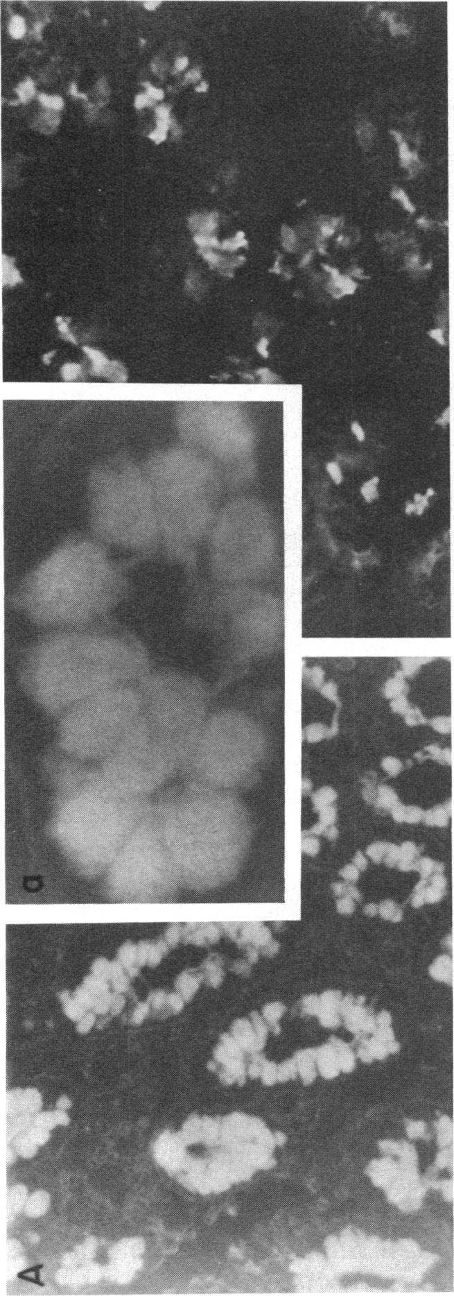
2 anti-hcm MAbs could not be defined. Spent MAb containing media or MAbs purified from ascites were prepared as described (20). Tissue samples. Pinch mucosal biopsy specimens were obtained from patients undergoing diagnostic flexible sigmoidoscopy, colonoscopy, or esophagogastroduodenoscopy at Massachusetts General Hospital, Boston, MA. Biopsy specimens were obtained at the same time that sampling was performed for routine histologic examination. Diagnostic classification of samples included in these studies as normal was made on the basis of the examining physicians' reports and the official interpretation of diagnostic biopsy specimens by members of the Pathology Department at Massachusetts General Hospital. Tissue samples from other sites (e.g., small intestine, gallbladder) were obtained from fresh surgical specimens. These studies were approved by the Human Studies Committee of Massachusetts General Hospital. Samples of colonic and small intestinal mucosa were obtained from outbred rats (Charles River Breeding Laboratories, Wilmington, MA) and New Zealand white rabbits, after sacrifice by intraperitoneal injection of sodium pentobarbital; samples from cotton-top tamarins (S. oedipus) were obtained after anesthetization as previously described (19). All samples were promptly processed for immunofluorescent studies as described below. Indirect immunofluorescence. Multiple frozen sections (2 Am) were prepared from tissue specimens embedded in OCT compound (Miles Laboratories, Naperville, IL) for indirect immunofluorescent (IIF) staining. Initial pilot studies showed no improvement in IlF staining characteristics in sections fixed in 1% formaldehyde before staining when compared with unfixed sections, and therefore prestaining fixation was not routinely employed. After equilibration at room temperature, one drop of anti-hcm MAb containing ascites, diluted 1:100 with phosphatebuffered saline (PBS), or spent anti-hcm MAb culture medium supernatant was added to cover individual tissue sections, and slides were placed in a moist chamber at room temperature for 30 min. Control sections were incubated with ascites or media derived from the parent NSl myeloma line. Subsequently, ascites or media were aspirated and sections were washed three times by immersion in excess PBS. After air drying, sections were routinely stained by addition of fluorescein isothiocyanate (FITC-conjugated rabbit anti-mouse Ig (Cappel Laboratories, Cochranville, PA) diluted 1:25 with PBS. In double-labeling experiments, staining was accomplished using FITC and rhodamine red-conjugated goat anti-mouse F(ab')2 specific for mouse immunoglobulin heavy chains (gamma or mu chain). Pilot studies demonstrated equivalent IIF staining in double-labeling experiments if conjugated antisera were added sequentially or simultaneously, and the latter was therefore used routinely. After incubation at room temperature for 30 min in a moist chamber, excess reagent was aspirated and sections were washed again as before. Tissue was counterstained with Evans Blue and fluorescent staining evaluated using a fluorescence microscope (Carl Zeiss, Inc., Thornwood, NY). Quantitative estimates of goblet cell and crypt staining were determined as the percentage of cells stained in 10 high power fields (40 X objective) or lower power fields (10 X objective), respectively. The coefficient of variation between fields was found to be ± 12%. Results In these studies, anti-hcm MAbs were used to examine tissue localization of HCM species by means of indirect immunofluorescent techniques. Among 23 anti-hcm MAbs, 17 demonstrated appreciable staining ofthe normal human colonic sigmoid mucosa by HF. The properties of anti-hcm MAbs, including their ability to effect immunofluorescent staining of colonic mucosa, are summarized in Table I. Of note, several anti-hcm MAbs (2, 10, 8, 12, 15) demonstrated significant IIF staining although they failed to recognize individual mucin species. Conversely, staining could not be demonstrated using these IIF techniques by six anti-hcm MAbs that bound specific combinations of mucin species in solid-phase RIA, including two MAbs that Table I. Properties ofanti-hcm Monoclonal Antibodies Mucin species Antigenic Immuno- MAb* specificityt Isotypes determinantl fluorescencel 1 IV, V IgGi IgM P + 3 I,II,III,IV,V IgG2B III, IV, V IgG2B IV, V, VI IgM V IgG2B III IgM UNK + 8 IV, VI IgGi P IgG2A UNK IgG1 P + 11 III, IV, V, VI IgM IgG2A UNK + 13 III, IV IgG2B IV IgM UNK IgG1 UNK + 16 V, VI IgM IV, V IgG1 P + 18 IV, V IgG2B 0-19 VI IgG1 UNK - 20 I, II, III, IV, V, VI IgM 0-21 III IgG2B UNK - 22 III, IV, V IgG II, VI IgG2B UNK - * Monoclonal antibodies demonstrating binding to unfractionated pure human colonic mucin in solid-phase sandwich RIA (20). t HCM species specificity determined by solid-phase sandwich assay using panel of polystyrene beads coated with separated mucin species as detailed in text (20). All MAbs bound unfractionated mucin. Isotypes of anti-hcm MAbs determined by sandwich RIA using heavy chain specific '251-labeled goat anti-mouse Ig or peroxidase immunoassay. 11 Antigenic determinant recognized by MAb: 0, oligosaccharide (see 20 for specific studies); P, protein (protease sensitive); UNK, unknown. Staining of goblet cells in normal human sigoid colonic mucosa assessed by indirect immunofluorescent techniques using FITC-conjugated rabbit anti-mouse Ig as described in Methods. Characteristics of staining depicted in Figs. 1 and 2 and Table II. bound single mucin species (MAbs 19 and 21). The failure to demonstrate staining by IIF did not seem to be associated with any particular HCM species. Several distinct patterns of staining ofindividual cells in the normal human sigmoid colon were observed when the various anti-hcm MAbs were studied. Many anti-hcm MAbs stained mucin droplets within the goblet cells in a dense homogeneous manner designated "diffuse" (Fig. 1 A). These MAbs usually also stained some of the extracellular materials overlying the mucosa, which presumably represented secreted mucin glycoproteins. A number of other MAbs stained intracellular mucin droplets, but in an inhomogeneous pattern, designated "speckled" (e.g., MAb 4, Fig. 1 B). Other MAbs stained the "peripheral" margin of intracellular mucin droplets (e.g., MAbs 7 and 10) as depicted in Fig. 1 C; while the remainder stained the goblet cell "cytoplasm" (MAb 15) or "apical" membrane (MAb 8) as illustrated in Fig. 1 D and 1 E, respectively. Staining patterns of 1264 D. K Podolsky, D. A. Fournier, and K E. Lynch


3 .- P Ali: a Identification ofcolonic Goblet Cell Subclasses 1265
.")
.")

4 individual cells were consistent within specimens; e.g., all cells stained by MAb 2 showed diffuse staining of the intracellular mucin droplet and not other staining patterns. Specific staining characteristics of each anti-hcm MAb were also consistent among mucosal samples ofnormal sigmoid colon from different patients (n = 9). The staining patterns of individual anti-hcm MAbs in normal human sigmoid colon are summarized in Table II. There were no consistent associations between species specificities ofmabs as determined by solid-phase RIA and particular patterns of cellular IIF staining (cf. Tables I and II). The latter point is illustrated by MAbs 4 and 5, which both bind species IV yet showed different cellular staining patterns. In this context, it should be noted that these MAbs recognize different and distinct epitopes as indicated by the different patterns of binding to other HCM species in solid-phase assays. Some anti-hcm MAbs that were specific for single HCM species failed to demonstrate any appreciable 1IF staining; and MAbs that were positive for IIF staining but failed to bind separated mucin fractions in solid-phase RIA, demonstrated a range of staining patterns. In addition to the differences in the manner of staining of individual cells, anti-hcm MAbs varied in the proportion of goblet cells stained within and between individual colonic crypts as shown in the representative photomicrographs of Fig MAbs were found to effect "uniform" goblet cell staining defined as >95% of cells stained in >95% of crypts of normal sigmoid mucosa (e.g., MAb 3; Fig. 2 A). Three anti-hcm MAbs stained goblet cells in every crypt (>95%) but within individual crypts not all goblet cells were stained (<70%) (e.g., MAb 7; Fig. 2 B), a crypt staining pattern designated "dispersed." Finally, four anti-hcm MAbs stained mucosa in a "limited" pattern defined as staining <70% of individual cells, with fewer than 70% of crypts containing stained goblet cells. Thus, there was complete Table IL Pattern ofcellular Staining ofnormal Colonic Mucosa by Anti-HCM MAbs MAb* HCM species Staining pattern I IV, V Cytoplasmic 2 0 Diffuse 3 I, II, III, IV, V Diffuse 4 III, IV, V Speckled 5 IV, V, VI Diffuse 6 V Diffuse 7 III Peripheral 8 IV, VI Apical 9 0 Speckled 10 0 Peripheral 11 III, IV, V, VI Diffuse 12 0 Diffiuse 13 III, IV Peripheral 14 IV Speckled 15 0 Cytoplasmic 16 V, VI Peripheral 17 IV, V Peripheral * Anti-HCM MAbs with varying HCM species binding specificities. Staining pattern of normal sigmoid mucosa by indirect immunofluorescence using FITC-conjugated rabbit anti-mouse Ig as detailed in Methods. (n = 9); cellular pattern classification as depicted in Fig. 1 reflecting manner of staining of individual cells within crypts. Figure 2. Heterogeneity of IIF staining of colonic mucosa by anti- HCM MAbs. IIF staining was performed as detailed in Methods and legend to Fig. I using normal sigmoid mucosa. (A) Uniform staining of crypts (MAb 3); X 450 (>95% cells stained in >95% crypts). (B) Staining of goblet cells dispersed in all crypts in a peripheral pattern (staining in >95% crypts, in each crypt <70% cells stained) (MAb 7, see Fig. I C); X 450. (C) Staining of scattered goblet cells in limited number of crypts (staining in <70% crypts: in each crypt <70% cells stained: MAb 6). For control see Fig. I F; X 450. absence of staining in some crypts by MAbs exhibiting the "limited" staining pattern. Patterns of crypt staining observed for the library of anti- HCM MAbs, referred to as "uniform," "dispersed," and "limited," and the data from which these designations were derived, are detailed in Table III. Variations in the pattern of staining appeared unrelated to the manner of staining of individual cells (i.e., diffuse, speckled, peripheral, etc.) (Tables II and III). Furthermore, heterogeneity of goblet cell staining by those MAbs 1266 D. K Podolsky, D. A. Fournier, and K E. Lynch
 (% staining) Designation n=9 n=9 1 29±18 42±11 Limited 2 95±3 >99 Uniform 3")
5 Table III. Pattern ofcrypt Staining in Normal Colonic Mucosa by anti-hcm MAbs Stainingt Within crypts Whole crypts MAbs* (% staining) (% staining) Designation n=9 n=9 1 29±18 42±11 Limited 2 95±3 >99 Uniform Uniform 4 97±3 95±5 Uniform 5 96±4 >99 Uniform 6 41±9 94±2 Dispersed 7 24±12 >99 Dispersed 8 32±7 31±15 Limited 9 98±3 96±4 Uniform 10 51±14 22±8 Limited 11 > Uniform 12 > Uniform 13 97±2 93±4 Uniform 14 46± Dispersed 15 > Uniform 16 21±6 27±14 Limited 17 98±2 >99 Uniform * Anti-HCM MAbs with varying species binding specificities detailed in Table I and (23). f Indirect immunofluorescent staining of normal sigmoid mucosa using FITC-conjugated rabbit anti-mouse Ig as detailed in Methods. Values reflect mean±sd of percentage of cells stained in 10 high power fields (X 40 objective) within individual crypts ("within crypts") or of crypts containing stained cells within 10 low power fields (X 10 objective) "whole crypts". Staining characteristics of individual cells are summarized in Table II. yielding dispersed or limited patterns could not be associated with specific regions within colonic glands ofthe normal sigmoid colon, as indicated in Table IV. Thus, the percentage of cells stained in the upper half of crypts was equivalent to the extent of cellular staining within the lower crypts. Anti-HCM MAbs that were both mucin species-specific and exhibited IIF staining demonstrated either dispersed or limited staining patterns, with intense staining of some goblet cells and lack of staining of others (see Table I and III). This finding is consistent with the presence of compositionally and/or functionally discrete goblet cell subpopulations (see below). In contrast, less mucin species-specific anti-hcm MAbs, recognizing relatively small oligosaccharide structures, more commonly stained the mucosa in a diffuse and uniform pattern. To further assess the relationship of staining heterogeneity to mucin species content, double-labeling IIF studies were carried out using anti-mucin MAbs with mutually exclusive species specificities as determined by solid-phase assay. These studies depended upon the use of heavy chain-specific fluorescent probes to permit simultaneous assessment of IIF staining, and were therefore limited to comparison of MAbs of different Ig classes. Multispecies-specific anti-hcm MAbs could not be uniformly used to effectively assess the relative distribution ofthe different species recognized by that MAb. These latter MAbs could only be used in conjunction with other MAbs directed to nonoverlapping subsets of mucin species. As demonstrated in a representative photomicrograph (Fig. 3 A), goblet cells exhibited simultaneous staining for two anti- HCM MAbs of differing species specificity, which suggests that these cells contain more than one mucin. Therefore, individual goblet cells seemed to be able to produce more than one mucin species. Note that anti-hcm MAbs used in double staining studies included those with demonstrated specificity for more extended intrinsic structural components of HCM (i.e., whole oligosaccharide side chain), and these served as reliable probes for the presence of specific HCM species. Nonetheless, pairs of mucin species were not invariably associated in every goblet cell; some goblet cells in the same tissue section were stained by only one of the pair of anti-hcm MAbs. Additional doublelabeling experiments suggested that some mucin species were found in mutually exclusive subpopulations of goblet cells; e.g., simultaneous incubation with MAbs selective for HCM species III (MAb 7) and V (MAb 6) showed staining of different goblet cells by the two probes (Fig. 3 B). In summary, it appeared that species III did not coexist with IV or V within single goblet cells, but seemed to be restricted to nonoverlapping goblet cell populations. However, goblet cells containing each of these species did consistently contain other remaining HCM species (I, II, and VI). Collectively, the double IIF staining studies using pairs of anti-hcm MAb allowed delineation of a number of subsets of goblet cells distinguished by their content of different combinations of HCM species (see Table V). None of these subpopulations were localized to specific areas of the mucosal surface or crypts, but rather, they appeared dispersed throughout the colonic surface. In view of the limitations of the IIF technique described above, the variety of sub- Table IV. Anti-HCM MAb Goblet Cell Staining within Crypts MAb* goblet cell staining (%) Region Upper crypt 27±15 98±2 >99 98±2 >99 44±6 25±0 36± ±9 > ±2 49±7 >99 15±8 97±2 Lower crypt 33±9 94±4 >99 96±2 97±3 40±5 21±11 30±4 >99 45±8 >99 >99 98±2 38±9 >99 23±7 100 * Anti-HCM MAbs with varying HCM species binding specificites summarized in Table I. Indirect immunofluorescent staining assessed as described in Methods and legend to Table III. Values represent mean±sd of percentage of goblet cells stained derived from >10 crypts from each of nine specimens of normal sigmoid colonic mucosa. Upper crypt arbitrarily defined as upper 50% of gland and lower crypt as base and lower 50% estimated from overall crypt length. Identification ofcolonic Goblet Cell Subclasses 1267

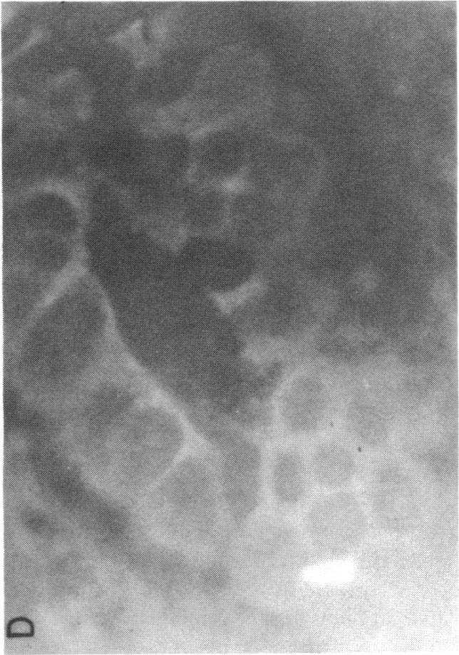
 Double labeling with MAb 6 (IgG specific for HCM species V) and MAb 7 (IgM specific for HCM species III) X 450.")

6 A Figure 3. Double 11F staining of normal colonic mucosa using two anti-hcm MAbs. Normal human mucosa was incubated simultaneously with two MAbs, one of IgG class and the other of IgM class, followed by sequential incubation with FITC conjugated goat antimouse IgG and rhodamine red conjugated F(ab')2 fragment goat antimouse IgM as detailed in Methods. (A) Double labeling with MAb 6 (IgG recognizing HCM species V) and MAb 14 (IgM recognizing HCM species IV) X 1, 150. Note that goblet cells are stained by both MAbs. (B) Double labeling with MAb 6 (IgG specific for HCM species V) and MAb 7 (IgM specific for HCM species III) X 450. Note goblet cells showing mutually exclusive staining. classes of goblet cells summarized in Table V must represent a minimum estimate of the extent of goblet cell heterogeneity, although collectively they accounted for 72±1 1% of goblet cells in normal colonic mucosa. Subsequently, IIF staining by anti-hcm MAbs was assessed in tissue samples from different regions ofthe colon. As indicated in Table VI, staining patterns were nearly consistent throughout the colon. However, a few instances of statistically significant reductions were encountered, e.g., staining by MAbs 1 and 12 was diminished in the transverse colon and staining by MAbs 1, 7, and 12 was reduced in the cecum. Staining by 14 of the anti-hcm MAbs was comparable in tissue samples from all areas ofthe large bowel. Cellular and crypt staining characteristics of each anti-hcm MAb were found to be the same in mucosal Table V. Colonic Goblet Cell Subpopulations Defined by HCM Species HCM specie4 Goblet cell type* (I)t (II)* III IV V VI 1 ~~(+) (+) + 2 (+) (+) + 3 (+) (+) + 4 (+) (+) + 5 (+) (+) + 6 (+) (+) (+) (+) + + * Goblet cell types defined by presence of the indicated HCM species in double IIF staining experiments as described in text using MAbs summarized in Table I. t The presence of HCM species I and II could not be determined independently of other species with available MAbs. samples from the other areas of the colon as those observed in the sigmoid colon (summarized in Tables II and III). Therefore, despite complex heterogeneity of colonic goblet cells at the microscopic level, the mixture of subpopulations appeared to be generally consistent throughout the large intestine. However, there was a gradient in representation of goblet cells staining by MAbs directed against HCM species IV (i.e., MAbs 1, 3, 4, 5, 8, 11, 13, 14, and 17). The proportion of cells containing HCM species IV appeared to increase progressively in more distal sites, with greatest numbers of positive cells in the rectum and sigmoid colon. However, the difference between proximal and distal colonic mucosa in the aggregate extent of staining by these MAbs did not achieve statistical significance (P > 0.05 and <0.10) and despite this possible gradient, there were still a large number of goblet cells that contained species IV in more proximal regions ofthe colon. Because these techniques permit only a semiquantitative assessment of mucin species, it is possible that there may be subtle regional variations in the number of cells stained by each MAb and/or the quantity of mucin glycoprotein antigen present within individual goblet cells. Finally, the similarity ofhuman colonic mucin glycoprotein to that in goblet cells in other sites of the human gastrointestinal tract as well as other animal species was examined using the library of anti-hcm MAbs and IIF techniques. As indicated in Table VII, a continuum of cross-reactivity between human colonic mucin and goblet cells in other areas of the gastrointestinal tract was observed. Overall, there was a progressive rise in the number of anti-hcm MAbs staining goblet cells in more distal areas of the digestive tract. While only one anti-hcm MAb stained gastric mucin, eight MAbs were found to stain goblet cells in the human ileum. Although there was some similarity between distal small intestinal goblet cells and colonic mucin as assessed by IIF, a number of the anti-hcm MAbs stained only the large intestinal cells. In this respect it is noteworthy that there appeared to be closer antigenic similarity between human 1268 D. K. Podolsky, D. A. Fournier, and K E. Lynch
7 Table VI. Regional Variations in Immunofluorescent Staining ofcolonic Mucin by Anti-HCM MAbs Anti-HCM MAb goblet cell staining Region % % %6 % % % % % % % % % % % % % % Rectum (n = 9) 32±12 92± ±2 93±5 38±12 21±7 43±8 95±4 46±9 >99 >99 98±2 50±11 >99 18±7 95±3 Sigmoid (n = 9) 29±18 95± ±3 96±4 42±9 24±12 32±7 98±3 51±14 >99 >99 97±2 46±10 >99 21±6 98±2 Transverse (n = 6) <5t >99 >99 95±3 92±6 33±6 19±6 37±9 92±9 44±10 92±10 85±9 89±8 39±7 >99 27±29 87±9 Cecum (n = 6) <5t 97±3 >99 96±2 94±7 80±8 <5 25±10 96±10 57±12 87±12 6±5t 93±7 33±8 >99 25±10 92±5 * Indirect immunofluorescent staining of goblet cells as discussed in Methods, expressed as percent±sd cells stained per 10 high power field/specimen. Pattern of staining for individual MAbs as detailed in Tables II and III. * P < P < colonic goblet cells and those in the colon of a non-human primate (S. oedipeus, cotton top tamarin) and rodents than more proximal sites of the human gastrointestinal tract as judged by the staining of some anti-hcm MAbs (Table VII). These data suggest that there are structural features of mucin glycoproteins that may be organ-specific and presumably related to colonic function. However, as shown in Table VII, despite the similarities of staining by a number of anti-hcm MAbs in colonic mucosa from humans and a variety of animal species, concordance was not uniform and several MAbs appeared to recognize determinants expressed only in the human large intestine. Discussion It has long been recognized on the basis of histochemical studies that mucin glycoproteins are the most abundant products of colonic goblet cells (21-24). Early studies suggested that these substances were heterogeneous but it was unclear whether polydispersity detected by conventional analytic tools (e.g., sedimentation centrifugation) reflected important biological diversity (25-29). More recent studies from this laboratory showed the presence of at least six chromatographically distinct HCM subclasses (9, 18). Partial structural analysis ofthe oligosaccharides of the most abundant HCM species has confirmed the presence of some distinctive structural components on each ofthese HCM species (30). The recent development of a library of anti-hcm MAbs with defined HCM species specificities and defined antigenic determinants has further substantiated the presence of discrete HCM species (20). Our library of anti-hcm MAbs, with a range of defined species binding patterns, has permitted evaluation ofthe cellular basis of HCM species distribution in colonic mucosa using IIF. Table VII. Tissue Immunofluorescence using Anti-HCM MAbs Anti-HCM MAbs Species/Tissue Human Colon Rectum (n = 9) Sigmoid (n = 9) Transverse (n = 6) Cecum(n = 6) Ileum (n = 7) Duodenum/Jejunum (n = 4) Stomach (n = 4) Gallbladder (n = 3) 100 Rat Colon (n = 3) Small intestine (n = 3) Rabbit Colon (n = 3) Small intestine (n = 3) Monkey Colon(n = 2) * Properties of individual MAbs described in Table I. Pattern of staining by individual MAbs described in Table II. Data expressed as percentage of tissue specimens exhibiting cellular staining. Blank indicates no detectable specific immunofluorescence. Identification ofcolonic Goblet Cell Subclasses 1269

8 Anti-HCM MAbs with limited or unique species specificity in solid-phase binding assays stained goblet cells selectively within the mucosa, indicating that each individual HCM species is not produced by all goblet cells. Conversely, goblet cells did show simultaneous staining by MAbs directed against distinct species, which suggests that individual goblet cells produce more than a single mucin specie. However, some combinations ofhcm species were never observed within the same goblet cells, indicating their production by mutually exclusive subpopulations. On the basis of these observations, it appears that colonic mucosa contains subpopulations of morphologically similar goblet cells, which produce distinctive combinations of mucin species. Goblet cell subpopulations defined by anti-hcm MAbs were distributed throughout the colonic crypts. Each crypt appeared to contain all types ofgoblet cells, and similar patterns ofgoblet cell heterogeneity were found in mucosa from all regions of the large bowel. Thus, the organization of goblet cell heterogeneity does not appear to involve regional or glandular concentration ofparticular cell types. These observations suggest that all HCM species or the coordinate pairs of HCM species produced by different goblet cell populations are important for normal colonic function. A number of MAbs that bound HCM in solid-phase assays failed to exhibit immunofluorescence in tissue studies, while a number of anti-hcm MAbs stained goblet cells specifically, yet did not recognize separated HCM species. These disparities likely reflect limitations of the methods employed to detect antigenic structures in either solid-phase binding or IIF assays. Failure to observe staining by IIF could reflect lack of accessibility of the antigenic structures or unfavorable configurations related to spatial relationships, suboptimal fixation, or intrinsic properties of the MAbs. Therefore, failure of staining or alterations in intensity of fluorescence cannot be construed as unequivocal proof of the absence of the relevant structures. Findings obtained with anti-hcm MAbs differ from those of earlier studies using lectins, histochemical stains, or conventional antisera, which suggested the presence of gradations of mucin glycoproteins both along the length of the colon (e.g., increasing acidic mucosubstances in the left colon and neutral in the right) or within individual colonic crypts (2, 6, 15, 16, 31-33). However, these earlier approaches used reagents directed against limited peripheral structural components, such as sulfate or terminal carbohydrate residues. Solid-phase RIA binding studies with anti-hcm MAbs suggest that probes which recognize only limited peripheral structural determinants may lead to distinctions unrelated to significant structural differences. Interestingly, anti-hcm MAbs directed to nonspecific peripheral oligosaccharide determinants showed some regional variations in staining patterns comparable to those observed with these earlier methods. These findings emphasize the importance of defining the structural antigenic determinants specified by probes before the biological significance ofstaining or binding phenomena can be meaningfully assessed. Despite the differences between patterns of mucin heterogeneity observed with anti-hcm MAbs and other methods within the colonic mucosa, the increasing cross-reactivity ofanti- HCM MAbs at progressively more distal sites of the gastrointestinal tract is comparable to the results of earlier studies (29, 34). The present studies confirm the impression that some structural features are common to all gastrointestinal tract mucin, while other components of mucin may represent organ specific determinants. Note that the similarity between animal and human colonic mucin was greater than that observed between human colon and more proximal areas of the human digestive tract, which supports the concept that colonic mucin structure is related to specific colonic functions. Although no HCM species was found to be localized to a specific region of the colon, HCM species IV containing goblet cells appeared to be present in greater numbers at more distal sites although the difference did not achieve the level of statistical significance. The distribution of mucin species IV containing goblet cells is interesting in view ofthe demonstrated association between reductions in this component and ulcerative colitis, a disorder in which there is preferential involvement of the distal colon. This observation raises the intriguing possibility that there may be a reduction or functional alteration of particular goblet cell subpopulations in association with specific disease processes; and studies are currently underway using these techniques to examine goblet cell heterogeneity in patients with ulcerative colitis as well as in other disorders. However, the limitations of the essentially qualitative methods used in these studies should be emphasized. The present methods do not precisely quantify all anti-hcm MAb defined goblet subtypes. They also do not quantify the amount of each substance within individual cells. Despite the demonstrated association of a particular disease process with selective reduction in one mucin species, the functional roles of discrete mucin species remain unknown. Neutra and co-workers (35-38) have demonstrated that mucin production and secretion may be subject to a variety of neural and hormonal control mechanisms. They have suggested that patterns of responsiveness to various mediators may define functional subclasses of colonic goblet cells. It will be interesting to determine whether the goblet cells producing different HCM species are subject to different control mechanisms. Insight into the functional significance ofthese distinctions will also depend on more detailed understanding of the peptide and oligosaccharide structures of HCM species. Anti-mucin MAbs may facilitate attempts to prepare enriched subpopulations of colonic goblet cells. Collectively, the present studies indicate the presence of previously unappreciated heterogeneity among colonic goblet cells. Acknowledgments The authors gratefully acknowledge the excellent technical assistance of Katharina Kirsch. The authors thank Dr. Kurt J. Isselbacher for his support and thoughtful review of this manuscript. These studies were supported by a grant from the National Foundation for Ileitis and Colitis. References 1. Chopra, D. P., K.-Y. Yeh, and R. W. Brockman Isolation and characterization of epithelial cell types from the normal rat colon. Cancer Res. 41: Shamsuddin, A. K. M., and B. F. Trump Colon epithelium I. Light microscopic, histochemical, and ultrastructural features ofnormal colon epithelium of male Fischer 344 rats. J. Natl. Cancer Inst. 66: Lev, R., and D. Orlic Histochemical and radioautographic studies of normal human fetal colon. Histochemistry. 39: Roediger, W. E. W., and S. C. Truelove Method ofpreparing isolated colonic epithelial cells for metabolic studies. Gut Filipe, M. I., and I. Dawson The diagnostic value of mu D. K Podolsky, D. A. Fournier, and K. E. Lynch

9 cosubstances in rectal biopsies from patients with ulcerative colitis and Crohn's disease. Gut. 11: Jacobs, L. R., D. DeFontes, and K. L. Cox Cytochemical localization of small intestinal glycoconjugates by lectin histochemistry in controls and subjects with cystic fibrosis. Dig. Dis. Sci. 28: Dawson, P. A., J. Patel, and M. I. Filipe Variations in sialomucins in the mucosa of the large intestine in malignancy. A quantimet and statistical analysis. Histochem. J. 10: Boland, C. R., P. Lance, B. Levin, R. H. Riddell, and Y. S. Kim Lectin binding indicates an abnormality of the goblet cell glycoconjugates in ulcerative colitis. Gastroenterology. 82: Podolsky, D. K., and K. J. Isselbacher Characterization of monoclonal antibodies to serum galactosyltransferase. Proc. Natl. Acad. Sci. USA. 81: Litinsky, C. M., and R. H. Riddell Patterns of mucin secretion in neoplastic and nonneoplastic diseases of the colon. Hum. Pathol. 12: Boland, C. R., P. Lance, B. Levin, R. H. Riddell, and Y. S. Kim Abnormal goblet cell glycoconjugates in rectal biopsies associated with an increased risk of neoplasia in patients with ulcerative colitis: early results of a prospective study. Gut. 25: Filipe, M. I Mucins in the gastrointestinal epithelium. A review. Invest Cell Pathol. 2: Allen, A The structure and function of gastrointestinal mucus in gastrointestinal mucosal protection. J. H. Wilkins, editors. William & Wilkins Co., Baltimore Silberberg, A., and F. A. Meyer Structure and function of mucus. Adv. Exp. Med. Biol. 144: Gad, A A histochemical study of human alimentary tract mucosubstances in health and disease. II. Inflammatory conditions. Br. J. Cancer 23: Yonezawa, S., T. Nakamura, S. Tanaka, and E. Sato Glycoconjugate with Ulex europaeus agglutinin- 1 binding sites in normal mucosa, adenoma and carcinoma of the human large bowel. J. Natl. Cancer Inst. 69: Jacobs, L. R., and P. W. Huber Regional distribution and alterations of lectin binding to colorectal mucin in mucosal biopsies from controls and subjects with inflammatory bowel disease. J. Clin. Invest. 75: Podolsky, D. K., and K. J. Isselbacher Glycoprotein composition of colonic mucosa. Gastroenterology. 87: Podolsky, D. K., J. L. Madara, W. King, P. Sehgal, R. Moore, and H. S. Winter Colonic mucin in primates: selected alterations in spontaneous colitis in cotton top tamarins. Gastroenterology. 88: Podolsky, D. K., K. Lynch, and D. A. Fournier Development of anti-human colonic mucin monoclonal antibodies. Characterization of multiple colonic mucin species. J. Clin. Invest. 77: O'Gorman, T. A., and J. T. LaMont Glycoprotein synthesis and secretion in human colonic cancers and normal colonic mucosa. Cancer. 38: MacDermott, R. P., R. M. Donaldson, and J. S. Trier Glycoprotein synthesis and secretion by mucosal biopsies of rabbit colon and human rectum. J. Clin. Invest. 54: Neutra, M. R., R. J. Grand, and J. S. Trier Glycoprotein synthesis, transport, and secretion by epithelial cells of human rectal mucosa. Lab. Invest. 36: Kim, Y. S., and R. Isaacs Glycoprotein metabolism in inflammatory and neoplastic diseases of the human colon. Cancer. 35: Marshall, T., and A. Allen The isolation and characterization of the high-molecular weight glycoprotein from pig colonic mucous. Biochem. J. 173: Allen, A., A. Bell, M. Mantle, and J. P. Pearson The structure and physiology ofgastrointestinal mucus. Adv. Exp. Med. Biol. 144: Forstner, G., A. Wesley, and J. Forstner Clinical aspects of gastrointestinal mucus. Adv. Exp. Med. Biol. 144: Fahim, R. E. F., G. G. Forstner, and J. F. Forstner Heterogeneity of rat goblet cell mucin before and after reduction. Biochem. J. 209: Gold, D. V., D. Shochat, and F. Miller Protease digestion of colonic mucin: evidence for the existence of two immunochemically distinct mucins. J. Biol. Chem. 255: Podolsky, D. K Oligosaccharide structures of human colonic mucin. J. Biol. Chem. 260: Gold, D. V Immunoperoxidase localization of colonic mucoprotein antigen in neoplastic tissues. Cancer Res. 41: Etzler, M. E Lectins as probes in studies of intestinal glycoproteins and glycolipids. Am. J. Clin. Nutr. 32: Boland, C. R., C. K. Montgomery, and Y. S. Kim Alterations in human colonic mucin occurring with cellular differentiation and malignant transformation. Proc. Natl. Acad. Sci. USA. 79: Bara, J., F. Loisillier, and P. Burtin. 198?. Antigens of gastric and intestinal mucous cells in human colonic tumors. Br. J. Cancer. 41: Specian, R. N., and M. R. Neutra Regulation of intestinal goblet cell secretion I: role ofparasympathetic stimulation. Am. J. Physiol. 242(Gastrointest. Liver Physiol. 5):G370-G Neutra, M. R., R. L. J. O'Malley, and R. D. Specian Regulation of intestinal goblet cell secretion. II. A survey of potential secretagogues. Am. J. Physiol. 242(Gastrointest. Liver Physiol. 5):G280- G Phillips, T. E., T. H. Phillips, and M. R. Neutra Regulation of intestinal goblet cell secretion. III. The isolated intestinal epithelium. Am. J. Physiol. 247(Gastrointest. Liver Physiol. 1O):G674-G Phillips, T. E., T. H. Phillips, and M. R. Neutra Regulation of intestinal goblet cell secretion. IV. Electrical field stimulation in vitro. Am. J. Physiol. 247(Gastrointest. Liver Physiol. 1O):G682-G687. Identification ofcolonic Goblet Cell Subclasses 1271
IMMUNOCHEMICAL TECHNIQUES
 24 IMMUNOCHEMICAL TECHNIQUES 24.1 INTRODUCTION All vertebrates have advanced immune system. The more complex the organism the more advanced the immune system. The immune system of mammals has evolved over
Mouse Monoclonal Antibody Isotyping Reagents
 Mouse Monoclonal Antibody Isotyping Reagents Catalog Number: SEK003 Storage Temperature: 2-8 C Fax : +86-10-58628220 Tel : +86-400-890-9989 http://www.sinobiological.com Description Mouse Monoclonal Antibody
LAMININ. For Immunohistochemical Demonstration of Laminin in Paraffin-embedded and Frozen Human Tissue Sections Stock No. IMMH-7
 LAMININ For Immunohistochemical Demonstration of Laminin in Paraffin-embedded and Frozen Human Tissue Sections Stock No. IMMH-7 TABLE OF CONTENTS BACKGROUND AND PRINCIPLE... 4 REAGENTS AND EQUIPMENT PROVIDED...
PROF. DR. ASMAA HUSSEIN DIRECTOR OF THE MOLECULAR BIOLOGY RESEARCH UNIT
 BY PROF. DR. ASMAA HUSSEIN DIRECTOR OF THE MOLECULAR BIOLOGY RESEARCH UNIT Immunoassay are based on the strong and highly specific interaction occurring between antigens (Ag)) and antibodies (Ab). Ag Ab
QImaging Camera Application Notes Multicolor Immunofluorescence Imaging
 QImaging Camera Application Notes Multicolor Immunofluorescence Imaging In order to image localization of intracellular proteins with high specificity, it is frequently necessary to multiplex antibody
Staining Techniques. Staining Techniques. There are many dyes. Histochemical Stains: chemical reactions. Feulgen reaction -DNA
 Staining Techniques There are many dyes. http://medinfo.ufl.edu/~dental/denhisto/stains.html Examples: Sudan black -Lipids Myelinated axons- blue ihcworld.com/imagegallery/displayimage.php?al... Weigert
666 THE JOURNAL OF CELL BIOLOGY' VOLUME 71, 1976" pages
 ph-dependent BINDING OF IMMUNOGLOBULINS TO INTESTINAL CELLS OF THE NEONATAL RAT RICHARD RODEWALD. From the Department of Biology, University of Virginia, Charlottesville, Virginia 22901. Neonatal rats
Application Note AN001
 Testing hybridoma supernatants with the Spots On Dots Antibody Screening Kit Application Note AN1 Table of Contents Overview... 2 Figure 1. Screening of hybridomas raised against peptide antigens... 3
SPHERO TM Coated Particles
 SPHERO TM Coated Particles Manufactured by either passive adsorption or covalent coupling depending upon the intended application Stable for several years under proper storage condition Available in a
Antibodies to Animal Cells and Serum Proteins
 ANTIBODIES TO ANIMAL CELLS AND SERUM PROTEINS Antibodies to Animal Cells and Serum Proteins Antibodies to Animal Cellular Antigens Antibodies to Mouse Brain Antibodies to Animal Serum Proteins INTERNATIONAL
How to run Alpha assay: How to setup an Alpha assay Make your own assay!
 How to run Alpha assay: How to setup an Alpha assay Make your own assay! 1 2009 PerkinElmer AlphaLISA kits - recommendations before starting the assay Samples: Phenol red and hemoglobin: choose AlphaLISA
1. Cross-linking and cell harvesting
 ChIP is a powerful tool that allows the specific matching of proteins or histone modifications to regions of the genome. Chromatin is isolated and antibodies to the antigen of interest are used to determine
Human IgG Antigen ELISA Kit
 Human IgG Antigen ELISA Kit Catalog No: IHUIGGKT Lot No: SAMPLE INTENDED USE This human immunoglobulin G antigen assay is intended for the quantitative determination of total human IgG antigen in serum,
Suppression of Polyclonal Immunoglobulin Production by M-proteins Shows Isotype Specificity
 274 Annals of Clinical & Laboratory Science, vol. 31, no. 3, 2001 Suppression of Polyclonal Immunoglobulin Production by M-proteins Shows Isotype Specificity Liang Wang and David C. Young Department of
Experience the Mabtech ELISA
 Experience the Mabtech ELISA Mabtech provides well-designed ELISA kits for specific, sensitive, and robust quantification of analytes in solution. A comprehensive selection of ELISA kits is available for
TheraLin. Universal Tissue Fixative Enabling Molecular Pathology
 TheraLin Universal Tissue Fixative Enabling Molecular Pathology TheraLin Universal Tissue Fixative Enabling Molecular Pathology Contents Page # TheraLin Universal Tissue Fixative 3 Introduction 5 Easy
Chapter 17: Immunization & Immune Testing. 1. Immunization 2. Diagnostic Immunology
 Chapter 17: Immunization & Immune Testing 1. Immunization 2. Diagnostic Immunology 1. Immunization Chapter Reading pp. 505-511 What is Immunization? A method of inducing artificial immunity by exposing
IMMUNOPRECIPITATION (IP)
") 1 IMMUNOPRECIPITATION (IP) Overview and Technical Tips 2 CONTENTS 3 7 8 9 12 13 17 18 19 20 Introduction Factors Influencing IP General Protocol Modifications Of IP Protocols Troubleshooting Contact Us
1 IMMUNOPRECIPITATION (IP) Overview and Technical Tips 2 CONTENTS 3 7 8 9 12 13 17 18 19 20 Introduction Factors Influencing IP General Protocol Modifications Of IP Protocols Troubleshooting Contact Us
Cell & Tissue Staining Kit
 Cell & Tissue Staining Kit For the detection of goat, mouse, rabbit, rat, or sheep primary IgG Antibodies Size: 50 Tests HRP-DAB System Goat Kit (Catalog Number CTS008) Mouse Kit (Catalog Number CTS002)
Segments of the obstructed intestinal loops were fixed in 4% paraformaldehyde
 Supplementary text Supplementary materials and methods Histopathological examination Segments of the obstructed intestinal loops were fixed in 4% paraformaldehyde (PFA) and embedded in paraffin wax with
ApoTrack Cytochrome c Apoptosis ICC Antibody
 ab110417 ApoTrack Cytochrome c Apoptosis ICC Antibody Instructions for Use For the Immunocytochemistry analysis of cytochrome c and a mitochondrial marker (Complex Vα) in apoptotic cells and nonapoptotic
BCH 462. Single Radial Immunodiffusion and Immuno-electrophoresis
 BCH 462 Single Radial Immunodiffusion and Immuno-electrophoresis Immunoassays tests include: 1. Precipitation. 2. Agglutination. 3. Immunofluorescence. 4. Radioimmunoassay (RIA). 5. Enzyme-Linked Immuno
In Vitro Diagnostic Products
 In Vitro Diagnostic Products Rely on Rockland for Unparalleled Quality Diagnostic: Overview For over 50 years Rockland has provided a dedicated portfolio of general purpose reagents (GPR) used to collect,
An indirect haemagglutination test to detect serum antibodies to Giardia lamblia
 J. Biosci., Vol. 10, Number 4, December 1986, pp. 475-480. Printed in India. An indirect haemagglutination test to detect serum antibodies to Giardia lamblia K. N. JALAN, TUSHER MAITRA and RITA DAS Kothari
SANTA CRUZ BIOTECHNOLOGY, INC.
 TECHNICAL SERVICE GUIDE: Western Blotting 2. What size bands were expected and what size bands were detected? 3. Was the blot blank or was a dark background or non-specific bands seen? 4. Did this same
MOLECULAR RECOGNITION
 MOLECULAR RECOGNITION Bioanalytical Methods Classification 1. Biassay: molecular recognition, signal generation and detection in solution or on inert solid phase 2. Biosensor: molecular recognition system
Immunohistologic Study of Ulcerative Colitis With Monoclonal Antibodies Against Tumor-Associated and/or Differentiation Antigens
 GASTROENTEROLOGY 1988;95:686-93 Immunohistologic Study of Ulcerative Colitis With Monoclonal Antibodies Against Tumor-Associated and/or Differentiation Antigens HARRY S. COOPER and ZENON STEPLEWSKI Department
Assays for Immunogenicity: Are We There Yet?
 Assays for Immunogenicity: Are We There Yet? Mark Wener, MD Department of Laboratory Medicine & Rheumatology Division Department of Medicine University of Washington Seattle, WA 98195 wener@uw.edu Goals:
Purification Kits. Fast and Convenient PROSEP -A and PROSEP-G Spin Column Kits for Antibody Purification DATA SHEET
 Â Montage Antibody Purification Kits Fast and Convenient PROSEP -A and PROSEP-G Spin Column Kits for Antibody Purification DATA SHEET Available with immobilized Protein A or Protein G Easy-to-use Antibody
 Montage Antibody Purification Kits Fast and Convenient PROSEP -A and PROSEP-G Spin Column Kits for Antibody Purification DATA SHEET Available with immobilized Protein A or Protein G Easy-to-use Antibody
H-ferritin (Human) ELISA Kit
 ELISA Kit") H-ferritin (Human) ELISA Kit Catalog Number KA0211 96 assays Version: 04 Intended for research use only www.abnova.com Table of Contents Introduction... 3 Intended Use... 3 Background... 3 Principle of
H-ferritin (Human) ELISA Kit Catalog Number KA0211 96 assays Version: 04 Intended for research use only www.abnova.com Table of Contents Introduction... 3 Intended Use... 3 Background... 3 Principle of
Cytomics in Action: Cytokine Network Cytometry
 Cytomics in Action: Cytokine Network Cytometry Jonni S. Moore, Ph.D. Director, Clinical and Research Flow Cytometry and PathBioResource Associate Professor of Pathology & Laboratory Medicine University
Immunohistochemistry: Basics and Methods
 Immunohistochemistry: Basics and Methods Bearbeitet von Igor B Buchwalow, Werner Böcker 1st Edition. 2010. Buch. x, 153 S. Hardcover ISBN 978 3 642 04608 7 Format (B x L): 15,5 x 23,5 cm Gewicht: 445 g
Rat IGF-1 ELISA Kit (rigf-1-elisa)
") Rat IGF-1 ELISA Kit (rigf-1-elisa) Cat. No. EK0377 96 Tests in 8 x 12 divisible strips Background Insulin-like growth factor 1 (IGF-1), also known as somatomedin C, is a polypeptide protein hormone similar
Rat IGF-1 ELISA Kit (rigf-1-elisa) Cat. No. EK0377 96 Tests in 8 x 12 divisible strips Background Insulin-like growth factor 1 (IGF-1), also known as somatomedin C, is a polypeptide protein hormone similar
Immunohistochemistry: Basics and Methods
 Immunohistochemistry: Basics and Methods Igor B. Buchwalow l Werner Böcker Immunohistochemistry: Basics and Methods Prof. Dr. Igor B. Buchwalow Prof. Dr. Werner Böcker Gerhard-Domagk-Institut für Pathologie
Figure S6. Detection of anti-gfp antibodies in anti-dna and normal plasma without competition DNA--9
 Supplementary Information Ultrasensitive antibody detection by agglutination-pcr (ADAP) Cheng-ting Tsai 1 *, Peter V. Robinson 1 *, Carole A. Spencer 2 and Carolyn R. Bertozzi 3,4ǂ Department of 1 Chemistry,
Generic DELFIA Reagents
 AD0005P-12 (en) 1 Generic DELFIA Reagents For Research Use Only These instructions for use apply to the following reagents: AD0038 DELFIA Eu-N1 PY20 antibody 50 µg vial AD0039 DELFIA Eu-N1 PY20 antibody
Immunohistochemistry. How does it look like? When do we need IHC? When do we need IHC? In clinic: In research:
 Introduction How does it look like? Immunohistochemistry Smooth muscle actin Parvalbumin Distrophyn Sandrine Bichet Head of Molecular Histology Platform Signal versus background 06.03.2012 IHC basics Introduction
Immunoglobulins. Harper s biochemistry Chapter 49
 Immunoglobulins Harper s biochemistry Chapter 49 Immune system Detects and inactivates foreign molecules, viruses, bacteria and microorganisms Two components with 2 strategies B Lymphocytes (humoral immune
Gastrointestinal Cancer Antigen CA 19-9 ELISA Kit Protocol. (Cat. No.:EK )
") Gastrointestinal Cancer Antigen CA 19-9 ELISA Kit Protocol (Cat. No.:EK-310-17) 330 Beach Road, Burlingame CA Tel: 650-558-8898 Fax: 650-558-1686 E-Mail: info@phoenixpeptide.com www.phoenixpeptide.com
Gastrointestinal Cancer Antigen CA 19-9 ELISA Kit Protocol (Cat. No.:EK-310-17) 330 Beach Road, Burlingame CA Tel: 650-558-8898 Fax: 650-558-1686 E-Mail: info@phoenixpeptide.com www.phoenixpeptide.com
PeliClass human IgG subclass ELISA kit Enzyme-linked immunosorbent assay
 PeliClass human IgG subclass ELISA kit Enzyme-linked immunosorbent assay Catalog No: M1551 Size: six pre-coated 8-well strips for each of the four IgG subclasses Test description The PeliClass human subclass
Antigen-antibody reactions with labeled reagents
 Antigen-antibody reactions with labeled reagents Department of Immunology Faculty of Medicine University of Belgrade ANTIGEN ANTIBODY REACTIONS WITH LABELED REAGENTS Enzyme immunoassay Radioimmunoassay
CHAPTER 3 ANTIBODY STRUCTURE I
 CHAPTER 3 ANTIBODY STRUCTURE I See APPENDIX: (3) OUCHTERLONY ANALYSIS; (6), EQUILIBRIUM DIALYSIS; (7) CROSS-REACTIVITY Electrophoretic separation of serum proteins identifies the GAMMA-GLOBULIN fraction
Human IgG ELISA Kit. Strip well format. Reagents for up to 96 tests
 Human IgG ELISA Kit Strip well format. Reagents for up to 96 tests Catalog No. CS222A Quantity: 1 x 96 tests CS222B 5 x 96 tests Intended Use: Background: Assay Principle: This human immunoglobulin G antigen
Cyfra 21-1 IRMA. Product information Information about other products is available at: Userś Manual DE52100
 Product information Information about other products is available at: www.demeditec.com Userś Manual Cyfra 21-1 IRMA The CYFRA 21.1 IRMA system provides a direct in vitro quantitative determination of
Discovery and Humanization of Novel High Affinity Neutralizing Monoclonal Antibodies to Human IL-17A
 Discovery and Humanization of Novel High Affinity Neutralizing Monoclonal Antibodies to Human IL-17A Contacts: Marty Simonetti martysimonetti@gmail.com Kirby Alton kirby.alton@abeomecorp.com Rick Shimkets
Monoclonal antibody detection of Giardia lamblia cysts in human stool by direct immunofluorescence
 Journal of Wilderness Medicine 1,203-207 (1990) Monoclonal antibody detection of Giardia lamblia cysts in human stool by direct immunofluorescence S.C. ZELL*, M. BUDHRAJA, J.L. RIGGS and S.K. SORENSON
Immunological Techniques in Research and Clinical Medicine. Philip L. Cohen, M.D. Chief of Rheumatology, LKSOM 10 March 2016
 Immunological Techniques in Research and Clinical Medicine Philip L. Cohen, M.D. Chief of Rheumatology, LKSOM 10 March 2016 Antibodies Remarkable Tools for Research and Diagnosis You can make an antibody
Your Antibody Source. prosci-inc.com. Extensive Antibody Services Broad Antibody Catalog
 Your Source prosci-inc.com Extensive Services Broad Catalog Company Overview Established in 1998, ProSci Incorporated is a leading provider of high performance antibodies and custom antibody services.
Anti-Asian Sea bass (Lates calcarifer) IgM monoclonal antibody labelled with horseradish peroxidase. Product no: C2-HRP
 IgM monoclonal antibody labelled with horseradish peroxidase. Product no: C2-HRP") Anti-Asian Sea bass (Lates calcarifer) IgM monoclonal antibody labelled with horseradish peroxidase Product no: C2-HRP Product Description This monoclonal antibody (Mab) reacts with Asian Sea bass (Lates
Anti-Asian Sea bass (Lates calcarifer) IgM monoclonal antibody labelled with horseradish peroxidase Product no: C2-HRP Product Description This monoclonal antibody (Mab) reacts with Asian Sea bass (Lates
Immunoprecipitation Protocol
 Immunoprecipitation Protocol Immunoprecipitation is a general method to obtain the enrichment of a specific protein from tissue lysate and cell lysate. It can be used to purify a specific protein, to identify
Antibodies and Antigens In the blood bank
 Antibodies and Antigens In the blood bank 1 Nice game!! http://nobelprize.org/ 2 Karl Landsteiner discovered blood groups in 1901. Awarded Nobel Prize for Physiology or Medicine in 1930 3 Why we study
Mouse Factor XII Total ELISA Kit
 Mouse Factor XII Total ELISA Kit Catalog No: IMFXIIKT-TOT Lot No: SAMPLE INTENDED USE This mouse coagulation Factor XII antigen assay is intended for the quantitative determination of total Factor XII
WesternMAX Alkaline Phosphatase Chemiluminescent Detection Kits
 WesternMAX Alkaline Phosphatase Chemiluminescent Detection Kits Code N221-KIT N220-KIT Description WesternMAX Chemiluminescent AP Kit, Anti-Mouse Includes: Alkaline Phosphatase (AP) Conjugated Anti-Mouse
Immunoglobulins: Structure and Function
 Immunoglobulins: Structure and Function Immunoglobulins:Structure and Function Definition: Glycoprotein molecules that are produced by plasma cells in response to an immunogen and which function as antibodies
Serology as a Diagnostic Technique
 Serology as a Diagnostic Technique Characteristics of Any Diagnostic Techniques Any useful detection strategy must be: Specific: yield a positive response for only the target organism or molecule. Sensitive:
FERTILIZATION IN BROWN ALGAE
 jf. Cell Sci. 6, 13-18 (1983) 13 Printed in Great Britain Company of Biologists Limited 1983 FERTILIZATION IN BROWN ALGAE IV. APPEARANCE OF SPERM-SPECIFIC ANTIGENS ON FERTILIZED EGGS H. I. M. V. VITHANAGE*,
Basic Antibody Structure. Multiple myeloma = cancerous plasma cells Monomer = 150,000. Chapter 4. Immunoglobulin Structure and Function
 Chapter 4. Immunoglobulin Structure and Function. Functional Regions. Types of chains. Constant & Variable regions 4. Glycoprotein * * * Heavy chain= 446 aa Light chain= 4aa Each heavy and light chain
General Comments. Misinformation in Immunohistochemistry. Common Misinformation s in Immunohistochemistry 4/13/2017
 Common Misinformation s in Immunohistochemistry Tri State Meeting May 3, 2017 Steven Westra Reagent Product Specialist Leica Biosystems Misinformation in Immunohistochemistry Flood of New Markers Diagnostic
Supporting information. Single-cell and subcellular pharmacokinetic imaging allows insight into drug action in vivo
 Supporting information Single-cell and subcellular pharmacokinetic imaging allows insight into drug action in vivo Greg Thurber 1, Katy Yang 1, Thomas Reiner 1, Rainer Kohler 1, Peter Sorger 2, Tim Mitchison
Practical Applications of Immunology (Chapter 18) Lecture Materials for Amy Warenda Czura, Ph.D. Suffolk County Community College Eastern Campus
 Lecture Materials for Amy Warenda Czura, Ph.D. Suffolk County Community College Eastern Campus") Practical Applications of Immunology (Chapter 18) Lecture Materials for Amy Warenda Czura, Ph.D. Suffolk County Community College Eastern Campus Primary Source for figures and content: Tortora, G.J. Microbiology
Practical Applications of Immunology (Chapter 18) Lecture Materials for Amy Warenda Czura, Ph.D. Suffolk County Community College Eastern Campus Primary Source for figures and content: Tortora, G.J. Microbiology
Strategies for Assessment of Immunotoxicology in Preclinical Drug Development
 Strategies for Assessment of Immunotoxicology in Preclinical Drug Development Rebecca Brunette, PhD Scientist, Analytical Biology SNBL USA Preclinical Immunotoxicology The study of evaluating adverse effects
See external label 2 C-8 C Σ=96 tests Cat # 5201Z CARCINOEMBRYONIC ANTIGEN (CEA) ENZYME IMMUNOASSAYTEST KIT CEA ELISA. Cat # 5201Z
 ENZYME IMMUNOASSAYTEST KIT CEA ELISA. Cat # 5201Z") DIAGNOSTIC AUTOMATION, INC. 23961 Craftsman Road, Suite D/E/F, Calabasas, CA 91302 Tel: (818) 591-3030 Fax: (818) 591-8383 onestep@rapidtest.com technicalsupport@rapidtest.com www.rapidtest.com See external
DIAGNOSTIC AUTOMATION, INC. 23961 Craftsman Road, Suite D/E/F, Calabasas, CA 91302 Tel: (818) 591-3030 Fax: (818) 591-8383 onestep@rapidtest.com technicalsupport@rapidtest.com www.rapidtest.com See external
Immunofluorescence of organoids embedded in Basement Membrane Matrix
 Immunofluorescence of organoids embedded in Basement Membrane Matrix Sol Degese 1, Gabe Benton 1 1 Organoid Resource Lab (ORL), Trevigen, Inc., 8405 Helgerman Court, Gaithersburg, MD 20877 Introduction
Novocastra Liquid Mouse Monoclonal Antibody Nitric Oxide Synthase-1
 Novocastra Liquid Mouse Monoclonal Antibody Nitric Oxide Synthase-1 Product Code: NCL-L-NOS-1 Leica Biosystems Newcastle Ltd Balliol Business Park West Benton Lane Newcastle Upon Tyne NE12 8EW United Kingdom
Custom Antibodies Services. GeneCust Europe. GeneCust Europe
 GeneCust Europe Laboratoire de Biotechnologie du Luxembourg S.A. 2 route de Remich L-5690 Ellange Luxembourg Tél. : +352 27620411 Fax : +352 27620412 Email : info@genecust.com Web : www.genecust.com Custom
Research Biochemicals. ANTIBODIES TO PRION PROTEINS
 Research Biochemicals. ANTIBODIES TO PRION PROTEINS 2005_03 Research Biochemicals. TABLE OF CONTENTS MONOCLONAL AB 6H4 3 MONOCLONAL AB 34C9 4 RECOMBINANT BOVINE PRP 5 PRICE LIST 7 ORDER INFORMATION 8 2
Human Cancer Antigen 15-3 (CA 15-3) ELISA Kit
 ELISA Kit") Product Manual Human Cancer Antigen 15-3 (CA 15-3) ELISA Kit Catalog Numbers PRB- 5069 PRB- 5069-5 96 assays 5 x 96 assays FOR RESEARCH USE ONLY Not for use in diagnostic procedures Introduction Breast
Product Manual Human Cancer Antigen 15-3 (CA 15-3) ELISA Kit Catalog Numbers PRB- 5069 PRB- 5069-5 96 assays 5 x 96 assays FOR RESEARCH USE ONLY Not for use in diagnostic procedures Introduction Breast
For the quantitative detection of human IL6 in serum, plasma, cell culture supernatants and urine.
 m andw da a For the quantitative detection of human IL6 in serum, plasma, cell culture supernatants and urine. general information Catalogue Number Product Name Species cross-reactivity Range (calibration
HISTOCHEMICAL LOCALIZATION OF GASTRIC AND SMALL BOWEL MUCOSAL ENZYMES OF MAN, MONKEY, AND CHIMPANZEE
 GASTROENTEROLOGY Copyright 1967 by The Williams & Wilkins Co. Vol. 52, No.2, Part 1 Printed in U.S.A. HISTOCHEMICAL LOCALIZATION OF GASTRIC AND SMALL BOWEL MUCOSAL ENZYMES OF MAN, MONKEY, AND CHIMPANZEE
HOST DEFENSE SMALL GROUP PROBLEM SOLVING SESSION CLINICAL IMMUNOLOGIC ASSAYS-II
 HOST DEFENSE SMALL GROUP PROBLEM SOLVING SESSION CLINICAL IMMUNOLOGIC ASSAYS-II Monday, March 24, 2008 2:00 PM 4:00 PM Small Group Classrooms LEARNING GOAL Understanding in vitro assessment of immunologic
Manufactured by. Zyagen Barnes Canyon Road San Diego, CA 92121, USA
 Alkaline Phosphatase Immunohistochemistry Detection kits For detection of mouse, rabbit, goat, rat, sheep, chicken, guinea pig, and human primary antibodies Size: 500 Tests Catalog #: AK-011, Mouse Kit
ab EXPOSE Mouse and Rabbit Specific HRP/DAB Detection IHC Kit
 Version 7 Last updated 17 January 2018 ab80436 - EXPOSE Mouse and Rabbit Specific HRP/DAB Detection IHC Kit For the detection of a specific antibody bound to an antigen in tissue sections. This product
Electrophoresis and transfer
 Electrophoresis and transfer Electrophoresis Cation = positively charged ion, it moves toward the cathode (-) Anion = negatively charged ion, it moves toward the anode (+) Amphoteric substance = can have
Anti-Piscirickettsia salmonis monoclonal antibody. Product no: P05
 Anti-Piscirickettsia salmonis monoclonal antibody Product no: P05 Product Description The monoclonal antibody (Mab) against Piscirickettsia salmonis is specific for this bacterium. The specificity of the
Product Datasheet. Acetylcholinesterase/ACHE Antibody (HR2) NB Unit Size: 200uL. Store at -20C. Avoid freeze-thaw cycles.
 NB Unit Size: 200uL. Store at -20C. Avoid freeze-thaw cycles.") Product Datasheet Acetylcholinesterase/ACHE Antibody (HR2) NB300-528 Unit Size: 200uL Store at -20C. Avoid freeze-thaw cycles. Publications: 1 Protocols, Publications, Related Products, Reviews, Research
Product Datasheet Acetylcholinesterase/ACHE Antibody (HR2) NB300-528 Unit Size: 200uL Store at -20C. Avoid freeze-thaw cycles. Publications: 1 Protocols, Publications, Related Products, Reviews, Research
SAMPLE LITERATURE Please refer to included weblink for correct version.
 REVISED & UPDATED Edvo-Kit #269 Introduction to ELISA Reactions Experiment Objective: This experiment introduces concepts and methodologies of enzyme-linked immunosorbent assays (ELISA). See page 3 for
IgG TrueBlot Protocol for Mouse, Rabbit or Goatderived Antibodies - For Research Use Only
 IgG TrueBlot Protocol for Mouse, Rabbit or Goatderived Antibodies - For Research Use Only Introduction The IgG TrueBlot for mouse, rabbit, or goat-derived antibodies represents unique series of respective
RID 101: Basic Principles of Radial Immuno-Diffusion Everything you wanted to know about RID testing *But were afraid to ask*
 RID 101: Basic Principles of Radial Immuno-Diffusion Everything you wanted to know about RID testing *But were afraid to ask* Carl A. Ascoli, Ph.D., CSO 1 2/29/2016 RID 101: Basic Principles Immunologicals
ab CFSE Fluorescent Cell Labeling Kit
 ab113853 CFSE Fluorescent Cell Labeling Kit Instructions for Use For the durable fluorescent labeling of live cells for fluorescent microscopy and flow cytometry, population growth studies and within sample
rprotein A GraviTrap Protein G GraviTrap rprotein A/Protein G GraviTrap
 GE Healthcare Life Sciences Data file 28-9921-04 AA rprotein A GraviTrap Protein G GraviTrap rprotein A/Protein G GraviTrap Protein sample preparation rprotein A GraviTrap, Protein G GraviTrap, and rprotein
PARAFORMALDEHYDE FIXATION OF HEMATOPOIETIC CELLS FOR QUANTITATIVE FLOW CYTOMETRY (FACS) ANALYSIS 1
 ANALYSIS 1") Journal oflmmunological Methods, 47 (1981) 25--30 25 Elsevier/North-Holland Biomedical Press PARAFORMALDEHYDE FIXATION OF HEMATOPOIETIC CELLS FOR QUANTITATIVE FLOW CYTOMETRY (FACS) ANALYSIS 1 L.L. LANIER
Journal oflmmunological Methods, 47 (1981) 25--30 25 Elsevier/North-Holland Biomedical Press PARAFORMALDEHYDE FIXATION OF HEMATOPOIETIC CELLS FOR QUANTITATIVE FLOW CYTOMETRY (FACS) ANALYSIS 1 L.L. LANIER
ASCA IgA ELISA Kit. Catalog Number KA assays Version: 02. Intended for research use only.
 ASCA IgA ELISA Kit Catalog Number KA1076 96 assays Version: 02 Intended for research use only www.abnova.com Table of Contents Introduction... 3 Intended Use... 3 Background... 3 Principle of the Assay...
SynAbs S.A. Driving mab innovation for the IVD & BioPharma R&D markets
 SynAbs S.A. Driving mab innovation for the IVD & BioPharma R&D markets February 20, 2016 SynAbs in a nutshell (1/2) Rat monoclonal antibodies, service and catalog Innovative guinea pig monoclonals! Mouse
Human Rheumatoid Factor IgM ELISA Kit
 Product Manual Human Rheumatoid Factor IgM ELISA Kit Catalog Number PRB- 5066 PRB- 5066 96 assays 5 x 96 assays FOR RESEARCH USE ONLY Not for use in diagnostic procedures Introduction Rheumatoid arthritis
TECHNICAL BULLETIN. QUANTUM SIMPLY CELLULAR KIT Product Numbers QSC-20 AND QSC-100 Storage Temperature 2-8 C. Do Not Freeze
 QUANTUM SIMPLY CELLULAR KIT Product Numbers QSC-20 AND QSC-100 Storage Temperature 2-8 C. Do Not Freeze TECHNICAL BULLETIN Product Description The Quantum Simply Cellular Kit provides a convenient method
A Bridging Immunogenicity Assay Using SPARCL TM Technology
 A Bridging Immunogenicity Assay Using SPARCL TM Technology Wenhua F. Xie Lumigen Inc., a Beckman Coulter Company INTRODUCTION Evaluating the immune response in patients is an important aspect associated
Updated April 27, PRODUCT INSERT
 Updated April 27, 2009 1 PRODUCT INSERT Hypoxyprobe, Inc. 121 Middlesex Turnpike Burlington, MA 01803 USA www.hypoxyprobe.com Hypoxyprobe Gemini Kit Kit Contents: Solid pimonidazole HCl (Hypoxyprobe -1)
A Comparison of Three Immunoperoxidase Techniques for Antigen Detection in Colorectal Carcinoma Tissues1
 0022-1554/88/$3.30 The Journal of Histochemistry and Cytochemistry Copyright 1988 by The Histochemical Society. Inc. Vol. 36, No. 3, pp. 317-322, 1988 Printed:n USA. Technical Note A Comparison of Three
Product Information. Before you begin. Component A 1 vial of 30 ul vial of 300 ul each Glycerol. Tris
 Glowing Products for Science Mix-n-Stain Antibody Labeling Kits Size: 1 labeling per kit Storage: -20 o C Stability: Stable for at least 1 year from date of receipt when stored as recommended. Components:
Supporting Information
 Supporting Information Groschwitz et al. 10.1073/pnas.0906372106 SI Methods In vitro permeability. Caco2-bbe human intestinal adenocarcinoma cells (ATCC) were maintained in DMEM supplemented with 10% FCS,
Bioanalytical LC-MS of monoclonal antibody therapeutics
 Bioanalytical LC-MS of monoclonal antibody therapeutics William D van Dongen, product manager pharma bioanalysis small molecules immunogenicity 40 years experience in bioanalysis Bioanalytical Expertise
colorimetric sandwich ELISA kit datasheet
 colorimetric sandwich ELISA kit datasheet For the quantitative detection of mouse Interleukin 6 (IL6) concentrations in serum, plasma and cell culture supernatants. general information Catalogue Number
This Document Contains:
 This Document Contains: 1. In-Cell Western Protocol II. Cell Seeding and Stimulation Supplemental Protocol III. Complete Assay Example: Detailing the Seeding, Stimulation and Detection of the A431 Cellular
antigen." 2 Moreover, when mixed populations of normal and sensitive cells
 DELA YED HYPERSENSITIVITY IN VITRO: ITS MEDIATION BY CELL-FREE SUBSTANCES FORMED BY LYMPHOID CELL-ANTIGEN INTERACTION* BY JOHN R. DAVIDt DEPARTMENT OF MEDICINE, NEW YORK UNIVERSITY SCHOOL OF MEDICINE Communicated
(pp , these PROCEEDINGS) suggested to him to test the effects of the antiserum DESTRUCTION OF THE SYMPATHETIC GANGLIA IN MAMMALS BY
 suggested to him to test the effects of the antiserum DESTRUCTION OF THE SYMPATHETIC GANGLIA IN MAMMALS BY") 384 ZOOLOGY: LEVI-MONTALC'INI AND BOOKER PROC. N. A. S. * This work has been supported by a grant from the National Science Foundation and by a contribution from an Institutional grant of the American
384 ZOOLOGY: LEVI-MONTALC'INI AND BOOKER PROC. N. A. S. * This work has been supported by a grant from the National Science Foundation and by a contribution from an Institutional grant of the American
FD antigenic system by producing three mouse monoclonal. cell lines derived from it has been described (12); the three
; the three") Proc. Natl. Acad. Sci. USA Vol. 85, pp. 3965-3969, June 1988 Immunology Class 1 (unique) tumor antigens of human melanoma: Identification of unique and common epitopes on a 90-kDa glycoprotein (cell-surface
Proc. Natl. Acad. Sci. USA Vol. 85, pp. 3965-3969, June 1988 Immunology Class 1 (unique) tumor antigens of human melanoma: Identification of unique and common epitopes on a 90-kDa glycoprotein (cell-surface
Return to Web Version
 Return to Web Version PKH Linker Kits BioFiles 2007, 2.5, 22. PKH Linker Kits for Fluorescent Cell Labeling Features Achieve stable, uniform, intense, and reproducible fluorescent labeling of live cells
PAUL BURN, ABRAHAM KUPFER, AND S. J. SINGER. peripheral proteins in the cytoplasm bind to the exposed
 497 Proc. Nati. Acad. Sci. USA Vol. 85, pp. 497-501, January 1988 Cell Biology Dynamic membrane-cytoskeletal interactions: Specific association of integrin and talin arises in vivo after phorbol ester
Thymosin (Alpha-1) EIA
 EIA") Thymosin (Alpha-1) EIA For the quantitative determination of Thymosin alpha 1 in serum and thymus extracts. For Research Use Only. Not For Use In Diagnostic Procedures. Catalog Number: 30-9510 Size: 96
Thymosin (Alpha-1) EIA For the quantitative determination of Thymosin alpha 1 in serum and thymus extracts. For Research Use Only. Not For Use In Diagnostic Procedures. Catalog Number: 30-9510 Size: 96
Species predicted to react based on 100% sequence homology: Chicken, Bovine, Dog.
 1 of 5 11/1/2013 10:25 PM Product Pathways - Jak/Stat Pathway Phospho-Stat3 (Tyr705) Antibody #9131 Have you tried your application using our XP monoclonal antibodies? Try products: 9145 PhosphoSitePlus