Disease State Differentiation and Identification of Tuberculosis Biomarkers via Native Antigen Array Profiling
|
|
|
- Sharlene Kelley
- 5 years ago
- Views:
Transcription
1 MCP Papers in Press. Published on August 9, 2006 as Manuscript M MCP200 Disease State Differentiation and Identification of Tuberculosis Biomarkers via Native Antigen Array Profiling Mark J. Sartain 1, Richard A. Slayden 1, Krishna K. Singh 2, Suman Laal 2,3, and John T. Belisle 1* 1Mycobacteria Research Laboratories, Department of Microbiology, Immunology, and Pathology, Colorado State University, Fort Collins, CO, 80521; 2Department of Pathology, New York University School of Medicine, New York, NY, 10010; 3 VA New York harbor Health Care System, New York, NY Running Title: Disease State Biomarkers of Tuberculosis *Author for correspondence: John T. Belisle, Ph.D. Mycobacteria Research Laboratories Department of Microbiology, Immunology, and Pathology Colorado State University Fort Collins, CO jbelisle@colostate.edu Phone: (970) FAX: (970) Copyright 2006 by The American Society for Biochemistry and Molecular Biology, Inc.
2 Abbreviations 2-D, two-dimensional; AFB, acid-fast bacilli; AIEX, anion-exchange; AvSNR, averaged signalto-noise ratio; BCA, bicinchoninic assay; CFP, culture filtrate protein; HIV, human immunodeficiency virus; LAM, lipoarabinomannan; MAbs, monoclonal antibodies; MS/MS, tandem mass spectrometry; Mtb, Mycobacterium tuberculosis; MudPIT, multidimensional protein identification technology; NAvSNR, normalized averaged signal-to-noise ratio; ORF, open-reading frame; PPD, purified-protein derivative; RP, reversed phase; RT, room temperature; SD, standard deviation; SNR, signal-to-noise ratio; TB, tuberculosis; WCL, whole-cell lysate 2
3 Summary A critical element of tuberculosis control is early and sensitive diagnosis of infection and disease. Our laboratories recently showed that different stages of disease were distinguishable via two-dimensional western blot analyses of Mycobacterium tuberculosis culture filtrate proteins. However, this methodology is not suitable for high-throughput testing. Advances in protein microarray technology provide a realistic mechanism to screen a large number of serum samples against thousands of proteins to identify biomarkers of disease states. Techniques were established for separation of native M. tuberculosis cytosol and culture filtrate proteins, resulting in 960 unique protein fractions that were used to generate protein microarrays. Evaluation of serological reactivity from 42 patients in three tuberculosis disease states and healthy PPD+ individuals demonstrated that HIV negative cavitary- and noncavitary-tb patients recognized 126 and 59 fractions, respectively. Sera from HIV patients coinfected with TB recognized 20 fractions, of which five overlapped with those recognized by non-hiv TB patients and 15 were unique to the HIV+TB+ disease state. Identification of antigens within the reactive fractions yielded eleven products recognized by both cavitary- and noncavitary-tb patients, and four proteins (HspX, MPT64, PstS1, and TrxC) specific to cavitary-tb patients. Moreover, four novel B cell antigens (BfrB, LppZ, SodC, and TrxC) of human tuberculosis were identified. 3
4 Introduction Recent World Health Organization data show that the global incidence of tuberculosis (TB) is increasing at 1% per year and that there are an estimated 8.8 million new cases each year (1). One concerning fact is less than half of the 8.8 million estimated cases are diagnosed as smear positive. This underscores the need for a rapid, sensitive diagnostic test to aid TB control efforts. The development of such a test has proven to be one of the greatest challenges in TB research. In recent years there has been renewed interest in developing antibody-based diagnostics that utilize multiple antigens to achieve high levels of sensitivity and specificity (2). The success of a serodiagnostic test for TB hinges on its ability to detect multiple disease states, including pauci- and multibacillary forms, pediatric cases, and patients coinfected with human immunodeficiency virus (HIV). Previous work from our laboratories identified several antigens that provide high sensitivity and specificity when used in an ELISA format (3-9). Furthermore, this work highlighted differential antigen reactivity based on the disease state (3, 5, 6, 8-10). However, a complete analysis of patients serological reactivity to a large proportion of the Mycobacterium tuberculosis (Mtb) proteome is hindered by the inability to evaluate the reactivity of the nearly 4,000 predicted proteins of Mtb in a high-throughput fashion. Over the past several years, protein microarrays have shown considerable potential for detecting antigen-antibody interactions on a proteomic scale (11-13). As a proof-of-principle, Bacarese-Hamilton, et al. (14) immobilized recombinant antigens of various pathogens to glass slides, and human antibodies specific for each antigen were detected in sub-picogram amounts. This microarray assay also performed at the same level of efficiency as conventional ELISA- 4
5 based methods in differentiating between positive and negative sera. Micoarray technology has now been extended to characterize antibody responses generated upon vaccination with Yersinia pestis (15) and vaccinia virus (16). Protein microarrays accommodate thousands of individual antigens or antigen pools on a single slide, and automation allows for hundreds of slides to be generated at once. Moreover, this methodology is facile and allows reproducible screening of sera from a large number of individual patients. In the absence of a complete Mtb open reading frame library, methods to produce a first generation Mtb protein microarray based on native proteins were required. A multi-dimensional separation strategy was devised to efficiently resolve native proteins found in the cytosol and culture filtrate of Mtb. This resulted in 960 relatively simple protein fractions from two highly complex protein pools. These fractions were spotted to nitrocellulose slides and probed with sera from purified-protein-derivative positive (PPD+) healthy controls, cavitary-tb, noncavitary- TB, and HIV and Mtb coinfected patients. The resulting analyses corroborated our earlier twodimensional (2-D) immunoblot based experiments, confirming that a subset of antigens is recognized early in TB disease progression. Furthermore, four proteins specific for cavitary-tb patients were identified, and four novel antigens previously undetected by other methods were defined as serodiagnostic targets. Experimental Procedures: Preparation of Mtb Subcellular Fractions Mtb strain H37Rv was expanded from a 1-ml frozen stock (approximately 10 8 colonyforming units per ml) to 24 L of late log culture in glycerol-alanine salts medium (17). The culture supernatant was separated from the cells and processed to generate the culture filtrate 5
6 proteins (CFP) of Mtb as previously described (18). The Mtb H37Rv cells (88.9 g wet weight) were washed 3 times with PBS (ph 7.4), frozen at -70 C and inactivated with 24 kgy of γ- irradiation. Lysis of these cells was achieved by suspending in 44 ml of TSE buffer [10 mm Tris-HCl (ph 7.4), 150 mm NaCl, 1 mm EDTA] containing 0.06% DNase, 0.06% RNase, 0.07% pepstatin, 0.05% leupeptin and 20 μm PMSF and passing through a French Press five times at 1,500 psi. The resulting lysate was diluted with 1 vol of TSE buffer and centrifuged at 2,000 x g for 5 min to remove unbroken cells. The cytosol was obtained as the final supernatant of sequential centrifugations at 27,000 x g and 100,000 x g (19), and was dialyzed against 10 mm ammonium bicarbonate using a 3,500 Da molecular weight cut-off membrane. The protein concentrations of the cytosol and CFP were determined with the bicinchoninic acid (BCA) protein assay (20). Multi-dimensional protein fractionation Initial fractionation of the CFP (124 ml at 3.6 mg/ml) and cytosolic proteins (200 ml at 2.5 mg/ml) was achieved with sequential rounds of ammonium sulfate precipitation. Specifically, the CFP and cytosolic proteins were precipitated with 42% and 67%; and 29% and 44% saturated ammonium sulfate, respectively. Precipitated proteins were collected by centrifugation at 10,000 x g, 4ºC for 1 hr. All protein pellets were suspended in 20 mm Tris-HCl (ph 8.0). These suspensions and the final supernatants of the sequential precipitations were dialyzed against 20 mm Tris-HCl (ph 8.0), using a 3,500 Da molecular weight cutoff membrane, and the dialyzed protein solutions were concentrated where needed. To ensure the removal of contaminating nucleic acids, MgCl 2 (5 mm final conc.) and DNase and RNase (1.25% final conc.) were added to each fraction followed by incubation at 37ºC for 30 min. 6
7 The fractions obtained by ammonium sulfate precipitation were adjusted to 10% acetonitrile, and applied to a high-pressure liquid chromatography (HPLC) column (1 x 10 cm) packed with Source 15Q strong anion-exchange (AIEX) resin (Amersham Biosciences, Piscataway, NJ). Proteins were eluted with a step gradient of increasing concentrations of NaCl at a flow rate of 3.3 ml per min using a Waters 600E HPLC system (Waters Corp., Milford, MA). The eluted protein fractions were concentrated 100-fold, and exchanged into 20 mm ammonium bicarbonate by ultrafiltration. Protein concentrations were determined by the BCA assay, and fractions containing less than 1 mg protein were pooled. All other fractions were kept separate. The concentrated AIEX fractions were adjusted to 10% acetonitrile, applied to a HPLC Source 15RPC ST 4.6/100 column (Amersham) and the proteins eluted with an increasing linear gradient (10% to 70%) of acetonitrile. All fractions were dried in a speed-vac, suspended in 67 μl of 10 mm ammonium bicarbonate, and protein concentrations determined by the BCA assay. Human Sera and Antibodies Sera from the following groups of individuals were obtained with informed consent. (i) Twelve PPD-positive healthy individuals. Seven of these individuals were recent immigrants from countries where Mtb is endemic, many of whom had been vaccinated with Mycobacterium bovis BCG; the remaining five individuals were from the United States or western Europe and were not BCG-vaccinated. (ii) Nine noncavitary-tb patients with no recognizable cavitary lesions on chest X rays. These were acid-fast bacilli (AFB) sputum-smear-negative (6/9) or positive (3/9), culture positive patients attending the infectious disease clinic at the Manhattan VA medical center. 7
8 None of these patients were HIV-infected. These individuals were bled either prior to or within two weeks of the initiation of therapy for TB. (iii) Eleven cavitary-tb patients, with moderate-to-advanced cavitary lesions as determined by chest X rays. These were sputum smear AFB positive patients obtained from the Lala Ram Sarup Institute in New Delhi, India who were all bled prior to initiation of therapy for TB. None of these patients were HIV infected. (iv) Ten HIV-positive TB patients. These were sputum smear positive (7/10) or negative (3/10), culture-confirmed patients from the Manhattan VA. None of the patients had radiological evidence of cavitary lesions. All ten patients were known to possess antibodies to the CFP of Mtb when tested by ELISA in earlier studies (3, 6). These patients were bled either prior to or within two weeks of the initiation of therapy for TB. (v) Six HIV-positive TB-negative patients. These were asymptomatic, HIV-infected individuals from the Manhattan VA. All sera were preadsorbed with Escherichia coli lysates to remove cross-reactive antibodies to ubiquitous prokaryotic proteins as described earlier (4). Monoclonal antibodies (MAbs) and polyclonal sera against specific Mtb proteins were obtained from the Colorado State University TB Research Materials and Vaccine Testing Contract (NIH, NIAID NO1-AI-75320). The following antibodies and dilutions were used for both microarray analyses and immunoblots: IT-12 α-19 kda (1:20), IT-20 α-hspx (1:100), IT- 23 α-psts1 (1:20), IT-47 α-psts1 (1:20), IT-52 α-mpt51 (1:5), CS-35 α-lam (1:20), CS-49 α- HspX (1:100), CS-93 α-45 kda (1:20), and α-45 kda polyclonal sera (1:1000). 8
9 Protein Microarray Printing and Probing An aliquot (5 μg protein) of each multi-dimensional chromatography fraction was transferred to 384-well microtiter plates, dried, and solubilized in 25 μl FAST protein array print buffer (Schleicher & Schuell Bioscience, Keene, NH). The plates were centrifuged briefly (2,000 x g) to pellet any precipitate, and ~1 nl of the supernatants (0.2 mg per ml) was printed to nitrocellulose-coated FAST glass slides (Schleicher & Schuell) using Stealth SMP3 spotting pins (TeleChem International, Sunnyvale, CA, and a VersaArray Chipwriter Pro microarray contact printer (Bio-Rad Laboratories, Hercules, CA). Cytosolic proteins, CFP, the native 38-kDa PstS1 protein (Rv0934), and the six ammonium sulfate precipitation fractions were also printed in a dilution series of 1.6, 0.8, 0.4, 0.2, 0.1, 0.5, 0.25, and mg/ml. As negative controls, E. coli whole-cell lysate WCL was printed in the same dilution series, and FAST print buffer was printed alone. All samples were printed in triplicate, resulting in 3,768 total spots per slide. The slides were allowed to dry 1 hour at room temperature (RT) and stored at 4 C until use. Printed microarray slides were washed 10 min in commercial FAST protein array wash buffer (Schleicher & Schuell), and probed with individual serum (750 μl) diluted 1:100 in PBS (ph 7.4), 1% BSA for 1 h at RT. Slides were washed twice for 10 min in FAST protein array wash buffer and probed for 1 h at RT with Cy3-conjugated anti-human IgG (Sigma, St. Louis, MO) diluted 1:500 in FAST protein array wash buffer. Slides were again washed twice for 10 min, allowed to dry, and scanned using a VersArray Chipreader (Bio-Rad Laboratories, Hercules, CA). Probing of the microarray slides with MAbs or polyclonal sera was performed in the same manner, except Cy3 conjugated anti-mouse IgG or Cy5 conjugated anti- 9
10 rabbit IgG (Amersham Biosciences) were used, respectively, as the secondary antibody. Microarray analyses of individual patient s serum were repeated in triplicate, and one slide was used for each MAb or rabbit polyclonal sera. Microarray Data Analyses Microarray spot intensity values were quantified with TIGR Spotfinder software (21). Signal-to-Noise Ratios (SNRs) were calculated for each spot by dividing the raw intensity (sum of all pixels per spot) by the background intensity (local background median multiplied by spot area). Analysis of SNR reduced the local background variation or bias observed between individual slides. The mean SNR for each protein or protein fraction printed in triplicate was determined, resulting in an averaged SNR (AvSNR). To allow for direct patient-to-patient or slide-to-slide comparisons, all AvSNRs for a slide were normalized against the median AvSNR of all multidimensional chromatography fractions of the slide. The normalized AvSNR (NAvSNR) was based on the median AvSNR rather than the mean AvSNR since the median AvSNR was less affected by variations in reactivity between sera. For the microarray slides probed with MAbs or rabbit polyclonal sera, the AvSNR for each fraction was calculated. SDS-PAGE and Western blot analyses SDS-PAGE of multidimensional chromatography fractions was performed with 10% to 20% polyacrylamide gradient Tricine gels (Invitrogen, Carlsbad, CA) or 15% Tris-glycine gels (10 cm x 7.5 cm) (22). Protein staining was achieved with silver nitrate (23) or Coomassie Brilliant Blue R-250. For Western blot analyses, aliquots (3 μg) of selected fractions were resolved on 10% to 20% polyacrylamide gradient Tricine gels and electroblotted to nitrocellulose 10
11 membranes (17). The membranes were blocked with 3% nonfat milk in PBS (ph 7.2) for 2 h, washed with PBS containing 2% Tween 20, and exposed overnight to preabsorbed pooled sera from patients and control subjects diluted 1:200. The blots were washed with PBS containing 2% Tween 20, probed with alkaline phosphatase-conjugated anti-human IgG (1:2000, Sigma) for 1.5 h, and washed extensively. Antigen-antibody complexes were visualized by color development with 5-bromo-4-chloro-indoyl-phosphatase-nitroblue tetrazolium substrate (Kirkegaard & Perry Laboratories, Gaithersburg, MD). Mass Spectrometry Coomassie stained protein bands corresponding to those that reacted to patients sera on Western blots were excised, subjected to in-gel digestion with modified Trypsin (Roche Applied Science, Indianapolis, IN), and the resulting peptides were extracted with 60% acetonitrile, 0.1% TFA (24). Extracted peptides were applied to a capillary (0.2 x 50 mm) C18 reversed phase (RP) column (Microchom BioResources, Auburn, CA) and eluted with an increasing linear gradient (5% to 70%) of acetonitrile in 0.1% acetic acid using an Eldex MicroPro capillary HPLC system (Napa, CA) with a flow rate of 5 μl per min. The RP eluant was introduced directly into a ThermoFinnigan LCQ electrospray mass spectrometer (San Jose, CA) operated using Xcalibur software version 1.3, and the peptides were analyzed by tandem mass spectrometry (MS/MS). The electrospray needle was set at 4 kv with a N 2 sheath gas flow of 40, and a capillary temperature of 200 C. MS/MS was automatically performed on the most dominant ion of the previous scan and the normalized collision energy was set at 40%. BioWorks 3.1 turbosequest software (ThermoFinnigan) was used to match the MS/MS data of peptides to protein sequences extracted form the Mtb genome database (NC_000962) that 11
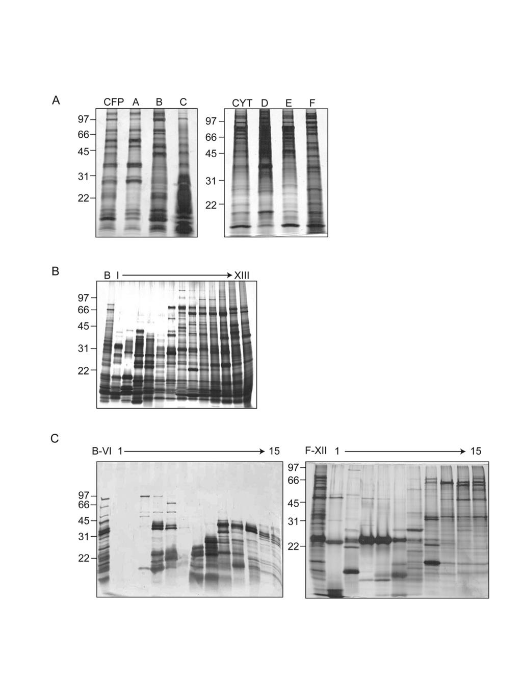
12 contained 3989 proteins. The software was set to evaluate peptides obtained by trypsin or chymotrypsin and GluC digegtion and to consider the oxidation of methionine (+16.0 amu) and the acrylamide modification of cysteine (+71.0 amu). The Scaffold software (Proteome Software, Portland, OR) that verifies peptide identifications made by SEQUEST and probabilistically validates the peptide and protein identifications was applied to all MS/MS sequencing results. Results Multi-dimensional protein fractionation resulted in the reduced complexity of mycobacterial protein pools. Recent advances in proteomics have resulted in the ability to separate and identify individual proteins or peptides from complex biological samples. Specifically, multidimensional chromatography of peptides, derived from tryptic digests of crude biological samples, followed by MS/MS analysis (MudPIT) has resulted in the experimental validation of substantial portions of theoretical proteomes (25). Recently, this approach was applied to Mtb and resulted in more than a threefold increase in the number of proteins previously identified by 2-D gel electrophoresis based methods (26). With goals similar to the MudPIT strategy, this study employed a multi-dimensional fractionation scheme (Fig. S1) to efficiently separate complex pools of intact mycobacterial proteins into relatively simple and enriched fractions that could be evaluated for serological reactivity in a high-throughput fashion. Aliquots of cytosolic proteins (500 mg) and CFPs (446 mg) from Mtb H37Rv were subjected to sequential ammonium sulfate precipitations, resulting in six fractions with nearly equal protein amounts (Fig. 1A). Subsequent AIEX chromatography expanded the fraction number to 78, with protein yields 12
13 varying from 160 μg to 13 mg per fraction (Fig. 1B). Those fractions containing 500 μg of protein were excluded from further separation (8 fractions), and fractions containing between 500 μg and 1 mg were pooled with neighboring fractions (10 fractions). This resulted in a total of 64 fractions that were applied to RP chromatography under mildly basic ph conditions. This multidimensional chromatography separation yielded a total of 960 fractions, and SDS-PAGE analysis revealed varied complexity among the fractions, with 1 to >20 proteins observed per fraction (Fig. 1C). Validation of the protein microarray format Each multidimensional chromatography fraction, as well as intermediate fractions and recombinant proteins were printed to nitrocellulose microarray slides. Sera from PPD+ healthy (n=12), noncavitary-tb (n=9), cavitary-tb (n=11), and HIV+TB+ (n=10) individuals were probed against the microarrays in triplicate, and for each slide the NAvSNR (see Materials and Methods) was calculated for each protein or protein fraction. The integrity of the protein microarray and validation of this platform was determined by assessing the reactivity of TB patients sera with selected protein fractions and purified proteins spotted on the microarray slides as controls, and comparing these data to published results obtained by plate ELISA (5, 6). To demonstrate specificity, the reactivity of TB patients sera to unfractionated CFP was compared to that of E. coli WCL and FAST print buffer. The CFP is known to contain numerous B cell antigens (2), and as expected, cavitary-tb patient sera displayed significantly greater reactivity to CFP than to either buffer alone (p-value = 0.001) or E. coli WCL (p-value = 0.002) (Fig. 2A). Although patient-to-patient variability in reactivity to unfractionated CFP was 13
14 observed, each individual cavitary-tb patient s serum recognized CFP more strongly than they did buffer or E. coli WCL. A second validation control was performed with purified native 38-kDa protein PstS1/Rv0934, a previously characterized B cell antigen (2). Reactivity to PstS1 was compared among the four patient groups used in this study (Fig. 2B). Sera from TB patients showed greater reactivity than that from PPD+ healthy individuals. Furthermore, when evaluated using the mean NAvSNR of PPD+ healthy controls plus three times the standard deviation (SD) as a cutoff, the number of patients sera demonstrating a positive response to PstS1 was greatest for cavitary-tb (72%), followed by noncavitary-tb (33%), and HIV+TB+ (30%) patients. These results concurred with data previously obtained via traditional ELISA platforms (5, 6). The PstS1 was also printed at multiple concentrations and a dose-dependent antibody response was observed (data not shown). TB Disease States React to a Defined Group of Antigens To evaluate patterns of serological reactivity the NAvSNR values of each fraction were averaged for all patients within a disease state, and expressed as a ratio over the corresponding fraction s averaged PPD+ healthy control NAvSNR (Fig. 3A). Fractions originating from the culture filtrate (fractions 1-525) were more antigenic overall than those derived from the cytosol (fractions ). Additionally, differences in reactivity based on disease state were observed. This was most pronounced when comparing HIV+TB+ patients to cavitary- or noncavitary-tb patients. The HIV+TB+ sera generally displayed poor reactivity, except for a distinct cluster of CFP fractions that were not well recognized by sera from other disease states (Fig. 3A). 14
15 To simplify patterns of antigen reactivity and to further assess similarities and differences in antigen recognition between disease states, a cutoff value of the mean NAvSNR of PPD+ healthy controls plus three SD was established for each fraction. Fractions displaying NAvSNR values greater than the cutoff value in 40% or more of the individuals in a disease state were selected and organized in a Venn diagram (Fig. 3B). Using these criteria, 145 of the 960 fractions (15%) were identified as having significant serological reactivity. As shown in Figure 3B, cavitary-tb patients recognized 126 fractions, while noncavitary-tb patients showed significant reactivity to 59 fractions, of which 55 were also recognized by cavitary-tb patients. The remaining four fractions were unique to noncavitary-tb patients. The pattern of fractions recognized by HIV+TB+ patients was less complex than that of either cavitary or noncavitary patients, and the overlap with these other two patient groups was minimal (Fig. 3B). HIV+TB+ patients recognized 20 fractions, of which five overlapped with those recognized by cavitary-tb patients, and of this latter group two also reacted to noncavitary-tb patients sera. Interestingly the 15 fractions that demonstrated significant reactivity to only HIV+TB+ patients sera possessed very similar separation characteristics: 1) they originated from the CFP and did not precipitate with 67% ammonium sulfate, and 2) they bound strongly to the AIEX column, but weakly to the RP column. In Figure 3A these 15 fractions correspond to the cluster of CFP fractions that showed enhanced reactivity with HIV+TB+ patients sera. To confirm that the antigen(s) in these fractions were specific to TB rather than HIV infection, the reactivity of three of the 15 fractions was assessed with HIV+TB- patients sera, and no antibody responses significantly greater than that of PPD+ healthy controls were observed. (data not shown) 15
16 Antigen identification of reactive fractions confirmed the reactivity to known B-cell antigens To identify individual antigens with the greatest utility in a serodiagnostic assay additional stringency was applied to the array data, further reducing the number of fractions demonstrating significant reactivity from 145 to 105. This was achieved by restricting analyses of the cavitary-tb specific fractions to those that yielded significant serological reactivity ( 3X SD above PPD+ mean) with 55% (6 of 11) or greater of cavitary-tb patients. It was also recognized that lipoarabinomannan (LAM), a well-characterized B cell antigen (27), would be present in some of the multidimensional fractions and was expected to be serologically dominant. Thus, microarray slides were probed with the CS-35 MAb specific for LAM (Fig. 4). Of the 105-targeted fractions 24 were found to contain LAM. The serological dominance of LAM was confirmed with conventional one-dimensional immunoblotting using pooled TB patients sera (Fig. 4). It is interesting to note that the LAM containing fractions consistently yielded microarray spots with the highest fluorescent intensities when probed with patients sera. Most of the PPD+ healthy control patients sera also reacted against these fractions. However, the reactivity to TB patients sera was significantly stronger than that of the healthy PPD+ control sera (Table 1). The twenty-four LAM-containing fractions were excluded from further antigen analyses. Molecular identities of the serologically active native proteins within the remaining fractions were obtained by two methods (Fig. S1). The first of these utilized Western blot analyses with patients sera compared alongside the reactivity to MAbs or polyclonal sera specific for five Mtb proteins (Fig. 5A). The second approach determined antigen composition of a fraction by Western blot analysis with patients sera and identification of the corresponding Coomassie-stained protein band via MS/MS (Fig. 5B and C). This combination of MS and 16
17 antibody-based identification strategies resulted in an assigned antigen composition for 38 of the 81 remaining fractions. Ten proteins previously found to be human B cell antigens (2) accounted for all or part of the serological activity of 26 fractions (Table 2). MAbs were used as the sole evidence to identify the presence of the 38-kDa PstS1 antigen, the 19-kDa lipoprotein antigen, and the 14- kda HspX antigen in four reactive fractions. Specifically, the probing of a microarray slide with MAbs IT-23 and IT-47 identified PstS1 in fraction C-II-11. Western blot analyses with IT-47, cavitary-tb sera, and PPD+ healthy control sera also revealed a single protein that reacted with IT-47 and cavitary-tb patients sera, but not sera of PPD+ healthy control individuals (Fig. 5A). In this same manner, the 19-kDa was found to be a reactive product of fractions B-I-7 and C-I- 14, and HspX contributed to the reactivity of fraction B-V-9. It was also noted that fractions B- I-7 and C-I-14 each contained a second reactive product of 12 kda and 45 kda, respectively, and fraction B-V-9 contained two additional reactive proteins of 7 and 10 kda. The identity of these unknown proteins was not determined due to insufficient protein quantities. The remaining seven previously described protein antigens (45 kda Apa, 30 kda Ag85A and B, GlcB, Rv3881c, SecE2, and MPT64) were identified by MS/MS analyses and some of these were confirmed by reactivity to MAbs (Table 2 and Table S1). For some fractions, after Western blot analyses with patients sera a sufficient amount of material was not available for protein identification. Thus, protein identity was obtained by MS/MS analysis of an adjacent fraction possessing a reactive band at the same molecular weight. This was done to identify the 45 kda Apa in one fraction, Ag85B in four fractions, Ag85A in two fractions, GlcB in four fractions, SecE2 in three fractions, and Rv3881c in one fraction (Table 2). 17
18 Discovery of novel B-cell antigens with potential serodiagnostic roles The MS/MS-based approach to antigen identification led to the elucidation of four new Mtb B cell antigens (Table 3). The first of these, SodC (Rv0432), a 27-kDa Cu/Zn superoxide dismutase (28), was the sole reactive constituent of four fractions originating from the CFP pool (B-III-3, B-III-4, B-IV-3, and B-IV-4), and was significantly recognized by both noncavitary- (five out of nine) and cavitary-tb (eight out of eleven) sera (Fig. 5B). MS/MS analysis of the corresponding protein in fraction B-III-4 resulted in 42% amino-acid coverage of the predicted protein sequence encoded by open-reading frame (ORF) rv0432 (Fig. 5C). LppZ (Rv3006) was identified as the only reactive product of five fractions (A-III-5, A- IV-4, A-IV-5, A-IV-6, and A-V-5) originating from the CFP pool, and was strongly recognized by noncavitary- (four out of nine) and cavitary-tb (six out of eleven) sera. These fractions were generated under similar separation conditions, and SDS-PAGE showed all possessed a dominant 45-kDa product. MS/MS analysis of the corresponding protein in fraction A-IV-5 resulted in 37% amino-acid coverage of the predicted protein sequence encoded by ORF rv3006. Bfrb (Rv3841), a putative iron-storage protein (29), and a previously described T cell antigen (30), was identified as the reactive protein in two fractions (A-VII-1 and A-VIII-1) with significant reactivity to four out of nine noncavitary- and six out of eleven cavitary-tb sera. MS/MS analysis of the 20-kDa reactive product in fraction A-VIII-1 resulted in 37% amino-acid coverage of the predicted protein sequence encoded by ORF rv3841, and analysis of the silverstained polyacrylamide gels revealed the 20-kDa band was the sole protein constituent in both fractions. TrxC (Rv3914), a 12-kDa thioredoxin (31), was found along with the 45-kDa Apa antigen to account for the serological reactivity of fraction B-II-9. In contrast to the other novel 18
19 antigen containing fractions, fraction B-II-9 was only significantly recognized by cavitary-tb patients sera (six out of eleven). MS/MS analysis of the 12-kDa protein resulted in 29% aminoacid coverage of the predicted protein sequence encoded by ORF rv3914. Whether or not the significant reactivity of fraction B-II-9 was due to TrxC or the strongly seroreactive 45-kDa antigen (6) could not be determined. When serological data were combined for those fractions containing the four novel antigens it was found that 56% and 91% of the noncavitary- and cavitary-tb patients sera, respectively, showed positive reactivity. In comparison, when the data was combined for all fractions containing novel and previously identified protein antigens, 78% of noncavitary and 91% of cavitary patients displayed reactivity. Of considerable interest were the 15 fractions that demonstrated significant reactivity with only HIV+TB+ patients sera. Western blot analysis of these fractions with HIV+TB+ patients sera failed to demonstrate reactive bands. Furthermore, there were no protein bands in common between these fractions when analyzed by SDS-PAGE and silver staining, but treatment of these fractions with pronase (10 μg/ml for 60 min) prior to microarray printing significantly abrogated reactivity to patients sera. Together these observations suggest that: 1) a single protein antigen may not be responsible for the reactivity of these 15 fractions, 2) a common antigen such as a small peptide may be responsible for the reactivity, or 3) the reactivity is due to a non-proteinaceous bacterial product complexed with protein. 19
20 Discussion Previous studies from our laboratories employed 2-D immunoblotting to characterize the profile of Mtb proteins recognized by TB patients sera (5, 10). However, this methodology is not well suited for analysis of large numbers of sera due to problems of reproducibility, difficulty in quantifying the results, and refractivity to high-throughput analyses. Protein microarrays offer a means by which a large number of sera can be analyzed not only to identify serologically reactive proteins, but to establish antigen recognition profiles based on the state or severity of disease (16). At present a complete recombinant protein library of Mtb does not exist. Therefore, to perform protein microarray studies for TB a novel approach involving a robust fractionation strategy that yielded 960 native protein fractions was utilized. The availability of such arrays allowed us to address differences in the patterns of antigens recognized by individuals exhibiting various forms of TB. Specifically, TB patients sera recognized a much greater number of protein fractions than did sera of PPD+ healthy controls and patients with noncavitary-tb recognized only a subset (44%) of the fractions that reacted to patients with advanced, cavitary disease. This pattern of reactivity agrees well with our previous results obtained by 2-D Western blot analysis where only three to four CFPs reacted to sera of PPD+ healthy individuals and where 12 of the 26 cavitary-tb reactive proteins (46%) were recognized by noncavitary-tb patients (5, 10). The inclusion of antigen identification into these current studies enabled a more in-depth assessment of the overlap between the three disease states (cavitary, noncavitary and HIV+TB+) studied. Of the 55 fractions recognized by both cavitary- and noncavitary-tb patients, 11 antigens (LAM, the 45 kda Apa protein, the 19-kDa LpqH protein, Ag85A, Ag85B, Bfrb, GlcB, LppZ, Rv3881c, SecE2, and SodC) were identified as being serologically dominant. Of 20
21 particular interest is the fact that all five fractions possessing GlcB as the reactive species were recognized by both noncavitary- and cavitary-tb patients, confirming previous reports that this antigen is recognized early in disease progression (5, 6). Our analyses also identified a total of 68 fractions recognized exclusively by cavitary-tb patients sera. Most of the antigens represented by these fractions (the 45-kDa Apa protein, Ag85B, LppZ, SecE2, SodC, LAM, Rv3881c, and the 19-kDa protein) overlapped with those recognized by both cavitary- and noncavitary-tb patients. However, four antigens (38 kda PstS1 protein, HspX, MPT64, and TrxC) were recognized only by cavitary-tb patients sera, thus, providing several antigens that may be useful in demarcating cavitary- and noncavitary-tb patients. The identification of the 38-kDa PstS1 as a cavitary-tb specific antigen concurs with previous reports that this antigen is recognized predominantly by patients with advanced disease (5, 6) Previous experimental approaches have failed to identify an antigen that distinguishes noncavitary-tb patients from individual with other stages of the disease. In this study four native protein fractions were designated as noncavitary-tb specific based on our selection parameters. A more in-depth inspection of these fractions, however, suggests they are likely not noncavitary-tb specific because these four fractions were each recognized by four of eleven (36%) cavitary-tb patients, just missing the 40% cutoff used to construct the Venn diagram. Additionally, the 45-kDa Apa protein and the 19-kDa protein were found to be the antigens responsible for the reactivity in three of these four fractions, and these same antigens were also found in five fractions recognized by sera of cavitary-tb patients. Thus, a noncavitary-tb specific antigen remains elusive and it appears that regardless of the methodology employed noncavitary-tb patients react with a subset of those antigens recognized by cavitary-tb patients. 21
22 While the overall data and conclusions obtained through these microarray-based studies were consistent with earlier work there were a few discrepancies with studies based on 2-D immunoblots (5, 10). In contrast to the qualitative 2-D immunoblot data the results obtained with microarrays were quantitative. Thus, the selection of reactive antigens or fractions was based on the percentage of sera with a NAvSNR value greater than an experimentally determined cutoff. This led to the exclusion of two antigens previously identified by 2-D immunoblots, but which fell outside the criteria set in this study to define significant reactivity. This was most notable with GlcB and its reactivity to HIV+TB+ patients sera. GlcB was previously shown to react with HIV+TB+ sera in both 2-D immunoblot and ELISA formats (3, 10). However, when the microarray data set was quantified the percentage of HIV+TB+ patients that recognized GlcB containing fractions was just below the 40% cutoff. Previous work shows that sera from this group of patients reacts to the same set of CFP as recognized by noncavitary-tb patients (10). However, our present work defines fractions containing an unidentifable antigen as the only material with signifcant reactivity to HIV+TB+ patients sera. The exceptionally strong response of these fractions with HIV+TB+ sera likely led to a bias in data analysis for this patient group. A second difference with the 2-D Western blot data was the failure to define MPT51 as a dominant antigen. By 2-D PAGE MPT51 readily separates from LAM and is recognized as a dominant antigen in multiple disease states (10). Probing the native protein array slide with the MPT51 specfic MAb (IT52) demonstrated that MPT51 cofractionated with LAM (data not shown). Since LAM-containing fractions were excluded from further protein antigen analyses, MPT51 was not designated as a significant serological antigen. In our previous work there were approximately 12 protein spots that reacted to patients sera by 2-D immunoblot, but were unidentifiable (10). Although it is possible that some of these 2-D protein spots correspond to 22
23 the four novel antigens identified in our current studies, it is not possible to draw such conclusions without further analyses. Over the past two decades molecular identification of the dominant B and T cell protein antigens of Mtb was achieved through a myriad of techniques and approaches (2, 32, 33) This current application of protein microarrays to TB serodiagnostics has allowed for identification of four novel antigens previously unnoticed by other methods. Two of these antigens, SodC and LppZ, are believed to undergo post-translational modification. SodC was experimentally shown to be lipid-modified and associated with the bacterial membrane (34). This protein also contains three predicted glycosylation sites, and the 40-aa N-terminal fragment of SodC was found to bind Concanavalin A when fused to the 19-kDa-leader sequence and expressed in M. smegmatis (35). A method for predicting gram-positive lipoprotein motifs has also identified LppZ as a probable lipoprotein (36), and similar to SodC, this protein sequence also contains three predicted glycosylation sites (35). The native and recombinant forms of several Mtb proteins have been directly compared, and it is becoming increasingly clear that recombinant forms often lack modifications or conformational epitopes required for immunological recognition (6, 37, 38). The discovery of SodC and LppZ as B cell antigens adds to the growing list of posttranslationally modified antigens of Mtb (39) and illustrates the need for a recombinant protein production system that mimics native protein structures. One of the other novel B cell antigens described in this work (BfrB) was previously shown by us as a dominant T cell antigen for mice experimentally infected with Mtb (30), and this current work reveals that BfrB is also antigenic in human disease. Thus, like many other Mtb antigens, BfrB is strongly recognized by both the cellular and humoral arms of the immune system and could be targeted as either a vaccine candidate or diagnostic tool. The final B cell 23
24 antigen newly recognized in this work (TrxC) was originally identified by Nagai et al. (40) as MPT46, a major product of the Mtb culture filtrate. Further work established MPT46 as a thioreductase (31). Although this protein was previously studied, the use of protein array technology has provided the first evidence of its antigenic potential. Through the work of multiple laboratories greater than sixteen Mtb proteins have been identified as potential serodiagnostic antigens and most of these proteins are associated with the CF (2). However, despite efforts to enrich for proteins of low abundance, we recognize that a relatively small number of novel antigens were identified in this study. The inclusion of denaturating agents may have further improved protein resolution and increased antigen detection; however, such an approach might also destroy conformational epitopes. An alternative explanation for the low number of novel antigens identified is that the discovery of new serodiagnostic antigens from in vitro grown Mtb H37Rv cytosolic and CF protein pools is nearly exhausted. Nevertheless, the cell envelope protein pool of M. tuberculosis is underexploited for serodiagnostic antigen discovery, and the methodologies described in this work lay a foundation for B cell antigen discovery with this fraction. It is likely that further antigen discovery will also be realized once microarray technology is applied to a complete Mtb recombinant protein library. Our earlier work on the serological response to Mtb proteins in human disease indicated considerable homogeneity in this response among TB patients (10). The previously accepted dogma of heterogeneity in antigen recognition likely resulted from the differences in the immunological response among the various states of the disease and poor immunological reactivity of Mtb recombinant proteins expressed in E. coli (41). The use of protein arrays has confirmed our previous observations and hypotheses. Moreover, the ability to assess antigen 24
25 recognition profiles between disease states allowed for the identification of several proteins recognized by both cavitary- and noncavitary-tb patients and at least four proteins that appear diagnostic of cavitary-tb. Thus, from these data it will be possible to develop and assess targeted protein arrays that will not only enable serodiagnosis of TB, but also the determination of disease severity. 25
26 Acknowledgements: We would like to thank Dr. Karen Dobos for her thoughtful discussions on protein separation strategies and Mr. Preston Hill for technical assistance in mass spectrometry. This work was supported by the NIH, NIAID grant RO1 AI and Contract NO1 AI-75320, and the Monfort Professor award to Dr Belisle. 26
27 References 1. World Health Organization (2005) Global tuberculosis control - surveillance, planning, financing (WHO Report 2005), Geneva 2. Laal, S. and Y.A., S. (2005) Immune-Based Methods, in Tuberculosis and the Tubercle Bacillus, pp , ASM Press, Washington, D.C. 3. Laal, S., Samanich, K.M., Sonnenberg, M.G., Belisle, J.T., O'Leary, J., Simberkoff, M.S. and Zolla-Pazner, S. (1997) Surrogate marker of preclinical tuberculosis in human immunodeficiency virus infection: antibodies to an 88-kDa secreted antigen of Mycobacterium tuberculosis. J Infect Dis. 176, Laal, S., Samanich, K.M., Sonnenberg, M.G., Zolla-Pazner, S., Phadtare, J.M. and Belisle, J.T. (1997) Human humoral responses to antigens of Mycobacterium tuberculosis: immunodominance of high-molecular-mass antigens. Clin Diagn Lab Immunol. 4, Samanich, K.M., Belisle, J.T., Sonnenberg, M.G., Keen, M.A., Zolla-Pazner, S. and Laal, S. (1998) Delineation of human antibody responses to culture filtrate antigens of Mycobacterium tuberculosis. J Infect Dis. 178, Samanich, K.M., Keen, M.A., Vissa, V.D., Harder, J.D., Spencer, J.S., Belisle, J.T., Zolla-Pazner, S. and Laal, S. (2000) Serodiagnostic potential of culture filtrate antigens of Mycobacterium tuberculosis. Clin Diagn Lab Immunol. 7, Singh, K.K., Dong, Y., Hinds, L., Keen, M.A., Belisle, J.T., Zolla-Pazner, S., Achkar, J.M., Nadas, A.J., Arora, V.K. and Laal, S. (2003) Combined use of serum and urinary antibody for diagnosis of tuberculosis. J Infect Dis. 188, Singh, K.K., Dong, Y., Belisle, J.T., Harder, J., Arora, V.K. and Laal, S. (2005) Antigens of Mycobacterium tuberculosis recognized by antibodies during incipient, subclinical tuberculosis. Clin Diagn Lab Immunol. 12, Singh, K.K., Dong, Y., Patibandla, S.A., McMurray, D.N., Arora, V.K. and Laal, S. (2005) Immunogenicity of the Mycobacterium tuberculosis PPE55 (Rv3347c) protein during incipient and clinical tuberculosis. Infect Immun. 73, Samanich, K., Belisle, J.T. and Laal, S. (2001) Homogeneity of antibody responses in tuberculosis patients. Infect Immun. 69, Joos, T.O., Schrenk, M., Hopfl, P., Kroger, K., Chowdhury, U., Stoll, D., Schorner, D., Durr, M., Herick, K., Rupp, S., Sohn, K. and Hammerle, H. (2000) A microarray enzyme-linked immunosorbent assay for autoimmune diagnostics. Electrophoresis. 21, Madoz-Gurpide, J., Wang, H., Misek, D.E., Brichory, F. and Hanash, S.M. (2001) Protein based microarrays: a tool for probing the proteome of cancer cells and tissues. Proteomics. 1, Kersten, B., Feilner, T., Kramer, A., Wehrmeyer, S., Possling, A., Witt, I., Zanor, M.I., Stracke, R., Lueking, A., Kreutzberger, J., Lehrach, H. and Cahilll, D.J. (2003) Generation of Arabidopsis protein chips for antibody and serum screening. Plant Mol Biol. 52, Bacarese-Hamilton, T., Bistoni, F. and Crisanti, A. (2002) Protein microarrays: from serodiagnosis to whole proteome scale analysis of the immune response against pathogenic microorganisms. Biotechniques. Suppl,
28 15. Li, B., Jiang, L., Song, Q., Yang, J., Chen, Z., Guo, Z., Zhou, D., Du, Z., Song, Y., Wang, J., Wang, H., Yu, S. and Yang, R. (2005) Protein microarray for profiling antibody responses to Yersinia pestis live vaccine. Infect Immun. 73, Davies, D.H., Liang, X., Hernandez, J.E., Randall, A., Hirst, S., Mu, Y., Romero, K.M., Nguyen, T.T., Kalantari-Dehaghi, M., Crotty, S., Baldi, P., Villarreal, L.P. and Felgner, P.L. (2005) Profiling the humoral immune response to infection by using proteome microarrays: high-throughput vaccine and diagnostic antigen discovery. Proc Natl Acad Sci U S A. 102, Sonnenberg, M.G. and Belisle, J.T. (1997) Definition of Mycobacterium tuberculosis culture filtrate proteins by two-dimensional polyacrylamide gel electrophoresis, N- terminal amino acid sequencing, and electrospray mass spectrometry. Infect Immun. 65, Dobos, K., Khoo, K., Swiderek, K., Brennan, P. and Belisle, J. (1996) Definition of the full extent of glycosylation of the 45-kilodalton glycoprotein of Mycobacterium tuberculosis. J. Bacteriol. 178, Hirschfield, G.R., McNeil, M. and Brennan, P.J. (1990) Peptidoglycan-associated polypeptides of Mycobacterium tuberculosis. J Bacteriol. 172, Smith, P.K., Krohn, R.I., Hermanson, G.T., Mallia, A.K., Gartner, F.H., Provenzano, M.D., Fujimoto, E.K., Goeke, N.M., Olson, B.J. and Klenk, D.C. (1985) Measurement of protein using bicinchoninic acid. Anal Biochem. 150, Saeed, A.I., Sharov, V., White, J., Li, J., Liang, W., Bhagabati, N., Braisted, J., Klapa, M., Currier, T., Thiagarajan, M., Sturn, A., Snuffin, M., Rezantsev, A., Popov, D., Ryltsov, A., Kostukovich, E., Borisovsky, I., Liu, Z., Vinsavich, A., Trush, V. and Quackenbush, J. (2003) TM4: a free, open-source system for microarray data management and analysis. Biotechniques. 34, Laemmli, U.K. (1970) Cleavage of structural proteins during the assembly of the head of bacteriophage T4. Nature. 227, Morrissey, J.H. (1981) Silver stain for proteins in polyacrylamide gels: a modified procedure with enhanced uniform sensitivity. Anal Biochem. 117, Hellman, U., Wernstedt, C., Gonez, J. and Heldin, C.H. (1995) Improvement of an "In- Gel" digestion procedure for the micropreparation of internal protein fragments for amino acid sequencing. Anal Biochem. 224, McDonald, W.H. and Yates, J.R., 3rd (2002) Shotgun proteomics and biomarker discovery. Dis Markers. 18, Mawuenyega, K.G., Forst, C.V., Dobos, K.M., Belisle, J.T., Chen, J., Bradbury, E.M., Bradbury, A.R. and Chen, X. (2005) Mycobacterium tuberculosis functional network analysis by global subcellular protein profiling. Mol Biol Cell. 16, Sada, E., Brennan, P.J., Herrera, T. and Torres, M. (1990) Evaluation of lipoarabinomannan for the serological diagnosis of tuberculosis. J Clin Microbiol. 28, Wu, C.H., Tsai-Wu, J.J., Huang, Y.T., Lin, C.Y., Lioua, G.G. and Lee, F.J. (1998) Identification and subcellular localization of a novel Cu,Zn superoxide dismutase of Mycobacterium tuberculosis. FEBS Lett. 439, Cole, S.T., Brosch, R., Parkhill, J., Garnier, T., Churcher, C., Harris, D., Gordon, S.V., Eiglmeier, K., Gas, S., Barry, C.E., 3rd, Tekaia, F., Badcock, K., Basham, D., Brown, D., Chillingworth, T., Connor, R., Davies, R., Devlin, K., Feltwell, T., Gentles, S., Hamlin, 28
29 N., Holroyd, S., Hornsby, T., Jagels, K., Krogh, A., McLean, J., Moule, S., Murphy, L., Oliver, K., Osborne, J., Quail, M.A., Rajandream, M.A., Rogers, J., Rutter, S., Seeger, K., Skelton, J., Squares, R., Squares, S., Sulston, J.E., Taylor, K., Whitehead, S. and Barrell, B.G. (1998) Deciphering the biology of Mycobacterium tuberculosis from the complete genome sequence. Nature. 393, Covert, B.A., Spencer, J.S., Orme, I.M. and Belisle, J.T. (2001) The application of proteomics in defining the T cell antigens of Mycobacterium tuberculosis. Proteomics. 1, Wieles, B., Nagai, S., Wiker, H.G., Harboe, M. and Ottenhoff, T.H. (1995) Identification and functional characterization of thioredoxin of Mycobacterium tuberculosis. Infect Immun. 63, Andersen, P. and Doherty, T.M. (2005) TB subunit vaccines--putting the pieces together. Microbes Infect. 7, Reed, S. and Lobet, Y. (2005) Tuberculosis vaccine development; from mouse to man. Microbes Infect. 7, D'Orazio, M., Folcarelli, S., Mariani, F., Colizzi, V., Rotilio, G. and Battistoni, A. (2001) Lipid modification of the Cu,Zn superoxide dismutase from Mycobacterium tuberculosis. Biochem J. 359, Herrmann, J.L., Delahay, R., Gallagher, A., Robertson, B. and Young, D. (2000) Analysis of post-translational modification of mycobacterial proteins using a cassette expression system. FEBS Lett. 473, Sutcliffe, I.C. and Harrington, D.J. (2004) Lipoproteins of Mycobacterium tuberculosis: an abundant and functionally diverse class of cell envelope components. FEMS Microbiol Rev. 28, Romain, F., Horn, C., Pescher, P., Namane, A., Riviere, M., Puzo, G., Barzu, O. and Marchal, G. (1999) Deglycosylation of the 45/47-kilodalton antigen complex of Mycobacterium tuberculosis decreases its capacity to elicit in vivo or in vitro cellular immune responses. Infect Immun. 67, Zanetti, S., Bua, A., Delogu, G., Pusceddu, C., Mura, M., Saba, F., Pirina, P., Garzelli, C., Vertuccio, C., Sechi, L.A. and Fadda, G. (2005) Patients with pulmonary tuberculosis develop a strong humoral response against methylated heparin-binding hemagglutinin. Clin Diagn Lab Immunol. 12, Belisle, J.T., Braunstein, M., Rosenkrands, I. and Andersen, P. (2005) The Proteome of Mycobacterium tuberculosis, in Tuberculosis and the Tubercle Bacillus, pp , ASM Press, Washington, D.C. 40. Nagai, S., Wiker, H.G., Harboe, M. and Kinomoto, M. (1991) Isolation and partial characterization of major protein antigens in the culture fluid of Mycobacterium tuberculosis. Infect Immun. 59, Lyashchenko, K., Colangeli, R., Houde, M., Al Jahdali, H., Menzies, D. and Gennaro, M.L. (1998) Heterogeneous antibody responses in tuberculosis. Infect Immun. 66,
30 Figure Legends Figure 1. Mtb CFP and cytosol proteins were subjected to a multi-dimensional separation scheme. (A) Silver stained SDS-PAGE analysis of fractionation obtained by ammonium-sulfate precipitation of CFP and cytosol. CFP, unfractionated CFP; lane A, 42% ammonium-sulfate precipitate of the CFP; lane B, 67% ammonium-sulfate precipitate of the CFP; lane C, 67% ammonium-sulfate soluble material of the CFP; CYT, unfractionated cytosol; lane D, 29% ammonium-sulfate precipitate of the cytosol; lane E, 44% ammonium-sulfate precipitate of the cytosol; and lane F, 44% ammonium-sulfate soluble material of the cytosol. (B) Silver stained SDS-PAGE of strong AIEX chromatography fractions using the 67% ammonium-sulfate precipitate of the CFP as an example. Lane B, unfractionated material; lanes I-XIII are the sequential AIEX fractions. (C) Silver stained SDS-PAGE of RP chromatography fractions using the B-VI AIEX fraction of the 67% ammonium-sulfate precipitate of the CFP and the F-XII AIEX fraction of the 44% ammonium-sulfate soluble material of the cytosol as examples. Lane B-VI and F-XII, unfractionated material; lanes 1-15 are the sequential RP fractions. Figure 2. Validation of protein microarray integrity. (A) Reactivity of cavitary-tb patients sera against buffer ( ), E. coli WCL ( ), and Mtb CFP ( ). (B) Reactivity of PPD+ healthy controls ( ), HIV+TB+ ( ), noncavitary-tb ( ), and cavitary-tb ( ) patients sera against spotted native, purified 38-kDa PstS1. The patient-averaged NAvSNR +3 SD was obtained by use of sera from healthy PPD+ control subjects, shown as the horizontal dashed line, and was used as the cutoff to determine positive reactivity. 30
31 Figure 3. Global Analysis of HIV+TB+, noncavitary-tb, and cavitary-tb patient reactivity against all 960 protein fractions. (A) Each fraction s NAvSNR was averaged for all patients in each disease state, and expressed as a ratio over the corresponding fraction s averaged healthy PPD+ NAvSNR. Culture filtrate fractions (1-525), Cytosolic fractions ( ) (B) A Venn diagram displaying the number of fractions with significant reactivity to sera from each disease state and the relatedness of these serological responses. The fractions included in the Venn diagram were recognized by 40% of patients. Figure 4. Identification of reactive fractions containing lipoarabinomannan as the serodominant antigen. Shown are representative arrays probed with serum from a single cavitary-tb patient, and the LAM specific MAb CS-35. The inset is a Western blot of fraction C-I-1 probed with pooled cavitary-tb patients sera. Figure 5. Antibody and MS based identification of antigens composing reactive fractions. (A) Western blot analyses of fraction C-II-11 with MAb IT-27, pooled cavitary-tb patients sera, and pooled PPD+ healthy controls sera (left to right) demonstrate the 38-kDa PstS1 protein as the dominant antigen in this fraction. (B) Fraction B-III-4 was analyzed (left to right) by SDS- PAGE and coomassie-staining, and Western blot with pooled sera fron cavitary-tb, noncavitary- TB, and healthy PPD+ individuals to demostrate a 28-kDa protein as the dominant antigen. (C) Peptide sequences resolved by MS/MS of the coomassie-stained 28-kDa protein band after trypsin digestion identified the antigen as SodC. 31
32 Table 1. Patient reactivity against LAM-containing fractions LAM-Reactivity Ratio a Fractionation Conditions (X,Y,Z) b PPD+ HIV+ TB Noncavitary TB Cavitary TB A-III A-IV A-IV A-V A-IX A-X/XI B-III B-III B-IV B-I C-III C-IV C-I C-I C-I C-I D-II/III/IV E-II E-III F-II F-II F-IV F-I F-I a Disease state-averaged NAvSNR for the fraction / Disease state-averaged NAvSNR for the all 960 fractions. b Key to fraction designation: X = Ammonium Sulfate cut A-F (see Materials and Methods), Y = AIEX fraction (13 sequential elution fractions, Roman Numeral I to XIII), "/" denotes pooled fractions, Z = RP-HPLC fraction (sequential elutions 1 through 15). 33
33 Table 2. Previously characterized antigens contained in serologically reactive fractions Antigen(s) Patient Reactivity b Fractionation Conditions (X,Y,Z) a HIV+TB+ Noncavitary TB Cavitary TB 38-kDa PstS1/Rv0934 c C-II-11 1/10 (10%) 1/9 (11%) 6/11 (55%) 45-kDa Apa/ModD/Rv1860 d A-II-11 2/10 (20%) 4/9 (44%) 4/11 (36%) A-III-9 2/10 (20%) 4/9 (44%) 5/11 (44%) A-III-11 2/10 (20%) 4/9 (44%) 7/11 (64%) B-III-10 2/10 (20%) 4/9 (44%) 4/11 (36%) Ag85B/Rv1886c e B-VI-13 3/10 (30%) 6/9 (67%) 6/11 (55%) B-VI-14 3/10 (30%) 3/9 (33%) 7/11 (64%) A-VI-15 2/10 (20%) 4/9 (44%) 5/11 (44%) GlcB/Rv1837c e A-VI-3 3/10 (30%) 4/9 (44%) 6/11 (55%) A-VI-4 2/10 (20%) 4/9 (44%) 6/11 (55%) A-VII-3 3/10 (30%) 4/9 (44%) 6/11 (55%) A-VIII-3 2/10 (20%) 4/9 (44%) 6/11 (55%) A-VIII-4 2/10 (20%) 4/9 (44%) 5/11 (44%) LAM c A-III-4 2/10 (10%) 4/9 (44%) 6/11 (55%) A-IV-1 1/10 (10%) 5/9 (56%) 6/11 (55%) A-IV-3 2/10 (20%) 4/9 (44%) 6/11 (55%) A-IX-1 3/10 (30%) 5/9 (56%) 6/11 (55%) A-V-1 3/10 (30%) 5/9 (56%) 6/11 (55%) A-X/XI-1 3/10 (30%) 4/9 (44%) 7/11 (64%) B-I-2 2/10 (20%) 4/9 (44%) 6/11 (55%) B-III-2 1/10 (10%) 4/9 (44%) 6/11 (55%) B-III-7 2/10 (20%) 3/9 (33%) 6/11 (55%) B-IV-1 0/10 (0%) 3/9 (33%) 7/11 (64%) C-I-1 0/10 (0%) 1/9 (11%) 6/11 (55%) C-I-2 0/10 (0%) 3/9 (33%) 6/11 (55%) C-I-4 2/10 (20%) 5/9 (56%) 6/11 (55%) C-I-13 2/10 (20%) 3/9 (33%) 5/11 (55%) C-III-1 2/10 (20%) 4/9 (44%) 6/11 (55%) C-IV-1 2/10 (20%) 4/9 (44%) 6/11 (55%) D-II/III/IV-1 0/10 (0%) 5/9 (56%) 7/11 (64%) E-II-1 0/10 (0%) 3/9 (33%) 6/11 (55%) E-III-1 2/10 (20%) 3/9 (33%) 6/11 (55%) F-I-1 0/10 (0%) 4/9 (44%) 7/11 (64%) F-I-2 2/10 (20%) 5/9 (56%) 6/11 (55%) F-II-2 2/10 (20%) 4/9 (44%) 7/11 (55%) F-II-3 2/10 (20%) 4/9 (44%) 7/11 (55%) F-IV-1 2/10 (20%) 3/9 (33%) 7/11 (64%) Rv3881c e A-VII-10 2/10 (20%) 3/9 (33%) 7/11 (64%) A-VII-11 2/10 (20%) 4/9 (44%) 7/11 (64%) SecE2/Rv0379 e A-I-4 2/10 (20%) 4/9 (44%) 5/11 (44%) B-I-4 3/10 (30%) 5/9 (56%) 5/11 (44%) F-I-3 2/10 (20%) 3/9 (33%) 6/11 (55%) A-I-3 1/10 (10%) 4/9 (44%) 6/11 (55%) Ag85A/Rv3804c & A-I-14 2/10 (20%) 3/9 (33%) 6/11 (55%) Ag85B/Rv1886c e A-I-13 2/10 (20%) 4/9 (44%) 5/11 (44%) 34
34 A-VI-12 2/10 (20%) 2/9 (22%) 6/11 (55%) 45 kda & MPT64/Rv1980c d A-III-10 2/10 (20%) 3/9 (33%) 6/11 (55%) HspX/Rv2031c & 2 unknowns c B-V-9 2/10 (20%) 0/9 (0%) 6/11 (55%) 19-kDa/Rv3763 & 1 unknown c B-I-7 2/10 (20%) 4/9 (44%) 4/11 (36%) C-I-14 2/10 (20%) 3/9 (44%) 6/11 (55%) a Key to fraction designation: X = Ammonium Sulfate cut A-F (see Materials and Methods), Y = AIEX fraction (13 sequential elution fractions, Roman Numeral I to XIII), "/" denotes pooled fractions, Z = RP-HPLC fraction (sequential elutions 1 through 15). b Number (Percentage) of patient sera significantly reactive against fraction in question. Significant is > 3 X SD above PPD+ mean. c Antigen identification based on reactivity to an antigen-specific MAb. d Antigen identification based on reactivity to an antigen-specific MAb and MS/MS analyses. e Antigen identification based on MS/MS analyses. Detailed MS/MS results are provided in Supplementary Table 1. 35
35 Table 3. Novel antigens composing reactive fractions Patient Reactivity b Antigen(s) Fractionation Conditions (X,Y,Z) a HIV+TB+ Noncavitary TB Cavitary TB MS/MS c Bfrb/Rv3841 A-VII-1 3/10 (30%) 4/9 (44%) 6/11 (55%) A-VIII-1 3/10 (30%) 4/9 (44%) 6/11 (55%) (37) LppZ/Rv3006 A-III-5 2/10 (20%) 3/9 (33%) 6/11 (55%) A-IV-4 2/10 (20%) 4/9 (44%) 5/11 (44%) A-IV-5 2/10 (20%) 3/9 (33%) 6/11 (55%) (37) A-IV-6 2/10 (20%) 3/9 (33%) 6/11 (55%) A-V-5 2/10 (20%) 4/9 (44%) 5/11 (44%) SodC/Rv0432 B-III-3 2/10 (20%) 4/9 (44%) 6/11 (55%) B-III-4 2/10 (20%) 5/9 (56%) 8/11 (73%) (42) B-IV-3 2/10 (20%) 4/9 (44%) 7/11 (64%) B-IV-4 3/10 (30%) 3/9 (33%) 7/11 (64%) Apa/Rv1860 d & TrxC/Rv3914 B-II-9 2/10 (20%) 3/9 (33%) 6/11 (55%) (29) e a Key to fraction designation: X = Ammonium Sulfate cut A-F (see Materials and Methods), Y = AIEX fraction (13 sequential elution fractions, Roman Numeral I to XIII), "/" denotes pooled fractions, Z = RP-HPLC fraction (sequential elutions 1 through 15). b Number (Percentage) of patient sera significantly reactive against fraction in question. Significant is > 3 X SD above PPD+ mean. c Antigen identification based on LC-ESI-MS/MS analysis. Percent amino acid sequence coverage is in parentheses. d The Apa was identified with the MAb CS-93. e The percent amino acid coverage is for TrxC. 36
36
37
38 Average Normalized SNR / PPD + SNR A * Noncavitary-TB Cavitary-TB CF fractions CYT fractions HIV + TB+ B 4 Noncavitary-TB Cavitary-TB 3 HIV + TB+


39 C.I CAV Cavitary CS-35
Proteomics and Vaccine Potency Testing
 Proteomics and Vaccine Potency Testing Louisa B. Tabatabai, PhD Respiratory Diseases of Livestock Research Unit National Animal Disease Center, ARS, USDA, Ames, Iowa What is proteomics Methodologies of
Lecture 5: 8/31. CHAPTER 5 Techniques in Protein Biochemistry
 Lecture 5: 8/31 CHAPTER 5 Techniques in Protein Biochemistry Chapter 5 Outline The proteome is the entire set of proteins expressed and modified by a cell under a particular set of biochemical conditions.
Application Note AN001
 Testing hybridoma supernatants with the Spots On Dots Antibody Screening Kit Application Note AN1 Table of Contents Overview... 2 Figure 1. Screening of hybridomas raised against peptide antigens... 3
RheB Activation Assay Kit
 A helping hand for your research Product Manual Configuration-specific Monoclonal Antibody Based RheB Activation Assay Kit Catalog Number: 81201 20 assays NewEast Biosciences 1 FAX: 610-945-2008 Table
SOP: PP021.6 Modified: 2/23/2017 by MCL Preparation of Purified Ag85 Individual Components (a, b, c)
") SOP: PP021.6 Modified: 2/23/2017 by MCL Preparation of Purified Ag85 Individual Components (a, b, c) Materials and Reagents: 1. Culture filtrate proteins (CFP) from M. tuberculosis, 300-600 mg 2. Ammonium
SOP: PP021.6 Modified: 2/23/2017 by MCL Preparation of Purified Ag85 Individual Components (a, b, c) Materials and Reagents: 1. Culture filtrate proteins (CFP) from M. tuberculosis, 300-600 mg 2. Ammonium
Cdc42 Activation Assay Kit
 A helping hand for your research Product Manual Configuration-specific Monoclonal Antibody Based Cdc42 Activation Assay Kit Catalog Number: 80701 20 assays 1 Table of Content Product Description 3 Assay
Rab5 Activation Assay Kit
 A helping hand for your research Product Manual Configuration-specific Monoclonal Antibody Based Rab5 Activation Assay Kit Catalog Number: 83701 20 assays 24 Whitewoods Lane 1 Table of Content Product
Arf6 Activation Assay Kit
 A helping hand for your research Product Manual Configuration-specific Monoclonal Antibody Based Arf6 Activation Assay Kit Catalog Number: 82401 20 assays NewEast Biosciences 1 Table of Content Product
WesternMAX Alkaline Phosphatase Chemiluminescent Detection Kits
 WesternMAX Alkaline Phosphatase Chemiluminescent Detection Kits Code N221-KIT N220-KIT Description WesternMAX Chemiluminescent AP Kit, Anti-Mouse Includes: Alkaline Phosphatase (AP) Conjugated Anti-Mouse
Samhita et al., Figure S1
 amhita et al., Figure 1 22 o C o C 37 o C 42 o C Figure 1: Growth of E. coli strains KL16 and KL16 metzwv. Replicates of E. coli strains KL16 and KL16 metzwv were grown overnight in LB. A loopful of culture
Solutions to 7.02 Quiz II 10/27/05
 Solutions to 7.02 Quiz II 10/27/05 Class Average = 83 Standard Deviation = 9 Range Grade % 87-100 A 43 74-86 B 39 55-73 C 17 > 54 D 1 Question 1 (56 points) While studying deep sea bacteria, you discover
IgG TrueBlot Protocol for Mouse, Rabbit or Goatderived Antibodies - For Research Use Only
 IgG TrueBlot Protocol for Mouse, Rabbit or Goatderived Antibodies - For Research Use Only Introduction The IgG TrueBlot for mouse, rabbit, or goat-derived antibodies represents unique series of respective
Gα 13 Activation Assay Kit
 A helping hand for your research Product Manual Configuration-specific Monoclonal Antibody Based Gα 13 Activation Assay Kit Catalog Number: 80401 20 assays NewEast Biosciences 1 Table of Content Product
SUPPLEMENTAL MATERIAL. Supplemental Methods:
 SUPPLEMENTAL MATERIAL Supplemental Methods: Immunoprecipitation- As we described but with some modifications [22]. As part of another ongoing project, lysate from human umbilical vein endothelial cells
Western blotting technique: principle, procedure and application
 Western blotting technique: principle, procedure and application The term blotting refers to the transfer of biological samples from a gel to a membrane and their subsequent detection on the surface of
Comparison of different methods for purification analysis of a green fluorescent Strep-tag fusion protein. Application
 Comparison of different methods for purification analysis of a green fluorescent Strep-tag fusion protein Application Petra Sebastian Meike Kuschel Stefan Schmidt Abstract This Application Note describes
METHODS IN CELL BIOLOGY EXAM II, MARCH 26, 2008
 NAME KEY METHODS IN CELL BIOLOGY EXAM II, MARCH 26, 2008 1. DEFINITIONS (30 points). Briefly (1-3 sentences, phrases, word, etc.) define the following terms or answer question. A. depot effect refers to
Extracting Pure Proteins from Cells
 Extracting Pure Proteins from Cells 0 Purification techniques focus mainly on size & charge 0 The first step is homogenization (grinding, Potter Elvejhem homogenizer, sonication, freezing and thawing,
Protein A Agarose Immunoprecipitation Kit
 Protein A Agarose Immunoprecipitation Kit Catalog Number KA0568 20 Reactions Version: 01 Intended for research use only www.abnova.com Table of Contents Introduction... 3 Background... 3 General Information...
ReliaBLOT TM. IP/Western Blot Reagents Cat. No. WB120, rabbit
 ReliaBLOT TM Introduction: IP/Western Blot Reagents Cat. No. WB120, rabbit ReliaBLOT TM IP/Western Blot Reagents and Procedures (patent pending) provide an improved method for the detection of immunoprecipitated
ab Ran Activation Assay Kit
 ab173247 Ran Activation Assay Kit Instructions for Use For the simple and fast measurement of Ran activation. This product is for research use only and is not intended for diagnostic use. Version 1 Last
Electrophoresis and transfer
 Electrophoresis and transfer Electrophoresis Cation = positively charged ion, it moves toward the cathode (-) Anion = negatively charged ion, it moves toward the anode (+) Amphoteric substance = can have
SensoLyte Anti-alpha-Synuclein Quantitative ELISA Kit (Human/Mouse/Rat) *Colorimetric*
 *Colorimetric*") Catalog # Kit Size SensoLyte Anti-alpha-Synuclein Quantitative ELISA Kit (Human/Mouse/Rat) *Colorimetric* AS-55550 One 96-well strip plate This kit is optimized to detect human/mouse/rat alpha-synuclein
Catalog # Kit Size SensoLyte Anti-alpha-Synuclein Quantitative ELISA Kit (Human/Mouse/Rat) *Colorimetric* AS-55550 One 96-well strip plate This kit is optimized to detect human/mouse/rat alpha-synuclein
SUPPLEMENTARY MATERIAL
 SUPPLEMENTARY MATERIAL Purification and biochemical characterization of acid phosphatase-i from seeds of Nelumbo nucifera Sanaullah Khan a*, Shahnaz Asmat c, Sajida Batool a, Mushtaq Ahmed b a Department
IMMUNOPRECIPITATION TROUBLESHOOTING TIPS
 IMMUNOPRECIPITATION TROUBLESHOOTING TIPS Creative Diagnostics Abstract Immunoprecipitation (IP) is the technique of precipitating a protein antigen out of solution using an antibody that specifically binds
Gα i Activation Assay Kit
 A helping hand for your research Product Manual Configuration-specific Monoclonal Antibody Based Gα i Activation Assay Kit Catalog Number 80301 20 assays NewEast Biosciences, Inc 1 Table of Content Product
Question 2 (15 points)
") Question 2 (15 points) Your undergraduate TAs were not very successful at performing the Protein Biochemistry experiments during Run-Through Week. Describe how the following mistakes would affect the purification
Question 2 (15 points) Your undergraduate TAs were not very successful at performing the Protein Biochemistry experiments during Run-Through Week. Describe how the following mistakes would affect the purification
Practical Applications of Immunology (Chapter 18) Lecture Materials for Amy Warenda Czura, Ph.D. Suffolk County Community College Eastern Campus
 Lecture Materials for Amy Warenda Czura, Ph.D. Suffolk County Community College Eastern Campus") Practical Applications of Immunology (Chapter 18) Lecture Materials for Amy Warenda Czura, Ph.D. Suffolk County Community College Eastern Campus Primary Source for figures and content: Tortora, G.J. Microbiology
Practical Applications of Immunology (Chapter 18) Lecture Materials for Amy Warenda Czura, Ph.D. Suffolk County Community College Eastern Campus Primary Source for figures and content: Tortora, G.J. Microbiology
1. Cross-linking and cell harvesting
 ChIP is a powerful tool that allows the specific matching of proteins or histone modifications to regions of the genome. Chromatin is isolated and antibodies to the antigen of interest are used to determine
Separose 4 Fast Flow beads stored in 100% isopropanol (Amersham. Bioscience) were washed four times with 1 mm HCl and twice with coupling buffer
 were washed four times with 1 mm HCl and twice with coupling buffer") Supporting Information Materials and Methods Affinity chromatography. Three-hundred microliters of N-hydroxysuccinimideactivated Separose 4 Fast Flow beads stored in 100% isopropanol (Amersham Bioscience)
Supporting Information Materials and Methods Affinity chromatography. Three-hundred microliters of N-hydroxysuccinimideactivated Separose 4 Fast Flow beads stored in 100% isopropanol (Amersham Bioscience)
Fig. S1. CrgA intracellular levels in M. tuberculosis. Ten and twenty micrograms of
 Supplementary data Fig. S1. CrgA intracellular levels in M. tuberculosis. Ten and twenty micrograms of cell free protein lysates from WT M. tuberculosis (Rv) together with various known concentrations
Application Note 18 RNA/DNA/Protein Sample Preparation METHODS AND MATERIALS INTRODUCTION
 Application Note 18 /DNA/Protein Sample Preparation Sequential Purification of, DNA and Protein from a Single Sample using 's /DNA/Protein Purification Kit and Comparison to a Market B. Lam, PhD 1, C.
The preparation of native chromatin from cultured human cells.
 Native chromatin immunoprecipitation protocol The preparation of native chromatin from cultured human cells. All solutions need to be ice cold. Sucrose containing solutions must be made up fresh on the
MagExtactor -His-tag-
 Instruction manual MagExtractor-His-tag-0905 F0987K MagExtactor -His-tag- Contents NPK-701 100 preparations Store at Store at 4 C [1] Introduction [2] Components [3] Materials required [4] Protocol3 1.
Index 1. Product Description 2. Purification Procedure 3. Troubleshooting 4. Ordering Information
 High Affinity Ni-Charged Resin Cat. No. L00223 Technical Manual No. TM0217 Version 07132010 Index 1. Product Description 2. Purification Procedure 3. Troubleshooting 4. Ordering Information 1. Product
Microarray Industry Products
 Via Nicaragua, 12-14 00040 Pomezia (Roma) Phone: +39 06 91601628 Fax: +39 06 91612477 info@lifelinelab.com www.lifelinelab.com Microarray Industry Products Page 10 NBT / BCPIP Chromogenic phosphatase
Protein Purification and Characterization Techniques. Nafith Abu Tarboush, DDS, MSc, PhD
 Protein Purification and Characterization Techniques Nafith Abu Tarboush, DDS, MSc, PhD natarboush@ju.edu.jo www.facebook.com/natarboush Extracting Pure Proteins from Cells Purification techniques focus
Strategies in proteomics
 Strategies in proteomics Systems biology - understand cellpathways, network, and complex interacting (includes Genomics, Proteomics, Metabolomics..) Biological processes - characterize protein complexes,
FOR RESEARCH USE ONLY. NOT FOR HUMAN OR DIAGNOSTIC USE.
 User Protocol 122643 Rev. 12 May 2005 JSW Page 1 of 7 ProteoExtract Albumin/IgG Removal Kit, Maxi Cat. No. 122643 1. Introduction One of the major challenges in functional proteomics is the handling of
Ab SpinTrap Ab Buffer Kit
 GE Healthcare Data File 28-9020-30 AB Protein Sample Preparation Ab SpinTrap Ab Buffer Kit The Ab SpinTrap and the Protein G HP SpinTrap are identical columns. The difference between the article numbers
Appendix. Table of contents
 Appendix Table of contents 1. Appendix figures 2. Legends of Appendix figures 3. References Appendix Figure S1 -Tub STIL (41) DVFFYQADDEHYIPR (55) (64) AVLLDLEPR (72) 1 451aa (1071) YLNENQLSQLSVTR (1084)
DETERMINATION OF SERUM FERRITIN GLYCOSYLATION IN HYPERFERRITINEMIA
 DETERMINATION OF SERUM FERRITIN GLYCOSYLATION IN HYPERFERRITINEMIA ASSOCIATED TO IRON OVERLOAD AND INFLAMMATION. Bethina Isasi Gasser 1,2 1 Medical University of Innsbruck, Department of Medicine II Gastroenterology
Nitrotyrosine IP Kit
 Nitrotyrosine IP Kit Item No. 601220 www.caymanchem.com Customer Service 800.364.9897 Technical Support 888.526.5351 1180 E. Ellsworth Rd Ann Arbor, MI USA TABLE OF CONTENTS GENERAL INFORMATION 3 Materials
For quantitative detection of mouse IGF-1 in serum, body fluids, tissue lysates or cell culture supernatants.
 Mouse IGF-1 ELISA Kit (migf-1-elisa) Cat. No. EK0378 96 Tests in 8 x 12 divisible strips Background Insulin-like growth factor 1 (IGF-1), also known as somatomedin C, is a polypeptide protein hormone similar
Antibodies against PCNA were previously described [1]. To deplete PCNA from Xenopus egg
![Antibodies against PCNA were previously described [1]. To deplete PCNA from Xenopus egg](/thumbs/84/90687567.jpg "Antibodies against PCNA were previously described [1]. To deplete PCNA from Xenopus egg") Supplementary information Supplementary methods PCNA antibody and immunodepletion Antibodies against PCNA were previously described [1]. To deplete PCNA from Xenopus egg extracts, one volume of protein
Supplementary information Supplementary methods PCNA antibody and immunodepletion Antibodies against PCNA were previously described [1]. To deplete PCNA from Xenopus egg extracts, one volume of protein
Mouse Factor XII Total ELISA Kit
 Mouse Factor XII Total ELISA Kit Catalog No: IMFXIIKT-TOT Lot No: SAMPLE INTENDED USE This mouse coagulation Factor XII antigen assay is intended for the quantitative determination of total Factor XII
Rat IGF-1 ELISA Kit (rigf-1-elisa)
") Rat IGF-1 ELISA Kit (rigf-1-elisa) Cat. No. EK0377 96 Tests in 8 x 12 divisible strips Background Insulin-like growth factor 1 (IGF-1), also known as somatomedin C, is a polypeptide protein hormone similar
Rat IGF-1 ELISA Kit (rigf-1-elisa) Cat. No. EK0377 96 Tests in 8 x 12 divisible strips Background Insulin-like growth factor 1 (IGF-1), also known as somatomedin C, is a polypeptide protein hormone similar
A Highly Accurate Mass Profiling Approach to Protein Biomarker Discovery Using HPLC-Chip/ MS-Enabled ESI-TOF MS
 Application Note PROTEOMICS METABOLOMICS GENOMICS INFORMATICS GLYILEVALCYSGLUGLNALASERLEUASPARG CYSVALLYSPROLYSPHETYRTHRLEUHISLYS A Highly Accurate Mass Profiling Approach to Protein Biomarker Discovery
PEPperCHIP Immunoassay Protocol
 Support: Phone: +49 6221 7264489 Fax: +49 6221 7264475 Email: Website: Address: pepperchip@pepperprint.com www.pepperprint.com PEPperPRINT GmbH, Rischerstr. 12, 69123 Heidelberg, Germany Table of Content:
Cyno Monkey IgG Antigen ELISA Kit
 Cyno Monkey IgG Antigen ELISA Kit Catalog No: ICYIGGKT Lot No: SAMPLE INTENDED USE This cynomolgus macaque (Macaca fascicularis) monkey Immunoglobulin G (IgG) antigen assay is intended for the quantitative
TECHNICAL BULLETIN. HIS-Select HF Nickel Affinity Gel. Catalog Number H0537 Storage Temperature 2 8 C
 HIS-Select HF Nickel Affinity Gel Catalog Number H0537 Storage Temperature 2 8 C TECHNICAL BULLETIN Product Description HIS-Select High Flow (HF) is an immobilized metal-ion affinity chromatography (IMAC)
BCH 462. Western Blot
 BCH 462 Western Blot Blotting Immunoassay: A test that uses antibody and antigen complexes [immuno-complexes] as a means of generating measurable results. Antigens [Ag]: A substance that when introduced
Identification of Unknown Target Antigens using PhyNexus Technology. Claudia Fieger Tim Hotaling June 2, 2009 June 4, 2009
 Identification of Unknown Target Antigens using PhyNexus Technology Claudia Fieger Tim Hotaling June 2, 2009 June 4, 2009 Today s Presentation MacroGenics Proprietary Platform Technologies Generation of
Orexin A (HUMAN, MOUSE, RAT, PORCINE, OVINE,
 Orexin A (HUMAN, MOUSE, RAT, PORCINE, OVINE, BOVINE) Western Blot Kit Protocol (Catalog #WBK-003-30) PHOENIX PHARMACEUTICALS, INC. TABLE OF CONTENTS 1. Kit Contents...2 2. Storage...2 3. Introduction...3
Protein analysis. Dr. Mamoun Ahram Summer semester, Resources This lecture Campbell and Farrell s Biochemistry, Chapters 5
 Protein analysis Dr. Mamoun Ahram Summer semester, 2015-2016 Resources This lecture Campbell and Farrell s Biochemistry, Chapters 5 Bases of protein separation Proteins can be purified on the basis Solubility
Rat Factor XII Total Antigen ELISA Kit
 Rat Factor XII Total Antigen ELISA Kit Catalog No: IRFXIIKT-TOT Lot No: SAMPLE INTENDED USE This rat coagulation Factor XII antigen assay is intended for the quantitative determination of total Factor
Electronic Supplementary Information. and purified according to previously published procedures(1). GlcNAc and Phos-FLAG were
. GlcNAc and Phos-FLAG were") Electronic Supplementary Information Experimental details: Synthesis and purification of sugars. GlcN, 4 GlcN, and 4 GlcN were synthesized and purified according to previously published procedures(1).
Electronic Supplementary Information Experimental details: Synthesis and purification of sugars. GlcN, 4 GlcN, and 4 GlcN were synthesized and purified according to previously published procedures(1).
Armenian Hamster IgG Antigen ELISA Kit
 Armenian Hamster IgG Antigen ELISA Kit Catalog No: IAHTIGGKT Lot No: SAMPLE INTENDED USE This Armenian hamster Immunoglobulin G (IgG) antigen assay is intended for the quantitative determination of total
T-cell response. Taken from NIAID: s.aspx
 T-cell receptor T-cell response 1. Macrophage or dendritic cell digest antigen bacteria, virus 2. Fragments of Ag bind to major histo-compatiblity (MHC) proteins in macrophage. 3. MHC I-Ag fragment expressed
Glutathione Resin. (Cat. # , , , ) think proteins! think G-Biosciences
 think proteins! think G-Biosciences") 191PR 05 G-Biosciences 1-800-628-7730 1-314-991-6034 technical@gbiosciences.com A Geno Technology, Inc. (USA) brand name Glutathione Resin (Cat. # 786 280, 786 310, 786 311, 786 312) think proteins! think
191PR 05 G-Biosciences 1-800-628-7730 1-314-991-6034 technical@gbiosciences.com A Geno Technology, Inc. (USA) brand name Glutathione Resin (Cat. # 786 280, 786 310, 786 311, 786 312) think proteins! think
Human IgG Antigen ELISA Kit
 Human IgG Antigen ELISA Kit Catalog No: IHUIGGKT Lot No: SAMPLE INTENDED USE This human immunoglobulin G antigen assay is intended for the quantitative determination of total human IgG antigen in serum,
RhoC Activation Assay Kit
 Product Manual RhoC Activation Assay Kit Catalog Number STA-403-C 20 assays FOR RESEARCH USE ONLY Not for use in diagnostic procedures Introduction Small GTP-binding proteins (or GTPases) are a family
Automated platform of µlc-ms/ms using SAX trap column for
 Analytical and Bioanalytical Chemistry Electronic Supplementary Material Automated platform of µlc-ms/ms using SAX trap column for highly efficient phosphopeptide analysis Xionghua Sun, Xiaogang Jiang
Protocol(Research use only)
") Immunohistochemistry (without pretreatment) p2 Immunohistochemistry (Microwave pretreatment) p3 Immunohistochemistry (Autoclave pretreatment) p4 Immunohistochemistry (Trypsin pretreatment) p5 Immunohistochemistry
Immunohistochemistry (without pretreatment) p2 Immunohistochemistry (Microwave pretreatment) p3 Immunohistochemistry (Autoclave pretreatment) p4 Immunohistochemistry (Trypsin pretreatment) p5 Immunohistochemistry
KPL LumiGLO Reserve Chemiluminescent Substrate
 DESCRIPTION KPL LumiGLO Reserve contains a luminol-based chemiluminescent substrate designed for use with peroxidase-labeled (HRP) reporter molecules. KPL LumiGLO Reserve offers improvements in the way
RayBio Human IgG1 ELISA Kit
 RayBio Human IgG1 ELISA Kit Catalog #: ELH-IGG1 User Manual Last revised April 15, 2016 Caution: Extraordinarily useful information enclosed ISO 13485 Certified 3607 Parkway Lane, Suite 100 Norcross, GA
CHAPTER 5: TECHNIQUES IN PROTEIN BIOCHEMISTRY
 CHAPTER 5: TECHNIQUES IN PROTEIN BIOCHEMISTRY CHEM 527 September 13, 2012 An understanding of the proteome is acquired by isolating, characterizing and cataloging proteins. In some, but not all, cases,
BACTERIAL PRODUCTION EXPRESSION METHOD OVERVIEW: PEF # GENE NAME EXPRESSION VECTOR MOLECULAR WEIGHT kda (full-length) 34.
 34.") BACTERIAL PRODUCTION PEF # GENE NAME EXPRESSION VECTOR MOLECULAR WEIGHT 2015-XXXX XXXX pet-32a 50.9 kda (full-length) 34.0 kda (cleaved) EXPRESSION METHOD OVERVIEW: Plasmid DNA was transformed into BL21
BACTERIAL PRODUCTION PEF # GENE NAME EXPRESSION VECTOR MOLECULAR WEIGHT 2015-XXXX XXXX pet-32a 50.9 kda (full-length) 34.0 kda (cleaved) EXPRESSION METHOD OVERVIEW: Plasmid DNA was transformed into BL21
Computing with large data sets
 Computing with large data sets Richard Bonneau, spring 2009 Lecture 14 (week 8): genomics 1 Central dogma Gene expression DNA RNA Protein v22.0480: computing with data, Richard Bonneau Lecture 14 places
An effective platform for purification of IgM monoclonal antibodies using Hydroxyapatite
 An effective platform for purification of IgM monoclonal antibodies using Hydroxyapatite Frank Hensel, Patrys, GmbH Pete Gagnon, Validated Biosystems 5th International Conference on Hydroxyapatite and
TECHNICAL BULLETIN. Ni-CAM HC Resin High Capacity Nickel Chelate Affinity Matrix. Product No. N 3158 Storage Temperature 2 8 C
 Ni-CAM HC Resin High Capacity Nickel Chelate Affinity Matrix Product No. N 3158 Storage Temperature 2 8 C TECHNICAL BULLETIN Product Description Ni-CAM affinity resin (Ni-CAM) is an immobilized metal-ion
IgG1 (Human) ELISA Kit
 ELISA Kit") IgG1 (Human) ELISA Kit Catalog Number KA1730 96 assays Version: 02 Intended for research use only www.abnova.com Table of Contents Introduction... 3 Background... 3 Principle of the Assay... 3 General
IgG1 (Human) ELISA Kit Catalog Number KA1730 96 assays Version: 02 Intended for research use only www.abnova.com Table of Contents Introduction... 3 Background... 3 Principle of the Assay... 3 General
2-D Fractionation Kit
 GE Healthcare 2-D Fractionation Kit Ettan sample preparation kits and reagents. Product Booklet Code: 80-6501-04 Page finder 1. Legal 3 2. Handling 4 2.1. Safety warnings and precautions 4 2.2. Storage
PROCEDURE FOR USE NICKEL NTA Magnetic Agarose Beads (5%)
") 1 AFFINITY HIS-TAG PURIFICATION PROCEDURE FOR USE NICKEL NTA Magnetic Agarose Beads (5%) DESCRIPTION Nickel NTA Magnetic Agarose Beads are products that allow rapid and easy small-scale purification of
1 AFFINITY HIS-TAG PURIFICATION PROCEDURE FOR USE NICKEL NTA Magnetic Agarose Beads (5%) DESCRIPTION Nickel NTA Magnetic Agarose Beads are products that allow rapid and easy small-scale purification of
This Document Contains:
 This Document Contains: 1. In-Cell Western Protocol II. Cell Seeding and Stimulation Supplemental Protocol III. Complete Assay Example: Detailing the Seeding, Stimulation and Detection of the A431 Cellular
Cross Linking Immunoprecipitation
 301PR 03 G-Biosciences 1-800-628-7730 1-314-991-6034 technical@gbiosciences.com A Geno Technology, Inc. (USA) brand name Cross Linking Immunoprecipitation Utilizes Protein A/G Agarose& DSS for Antibody
Checkpoint Kinase Activity Immunoblot Kit
 Product Manual Checkpoint Kinase Activity Immunoblot Kit Catalog Number STA- 413 20 assays FOR RESEARCH USE ONLY Not for use in diagnostic procedures Introduction Cdc25C is a protein phosphatase responsible
His Tag ELISA Detection Kit
 His Tag ELISA Detection Kit Technical Manual No. TM0635 Version 12242015 Product Name Cat.No Size His Tag ELISA Detection Kit L00436 1 plate (8 wells x 12 strips) The product is used for rapid and high
Protocol for in vitro transcription
 Protocol for in vitro transcription Assemble the reaction at room temperature in the following order: Component 10xTranscription Buffer rntp T7 RNA Polymerase Mix grna PCR DEPC H 2 O volume 2μl 2μl 2μl
Purification: Step 1. Lecture 11 Protein and Peptide Chemistry. Cells: Break them open! Crude Extract
 Purification: Step 1 Lecture 11 Protein and Peptide Chemistry Cells: Break them open! Crude Extract Total contents of cell Margaret A. Daugherty Fall 2003 Big Problem: Crude extract is not the natural
Purification: Step 1. Protein and Peptide Chemistry. Lecture 11. Big Problem: Crude extract is not the natural environment. Cells: Break them open!
 Lecture 11 Protein and Peptide Chemistry Margaret A. Daugherty Fall 2003 Purification: Step 1 Cells: Break them open! Crude Extract Total contents of cell Big Problem: Crude extract is not the natural
IR-Blot Secondary antibodies Rev01
 0 About us Cyanagen is a biotech company located in Bologna, dedicated to research, development and production of reagents for molecular diagnostic since 2003 and one of the leading companies in the field
Protein electrophoresis: Introduction to SDS-PAGE
 Protein electrophoresis: Introduction to SDS-PAGE Aim: -Separation of proteins in an electric field by electrophoresis. Purposes: -Estimation of molecular masses -Relative abundances of major proteins
Chapter 6. Techniques of Protein and Nucleic Acid Purification
 Chapter 6 Techniques of Protein and Nucleic Acid Purification Considerations in protein expression and purification Protein source Natural sources Recombinant sources Methods of lysis and solubilization
Week 1: Protein isolation and quantification
 Week 1: Protein isolation and quantification Objective The objective of this lab exercise is to obtain protein samples from fruit fly larvae, BCS and FBS, all of which are then quantitated in the preparation
Phos-tag beads as an immunoblotting enhancer for selective detection of phosphoproteins in cell lysates
 Notes & Tips Phos-tag beads as an immunoblotting enhancer for selective detection of phosphoproteins in cell lysates Emiko Kinoshita-Kikuta, Eiji Kinoshita *, and Tohru Koike Department of Functional Molecular
Technical Note Detection of post-immunoprecipitation proteins by Western blot using the Quick Western Kit IRDye 680RD
 Technical Note Detection of post-immunoprecipitation proteins by Western blot using the Quick Western Kit IRDye 680RD Developed for: Aerius, Odyssey Classic, Odyssey CLx and Odyssey Sa Imaging Systems
IR-Blot Secondary antibodies Rev00
 0 About us Cyanagen is a biotech company located in Bologna, dedicated to research, development and production of reagents for molecular diagnostic since 2003 and one of the leading companies in the field
LumiPico ECL Kit. ShineGene. User Manual. For Western Blot. Cat.Nos.ZK00901(12.5ml 2) ZK00902(50.0ml 2) LumiPico ECL Kits User Manual
 ZK00902(50.0ml 2) LumiPico ECL Kits User Manual") LumiPico ECL Kits User Manual ShineGene LumiPico ECL Kit For Western Blot User Manual Cat.Nos.ZK00901(12.5ml 2) ZK00902(50.0ml 2) USD44.46 USD175.20 Published 24 Feb 2007 ShineGene LumiPico ECL Kits User
LumiPico ECL Kits User Manual ShineGene LumiPico ECL Kit For Western Blot User Manual Cat.Nos.ZK00901(12.5ml 2) ZK00902(50.0ml 2) USD44.46 USD175.20 Published 24 Feb 2007 ShineGene LumiPico ECL Kits User
1. QUANTITY OF LYSATE 2. LYSIS BUFFER
 SAMPLE PREPARATION 1. QUANTITY OF LYSATE The amount of protein requested for the Kinex KAM-880 Antibody Microarray service is 100 µg per sample at an approximate concentration of 2 mg/ml. If your samples
Strep-Spin Protein Miniprep Kit Catalog No. P2004, P2005
 INSTRUCTION MANUAL Strep-Spin Protein Miniprep Kit Catalog No. P2004, P2005 Highlights Fast protocol to purify Strep-tagged proteins from cell-free extracts Screen your recombinant colonies directly for
Polyclonal ARHGAP25 antibody was prepared from rabbit serum after intracutaneous
 Preparation and purification of polyclonal antibodies Polyclonal ARHGAP25 antibody was prepared from rabbit serum after intracutaneous injections of glutathione S-transferase-ARHGAP25-(509-619) (GST-coiled
Anti-CLOCK (Mouse) mab
 mab") Page 1 For Research Use Only. Not for use in diagnostic procedures. Anti-CLOCK (Mouse) mab CODE No. D349-3 CLONALITY CLONE ISOTYPE QUANTITY SOURCE IMMUNOGEN FORMURATION STORAGE Monoclonal CLSP4 Mouse IgG1
Page 1 For Research Use Only. Not for use in diagnostic procedures. Anti-CLOCK (Mouse) mab CODE No. D349-3 CLONALITY CLONE ISOTYPE QUANTITY SOURCE IMMUNOGEN FORMURATION STORAGE Monoclonal CLSP4 Mouse IgG1
Western Blot Tool Box
 Western Blot Tool Box BOX12/BOX12-03 V1.1 Store at 2-8 C For research use only Introduction The Western Blot Tool Box is designed to conveniently provide reagents/buffers needed for Western blotting, from
Strep-Spin Protein Miniprep Kit Catalog No. P2004 & P2005
 INSTRUCTION MANUAL Strep-Spin Protein Miniprep Kit Catalog No. P2004 & P2005 Highlights Fast & Simple: Purify Strep-tagged proteins from cell-free extracts using a spin-column in 7 minutes High-Quality:
IMMUNOPRECIPITATION (IP)
") 1 IMMUNOPRECIPITATION (IP) Overview and Technical Tips 2 CONTENTS 3 7 8 9 12 13 17 18 19 20 Introduction Factors Influencing IP General Protocol Modifications Of IP Protocols Troubleshooting Contact Us
1 IMMUNOPRECIPITATION (IP) Overview and Technical Tips 2 CONTENTS 3 7 8 9 12 13 17 18 19 20 Introduction Factors Influencing IP General Protocol Modifications Of IP Protocols Troubleshooting Contact Us
Peptide libraries: applications, design options and considerations. Laura Geuss, PhD May 5, 2015, 2:00-3:00 pm EST
 Peptide libraries: applications, design options and considerations Laura Geuss, PhD May 5, 2015, 2:00-3:00 pm EST Overview 1 2 3 4 5 Introduction Peptide library basics Peptide library design considerations
CONTRACTING ORGANIZATION: Albert Einstein College of Medicine Bronx, NY 10461
 AD Award Number: W81XWH-08-1-0011 TITLE: Defining B. Anthracis Protective Antigen Antigenic Domains PRINCIPAL INVESTIGATOR: Arturo Casadevall, M.D., Ph.D. CONTRACTING ORGANIZATION: Albert Einstein College
special offers from your protein biology resource
 special offers from your protein biology resource Pop open your cells, extract your proteins, purify, quantify and express them. Seeking knowledge about proteins with Thermo Scientific Protein Research
Western Blot Protocol
 Western Blot Protocol Western blotting (WB) is the most widely performed immunoassay and is the best initial validation technique used to identify proteins of interest within a tissue homogenate or cell