Materials and methods
|
|
|
- Anabel Glenn
- 5 years ago
- Views:
Transcription
1 Materials and methods Materials Precut wafers (P/Boron<100>, SI-MAT) were purchased from Litcon AB (Sweden). PDDA (Poly(diallyldimethylammonium chloride)), PSS (Poly(sodium-4-styrenesulfonate)) and Octadecylmercaptan were purchased from Sigma. PAX-XL60 (polyaluminium chloride) was purchased from Kemira miljø (Denmark). Polystyrene colloidal particles, sulphate latex diameter 0.1µm, 0.2µm, 0.5µm and 1µm was from Invitrogen (US). Buffers were prepared with MQ-water (MilliQ Gradient, Millipore) and filtered through a 0.2µm pore filter prior to use. The buffers used was HEPES 10mM at ph7.4 for the PLL-g-PEG and TRIS 10mM (Tris hydroxylmethyl)aminomethane) 2.7mM KCl, 137mM NaCl ph7.4 for the protein. Bovine Serum Albumin (BSA) was purchased from Sigma-Aldrich (cell culture tested 96% purity). Fibronectin (Sigma- Aldrich, Denmark) was stored at 4 C and sterile filtered prior to use. Vitronectin (R&D systems, UK) was stored at -20 C until dissolved and used. PLL(20)-g[3.5]-PEG(2) (SurfaceSolutions, Switzerland) was dissolved in HEPES buffer to a concentration of 0.25 mg/ml and sterile filtered before use. For immunofluorescence, primary antibody for vinculin, rhodamine-labelled Phalloidin and DAPI (4,6-diamidino-2-phenylindole) were from Sigma and secondary antibody for vinculin (Alexa Flour 488 F(ab')2 fragment of goat anti-mouse IgG (H+L)) was from Invitrogen. Sample preparation Samples were prepared on precut oxidized silicon wafers and coated by 4nmTi and 30nm Au (RF magnetron sputtering (home made), 2x10⁻³ mbar argon pressure, Ti deposition rate 1nm/s (6.45 Watt/cm 2 ), Au deposition rate 2.2 nm/s (2.5 Watt/cm 2 ). Gold coated wafers were cleaned by UV/ozone for 1h prior to use followed by immersing in MQ-water for 1h after UV/ozone treatment to allow for the Au 2 O 3 formed 1 to be reduced back to Au 0. Thiolation (35mM) was performed in ethanol (p.a grade, Merck) for a minimum of 12 hours. After the assembly the surfaces were sonicated in ethanol and MQ-water and subsequently dried under a stream of nitrogen. Nanostructured samples were fabricated using the basic principles of hole mask lithography. 2 The modified procedure as follows: A triple layer of PDDA (2% in MQ), PSS (2% in MQ) and PAX- XL60 (5% in MQ) was deposited onto which colloidal assembly of polystyrene particles were made (0.2% in MQ for µm, 0.5% for µm, 1% for 0.8µm and 2% for 1-3µm particles). After particle deposition, the samples were carefully rinsed, transferred without dewetting to a pressure chamber with MQ-water in which they were heated to 120 C (130 C for 1µm particles and 140 C for 3µm particles). 2nm Ti and 11nm SiO 2 was evaporated coated onto the sample (3kW Multiple Crucible Linear e-gun, Port Townsend, US. Ti deposition rate 0.5-1Å/s, SiO 2 deposition rate 1-10Å/s). The particles were removed by tapestripping and sonication in ethanol and MQ-water. The samples were characterized using Scanning Electron Microscopy (NovaSEM 600 FEI company, the Netherlands) to determine hole size and spacing and Atomic Force Microscopy (Nanoscope III, Veeco Instruments) to determine the depth of the holes. Cell culture: The C2C12 myoblast cells (from American Type Culture Collection ) were maintained in Dulbecco s modified Eagle s Medium with Glutamax (DMEM) supplemented with 10% fetal bovine serum, 50 U/ml penicillin, 50 µg/ml streptomycin and 1 mm sodium pyrovate (all from Invitrogen). For cell adhesion experiments to substrates, cells were always used at the same passage number to reduce variability.
2 Preparation of protein nanopatterns: During all sample transfers, washing, cell seeding and the staining procedure precautions were taken to avoid dewetting of the samples. Immediately after thiolation (2mM octadecylmercaptan (Sigma) in ethanol, p.a. 12h and subsequent sonication in ethanol and water) and sterilization in 70% EtOH of the samples they were transferred to 48 wells plates containing 10mM HEPES ph 7,4 (buffer 1). After an initial incubation in 0,25mg/ml PLL-g-PEG in buffer 1 for 30 min the sample were washed once in buffer 1 and once in 10mM Tris, 2,7mM KCl, 137mM NaCl, ph 7,4 (buffer 2). Then samples were incubated with either bovine fibronectin (F-1141, Sigma) 20µg/ml (45nM); bovine vitronectin (2348-VN, R&D Systems) 5µg/ml (67nM) or human vitronectin (2349-VN, R&D Systems) 5µg/ml (67nM), all diluted in buffer 2. Human vitronectin was utilized in a limited set of experiments to visualize vitronectin via immunostaining. Good staining was achieved for human vitronectin but we could not identify a good commercial antibody that worked against bovine vitronectin. We did not observe differences in cellular response to bovine versus human vitronectin. Next day the samples were washed once in buffer 2, blocked with 2% BSA in buffer 2 for 30 min at room temperature and washed twice in buffer 2 before the C2C12 cells were seeded at a density of 8000 cells/cm 2 in DMEM with 50 U/ml penicillin, 50 µg/ml streptomycin, 1mM sodium pyrovate and 0.1% BSA (A 3803, Sigma). Immunofluorescence Without removing the media cells were first gently supplemented with percoll solution (73% percoll, 0,9% NaCl) for washing and next fixed in 6,4% para formaldehyde in 80% percoll for 30 min before washing in PBS. After permeabilization in 0.1% Triton X 100 in PBS (T-PBS) for 10 min and incubation with 2% BSA in T-PBS for 2 hours, the cells were incubed with primary antibodies for 1.5 hours. The cells were then washed 3 times in T-PBS followed by addition of the secondary antibodies and incubation for 1 hour. Simultaneously, DAPI (4,6-diamidino -2- phenylindole) (Sigma-Aldrich) was added for nuclear staining and when stated in the text rhodamine-labelled Phalloidin (p1951 Sigma-Aldrich) for staining of actin fibers. After washing three times in T-PBS the wafers were analyzed by automated fluorescence microscopy as described. Primary antibodies, all diluted in T-PBS: anti-zyxin (sc-6437, goat polyclonal IgG, Santa Cruz Biotechnology) diluted 1:400; anti-vinculin (Clone hvin-1 mouse Ascites fluid, Sigma-Aldrich) diluted 1: 800; anti-vitronectin (MAB 2349 mouse monoclonal, R&D Systems) diluted 1:50, antiintegrin αv (AB 1923, rabbit polyclonal, Millipore) diluted 1:500; anti-integrin α5 (AB 1928, rabbit polyclonal, Millipore) diluted 1:500; anti-fibronectin (F 3648, rabbit polyclonal, Sigma) diluted 1:500. Secondary antibodies, all diluted in T-PBS; Alexa Flour 488-Goat Anti Mouse IgG (1:400) (Molecular Probes, Invitrogen), Rhodamine (TRITC)-conjugated Donkey Anti Rabbit (1:200) ( , Jackson ImmunoResearch) and Rhodamine (TRITC)-conjugated Donkey Anti Goat (1:200) ( , Jackson ImmunoResearch). Microscopy and data analysis The stained samples were imaged in PBS using a motorized Leica DM6000B microscope with water immersion objectives. At least 3 pictures were taken for each magnification on each sample. The images with 10x magnification were randomly chosen on the sample using preset stage moves, whereas the 63x magnification images were manually chosen as representative (spread cells if available) of the population on the surface. The acquired 10x images, stored in the Leica IM500 database, were automatically analyzed by a Leica Qwin macro to determine the cell number and total cell area. Prior to the automatic analysis, a color threshold was set manually for the images.
3 Every picture was manually inspected to correct for cases were the nuclei of cells were so close that the software could not separate them. 3 Statistics Quantitative data is displayed showing average and standard deviations. Significant differences were judged using students T-test with t<0.05 representing a significant difference. Significant differences are only indicated for each data point compared to the homogenous surfaces for FN and VN and for the significance between FN and VN on the same sample type. Preparation of cells for SEM After fixing, immunoflorescence staining and microscopy, the cells were dehydrated in a graded series of ethanol (25%, 50%, 70%, 85%, 95% and 2 x 100%) for 5 min each. Dehydrated samples were dried under a stream of nitrogen and imaged by SEM. Supplementary results Table S1: Characteristic properties of patterned substrates Sample Measured diameter (nm) Estimated number of integrins per protein patch Measured characteristic distance center to center (nm) 0.1µm ± µm ± µm ± µm ± µm ± µm ± µm ± µm ± Calculated characteristic distance edge to edge (nm) The characteristic spacing of the holes on each sample type was determined by analysis of 4 SEM images from one sample of each type. The images were used to identify the center of each hole which was then used to calculate the hole radial distribution functions for each image and the average peak position marks the characteristic spacing, whereas the average full width at half maximum of the peak gives the error bars presented above. The estimate for the number of integrins per protein patch is based on the areas of each patch and from an estimate of 300 integrins per square micron from reference 4. We have quantified the binding of fibronectin and vitronectin to homogeneous surfaces under the same conditions utilising the Quartz Crystal Microbalance with Dissipation. We obtain an estimated minimum of 1400 fibronectin molecules or vitronectin molecules per micron squared. We use the Sauerbrey equation 4 to calculate the wet mass, and subsequently calculate the dry mass by assuming the density of the dry protein to be 1.3 g/cm 3 and a protein film density of 1.1 g/cm 3 (which is an estimate in line with literature values). 5-7 These results indicate that there are roughly 4 times more fibronectins or 40 times more vitronectins per patch than the number of integrins that can fit over the patch. So we believe that the number of ligands does not play a significant role in determining the cell binding properties.

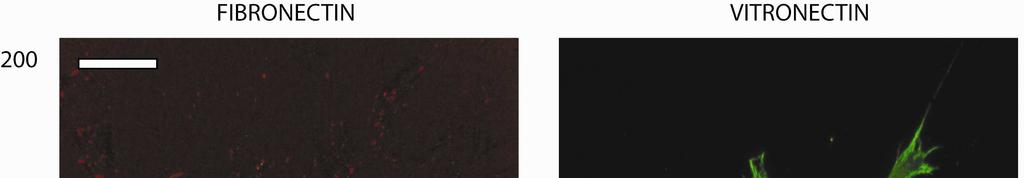
4 Figure S1: A range of sizes of FN patterns. Size of pattern indicated in picture (nm), scalebar 20µm cell number cells/mm FN VN BSA Au SiO2 evap
5 Area/cell of spread cells µm 2 /spread cell 1000 FN VN Au Figure S2: Quantitative data from full repeat showing cell number and area of spread cells for each sample type. The cell area was averaged only from cells interacting substantially with the substrate with round cells below a threshold size excluded. Standard error means are displayed. The data is derived from 4 images per sample using a 10x objective, and 4 samples per condition. Significant differences (t<0.05) between each sample and the flat control (Au) are denoted with a star, and between FN and VN on the same sample type with a cross.

6

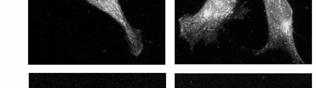
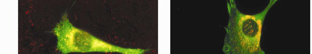
7 Figure S3: Zoom in to regions of interest in pictures displayed in figure 3 (main paper). Red stains actin and green vinculin. Scalbar 10µm. Figure continued from previous page.


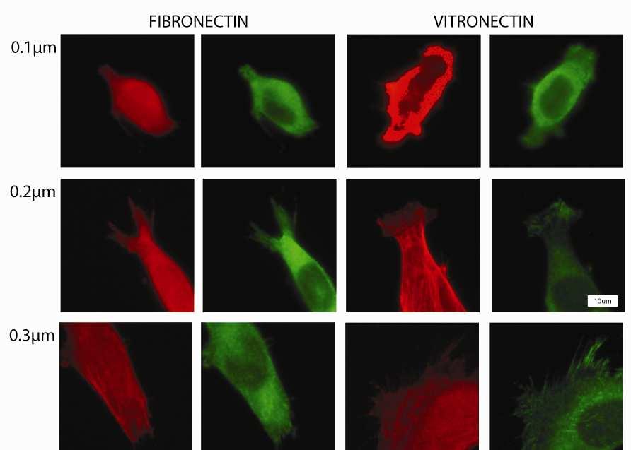
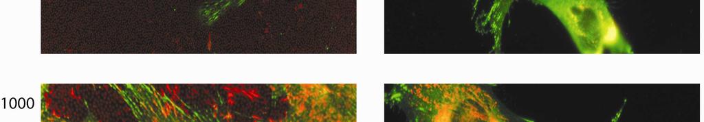
8 Figure S4: Fluorescent microscopy images using 40x objective with a 1.6x optical zoom from the repeat experiment. High magnification images of 0.1µm patterns not included here. Blue staining the nucleus, red the actin cytoskeleton and green vinculin. The patch size is displayed for each pair of images (FN and VN). Scale bar 30µm


.")
9 Figure S5: Fluorescent microscopy images using 40x objective with a 1.6x optical zoom from the experiment presented in main paper. Blue staining the nucleus, red staining zyxin and green vinculin. The patch size is displayed for each pair of images (FN and VN). Scale bar 30µm

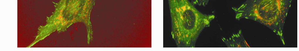
10 Figure S6: Cells on 0.5µm surfaces coated with FN or VN. Stained against zyxin. Scale bar 20µm


11


12
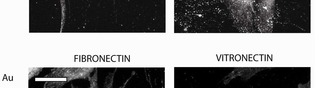
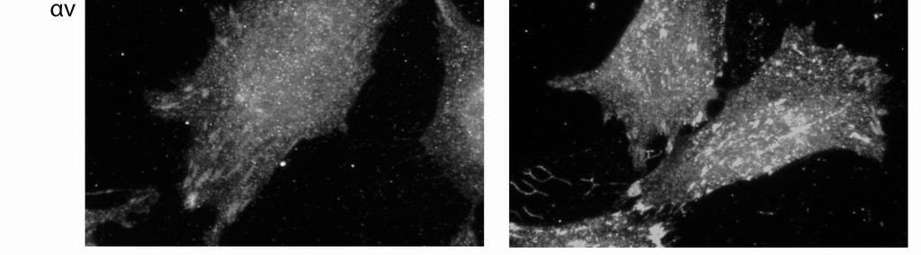
13 Figure S7: Anti-integrin α 5 or α v on FN and VN patterns and homogenous controls. The size of pattern (200, 500, 800nm or Au=homogenous control) is indicated top left of each figure panel. Scale bar 20µm.


14
 is indicated top left of each figure panel. Scale bar 20µm.")
15 Figure S8: Examples of Anti-FN (red) and anti-vinculin (green) staining of cells on FN and VN patterns and homogenous controls. The size of pattern (200, 500, 1000nm or Au=homogenous control) is indicated top left of each figure panel. Scale bar 20µm. Figure S9: Florescence microscopy image showing vinculin (green) and the underlying pattern of 1µm in diameter FN patches (red). Table S2 Bridging focal adhesion analysis: Length and aspect ratio estimates for vinculin stained bridging focal adhesions in adherent cells Sample type Length SEM Width SEM Aspect SEM (µm) (µm) ratio VN Homogeneous FN Homogeneous VN FN VN FN The cells were in general characterised by having a broad distribution of focal adhesions. For the nanopatterned surfaces the majority of the vinculin stained domains corresponded to a single focal adhesion limited by the patch size. In addition we observe bridging events where vinculin (and zxyin) stained adhesion bridge several patches. The characteristics of bridging focal adhesions were quantified by measuring the length and aspect ratio of the six longest focal adhesions per field of view (from images taken at 63 times magnification - 40 times magnification with a 1.6 optical zoom typically 2-4 cells). The results thus are biased to long focal adhesions. The data are measured from 4 images from each of 4 samples and displayed as an average and a standard error mean of all images quantifying the variation from image to image. The results show a large variation which is not so surprising given the diversity of cellular response. There is a clear result that the length of the longest focal adhesions on the FN200 is around 1.2 microns in length with a low aspect ratio and
16 substantially lower that those at 800nm and homogeneous patterns, The nanopatterned surfaces show shorter adhesions than homogeneous VN or FN samples. These adhesions also show lower aspect ratio. Cells at the VN200 sample have very similar adhesions compared to at VN800 and FN800 patterns and are substantially longer with higher aspect ratio than for cells at FN200. These results support the idea that bridging events are occurring differently at VN200 patterns compared to FN200 patterns. The width of the adhesions shows a trend of narrower adhesions for smaller pattern sizes. However the resolution of our microscope (~500nm) precludes any further analysis. References 1. Krozer, A.; Rodahl, M. Journal of Vacuum Science and Technology a-vacuum Surfaces and Films 1997, 15, (3), Fredriksson, H.; Alaverdyan, Y.; Dmitriev, A.; Langhammer, C.; Sutherland, D. S.; Zaech, M.; Kasemo, B. Advanced Materials 2007, 19, (23), Lovmand, J.; Justesen, E.; Foss, M.; Lauridsen, R. H.; Lovmand, M.; Modin, C.; Besenbacher, F.; Pedersen, F. S.; Duch, M. Biomaterials 2009, 30, (11), Sauerbrey, G. Zeitschrift Fur Physik 1959, 155, (2), Hovgaard, M. B.; Rechendorff, K.; Chevallier, J.; Foss, M.; Besenbacher, F. Journal of Physical Chemistry B 2008, 112, (28), Reimhult, E.; Larsson, C.; Kasemo, B.; Hook, F. Analytical Chemistry 2004, 76, (24), Voros, J. Biophysical Journal 2004, 87, (1),
Protein patterning on hydrogels by direct microcontact. printing: application to cardiac differentiation SUPPLEMTARY INFORMATION
 Electronic Supplementary Material (ESI) for RSC Advances. This journal is The Royal Society of Chemistry 2014 Protein patterning on hydrogels by direct microcontact printing: application to cardiac differentiation
restricted by the nanoscale spatial distribution of ICAM1
 1 Supporting information: 2 3 4 Podosome formation and development in monocytes restricted by the nanoscale spatial distribution of ICAM1 5 6 7 Andreas S. Andersen 1, Hüsnü Aslan 1, Mingdong Dong 1, Xingyu
Beta3 integrin promotes long-lasting activation and polarization of Vascular Endothelial Growth Factor Receptor 2 by immobilized ligand
 SUPPLEMENTAL FIGURES Beta3 integrin promotes long-lasting activation and polarization of Vascular Endothelial Growth Factor Receptor 2 by immobilized ligand C. Ravelli et al. FIGURE S. I Figure S. I: Gremlin
CD93 and dystroglycan cooperation in human endothelial cell adhesion and migration
 /, Supplementary Advance Publications Materials 2016 CD93 and dystroglycan cooperation in human endothelial cell adhesion and migration Supplementary Materials Supplementary Figure S1: In ECs CD93 silencing
Interaction of Cells with Patterned Reactors Chuntao Zhu, a,b Essi M. Taipaleenmäki, b Yan Zhang, b Xiaojun Han, *,a and Brigitte Städler *,b
 Electronic Supplementary Material (ESI) for Biomaterials Science. This journal is The Royal Society of Chemistry 2017 Supporting Information Interaction of Cells with Patterned Reactors Chuntao Zhu, a,b
Immunofluorescence Confocal Microscopy of 3D Cultures Grown on Alvetex
 Immunofluorescence Confocal Microscopy of 3D Cultures Grown on Alvetex 1.0. Introduction Immunofluorescence uses the recognition of cellular targets by fluorescent dyes or antigen-specific antibodies coupled
Supplementary Materials and Methods
 Supplementary Materials and Methods Reagents Supplementary Material (ESI) for Lab on a Chip RPMI medium, FBS, HEPES buffer solution, sodium pyruvate, penicillin, and streptomycin were obtained from Biological
Immunofluorescence Staining Protocol for 3 Well Chamber, removable
 Immunofluorescence Staining Protocol for 3 Well Chamber, removable This Application Note presents a simple protocol for the cultivation, fixation, and staining of cells using the 3 Well Chamber, removable.
SUPPLEMENTARY INFORMATION
 Biosynthesis of Luminescent Quantum Dots in an Earthworm S.R. Stürzenbaum, a# M. Hoeckner, a# A. Panneerselvam, b J. Levitt, b J.-S. Bouillard, b S. Taniguchi, b L.-A. Dailey, d R. Ahmad Khanbeigi, d E.
Segments of the obstructed intestinal loops were fixed in 4% paraformaldehyde
 Supplementary text Supplementary materials and methods Histopathological examination Segments of the obstructed intestinal loops were fixed in 4% paraformaldehyde (PFA) and embedded in paraffin wax with
Supplemental Information. Materials and methods.
 Supplemental Information Materials and methods. Cell culture. hmscs were isolated from bone marrow of 3 male donors, undergoing orthopedic surgery (mean age 69.7). Cells were cultured in high glucose DMEM
SUPPORTING INFORMATION
 Electronic Supplementary Material (ESI) for Nanoscale. This journal is The Royal Society of Chemistry 2015 SUPPORTING INFORMATION Chemical Sporulation and Germination: Cytoprotective Nanocoating of Individual
Immunofluorescence and phalloidin labeling of mammalian cells
 Immunofluorescence and phalloidin labeling of mammalian cells 2 Contents Materials for immunofluorescence and phalloidin labeling of mammalian cells...1 Immunofluorescence-labelling on cultivated adherent
RNA was isolated using NucleoSpin RNA II (Macherey-Nagel, Bethlehem, PA) according to the
 according to the") Supplementary Methods RT-PCR and real-time PCR analysis RNA was isolated using NucleoSpin RNA II (Macherey-Nagel, Bethlehem, PA) according to the manufacturer s protocol and quantified by measuring the
Supplementary Methods RT-PCR and real-time PCR analysis RNA was isolated using NucleoSpin RNA II (Macherey-Nagel, Bethlehem, PA) according to the manufacturer s protocol and quantified by measuring the
Supplementary Protocol. sirna transfection methodology and performance
 Supplementary Protocol sirna transfection methodology and performance sirna oligonucleotides, DNA construct and cell line. Chemically synthesized 21 nt RNA duplexes were obtained from Ambion Europe, Ltd.
TRIPLE (Insulin, Glucagon and EGFP) Immunofluorescence Staining Protocol in Pancreas Woogyun Choi 1, Randal J. Kaufman 2 and Sung Hoon Back 3*
 Immunofluorescence Staining Protocol in Pancreas Woogyun Choi 1, Randal J. Kaufman 2 and Sung Hoon Back 3*") TRIPLE (Insulin, Glucagon and EGFP) Immunofluorescence Staining Protocol in Pancreas Woogyun Choi 1, Randal J. Kaufman 2 and Sung Hoon Back 3* 1 School of Biological Sciences, University of Ulsan, Ulsan,
TRIPLE (Insulin, Glucagon and EGFP) Immunofluorescence Staining Protocol in Pancreas Woogyun Choi 1, Randal J. Kaufman 2 and Sung Hoon Back 3* 1 School of Biological Sciences, University of Ulsan, Ulsan,
Electronic Supplementary Information
 Electronic Supplementary Information A facile bottom-up route to self-assembled chitin nanofibers C. Zhong, A. Cooper, A. Kapetanovic, Z. Fang, M. Zhang, M. Rolandi Chitin Nanofiber Fabrication: Materials:
Online Supplement ALVEOLAR CELL SENESCENCE IN PATIENTS WITH PULMONARY EMPHYSEMA. Takao Tsuji, Kazutetsu Aoshiba, and Atsushi Nagai
 Online Supplement ALVEOLAR CELL SENESCENCE IN PATIENTS WITH PULMONARY EMPHYSEMA Takao Tsuji, Kazutetsu Aoshiba, and Atsushi Nagai MATERIALS AND METHODS Immunohistochemistry Deparaffinized tissue sections
2-step or indirect immunofluorescence 1. Substrate on which cells are plated: plastic vs. glass; coating vs. non
 Variables in standard immunostaining protocol 2-step or indirect immunofluorescence 1. Substrate on which cells are plated: plastic vs. glass; coating vs. non 2. Plating density: sparse vs. confluent 3.
Dynamic Re-organization of Individual Adhesion Nanoclusters in Living Cells by Ligand Patterned Surfaces**
 Supplementary information: SMALL Dynamic Re-organization of Individual Adhesion Nanoclusters in Living Cells by Ligand Patterned Surfaces** Ruth Diez-Ahedo, Davide Normanno, Olga Esteban, GertJan Bakker,
Supporting Information
 Electronic Supplementary Material (ESI) for Lab on a Chip. This journal is The Royal Society of Chemistry 2014 Supporting Information Cell culture. C2C12 cells (ATCC CRL 1772) were cultured in 75 cm 2
Promotion of HDF Cell Attachment and Proliferation
 Promotion of HDF Cell Attachment and Proliferation Objectives To qualitatively assess the effect of fibronectin (Fn) on HDF cell attachment Fn Attachment Assay To observe HDF cell proliferation and position
Supplementary Information. Facile fabrication of microsphere-polymer brush hierarchically. three-dimensional (3D) substrates for immunoassays
 substrates for immunoassays") Electronic Supplementary Material (ESI) for ChemComm. This journal is The Royal Society of Chemistry 2015 Supplementary Information Facile fabrication of microsphere-polymer brush hierarchically three-dimensional
Electronic Supplementary Material (ESI) for ChemComm. This journal is The Royal Society of Chemistry 2015 Supplementary Information Facile fabrication of microsphere-polymer brush hierarchically three-dimensional
Supporting Information for. Bull s Eye Janus Particles as Artificial Antigen-Presenting Cells for T Cell Activation
 Supporting Information for Bull s Eye Janus Particles as Artificial Antigen-Presenting Cells for T Cell Activation Bo Chen, Yilong Jia, Yuan Gao, Lucero Sanchez, Stephen M. Anthony, and Yan Yu * Department
SUPPLEMENTAL INFORMATION: 1. Supplemental methods 2. Supplemental figure legends 3. Supplemental figures
 Supplementary Material (ESI) for Lab on a Chip This journal is The Royal Society of Chemistry 2008 A microfluidics-based turning assay reveals complex growth cone responses to integrated gradients of substrate-bound
Elecrtonic Supplementary Information. Application of quantum dot barcodes prepared using biological self-assembly to multiplexed immunoassays
 Elecrtonic Supplementary Information Application of quantum dot barcodes prepared using biological self-assembly to multiplexed immunoassays Sakandar Rauf, Andrew Glidle and Jonathan M Cooper Department
SUPPLEMENTARY NOTE 3. Supplememtary Note 3, Wehr et al., Monitoring Regulated Protein-Protein Interactions Using Split-TEV 1
 SUPPLEMENTARY NOTE 3 Fluorescent Proteolysis-only TEV-Reporters We generated TEV reporter that allow visualizing TEV activity at the membrane and in the cytosol of living cells not relying on the addition
Neural Stem Cell Characterization Kit
 Neural Stem Cell Characterization Kit Catalog No. SCR019 FOR RESEARCH USE ONLY Not for use in diagnostic procedures USA & Canada Phone: +1(800) 437-7500 Fax: +1 (951) 676-9209 Europe +44 (0) 23 8026 2233
Supporting Information
 Supporting Information Self-Powered Electrical Stimulation for Enhancing Neural Differentiation of Mesenchymal Stem Cells on Graphene-Poly(3,4-ethylenedioxythiophene) Hybrid Microfibers Weibo Guo, 1,2
For labelling sub-cellular organelles in tissue sections, cell cultures and cell free experiments using our proprietary Red fluorescence probe
 ab112127 CytoPainter F-actin Staining Kit - Red Fluorescence Instructions for Use For labelling sub-cellular organelles in tissue sections, cell cultures and cell free experiments using our proprietary
Visualizing mechanical tension across membrane receptors with a fluorescent sensor
 Nature Methods Visualizing mechanical tension across membrane receptors with a fluorescent sensor Daniel R. Stabley, Carol Jurchenko, Stephen S. Marshall, Khalid S. Salaita Supplementary Figure 1 Fabrication
Whole Mount IHC Protocol
 Whole Mount IHC Protocol Authors: Ruth Sullivan, Ryan Trevena and Kyle Wegner Creation Date: 03/17/2016 All steps should be conducted with gentle agitation on an orbital shaker, unless otherwise instructed.
ApoTrack Cytochrome c Apoptosis ICC Antibody Kit: 2 color immunocytochemistry of cytochrome c and mitochondria.
 PROTOCOL ApoTrack Cytochrome c Apoptosis ICC Antibody Kit 1850 Millrace Drive, Suite 3A Eugene, Oregon 97403 MSA07 Rev.1 DESCRIPTION ApoTrack Cytochrome c Apoptosis ICC Antibody Kit: 2 color immunocytochemistry
SUPPLEMENTARY INFORMATION FIGURE 1 - 1
 SUPPLEMENTARY INFORMATION FIGURE 1-1 SUPPLEMENTARY INFORMATION FIGURE 2-2 SUPPLEMENTARY INFORMATION METHODS GST-Pull-Down. Cultures of E. Coli (BL21) were transformed with pgex (Clontech) and pgex recombinant
This Document Contains:
 This Document Contains: 1. In-Cell Western Protocol II. Cell Seeding and Stimulation Supplemental Protocol III. Complete Assay Example: Detailing the Seeding, Stimulation and Detection of the A431 Cellular
Methods Western blot analysis of plg Quantification of plasminogen accumulation by ELISA Immunohistochemical analysis
 Methods Western blot analysis of plg Wild-type mice first received a standardized burn wound and then were intravenously administered 2 mg of human plg (Omnio AB, Umeå, Sweden). 24 hours after wounding
Protocols for Neural Progenitor Cell Expansion and Dopaminergic Neuron Differentiation
 Protocols for Neural Progenitor Cell Expansion and Dopaminergic Neuron Differentiation In vitro neurological research presents many challenges due to the difficulty in establishing high-yield neuronal
Supplementary Material
 Supplementary Material Supplementary Methods Cell synchronization. For synchronized cell growth, thymidine was added to 30% confluent U2OS cells to a final concentration of 2.5mM. Cells were incubated
ApoTrack Cytochrome c Apoptosis ICC Antibody
 ab110417 ApoTrack Cytochrome c Apoptosis ICC Antibody Instructions for Use For the Immunocytochemistry analysis of cytochrome c and a mitochondrial marker (Complex Vα) in apoptotic cells and nonapoptotic
1. Goat Anti-Caspase-3 (CPP32) Antibody, R&D systems (cat #AF-605-NA), 0.5ug/ml
 Antibody, R&D systems (cat #AF-605-NA), 0.5ug/ml") Western Blot Antibodies: 1. Goat Anti-Caspase-3 (CPP32) Antibody, R&D systems (cat #AF-605-NA), 0.5ug/ml 2. Goat Anti-human LAP (TGF-b1) Antibody, R&D Systems (cat #AF-246-NA), 0.1-0.2 ug/ml 3. Rabbit
Western Blot Antibodies: 1. Goat Anti-Caspase-3 (CPP32) Antibody, R&D systems (cat #AF-605-NA), 0.5ug/ml 2. Goat Anti-human LAP (TGF-b1) Antibody, R&D Systems (cat #AF-246-NA), 0.1-0.2 ug/ml 3. Rabbit
Short hairpin RNA (shrna) against MMP14. Lentiviral plasmids containing shrna
 against MMP14. Lentiviral plasmids containing shrna") Supplemental Materials and Methods Short hairpin RNA (shrna) against MMP14. Lentiviral plasmids containing shrna (Mission shrna, Sigma) against mouse MMP14 were transfected into HEK293 cells using FuGene6
Supplemental Materials and Methods Short hairpin RNA (shrna) against MMP14. Lentiviral plasmids containing shrna (Mission shrna, Sigma) against mouse MMP14 were transfected into HEK293 cells using FuGene6
Immunohistochemistry guide
 Immunohistochemistry guide overview immunohistochemistry Overview Immunohistochemistry is a laboratory technique utilized for the visual detection of antigens in tissue. When working with cells this technique
ApoTrack Cytochrome c Apoptosis ICC Antibody Kit
 ab110417 ApoTrack Cytochrome c Apoptosis ICC Antibody Kit Instructions for Use For the Immunocytochemistry analysis of cytochrome c and a mitochondrial marker (Complex Vα) in apoptotic cells and non-apoptotic
THE BASICS OF IMMUNOHISTOCHEMISTRY
 THE BASICS OF IMMUNOHISTOCHEMISTRY Introduction Immunohistochemistry (IHC) identifies specific tissue components by means of a specific antigen/antibody reaction tagged with a visible label. IHC makes
Supplemental data. Supplemental Materials and Methods
 Supplemental data Supplemental Materials and Methods Transfection of plasmid. Transfection of plasmids into FRTL5 cells was performed using Lipofectamine LTX with Plus reagent (Invitrogen) according to
Alpha or beta human chorionic gonadotropin knockdown decrease BeWo cell fusion by
 Alpha or beta human chorionic gonadotropin knockdown decrease BeWo cell fusion by down-regulating PKA and CREB activation Sudha Saryu Malhotra 1, Pankaj Suman 2 and Satish Kumar Gupta 1 * 1 Reproductive
IncuCyte Live-Cell Immunocytochemistry Assay
 IncuCyte Live-Cell Immunocytochemistry Assay For cell surface marker analysis This protocol describes a solution for measuring immunocytochemistry in live-cells expressing a surface antigen of interest.
Mouse/Rat Neural Stem Cell Functional Identification Kit
 Mouse/Rat Neural Stem Cell Functional Identification Kit Catalog Number SC013 Reagents for the identification of mouse/rat Neural Stem Cells (NSCs) by in vitro functional differentiation. This package
Human IgG Antigen ELISA Kit
 Human IgG Antigen ELISA Kit Catalog No: IHUIGGKT Lot No: SAMPLE INTENDED USE This human immunoglobulin G antigen assay is intended for the quantitative determination of total human IgG antigen in serum,
Supplementary Information. for
 Electronic Supplementary Material (ESI) for ChemComm. This journal is The Royal Society of Chemistry 2014 Supplementary Information for Nanoslitting Phase-separated Block Copolymers by Solvent Swelling
Department of Mechanical Engineering, University of Washington, Seattle, WA, 98195
 Supplementary information Contact Angle Changes Induced by Immunocomplex Formation Jong-Hoon Kim, a Amy Q. Shen, a Kyong-Hoon Lee, b Gerard A. Cangelosi c and Jae-Hyun Chung* a a Department of Mechanical
HEK293A cells were cultured in high glucose (4.500 mg/l) Dulbecco s modified
 Dulbecco s modified") Additional methods: HEK293A and C7 cell cultures HEK293A cells were cultured in high glucose (4.500 mg/l) Dulbecco s modified Eagle s medium (DMEM) with GlutaMAX I (Invitrogen, Cergy Pontoise, France),
Additional methods: HEK293A and C7 cell cultures HEK293A cells were cultured in high glucose (4.500 mg/l) Dulbecco s modified Eagle s medium (DMEM) with GlutaMAX I (Invitrogen, Cergy Pontoise, France),
*Corresponding author. Tel: ;
 1 SUPPLEMENTARY DATA 2 3 4 5 6 7 8 9 10 11 Integrin 2 1 in nonactivated conformation can induce focal adhesion kinase signaling Maria Salmela 1, Johanna Jokinen 1,2, Silja Tiitta 1, Pekka Rappu 1, Holland
Supplemental material
 Supplemental material THE JOURNAL OF CELL BIOLOGY Taylor et al., http://www.jcb.org/cgi/content/full/jcb.201403021/dc1 Figure S1. Representative images of Cav 1a -YFP mutants with and without LMB treatment.
CytoGLOW. IKK-α/β. Colorimetric Cell-Based ELISA Kit. Catalog #: CB5358
 CytoGLOW IKK-α/β Colorimetric Cell-Based ELISA Kit Catalog #: CB5358 Please read the provided manual entirely prior to use as suggested experimental protocols may have changed. Research Purposes Only.
Supporting Information. A Fluorogenic Resveratrol-Confined Graphene Oxide For Economic and Rapid. Detection Of Alzheimer's Disease
 Supporting Information A Fluorogenic Resveratrol-Confined Graphene Oxide For Economic and Rapid Detection Of Alzheimer's Disease Xiao-Peng He, 1 Qiong Deng, 1 Liang Cai, 1 Chang-Zheng Wang, 1,2 Yi Zang,*,2
Supporting Information
 Electronic Supplementary Material (ESI) for RSC Advances. This journal is The Royal Society of Chemistry 2015 Supporting Information Materials BSA (bovine serum albumin, 99%), fluorescein isothiocyanate
Praktikum III: Experiment B
 Praktikum III: Experiment B Cell Patterning Using Micro Contact Printing Fall Term 2009 Amanda Hüsler, Katja Fröhlich, Philippe Knüsel, Schwarzenberger Michael Participants: Amanda Hüsler, Katja Fröhlich,
PRODUCT DATA SHEET. Carboxylated Fluorescent Gold Nanoparticles. Description. Characteristics
 PRODUCT DATA SHEET Carboxylated Fluorescent Gold Nanoparticles Description Cytodiagnostics carboxylated fluorescent gold nanoparticles is a unique product that combines our Cyto fluorescent dyes and gold
BoLISA BoNT Sandwich ELISA Protocol
 BoLISA BoNT Sandwich ELISA Protocol 55 S. Rosa Road, Suite 5 Madison, WI 5379-68-44-874 info@biosentinelpharma.com BioSentinel Part No: L7, Release Date: May, 7 BoLISA A BoNT/A Sandwich ELISA Detection
0.5% Triton X-100 for 5 min at room temperature. Fixed and permeabilized cells were
 1 Supplementary Methods Immunohistochemistry EBC-1 cells were fixed in 4% paraformaldehyde for 15 min at room temperature, followed by 0.5% Triton X-100 for 5 min at room temperature. Fixed and permeabilized
Frequently Asked Questions (FAQ)
") Frequently Asked Questions (FAQ) Matrigen Softwell Hydrogels Version 1.0 Contents General Questions... 3 Cell Culture and Experimental Questions... 6 Quality Control Technical Questions... 8 SoftTrac Products...
Frequently Asked Questions (FAQ) Matrigen Softwell Hydrogels Version 1.0 Contents General Questions... 3 Cell Culture and Experimental Questions... 6 Quality Control Technical Questions... 8 SoftTrac Products...
T H E J O U R N A L O F C E L L B I O L O G Y
 T H E J O U R N A L O F C E L L B I O L O G Y Supplemental material Monteiro et al., http://www.jcb.org/cgi/content/full/jcb.201306162/dc1 Figure S1. 3D deconvolution microscopy analysis of WASH and exocyst
Chicken IgY (IgG) ELISA Catalog Number:
 ELISA Catalog Number:") Chicken IgY (IgG) ELISA : 0801010 INTENDED USE Chicken IgY is the designation for the IgG-like molecule found in avian species. The IMMUNOtek Chicken IgY (IgG) ELISA is a rapid, easy to use enzyme linked
Chicken IgY (IgG) ELISA : 0801010 INTENDED USE Chicken IgY is the designation for the IgG-like molecule found in avian species. The IMMUNOtek Chicken IgY (IgG) ELISA is a rapid, easy to use enzyme linked
Zenon Goat IgG Labeling Kits
 Product Information Revised: 19 June 2007 Quick Facts Storage upon receipt: 2 6 C Protect from light Abs/Em: See Table 1 Unlabeled IgG antibody Zenon labeling reagent (labeled Fab fragment) Introduction
Impairment of Alveolar Macrophage Transcription in. Idiopathic Pulmonary Fibrosis. Online Data Supplement
 Impairment of Alveolar Macrophage Transcription in Idiopathic Pulmonary Fibrosis Online Data Supplement Ping Ren, Ivan O. Rosas, Sandra D. MacDonald, Hai-Ping Wu, Eric M. Billings, and Bernadette R. Gochuico
Topographical modulation of macrophage phenotype by shrink-film multi-scale. Department of Biomedical Engineering, University of California, Irvine
 Electronic Supplementary Material (ESI) for Biomaterials Science. This journal is The Royal Society of Chemistry 2016 Supplementary Information for Topographical modulation of macrophage phenotype by shrink-film
Cyno Monkey IgG Antigen ELISA Kit
 Cyno Monkey IgG Antigen ELISA Kit Catalog No: ICYIGGKT Lot No: SAMPLE INTENDED USE This cynomolgus macaque (Macaca fascicularis) monkey Immunoglobulin G (IgG) antigen assay is intended for the quantitative
Nonspecific binding of 10 nm Cy5-labeled DinB on nine different surfaces, measured by the number of DinB spots over an imaging area of 2,500 µm 2.
 Supplementary Figure 1 Nonspecific binding of 10 nm Cy5-labeled DinB on nine different surfaces, measured by the number of DinB spots over an imaging area of 2,500 µm 2. Free DinB was washed out using
TUNEL Universal Apoptosis Detection Kit (FITC-labeled)
") TUNEL Universal Apoptosis Detection Kit (FITC-labeled) Cat. No. L00427 Technical Manual No. TM0623 Version 03102011 I Description. 1 II Key Features.... 1 III Kit Contents.. 1 IV Storage.. 2 V Protocol..
TUNEL Universal Apoptosis Detection Kit (FITC-labeled) Cat. No. L00427 Technical Manual No. TM0623 Version 03102011 I Description. 1 II Key Features.... 1 III Kit Contents.. 1 IV Storage.. 2 V Protocol..
Supplementary Material (ESI) for Lab on a Chip This journal is The Royal Society of Chemistry 2007
 for Lab on a Chip This journal is The Royal Society of Chemistry 2007") Supplementary Material (ESI) for Lab on a Chip This journal is The Royal Society of Chemistry 2007 Fibronectin isolation and fluorescent labeling Human plasma Fn was isolated from fresh human plasma (Swiss
Supplementary Material (ESI) for Lab on a Chip This journal is The Royal Society of Chemistry 2007 Fibronectin isolation and fluorescent labeling Human plasma Fn was isolated from fresh human plasma (Swiss
SEM Immunocytochemistry for Cells & Materials
 SEM Immunocytochemistry for Cells & Materials R. Geoff Richards AO Research Institute, AO Foundation, Davos, Switzerland. Immunohistochemistry Immunocytochemistry can be performed on a biological specimen,
B. ADM: C. D. Apoptosis: 1.68% 2.99% 1.31% Figure.S1,Li et al. number. invaded cells. HuH7 BxPC-3 DLD-1.
 A. - Figure.S1,Li et al. B. : - + - + - + E-cadherin CK19 α-sma vimentin β -actin C. D. Apoptosis: 1.68% 2.99% 1.31% - : - + - + - + Apoptosis: 48.33% 45.32% 44.59% E. invaded cells number 400 300 200
Rat IgG ELISA Catalog #:
 INTENDED USE The IMMUNO-TEK Rat IgG ELISA* Kit is a rapid, easy to use enzyme linked immunosorbent assay (ELISA) designed for the measurement of rat IgG in rat serum, plasma, saliva, mucosa, cell culture
SensoLyte Anti-alpha-Synuclein Quantitative ELISA Kit (Human/Mouse/Rat) *Colorimetric*
 *Colorimetric*") Catalog # Kit Size SensoLyte Anti-alpha-Synuclein Quantitative ELISA Kit (Human/Mouse/Rat) *Colorimetric* AS-55550 One 96-well strip plate This kit is optimized to detect human/mouse/rat alpha-synuclein
Catalog # Kit Size SensoLyte Anti-alpha-Synuclein Quantitative ELISA Kit (Human/Mouse/Rat) *Colorimetric* AS-55550 One 96-well strip plate This kit is optimized to detect human/mouse/rat alpha-synuclein
IR-Blot Secondary antibodies Rev00
 0 About us Cyanagen is a biotech company located in Bologna, dedicated to research, development and production of reagents for molecular diagnostic since 2003 and one of the leading companies in the field
Immunohistochemistry with APAAPstaining Authors
 v Immunohistochemistry with APAAPstaining Authors S. Raffegerst Date 30-10-2007 Background Version 1.0 The immunohistochemistry method allows the identification of cells with expression of specific surface
Technical Manual No Version
 TUNEL Apoptosis Detection Kit Cat. No. L00301 (For Cryopreserved Tissue Sections, FITC-labled POD) Technical Manual No. 0269 Version 01132011 I Description. 1 II Key Features.... 1 III Kit Contents.. 1
Combined Digoxigenin-labeled in situ hybridization/ Immunohistochemistry protocol (for fixed frozen cryostat sections)
") Combined Digoxigenin-labeled in situ hybridization/ Immunohistochemistry protocol (for fixed frozen cryostat sections) A. Digoxigenin-UTP labeling of crna antisense probe Refer to laboratory protocol and
Combined Digoxigenin-labeled in situ hybridization/ Immunohistochemistry protocol (for fixed frozen cryostat sections) A. Digoxigenin-UTP labeling of crna antisense probe Refer to laboratory protocol and
Supporting Information
 Supporting Information Stavru et al. 0.073/pnas.357840 SI Materials and Methods Immunofluorescence. For immunofluorescence, cells were fixed for 0 min in 4% (wt/vol) paraformaldehyde (Electron Microscopy
A guide to selecting control, diluent and blocking reagents
 Specializing in Secondary Antibodies and Conjugates A guide to selecting control, diluent and blocking reagents Optimize your experimental protocols with Jackson ImmunoResearch Secondary antibodies and
A guide to selecting control, diluent and blocking reagents
 Specializing in Secondary Antibodies and Conjugates A guide to selecting control, diluent and blocking reagents Optimize your experimental protocols with Jackson ImmunoResearch Secondary antibodies and
SelectFX Alexa Fluor 488 Peroxisome Labeling Kit
 SelectFX Alexa Fluor 488 Peroxisome Labeling Kit Catalog no. S34201 Table 1. Contents and storage information. Material Amount Concentration Storage Stability SelectFX Alexa Fluor 488 Peroxisome Labeling
PRODUCT DATA SHEET. Carboxylated Gold Nanoparticles. Description. Features. Storage. Applications. Handling. Characteristics
 PRODUCT DATA SHEET Carboxylated Gold Nanoparticles Description Cytodiagnostics carboxylated gold nanoparticles are available with two different lengths of PEG surface spacers, i.e. 3000Da and 5000Da offering
Electric Supplement Information
 Electric Supplement Information Preparation of DNA-AuNPs Thiol-modified oligonucleotide (5 -Cy5- ATCTCGGCTCTGCTAGCGAAAAAAAAAA-(C3H6)-SH-3, 5 15.5 OD) was added to 13 nm citrate-stabilized AuNPs (~3 nmol
Supporting Information
 Electronic Supplementary Material (ESI) for Nanoscale. This journal is The Royal Society of Chemistry 215 Supporting Information Quantitative Description of Thermodynamic and Kinetic Properties of the
Anti-MOUSE IgG (H&L) (GOAT) Antibody DyLight 488 Conjugated (Min X Bv Ch Gt GP Ham Hs Hu Rb Rt & Sh Serum Proteins)
 (GOAT) Antibody DyLight 488 Conjugated (Min X Bv Ch Gt GP Ham Hs Hu Rb Rt & Sh Serum Proteins)") Anti-MOUSE IgG (H&L) (GOAT) Antibody DyLight 488 Conjugated (Min X Bv Ch Gt GP Ham Hs Hu Rb Rt & Sh Serum Proteins) - 610-141-121 Code: 610-141-121 Size: 100 µg Product Description: Anti-MOUSE IgG (H&L)
Anti-MOUSE IgG (H&L) (GOAT) Antibody DyLight 488 Conjugated (Min X Bv Ch Gt GP Ham Hs Hu Rb Rt & Sh Serum Proteins) - 610-141-121 Code: 610-141-121 Size: 100 µg Product Description: Anti-MOUSE IgG (H&L)
Artificial niche microarrays for probing single-stem-cell fate in high throughput
 Nature Methods Artificial niche microarrays for probing single-stem-cell fate in high throughput Samy Gobaa, Sylke Hoehnel, Marta Roccio, Andrea Negro, Stefan Kobel & Matthias P Lutolf Supplementary Figure
Cell viability. Cell viability was examined by MTT assay (Sigma-Aldrich).
.") Supplementary Materials Supplementary materials and methods Cell culture. Primary human dermal fibroblasts (DFs) were isolated from full-thickness skin samples. Tissue samples were dissected into small
Supplementary Materials Supplementary materials and methods Cell culture. Primary human dermal fibroblasts (DFs) were isolated from full-thickness skin samples. Tissue samples were dissected into small
. Viability of colonies was then assessed using the WST-1 reagent as described above, and normalized relative to untreated controls.
 Cell viability analysis in the absence of disaggregation To assess cell viability in the absence of disaggregation, quintuplicate samples of cells at 5 x 1 5 /ml were treated with mab (1 µg/ml) for 24
Contaminant bovine IgG assay
 ILA Application Note Contaminant bovine IgG assay INTRODUCTION Bovine IgG, a 16, Dalton protein, is one of the constituents of bovine serum. Fetal calf serum containing bovine IgG is commonly used in mammalian
Supporting Information
 Supporting Information Chan et al. 10.1073/pnas.0903849106 SI Text Protein Purification. PCSK9 proteins were expressed either transiently in 2936E cells (1), or stably in HepG2 cells. Conditioned culture
HUMAN IPSC CULTURE PROTOCOLS
 HUMAN IPSC CULTURE PROTOCOLS FOR TRAINING COURSE IXCELLS BIOTECHNOLOGIES USA, LLC 10340 CAMINO SANTA FE, SUITE C, SAN DIEGO, CA 92121 TABLE OF CONTENTS CHAPTER 1 BEFORE STARTING 2-7 SECTION 1.1 COATING
High-throughput Immunoassay through In-channel Microfluidic Patterning
 Supporting Information High-throughput Immunoassay through In-channel Microfluidic Patterning Chunhong Zheng, a,b Jingwen Wang, a Yuhong Pang, a,b Jianbin Wang, a Wenbin Li, c Zigang Ge,*,a and Yanyi Huang*,a,b
Atomic Force Microscope (AFM) analysis of GO and RGD-pyrene/GO complex
 analysis of GO and RGD-pyrene/GO complex") Supporting information Materials and apparatus 1-Pyrenebutyric acid (PBA), N,N-Diisopropylethylamine (DIEA), Dipyrrolidino(Nsuccinimidyloxy)carbenium hexafluorophosphate (HSPyU), N,N-dimethylformamide
Supporting information Materials and apparatus 1-Pyrenebutyric acid (PBA), N,N-Diisopropylethylamine (DIEA), Dipyrrolidino(Nsuccinimidyloxy)carbenium hexafluorophosphate (HSPyU), N,N-dimethylformamide
INOS. Colorimetric Cell-Based ELISA Kit. Catalog #: OKAG00807
 INOS Colorimetric Cell-Based ELISA Kit Catalog #: OKAG00807 Please read the provided manual entirely prior to use as suggested experimental protocols may have changed. Research Purposes Only. Not Intended
Investigation of the protective properties of glycosylphosphatidylinositol based vaccine candidates in a Toxoplasma gondii mouse challenge model
 Investigation of the protective properties of glycosylphosphatidylinositol based vaccine candidates in a Toxoplasma gondii mouse challenge model Sebastian Götze 1,2,, Anika Reinhardt 1,2,, Andreas Geissner
EGFR (Phospho-Ser695)
") Assay Biotechnology Company www.assaybiotech.com Tel: 1-877-883-7988 Fax: 1-877-610-9758 EGFR (Phospho-Ser695) Colorimetric Cell-Based ELISA Kit Catalog #: OKAG02090 Please read the provided manual entirely
Assay Biotechnology Company www.assaybiotech.com Tel: 1-877-883-7988 Fax: 1-877-610-9758 EGFR (Phospho-Ser695) Colorimetric Cell-Based ELISA Kit Catalog #: OKAG02090 Please read the provided manual entirely
FIGURE S1. Representative images to illustrate RAD51 foci induction in FANCD2 wild-type (wt) and
 and") Supplementary Figures FIGURE S1. Representative images to illustrate RAD51 foci induction in FANCD2 wild-type (wt) and mutant (mut) cells. Higher magnification is shown in Figure 1A. Arrows indicate cisplatin-induced
Supplementary Figures FIGURE S1. Representative images to illustrate RAD51 foci induction in FANCD2 wild-type (wt) and mutant (mut) cells. Higher magnification is shown in Figure 1A. Arrows indicate cisplatin-induced
IR-Blot Secondary antibodies Rev01
 0 About us Cyanagen is a biotech company located in Bologna, dedicated to research, development and production of reagents for molecular diagnostic since 2003 and one of the leading companies in the field