Blotting and Immunodetection. Multiplex Fluorescent Blot Detection: A Troubleshooting Guide
|
|
|
- Josephine Hardy
- 6 years ago
- Views:
Transcription

1 Blotting and Immunodetection? Multiplex Fluorescent Blot Detection: A Troubleshooting Guide
2

3 Bio-Rad s Handy Introduction to Fluorescent Blotting This guide is intended as a brief introduction to fluorescent western blotting for researchers currently using chemiluminescent or colorimetric detection. Most western blotting protocols can be readily adapted for fluorescent detection but may require optimization. Fluorescent detection offers several advantages compared to other methods: Multiplexing Use of multiple fluorophores for simultaneous detection of several target proteins makes stripping and reprobing unnecessary. Dynamic range A tenfold increase in dynamic range over chemiluminescent detection offers better linearity within detection limits. Quantitative Fluorescent detection is more quantitative than enzyme-based detection methods. Stability Most fluorescent molecules provide excellent stability, allowing blots to be archived and re-imaged at a later date.

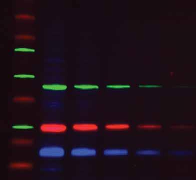
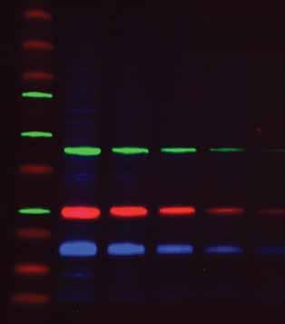
4 Tips for Fluorescent Blotting Antibody concentrations should be optimized by incubating the membrane in several dilutions of each antibody. Select the dilution that yields the highest signal-to-background ratio When adapting a chemiluminescent protocol for fluorescent detection, primary antibody concentrations may need to be increased; two- to fivefold increases are common. Secondary antibody concentrations may also have to be optimized; a good starting point is a 1:5,000 dilution. Check the manufacturer s recommendations when using specific antibodies In order to maximize the signal-tobackground ratio, use a membrane with low autofluorescence, such as the Immun-Blot low fluorescence (LF) PVDF membrane Many blocking buffers can be successfully used for fluorescent detection. We recommend 0.5 5% casein, up to 5% nonfat dry milk, or up to 3% BSA dissolved in TTBS Particulates in buffers can settle on membranes and create fluorescent artifacts. Use only high-quality reagents and filter sterilize all buffers Use blunt forceps to handle the membrane from the edges. Avoid scratching or creasing the membrane, which can produce artifacts during fluorescent detection Use a pencil to mark membranes because many inks fluoresce Bromophenol blue will fluoresce. Ensure that the dye front has migrated away from the sample, cut off the portion of gel containing the dye front, or omit bromophenol blue from the sample buffer It is not necessary to perform immunodetection in the dark; normal room lighting will not significantly photobleach fluorescently labeled antibodies. However, stocks of fluorescently labeled antibodies should be stored in the dark Use powder-free nitrile gloves when handling the membrane to minimize artifacts and fingerprints on the blot Signal-to-background ratio :250 1:500 1:1,000 1:2,000 Primary antibody dilution :250 1:500 1:1,000 1:2,000 Primary antibody dilution Optimizing antibody concentration is critical for best results. A gel was loaded with a protein sample containing transferrin (~80 kd) and blotted onto an Immun-Blot LF PVDF membrane. This blot was cut into strips and each strip was probed with a different concentration of primary antibody. The intensity of the transferrin band was compared to the background intensity of the membrane. For this antibody, a 1:500 dilution provides the optimal signal-to-background ratio. A high concentration of antibody increases background, yielding a lower overall signal-to-background ratio while low concentrations of antibody yield lower overall signal intensity. Higher antibody concentrations also increase nonspecific binding.
 give higher auto fluorescence.")
 and 650 (red) channels.")
 often give cross-reactivity, even when the antibodies are cross-adsorbed Use secondary antibodies that are highly")
5 Signal-to-background ratio Standard PVDF Membrane Immun-Blot LF PVDF Immun-Blot LF PVDF membrane Standard PVDF membranes (bottom right) give higher auto fluorescence. The Immun-Blot LF PVDF membrane (top right) has lower autofluorescence and higher signal-to-background ratios in each channel, allowing detection of lower quantities of target protein. Both blots were handled and imaged using the same parameters on the ChemiDoc MP system using the DyLight 549 (green) and 650 (red) channels. Standard PVDF membrane Tips for Multiplexing Use primary antibodies from different host species (for example, mouse and rabbit). Antibodies produced from two closely related species (such as rat and mouse) often give cross-reactivity, even when the antibodies are cross-adsorbed Use secondary antibodies that are highly cross-adsorbed against other species to avoid cross-reactivity Avoid cross-channel fluorescence by using fluorophore conjugates with optically distinct spectra Always optimize the detection of each target individually before simultaneously detecting multiple targets. Since some primary antibodies may be nonspecific and yield multiple bands on a blot, single target detection will help determine the banding pattern of each antibody prior to a multiplex experiment Most membranes show higher background with shorter wavelength excitation light. Detect your strongest target in the blue channel, your middle target in green, and reserve the red channel for your weakest target
 and soybean trypsin inhibitor (red signal in left panel) were run on a gel, blotted, and")
 is weakly detecting the mouse primary antibody (green arrow at 21 kd).")
6 Multiplex image Red channel Green channel Non cross adsorbed antibodies may bind nonspecifically and result in channel cross-talk. In this example, a protein sample containing glutathione S-transferase (green signal in left panel) and soybean trypsin inhibitor (red signal in left panel) were run on a gel, blotted, and immunodetected with a rabbit antibody against glutathione S-transferase (25 kd) and a mouse antibody against soybean trypsin inhibitor (21 kd). The non cross adsorbed anti-rabbit secondary antibody (green) is weakly detecting the mouse primary antibody (green arrow at 21 kd). Multiplexing with Stain-Free Gels Stain-free gels are formulated with a trihalo compound that becomes covalently bound to tryptophan residues in proteins when the gel is activated with UV light. The labeled tryptophan adducts then fluoresce when excited by UV light. This allows for the direct visualization of total protein samples in gels and on blots. The stain-free fluorescent adduct is excitable with short-wavelength excitation light and will be visible when imaging blue fluorophores. For multiplex blot applications, use the red and green channels for detecting proteins of interest and reserve the blue channel for total protein detection Since Qdot nanocrystals and stain-free imaging both utilize UV excitation, Qdots cannot be used with proteins activated in stain-free gels. If stain-free total protein imaging is desired, use fluorophores that are not excitable with UV light
7 Fluorophore Selection Recommended Fluorophores* Red Green Blue Number of Targets Fluor A Fluor B Fluor C 1 Qdot 705 or DyLight Qdot 705 or Qdot 605 or DyLight 650 DyLight 549 3** Qdot 705 or Qdot 605 or Qdot 525 or DyLight 650 DyLight 549 DyLight stain-free DyLight stain-free DyLight 650 DyLight 550 * These fluorophores have been validated for use with Bio-Rad s ChemiDoc MP system; other fluorophores with similar excitation and emission spectra may also be used. ** Note: Qdot secondary antibodies may not be available for all three of your host species. In this case, DyLight 650 can be replaced with Qdot 705. Compatible Fluorophores* Fluorophore Excitation Filter/Bandpass, nm Emission Filter/Bandpass, nm IR Red Green Blue UV Stain-free UV-B transilluminator 590/110 Cy2 Alexa 488 DyLight /30 530/28 (532/28) Fluorescein Qdot 525 Qdot /30 Qdot 625 Cy3 605/50 (607/50) Alexa /28 DyLight 549 or 550 Rhodamine Cy5 Alexa 647 DyLight 649 or /55 (697/55) Cy /30 Alexa 680 DyLight 680 IRDye 680 Qdot /30 * Note: This is a partial list. Other fluorophores with similar excitation and emission profiles will also be compatible.
8 Immunodetection Protocol Example Materials: Immun-Blot LF PVDF membrane with transferred proteins Methanol or ethanol (necessary for wetting/rewetting dry PVDF membranes) Blocking buffer (Tris-buffered saline with 0.05% Tween 20 and an additional 1% casein) Wash buffer (Tris-buffered saline with 0.05% Tween 20) Primary antibody, diluted in blocking buffer For multiplexing applications, add all primary antibodies to the same blocking solution Fluorescently labeled secondary antibody, diluted in wash buffer For multiplexing applications, add all secondary antibodies to the same solution Procedure: Wash buffer volumes should be at least 20 ml for mini blots and 100 ml for Criterion blots. Block and antibody solution volumes should be enough to cover the membrane. Generally, this is at least 10 ml for mini gel blots and 25 ml for Criterion blots. 1. After protein transfer, equilibrate membranes in wash buffer for 3 min. Dried PVDF membranes should be briefly rewet in methanol prior to equilibration in wash buffer. 2. Incubate the blot protein side up in blocking buffer for 1 hr with continuous agitation. 3. Incubate the blot in diluted primary antibody solution for 1 hr with continuous agitation. 4. Wash the blot in wash buffer 5 times for 5 min each with continuous agitation. 5. Incubate the blot in diluted secondary antibody solution for 1 hr with continuous agitation. 6. Wash the blot in wash buffer 6 times for 5 min each with continuous agitation. 7. If recommended for your imager or fluorescent conjugate, the blot may be imaged while still wet. Alternatively, dip the blot in methanol and allow to air dry for imaging or long-term storage. Storage of Fluorescently Detected Membranes After immunodetection, membranes may be stored for analysis at a later date. If reprobing is not desired, blots can be dipped for a few seconds in methanol before hanging them to dry. This displaces the water with methanol, shortening drying time and also reducing background. Blots can then be placed in a plastic sheet protector to prevent contamination. Store the blots in the dark or wrapped in foil to prevent photobleaching of fluorophores. Depending on the fluorophore, properly stored blots can be imaged months later without significant loss of signal. Buffers and Solutions Wash buffer, Tris-buffered saline with Tween 20 (TTBS) (20 mm Tris, 500 mm NaCl, 0.05% Tween 20, ph 7.4) Tris 2.4 g NaCl 29.2 g 10% Tween 20 5 ml Adjust volume to 1 L with water. Blocking buffer (20 mm Tris, 500 mm NaCl, 0.05% Tween 20, 1% casein, ph 7.4) TTBS Buffer 100 ml Casein 1 g
9 Troubleshooting Cross-channel fluorescence or extra bands Fluorophores not optically distinct. Ensure that secondary antibodies are conjugated to fluorophores with nonoverlapping emission spectra and that appropriate emission filters are used during image acquisitions Primary antibodies from same or related species. Check that each primary antibody in the multiplex set is from a different species (for example, mouse and rabbit). Avoid using two closely related species such as rat and mouse, as even cross-adsorbed antibodies from similar species may still cross-react Primary antibody may be nonspecific. Reduce concentration or use purified antibody Secondary antibody may be nonspecific. Use cross-adsorbed secondary antibodies. Run control omitting primary antibody If one signal of the multiplex set is much stronger than the others, the signal may be bleeding through to the other channels. Try reducing the concentration of the stronger primary antibody or swap the fluorophores on the secondary antibodies Low overall signal Antibody concentrations too low. When adapting a chemiluminescent protocol for fluorescent detection, antibody concentrations may have to be increased. Typically, a two- to fourfold increase is a good starting point Concentration of labeled antibody too high. Extremely high concentrations of labeled antibody may also yield low signal due to quenching via Förster resonance energy transfer (FRET) Incomplete transfer of proteins to blot. High molecular weight proteins may require a longer transfer time. Verify transfer efficiency by using stain-free gels and monitor gel and membrane post transfer Protein load on gel too low. Some proteins elute less readily from the gel and require higher loads of protein to produce satisfactory blots Uneven signal levels Air bubbles between gel and membrane. Air bubbles will produce blank spots on the membrane. Roll the gel and membrane to expel any trapped air during assembly of the transfer sandwich. Transfer artifacts can be monitored by using stain-free gels Agitation during antibody incubation may be insufficient. Ensure incubations occur with enough volume to completely cover the membrane and incubate and wash with continuous, vigorous agitation Use a membrane with low autofluorescence
10 Troubleshooting (contd.) High overall background Membranes tend to have higher inherent fluorescence with shorter wavelength excitation light. Detect the weakest target using the red channel Blocking may be insufficient. Increase the concentration of the blocking agent or duration of the blocking step, or try an alternate blocking agent Washing may be insufficient. Increase the number or duration of wash steps Antibody concentrations may be too high. The concentration of primary or secondary antibodies may have to be optimized Adding 0.01% SDS to the secondary antibody solution during incubation may help reduce nonspecific binding to the blot Uneven background levels PVDF membrane may have been improperly wetted or may have dried during handling. Ensure that the membrane is fully wetted in methanol prior to equilibration in aqueous solution and ensure that the membrane stays wet Wash steps may have occurred in insufficient volume. Ensure that incubations occur with enough volume to completely cover the membrane and wash with vigorous agitation Fluorescent artifacts seen on blot Scratches or creases on the membrane can produce artifacts. Always use blunt forceps to handle the membrane and grasp the membrane at the edges Fingerprints on membrane. Do not touch membranes with bare hands. Some gloves leave fluorescent residues on membranes. Use powder-free nitrile gloves and forceps to handle the membrane Particulate contaminants in solutions may appear as fluorescent speckles on the blot. Filter buffers and reagents to remove contamination Do not use a pen to mark the membrane as some inks fluoresce. Use a pencil instead Bromophenol blue can produce a fluorescent signal when transferred to the membrane. Ensure that the dye front has migrated away from the protein of interest or use a loading dye without bromophenol blue

11 BEGIN Electrophoresis and Blotting For additional suggestions on optimizing blotting conditions, refer to the Bio-Rad Protein Blotting Guide (Bulletin 2895). Protein Blotting Guide Order Information Catalog # Description Imaging System and Accessories ChemiDoc MP System, gel imaging system, PC or Mac, includes darkroom, UV transilluminator, epi-white illumination, camera, power supply, cables, Image Lab software ChemiDoc MP Red LED Module Kit, pkg of 2 epi-red LED modules and 1 red emission filter, for use with applications requiring red fluorophore detection ChemiDoc MP Green LED Module Kit, pkg of 2 epi-green LED modules and 1 green emission filter, for use with applications requiring green fluorophore detection ChemiDoc MP Blue LED Module Kit, pkg of 2 epi-blue LED modules and 1 blue emission filter, for use with applications requiring blue fluorophore detection Membranes Immun-Blot Low Fluorescence PVDF/Filter Paper Sets, pkg of 10, 7 x 8.5 cm, precut low fluorescence PVDF/filter paper for immunoblotting Immun-Blot Low Fluorescence PVDF/Filter Paper Sets, pkg of 20, 7 x 8.5 cm, precut low fluorescence PVDF/filter paper for immunoblotting Immun-Blot Low Fluorescence PVDF/Filter Paper Sets, pkg of 10, 8.5 x 13.5 cm, precut low fluorescence PVDF/filter paper for immunoblotting Immun-Blot Low Fluorescence PVDF/Filter Paper Sets, pkg of 20, 8.5 x 13.5 cm, precut low fluorescence PVDF/filter paper for immunoblotting Immun-Blot Low Fluorescence PVDF membrane, pkg of 1 roll, 28 cm x 3.8 m, low fluorescence PVDF membrane for immunoblotting
12 10-Well 10-Well 15-Well IPG/prep 12-Well 8+1-Well Description 30 μl 50 μl 15 μl 450 μl 20 μl 30 µl Mini-PROTEAN TGX Stain-Free Precast Gels 7.5% % % Any kd All formats are available as both ten packs (catalog numbers listed) and two packs. To order as a two pack, add an S to the end of the catalog number for the corresponding ten pack. 12+2*-Well 18-Well 26-Well Prep+2*-Well IPG+1*-Well Description 45 µl 30 µl 15 µl 800 µl 11 cm IPG Strip Criterion TGX Stain-Free Precast Gels** 7.5% % % % % % % % Any kd * Reference well accommodates 15 µl of markers/standards. ** Criterion TGX Stain-Free gels are sold as a single gel. Reagents % Tween 20, 1 L, detergent x Phosphate Buffered Saline with 1% Casein, 1 L, blocking reagent x Phosphate Buffered Saline, 100 ml, 10x PBS x Tris Buffered Saline with 1% Casein, 1 L, blocking reagent x Tris Buffered Saline, 1 L, 10x TBS Alexa Fluor and Qdot are trademarks of Life Technologies. DyLight is a trademark of Thermo Fischer Scientific. IRDye is a trademark of LI-COR Biosciences. Bio-Rad Laboratories, Inc. Life Science Group Web site USA Australia Austria Belgium Brazil Canada China Czech Republic Denmark Finland France Germany Greece Hong Kong Hungary India Israel Italy Japan Korea Malaysia Mexico The Netherlands New Zealand Norway Poland Portugal Russia Singapore South Africa Spain Sweden Switzerland Taiwan Thailand United Kingdom Bulletin 6166 Rev A US/EG Sig 0211
Multiplex Fluorescent Western Blot Starter Kit for the Bio- Rad ChemiDoc MP
 Page 1 of 7 INSTRUCTIONS: Z-310 Multiplex Fluorescent Western Blot Starter Kit for the Bio- Rad ChemiDoc MP Rockland Immunochemicals and Bio-Rad Laboratories have jointly developed an easy to use multiplex
Clarity and Clarity Max Western ECL Substrates
 Imaging and Max Western Substrates The Clear Choice in Chemiluminescent Western Blot Detection and Max Western Substrates Get. The Clear Choice in Chemiluminescent Western Blot Detection bio-rad.com/web/substrate
Immun-Star WesternC Chemiluminescent Kit
 Immun-Star WesternC Chemiluminescent Kit Instruction Manual For Use With Nitrocellulose and PVDF Membranes Catalog # 170-5070 For technical support, call your local Bio-Rad office, or in the US, call 1-800-4BIORAD
Imaging and Electrophoresis. Gel Doc EZ Imager. Accelerate your research. Push-button imaging in just 10 seconds.
 Imaging and Electrophoresis Gel Doc EZ Imager Accelerate your research. Push-button imaging in just 10 seconds. ready imaging fault EZ imaging... Pick your tray and get your results 1LOAD TRAY Smart Tray
ChemiDoc MP Imaging System
 Imaging ChemiDoc MP Imaging System Confidence in every step of your imaging workflow. ChemiDoc MP Imaging System Because results always matter. Bring a new level of capability and efficiency to your experiments
and Criterion TGX Stain-Free Precast Gels
 ELECTROPHORESIS Mini-PROTEAN and Criterion TGX Stain-Free Precast Gels Key Benefits: Complete protein separations, gel imaging, and data analysis in as little as 20 min Run times as short as 15 min Transfer
Experimental Protocol for Multiplex Fluorescent Blotting Using the ChemiDoc MP Imaging System
 Experimental Protocol for Multiplex Fluorescent Blotting Using the ChemiDoc MP Imaging System Protocol Bulletin 6570 Stefanie L. Ritter and Donald G. Rainie, Deparment of Behavioral Neuroscience and Psychiatric
western blotting tech
 western blotting tech note 6148 Transfer of High Molecular Weight Proteins to Membranes: A Comparison of Transfer Efficiency Between Blotting Systems Nik Chmiel, Bio-Rad Laboratories, Inc., 6000 James
IR-Blot Secondary antibodies Rev00
 0 About us Cyanagen is a biotech company located in Bologna, dedicated to research, development and production of reagents for molecular diagnostic since 2003 and one of the leading companies in the field
IR-Blot Secondary antibodies Rev01
 0 About us Cyanagen is a biotech company located in Bologna, dedicated to research, development and production of reagents for molecular diagnostic since 2003 and one of the leading companies in the field
Western Blot Analysis
 Odyssey Infrared Imaging System Western Blot Analysis Published October, 2004. The most recent version of this protocol is posted at http://biosupport.licor.com/support Doc# 988-07737 Odyssey Western Blot
StarBright Blue Fluorescent Secondary Antibodies
 StarBright Blue Fluorescent Secondary Antibodies Instruction Manual Catalog # Description 12005866 Goat Anti-Mouse IgG StarBright Blue 520, 400 µl 12005869 Goat Anti-Rabbit IgG StarBright Blue 520, 400
Western Blotting Products. A Complete Source for All Your Blotting Needs
 Western Blotting Products A Complete Source for All Your Blotting Needs A History of Innovation and Leadership A pioneer of western blotting apparatus, Bio-Rad has come to be considered the industry leader
Introduction. Important Product Information. Number Description. Table of Contents
 Super Signal Western ECL Substrate Number Description Super Signal Western ECL Substrate, Sufficient for 1000cm2 of membrane Kit Contents: Super Signal Western Solution A, 50mL Super Signal Western Solution
PrimePCR Pricing and Bulk Discounts
 Assay and Control Bulk Discounts 25% off 5 or more assays Primer Assay Pricing Product Catalog # Description List Price (EUR) Primer Assays (desalted) 10025636 Primer assay desalted, 200 reactions 96 10025637
PrimePCR Pricing and Bulk Discounts
 Assay and Control Bulk Discounts 25% off 5 or more assays Primer Assay Pricing Product Catalog # Description List Price (DKK) Primer Assays (desalted) 10025636 Primer assay desalted, 200 reactions DKK
Superior Performance of Laemmli PAGE with Extended Shelf Life
 ELECTROPHORESIS Mini-PROTEAN TGX Precast Gels Get superior results with the convenient and fast Mini-PROTEAN system: Direct drop-in for Laemmli system Inexpensive buffer system, low running costs Fast
Overview. Tools for Protein Sample Preparation, 2-D Electrophoresis, and Imaging and Analysis
 Expression Proteomics // Tools for Protein Separation and Analysis www.expressionproteomics.com 1 2 3 4 Overview Tools for Protein Sample Preparation, 2-D Electrophoresis, and Imaging and Analysis overview
Azure Biosystems Western Blotting Workflow
 Azure Biosystems Western Blotting Workflow PROBE PLAN SEPARATE ANALYZE VISUALIZE PLAN Plan your experiment and choose your detection method Chemiluminescent Western Blotting The most common method for
ProteinChip IMAC30 Array (Immobilized Metal Affinity Capture) Instruction Manual. Catalog #C
 Instruction Manual. Catalog #C") ProteinChip IMAC30 Array (Immobilized Metal Affinity Capture) Instruction Manual Catalog #C57-30078 For technical support, call your local Bio-Rad office, or in the US, call 1-800-4BIORAD (1-800-424-6723).
ProteinChip IMAC30 Array (Immobilized Metal Affinity Capture) Instruction Manual Catalog #C57-30078 For technical support, call your local Bio-Rad office, or in the US, call 1-800-4BIORAD (1-800-424-6723).
hfab Rhodamine Housekeeping Antibodies
 hfab Rhodamine Housekeeping Antibodies Catalog # Description 12004163 Anti-Actin hfab Rhodamine Antibody, 200 µl 12004164 Anti-Actin hfab Rhodamine Antibody, 40 µl 12004165 Anti-Tubulin hfab Rhodamine
ADDENDUM. Ready Gel System RESOURCE GUIDE. Applications:
 Ready Gel System RESOURCE GUIDE ADDENDUM This addendum includes new electrophoresis products introduced since the Ready Gel Resource Guide was published and updated information for a few products. Keep
PrimePCR Pricing and Bulk Discounts
 Assay and Control Bulk Discounts 25% off 5 or more assays Primer Assay Pricing Product Catalog # Description List Price (CHF) Primer Assays (desalted) 10025636 Primer assay desalted, 200 reactions CHF
Western Blotting Detection Reagents
 Electrophoresis Western Blotting Detection Reagents Maximize Western Blot Detection Solutions for Any Blotting Application Choose the Best Approach for Your Needs When it comes to western blot detection,
ProteOn XPR36 Quantikinetics: Antibody Concentration and Detailed Kinetic Analysis in a Single Experimental Cycle
 ProteOn XPR36 Quantikinetics: Antibody Concentration and Detailed Kinetic Analysis in a Single Experimental Cycle Gary Ross, Mingde Zhu, Ruben Luo, Bio-Rad Laboratories, Inc., 2 Alfred Nobel Drive, Hercules,
electrophoresis tech Versatile Separation Capabilities of the PROTEAN i12 IEF System
 electrophoresis tech note 6139 Versatile Separation Capabilities of the PROTEAN i12 IEF System ChengJun Sun, Adriana Harbers, Tom Berkelman, and Katy McGirr, Life Science Group, Bio-Rad Laboratories Inc.,
Technical Data Sheet. Sensido Plus ECL Substrate (HRP) Description. Advantages
 Description. Advantages") ECL Substrate (HRP) Catalog R13-VG08 Technical Data Sheet ADD: 7F, 72, Song-Te Rd., Taipei 110, Taiwan TEL : 886-2-2723-0886 www.recenttec.com Package Cat. Components Solution A (Substrate Solution) Solution
ECL Substrate (HRP) Catalog R13-VG08 Technical Data Sheet ADD: 7F, 72, Song-Te Rd., Taipei 110, Taiwan TEL : 886-2-2723-0886 www.recenttec.com Package Cat. Components Solution A (Substrate Solution) Solution
Optiblot ECL Ultra Detect Kit (1.2pg 2ng)
") ab133409 Optiblot ECL Ultra Detect Kit (1.2pg 2ng) Instructions for Use For enhanced chemiluminescent Western blotting. This product is for research use only and is not intended for diagnostic use. 1 Table
ab133409 Optiblot ECL Ultra Detect Kit (1.2pg 2ng) Instructions for Use For enhanced chemiluminescent Western blotting. This product is for research use only and is not intended for diagnostic use. 1 Table
Ordering Information. ProteOn XPR36 System. Protein Interaction Analysis
 Protein Interaction Analysis Ordering Information ProteOn XPR36 System The ProteOn XPR36 system is an optical biosensor capable of simultaneously measuring 36 individual molecular interactions. It integrates
Chromatography Column Performance and Data Analysis Success Guide. Hints and Tips for Better Purifications
 Chromatography Column Performance and Data Analysis Success Guide Hints and Tips for Better Purifications This Chromatography Success Guide provides practical advice on preparative chromatography and protein
ProteoMiner Protein Enrichment Technology
 Sample Preparation ProteoMiner Protein Enrichment Technology Digging Deeper in the Proteome Detect More Proteins With ProteoMiner Technology Than With Immunodepletion ProteoMiner Technology vs. an Agilent
Strep-tag Technology for Molecular Weight (MW) Determinations on Blots Using Precision Plus Protein Standards
 Determinations on Blots Using Precision Plus Protein Standards") blotting tech note 2847 Strep-tag Technology for Molecular Weight (MW) Determinations on Blots Using Precision Plus Protein Standards Introduction Bio-Rad has consistently provided innovative standards
blotting tech note 2847 Strep-tag Technology for Molecular Weight (MW) Determinations on Blots Using Precision Plus Protein Standards Introduction Bio-Rad has consistently provided innovative standards
ab Optiblot Fluorescent Western Blot Kit
 ab133410 Optiblot Fluorescent Western Blot Kit Instructions for Use For quantitative, multi-color fluorescent Western blotting. This product is for research use only and is not intended for diagnostic
SYNGENE APPLICATION TRAINING GUIDE
 SYNGENE APPLICATION TRAINING GUIDE Contents Page Lighting and Filter combinations 3 Examples.. 6 Chemiluminescence versus Fluorescence Western blot detection. 8 Film versus digital imaging Chemiluminescence
Odyssey Western Blotting Kits LT
 Odyssey Western Blotting Kits LT Developed for: Odyssey Family of Imagers Please refer to your manual to confirm that this protocol is appropriate for the applications compatible with your Odyssey Imager
Electrophoresis. Ready-to-Run Buffers and Solutions
 Electrophoresis Ready-to-Run Buffers and Solutions Bio-Rad is a premier provider of buffers and premixed reagents for life science research. We offer a variety of different products for all your protein
SmartSpec Plus spectrophotometer
 SmartSpec Plus spectrophotometer Simply Brilliant. The SmartSpec Plus has a more complete range of features and functions than many other benchtop spectrophotometers. It s a complete quantitation package
Pulsed Field Gel Electrophoresis
 Pulsed Field Gel Electrophoresis Protocol Bulletin 6225 Restriction Enzyme Digestion Restriction enzyme digestion is used to prepare DN for analysis or other processing steps. Guidelines about preparing
Easy-WESTERN-II Super
 Easy-WESTERN-II Super Primary Antibody Detection Reagent for Western Blots User Manual for High Sensitivity and Strong Signal Detection Immediately after receiving the kit, read the section titled COMPONENTS
Western BLoT Ultra Sensitive HRP Substrate
 Cat. # T7104A For Research Use Western BLoT Ultra Sensitive Product Manual Table of Contents I. Description... 3 II. Components... 3 III. Storage... 3 IV. Materials Required but Not Provided... 3 V. Precautions...
Sensido Advance ECL Substrate (HRP) Solution A (Substrate Solution) Solution B (Peroxide Solution)
 Solution A (Substrate Solution) Solution B (Peroxide Solution)") Sensido Advance ECL Substrate (HRP) ADD: 7F, 72, Song-Te Rd., Taipei 110, Taiwan TEL : 886-2-2723-0886 www.recenttec.com Order Info Cat. Components Volume Package R13-VG016-100 Solution A (Substrate Solution)
Sensido Advance ECL Substrate (HRP) ADD: 7F, 72, Song-Te Rd., Taipei 110, Taiwan TEL : 886-2-2723-0886 www.recenttec.com Order Info Cat. Components Volume Package R13-VG016-100 Solution A (Substrate Solution)
Product Catalog # Description List Price (JPY) Primer Assays (desalted)
 Primer Assays (desalted)") Assays and Controls Primer Assay Pricing Primer Assays (desalted) 10025636 Primer assay desalted, 200 reactions 16,000 (Wet-lab validated human, mouse, and rat) 10025637 Primer assay desalted, 1,000 reactions
Assays and Controls Primer Assay Pricing Primer Assays (desalted) 10025636 Primer assay desalted, 200 reactions 16,000 (Wet-lab validated human, mouse, and rat) 10025637 Primer assay desalted, 1,000 reactions
Optiblot ECL Detect Kit (23 pg 187 ng)
") ab133406 Optiblot ECL Detect Kit (23 pg 187 ng) Instructions for Use For enhanced chemiluminescent Western blotting. This product is for research use only and is not intended for diagnostic use. 1 Table
ab133406 Optiblot ECL Detect Kit (23 pg 187 ng) Instructions for Use For enhanced chemiluminescent Western blotting. This product is for research use only and is not intended for diagnostic use. 1 Table
Easy-WESTERN-II Super
 Easy-WESTERN-II Super Primary Antibody Detection Reagent for Western Blots User Manual for High Sensitivity and Strong Signal Detection Immediately after receiving the kit, read the section titled COMPONENTS
Nuvia HR-S Strong Cation Exchange Media Instruction Manual
 Nuvia HR-S Strong Cation Exchange Media Instruction Manual Catalog numbers 156-0511 156-0513 156-0515 156-0517 Please read these instructions before you use Nuvia HR-S cation exchange media. If you have
AzureSpectra Fluorescent Demo Kits
 AzureSpectra Fluorescent Demo Kits Quantitative fluorescent Western blotting demo kits DC0040-006 Long Protocol for Catalog Numbers AC2172 AC2173 AC2174 AC2175 AC2176 AC2177 AC2178 AC2179 AC2218 AC2219
Bio-Rad Laboratories BIOPLEX 2200 SYSTEM. BioPlex 2200 ANA Screen with MDSS. The first and only fully-automated, multiplexed ANA Screen
 Bio-Rad Laboratories BIOPLEX 2200 SYSTEM BioPlex 2200 ANA Screen with MDSS The first and only fully-automated, multiplexed ANA Screen Bio-Rad Laboratories BIOPLEX 2200 SYSTEM Like no other The BioPlex
Bio-Plex Pro Mouse Cytokine, Chemokine, and Growth Factor Assays
 Acute Phase Response Cancer Cardiovascular Disease Cytokine, Chemokines, Growth Factors Diabetes Gene Expression Genotyping Immunoglobulin Isotyping MicroRNA Expression Signal Transduction Toxicology Bio-Plex
protein interaction analysis bulletin 6299
 protein interaction analysis bulletin 6299 Guide to SPR Data Processing on the ProteOn XPR36 Protein Interaction Array System Ruben Luo, Bio-Rad Laboratories, Inc., 2000 Alfred Nobel Drive, Hercules, CA
Bio-Plex Pro Mouse Cytokine, Chemokine, and Growth Factor Assays
 Acute Phase Response Cancer Cardiovascular Disease Cytokine, Chemokines, Growth Factors Diabetes Gene Expression Genotyping Immunoglobulin Isotyping MicroRNA Expression Signal Transduction Toxicology Bio-Plex
Solutions for Western Blotting
 dies Azure B Biosystems lot Develo C minescent Sub Solutions for Western Blotting ec ary Fluorescent An bodies Me branes Buffers Blot Develop rotein stains Chemiluminescent Sub Antibodies Secondary Fluorescent
AzureSpectra Fluorescent Demo Kits
 AzureSpectra Fluorescent Demo Kits Quantitative fluorescent Western blotting demo kits www.azurebiosystems.com info@azurebiosystems.com Copyright 2017 Azure Biosystems Inc. All rights reserved. The Azure
Biotechnology Explorer
 Biotechnology Explorer Fish DNA Barcoding Kit Planning Guide explorer.bio-rad.com Catalog #166-5100EDU, Fish DNA Barcoding Kit Catalog #166-5115EDU, DNA Barcoding Sequencing Module Note: This document
Innovations To Meet Your Needs
 Innovations To Meet Your Needs Cooled CCD Camera 1340 x 1037 pixel resolution for greatest image quality 12-bit precision provides 3 orders of linear dynamic range Windows and Power Macintosh Software
protein interaction analysis tech note 5367
 protein interaction analysis tech note 5367 Rapid Optimization of Immobilization and Binding Conditions for Kinetic Analysis of Protein-Protein Interactions Using the ProteOn XPR36 Protein Interaction
Western BLoT Quant HRP Substrate
 For Research Use Western BLoT Quant Product Manual Table of Contents I. Description... 3 II. Components... 3 III. Storage... 3 IV. Materials Required but Not Provided... 4 V. Precautions... 5 VI. Protocol...
In-Gel Western Detection Using Near-Infrared Fluorescence
 In-Gel Western Detection Using Near-Infrared Fluorescence Developed for: Aerius, and Odyssey Family of Imagers Please refer to your manual to confirm that this protocol is appropriate for the applications
Get Your Fill. A full line of empty chromatography columns and accessories. Protein Purification
 Get Your Fill. A full line of empty chromatography and accessories. Protein Purification Bio-Rad s Empty Line Is Packed With Options From low-pressure chromatography to high-resolution separations, we
Western BLoT Hyper HRP Substrate
 For Research Use Western BLoT Hyper Product Manual Table of Contents I. Description... 3 II. Components... 3 III. Storage... 3 IV. Materials Required but Not Provided... 4 V. Precautions... 5 VI. Protocol...
Western BLoT Quant HRP Substrate
 For Research Use Western BLoT Quant Product Manual Table of Contents I. Description... 3 II. Components... 3 III. Storage... 3 IV. Materials Required but Not Provided... 4 V. Precautions... 5 VI. Protocol...
Western BLoT Chemiluminescence HRP Substrate
 Cat. # T7101A For Research Use Western BLoT Chemiluminescence Product Manual Table of Contents I. Description... 3 II. Components... 3 III. Storage... 3 IV. Materials Required but Not Provided... 4 V.
protein interaction analysis
 protein interaction analysis protocol guide 5821 Guide to Ligand Immobilization on the ProteOn XPR36 System Laura Moriarty, Bio-Rad Laboratories, Inc., 2000 Alfred Nobel Drive, Hercules, CA 94547 USA.
KPL SignaLOCK ChemiWestern Kits (Film and Imager Analysis)
") KPL SignaLOCK ChemiWestern Kits (Film and Imager Analysis) SignaLOCK HRP ChemiWestern Kit (Film) Catalog No. 54-53-00 SignaLOCK HRP ChemiWestern Kit (Imager) Catalog No. 54-54-00 SignaLOCK AP ChemiWestern
KPL SignaLOCK ChemiWestern Kits (Film and Imager Analysis) SignaLOCK HRP ChemiWestern Kit (Film) Catalog No. 54-53-00 SignaLOCK HRP ChemiWestern Kit (Imager) Catalog No. 54-54-00 SignaLOCK AP ChemiWestern
electrophoresis tech Mini-PROTEAN TGX Precast Gel: A Gel for SDS-PAGE with Improved Stability Comparison with Standard Laemmli Gels
 electrophoresis tech note 591 Mini-PROTEAN Precast Gel: A Gel for SDS-PAGE with Improved Stability Comparison with Standard Laemmli Gels Tom Berkelman, Shane Petersen, Chengjun Sun, and Sean Cater, Bio-Rad
Azure cseries. A new way to see the light. c600 c500 c400 c300
 Azure cseries A new way to see the light. c600 c500 c400 c300 Infrared Laser Excitation for Quantitative Western Blot Imaging in the NIR Improve Your Data Quality Imaging with infrared dyes offers signal
Chromatography Success Guide. Tips and Tricks for Successful Preparative Chromatography and Protein Purification
 Chromatography Success Guide Tips and Tricks for Successful Preparative Chromatography and Protein Purification This Chromatography Success Guide provides practical advice on preparative chromatography
SuperSignal West Pico Chemiluminescent Substrate
 INSTRUCTIONS SuperSignal West Pico Chemiluminescent Substrate 3747 N. Meridian Road P.O. Box 117 Rockford, IL 61105 34077 34078 34079 34080 0636.3 Number Description 34079 SuperSignal West Pico Chemiluminescent
Azure cseries. A new way to see the light. c600 c500 c400 c300 c200
 Azure cseries A new way to see the light. c6 c5 c4 c3 c2 Infrared Laser Excitation for Quantitative Western Blot Imaging in the NIR Improve Your Data Quality Imaging with infrared dyes offers signal stability
Radiance Plus. Long Protocol for Catalog Numbers. Chemiluminescent substrate for the most sensitive Western blotting experiments
 Radiance Plus Chemiluminescent substrate for the most sensitive Western blotting experiments Long Protocol for Catalog Numbers AC2102 Radiance Plus Sample, sufficient for 200 cm 2 membrane AC2103 Radiance
Purification of Recombinant Proteins on Nuvia TM cprime TM Hydrophobic Cation Exchange Media: A Simple Approach to Method Development
 Purification of Recombinant Proteins on Nuvia TM cprime TM Hydrophobic Cation Exchange Media: A Simple Approach to Method Development Xuemei M. He, Sherif Hanala, and Mark Snyder, Bio-Rad Laboratories,
Orexin A (HUMAN, MOUSE, RAT, PORCINE, OVINE,
 Orexin A (HUMAN, MOUSE, RAT, PORCINE, OVINE, BOVINE) Western Blot Kit Protocol (Catalog #WBK-003-30) PHOENIX PHARMACEUTICALS, INC. TABLE OF CONTENTS 1. Kit Contents...2 2. Storage...2 3. Introduction...3
Sale. Bio-Rad. See inside for an array of offers on: The
 Life Science Group The Bio-Rad 2013 Sale See inside for an array of offers on: Electrophoresis and Blotting Imaging Systems Thermal Cyclers Cell Counting PCR Reagents and Plastics Electroporation Immunoassay
Radiance. Long Protocol for Catalog Numbers. Chemiluminescent substrate for quantitative chemiluminescent Westerns
 Radiance Chemiluminescent substrate for quantitative chemiluminescent Westerns Long Protocol for Catalog Numbers AC2100 Radiance Sample, sufficient for 140 cm 2 membrane AC2101 Radiance, 150 ml, sufficient
Easy-WESTERN-II Quick
 Easy-WESTERN-II Quick Primary Antibody Detection Reagent for Western Blots User Manual for Quick Antigen Detection or Multi Antigen Detection Immediately after receiving the kit, read the section titled
SERVALight EosUltra Western Blot Chemiluminescence HRP Substrate Kit
 INSTRUCTION MANUAL SERVALight EosUltra Western Blot Chemiluminescence HRP Substrate Kit (Cat. No. 42586) SERVA Electrophoresis GmbH Carl-Benz-Str. 7 D-69115 Heidelberg Phone +49-6221-138400, Fax +49-6221-1384010
ab64254 Liquid Fast-Red Substrate Kit (75X)
") Version 1 Last updated 6 June 2018 ab64254 Liquid Fast-Red Substrate Kit (75X) For the immunohistochemical staining. This product is for research use only and is not intended for diagnostic use. Table
Version 1 Last updated 6 June 2018 ab64254 Liquid Fast-Red Substrate Kit (75X) For the immunohistochemical staining. This product is for research use only and is not intended for diagnostic use. Table
Amersham ECL start Western Blotting Detection Reagent
 Amersham ECL start Western Blotting Detection Reagent Product booklet Code: RPN3243 RPN3244 RPN3245 Page Finder 1. Legal 4 2. Description 5 2.1 Introduction 5 2.2 Design and features 6 2.3 Compatibility
electrophoresis tech Performance Comparison of the Experion Automated Electrophoresis System and SDS-PAGE for Protein Analysis
 electrophoresis tech note 5299 Performance Comparison of the Experion Automated Electrophoresis System and for Protein Analysis Karen Zhu, Marie Nguyen, William Strong, and Christina Whitman-Guliaev, Bio-Rad
Expression Proteomics // Tools for Protein Separation and Analysis. 2-D Electrophoresis Tools for Rapid, High-Resolution Protein Separations
 Expression Proteomics // Tools for Protein Separation and Analysis www.expressionproteomics.com 2-D Electrophoresis Tools for Rapid, High-Resolution Protein Separations 1 2 3 4 2-D ELECTROPHORESIS BIO-RAD
protein interaction analysis tech note 5540
 protein interaction analysis tech note 55 Screening, Ranking, and Epitope Mapping of Anti-Human IL-9 Supernatants Tsafrir Bravman, 1 Vered Bronner, 1 Daniel Laune, 2 Nicolas Novali, 2 Dominique Piquer,
More choices, better detection.
 3 Protein Detection More choices, better detection. Perform Western blotting experiments faster and easier with our new Thermo Scientific Pierce G2 Fast Blotter and accessories, and push your microscopy
*with VIVID Technology*
 WesternDot Secondary Antibody Conjugates *with VIVID Technology* Table 1 Contents and storage Material Amount Pack size Storage Stability WesternDot Secondary Antibody Conjugates 500 μl 25 blots 2 8 C
WesternBright TM ECL. For Catalog Numbers. User Manual. Chemiluminescent HRP Substrate
 User Manual WesternBright TM ECL Chemiluminescent HRP Substrate For Catalog Numbers K-12045-C20 20 ml, sufficient for 200 cm 2 K-12045-D20 200 ml, sufficient for 2000 cm 2 K-12045-D50 500 ml, sufficient
Biotechnology: A Laboratory Skills Course Second Edition
 Now Available Biotechnology: A Laboratory Skills Course Second Edition New edition Biotechnology: A Laboratory Skills Course, second edition, by J. Kirk Brown integrates concepts and hands-on laboratory
BioPlex 2200 System. Like no other. The first and only fully-automated, random access, multiplex testing platform. Bio-Rad Laboratories
 Bio-Rad Laboratories BIOPLEX 2200 SYSTEM BioPlex 2200 System Like no other. The first and only fully-automated, random access, multiplex testing platform Bio-Rad Laboratories BIOPLEX 2200 SYSTEM Fully
Western Blot Protocol
 Western Blot Protocol Western blotting (WB) is the most widely performed immunoassay and is the best initial validation technique used to identify proteins of interest within a tissue homogenate or cell
IMAC, GST, Desalting, and Cartridge Cleaning Buffer Kits. Instruction Manual. Catalog Number
 10017290B:4006073B.QXD 11/11/2009 10:51 AM Page CVR1 IMAC, GST, Desalting, and Cartridge Cleaning Buffer Kits Instruction Manual Catalog Number 10017290 For technical support, call your local Bio-Rad office
WesternBright Quantum
 User Manual WesternBright Quantum Chemiluminescent HRP Substrate For Catalog Numbers K-12042-C20 20 ml, sufficient for 200 cm 2 K-12042-D10 100 ml, sufficient for 1000 cm 2 K-12042-D20 200 ml, sufficient
Western Blot Tool Box
 Western Blot Tool Box BOX12/BOX12-03 V1.1 Store at 2-8 C For research use only Introduction The Western Blot Tool Box is designed to conveniently provide reagents/buffers needed for Western blotting, from
Standards for Electrophoresis and Blotting
 Electrophoresis and Blotting for Electrophoresis and Blotting A Diverse Portfolio for All Applications A Diverse Portfolio of Bio-Rad s standards provide an excellent means of monitoring electrophoresis
ONE-HOUR Western TM Multiplex Kit II
 ONE-HOUR Western TM Multiplex Kit II Technical Manual No. 0256 Version 06192009 I Description... 1 II Kit Contents.. 2 III Related Products 2 IV Key Features. 2 V Storage... 2 VI ONE-HOUR Multiplex Western
Strep-tag detection in Western blots
 Strep-tag detection in Western blots General protocol for the detection of Strep-tag fusion proteins Last date of revision April 2012 Version PR07-0010 www.strep-tag.com For research use only Important
WesternBright TM Quantum
 User Manual WesternBright TM Quantum Chemiluminescent HRP Substrate For Catalog Numbers K-12042-C20 20 ml, sufficient for 200 cm 2 K-12042-D10 100 ml, sufficient for 1000 cm 2 K-12042-D20 200 ml, sufficient
Immunohematology. IH-1000 Fully Automated System for ID-Cards
 Immunohematology IH-1000 Fully Automated System for ID-Cards IH-1000 The Ideal Solution for Immunohematology IH-1000 is a revolutionary instrument for immunohematological diagnostics which can be used
LumiGOLD ECL Western Blotting Detection Kit
 LumiGOLD ECL Western Blotting Detection Kit An Enhanced Chemiluminescence Western Blotting Detection Kit for HRP Cat. No.: SL100309-L Store at: 4 C for 18 months Kit Contents Cat. No.: SL100309-L, Reagent
Discover the Importance of Third Party Quality Control
 Bio-Rad Laboratories Q U A L I T Y C O N T R O L Discover the Importance of Third Party Quality Control Introduce Your Laboratory to an Independent Assessment Bio-Rad Laboratories Q U A L I T Y C O N T
Experion Automated Electrophoresis System
 Experion Automated Electrophoresis System Focus on the results, not the method. Experion Automated Electrophoresis System Experience Meets Innovation The Experion automated electrophoresis system is a
ProSignal Dura Juniper Creek Lane, San Diego, CA Phone: Fax:
 ProSignal Dura 8430 Juniper Creek Lane, San Diego, CA 92126 Phone: 800.789.5550 Fax: 888.789.0444 Email: support@geneseesci.com www.geneseesci.com User Manual ProSignal Dura Chemiluminescent HRP Substrate
EZ-ECL Chemiluminescence Detection Kit for HRP
 BIOLOGICAL INDUSTRIES India Contact: Life Technologies (India) Pvt. Ltd. 306, Aggarwal City Mall, Opposite M2K Pitampura, Delhi 110034 (INDIA). Ph: +91-11-42208000, 42208111, 42208222 Mobile: +91-9810521400
Bio-Plex suspension array system
 Multiplex Assays Bio-Plex suspension array system tech note 64 Profiling of Human, Canine, and Rat Urine Samples Using Bio-Plex Pro RBM Kidney Toxicity Assays L. Stephen, H. Schnaars, K. Marshall, H. Akey,