Toward a Recombinant Adeno-Associated Virus Origin of Replication
|
|
|
- Derek Nash
- 6 years ago
- Views:
Transcription
1 Toward a Recombinant Adeno-Associated Virus Origin of Replication F. Curtis Hewitt A dissertation submitted to the faculty of the University of North Carolina at Chapel Hill in partial fulfillment of the requirements for the degree of Doctor of Philosophy in the Curriculum of Genetics and Molecular Biology Chapel Hill 2009 Approved By: Advisor: Richard J. Samulski Reader: Christina Burch Reader: Blossom Damania Reader: Tal Kafri Reader: Dale Ramsden
2 2009 F. Curtis Hewitt ALL RIGHTS RESERVED ii
3 Abstract F. Curtis Hewitt Toward a Recombinant Adeno-Associated Virus Origin of Replication (Under the direction of Richard Jude Samulski) Adeno-associated virus (AAV) is widespread throughout the human population. Many of the traits responsible for this ubiquity also make AAV an exceptional gene delivery vehicle. However, the widespread nature of AAV creates the risk that recombinant AAV vectors could be mobilized into unintended tissues or the general population by wild type (wt) AAV replication machinery. Alarmingly, the majority of characterized AAV serotypes are capable of rescuing and replicating the AAV2-based vectors currently used in AAV clinical trials. There are a number of potential methods to prevent AAV vector mobilization. The origin of replication from a less prevalent human AAV serotype or a non-human serotype would decrease, but not eliminate, the risk of mobilization. Ultimately, AAV vectors must utilize an origin of replication incompatible with the Replication (Rep) proteins of any naturally occurring AAV serotype. Unfortunately, the nature of the AAV origin, the inverted terminal repeat (ITR), has been a barrier to understanding the mechanisms of replicative specificity necessary to synthesize a novel origin. By generating a panel of chimeric and mutant ITRs and Rep proteins between two serotypes of AAV, we have mapped two independent DNA-protein interfaces required to generate replicative specificity. In vivo replication assays demonstrated that for AAV2, three residues in the Rep active site are required to make specific contacts with the nicking site of ITR2. AAV5 has a unique interaction between a 12 nucleotide extended Rep binding element iii
4 and a 49 amino acid region of Rep5 containing two DNA binding interfaces. Structural models display significant differences between serotypes in these regions. Understanding the separation of these elements led to the creation of a recombinant origin of replication with properties independent of either parent serotype. This novel origin stands to prevent AAV vector mobilization and expand our understanding of the basic mechanisms of AAV replication. iv
5 Acknowledgements Without the support of family, the lab, and my committee, this work would not have been possible. I would like to thank Jude Samulski for pushing me to be a better scientist and for his optimistic outlook on both my positive and negative results. I would also like to thank my committee, Tal Kafri, Dale Ramsden, Blossom Damania, and Christina Burch who helped direct this work to a degree they may not recognize. Every member of the Samulski lab played a role in this project and never failed to assist me with any issue. I would like to thank Matt Hirsch in particular for our many scientific discussions, his critical reading, and the guidance he offered throughout my graduate career. I owe my work ethic, education, academic mindset, and so much more to my parents and the sacrifices they made. Finally, I thank my wife. She saw me through the highs and lows of graduate school, and our life and future together make all the hard work well worth it. v
6 Table of Contents List of tables...x List of figures...xii List of abbreviations... xiiii Chapter 1: Introduction...1 1A. AAV Virology...2 1B. AAV Replication...7 1C. Evolutionary Relationships of AAV Serotypes D. AAV as a Gene Therapy Vector E. AAV Vector Mobilization...23 Chapter 2: Creating a Cell Culture Assay for AAV Vector Mobilization A. Introduction B. Materials and Methods...30 B1. Plasmid Construction B2. Cell culture B3. Production of raav...32 B4. Hirt DNA Purification and Southern Blot Analysis B5. In Vitro Transduction Assay...33 B6. Quantitative PCR for Virus Titer...33 B6. Dot Blot for Virus Titer...34 B7. DNA Alignment and Generation of Phylogenetic Trees vi
7 B8. Mobilization of ITR2 and ITR5 Vector Genomes by an AAV2 Provirus...35 B9. Creation and Mobilization of AAV Vector Genomes from Stable GFP Expressing Cell Lines C. Results...36 C1. The AAV5 Origin of Replication is Unique Among Human Serotypes C2. Creation and Characterization of Rep5 Helper Constructs for Cap C3. ITR2 but not ITR5 Vectors can be Mobilized by a wt AAV2 Provirus...43 C4. Persisting AAV Vector Genomes can be Mobilized D. Discussion...49 Chapter 3: Mapping Protein/DNA Specificity of the AAV Origin of Replication A. Introduction B. Materials and Methods...59 B1. ITR Cloning...59 B2. Rep Cloning...59 B3. Western Blot Analysis...62 B4. Densitometry...62 B5. Molecular Modeling...63 B6. Plasmid Construction C. Results...63 C1. Characterization of Chimeric ITRs...63 C2. Nicking Stem Sequence and Height are Critical for ITR5 specificity...68 vii
8 C3. Spacer Length is Critical for ITR2, not ITR C4. The ITR5 Spacer Acts as a RBE for Rep C5. Identification of Regions of Rep Responsible for ITR Specificity C6. Characterization of Rep Regions Involved in ITR Specificity...84 C7. Comparison of Rep Region 1 across Serotypes...89 C8. Structure-Function Model of Rep-ITR Specificity D. Discussion...97 Chapter 4: Methods to Prevent Vector Mobilization A. Introduction B. Materials and Methods B1. Plasmid Construction B2. Transduction Assay B3. qpcr Titering for Mobilization C. Results C1. Non-Homologous ITR Vectors C2. Snake AAV C3. Novel Chimeric ITR as a raav Vector C4. Prevention of AAV Vector Mobilization by a Novel Chimeric ITR D. Discussion Chapter 5: Future Directions Appendix 1: Chimeric and Mutant Rep Proteins Created Appendix 2: Chimeric and Mutant ITRs Created viii
9 References ix
10 List of Tables Table 1. Prevalence of Human AAV Serotypes...12 Table 2. Homology Between AAV Serotypes...17 Table 3 Oligonucleotides Utilized for Chimeric Rep Cloning...61 x
11 List of Figures Figure 1. Organization of the AAV genome...3 Figure 2. Model of AAV genomic replication...8 Figure 3. Phylogenetic comparison between AAV serotypes Figure 4. Comparison between ITR2 and ITR Figure 5. AAV as a gene delivery vector Figure 6. Illustration of AAV Vector Mobilization Figure 7. The Rep5-ITR5 interaction is unique among the fully characterized AAV serotypes Figure 8. Creation and characterization of Rep5 helper constructs for Cap Figure 9. TR2 but not TR5 vectors are mobilized by latent wt AAV2 and Ad coinfection Figure 10. AAV genomes conferring long-term transgene expression in cultured cells can be rescued, replicated, and packaged Figure 11. AAV Vector Mobilization Assay Figure 12. Diagram of ITR synthesis Figure 13. Cloning and Characterization of Chimeric ITRs...67 Figure 14. Relation of Nicking Stem Height and Sequence to Rep-ITR Specificity...70 Figure 15. Effect of RBE-Nicking Stem Spacing on Rep-ITR Specificity Figure 16. The ITR5 Spacer Acts as a RBE for Rep Figure 17. Cloning and Characterization of Chimeric Reps...82 Figure 18. Characterization of Rep Regions Critical for ITR Specificity Figure 19. Alignment of Rep Region Figure 20. Chimeric Rep8 proteins are not compatible with ITR2 or ITR Figure 21. Model of Rep-ITR specificity xi
12 Figure 22. Replication of non-homologous ITR vectors Figure 23. Snake AAV possesses a unique origin of replication with respect to characterized human/primate AAV serotypes Figure 24. Vector yields and transduction potential of a novel ITR Figure 25. Specificity of the novel recombinant origin of replication extends to vector mobilization xii
13 List of Abbreviations AAV Ad bp Cap CMV CNS d DMEM ds GFP HEK ITR kb kda m moi nt orf pa PCR Pol qpcr Adeno-Associated Virus Adenovirus Base Pairs Capsid Protein Cytomegalovirus Central Nervous System Dimer Dulbecco s Modified Eagles Medium Double Strand Green Fluorescent Protein Human Embryonic Kidney Inverted Terminal Repeat Kilobases Kilodalton Monomer Multiplicity of infection Nucleotides Open Reading Frame Poly Adenylation Polymerase Chain Reaction Polymerase Quantitative PCR xiii
14 raav Rep RPE SAAV sc sitr srep ss TK vg wt Recombinant Adeno-Associated Virus Replication Protein Retinal Pigmented Epithelium Snake AAV Self Complimentary Snake ITR Snake Rep Single Strand Thimidine Kinase Vector Genome Wild Type xiv
15 Chapter 1: Introduction
16 1A. AAV Virology Adeno-associated virus (AAV) was discovered as a contaminating factor in adenoviral (Ad) preparations over 40 years ago (2). The potential of AAV for gene delivery has been the driving force behind research of the virus since then, due in large part to its lack of pathogenicity. While the requirement of a helper virus to replicate AAV once led to the label of defective virus, today the elegant and complex nature of the virus is better understood. Indeed, AAV has succeeded evolutionarily to such a great degree that it is now ubiquitous in many vertebrate species including our own. The 4.7kb single strand (ss) AAV genome is relatively simple (Figure 1). The genome is flanked by T-shaped inverted terminal repeats (ITRs) approximately 150 base pairs (bp) in length (4). Roughly half of the genome encodes four non-structural replication (Rep) genes from a single open reading frame (orf) via alternative splicing and an alternative transcriptional start site (58, 88). The p5 and p19 promoters drive transcription of the Rep gene products and harbor Rep binding sequences which allow Rep to auto-regulate its expression. The other half of the genome encodes three capsid (Cap) proteins from the same orf by virtue of alternative splicing. The Cap genes are transcribed from the p40 promoter which is trans-activated by the Rep protein (69). 2
 are produced from the p5 and p19 promoters and alternative splicing.")
17 Figure 1. Organization of the AAV genome. Two T-shaped ITRs flank the AAV genome. Four different Rep proteins (Rep78, Rep68, Rep52, and Rep40) are produced from the p5 and p19 promoters and alternative splicing. The p40 promoter drives expression of the three Cap proteins (VP1, VP2, and VP3) which are produced by alternative splicing. 3
18 The viral ITRs are the only cis-elements required for replication and encapsidation of the genome. Sequences within the ITR are bound site-specifically by Rep. The ATPdependent Rep helicase unwinds the ITR structure allowing the formation of a secondary hairpin, or nicking stem which is nicked by Rep in order to catalyze AAV replication (9). The complex secondary structure and high G-C content of the ITR make it highly recombinogenic. These features are necessary for the latent phase of the virus as well as for efficient correction or repair of mutated ITRs upon entry into the lytic phase. The AAV Rep proteins have multiple functions critical to the AAV replicative cycle. The N-terminus contains two separate DNA binding interfaces which bind specific sequences within the ITR. The Rep DNA binding domain also binds specifically to the three AAV promoter sequences as well as specific Ad promoters. This domain also contains the active site of the molecule. The active site interacts with the nicking stem of the ITR and contains the nucleophilic tyrosine molecule responsible for creating a site-specific nick (86). The C- terminus of Rep contains an oligomerization domain that participates in Rep multimerization on the ITR. The C-terminus also possesses residues which bind specifically to the viral capsid in an interaction required for encapsidation of the genome. Additionally, the C- terminus harbors an ATP-dependent SF3 DNA helicase required for both replication and encapsidation (20). It is believed that the Rep helicase motor bound to the pre-constructed capsid acts to thread the viral genome through a pore at the 5-fold axis of symmetry (43). The N-terminal DNA binding/nicking domain is contained exclusively by the large Rep proteins, Rep78 and Rep68 (Figure 1). These large Reps are necessary to catalyze replication due to their exclusive interaction with the ITR. Two small Rep proteins, Rep52 and Rep40, 4
19 transcribed from the p19 promoter, lack this domain. While these small Rep proteins are not necessary for replication, they are required for efficient encapsidation (43). Sixty of the AAV Cap proteins come together to form a 22 nm diameter particle with T=1 icosahedral symmetry. Three different Cap proteins are produced: VP1, VP2, and VP3 (Figure 1). An assembled capsid consists of these proteins in a 5:5:50 ratio, respectively. Encapsidation of the genome is believed to occur through a pore in the center of one of the twelve pentamers which make up the capsid (8). This pore is also thought to allow the extrusion of the unique N-terminus of the VP1 and VP2 subunits. The N-terminus of VP1 contains a phospholipase domain required for transduction and is only externalized after the virion has been internalized into the endosome (26, 110). The N-terminus of VP1 and VP2 harbor nuclear localization sequences necessary to target the virion to the nucleus for delivery of the genome (87). VP3 is the major structural protein and contains the receptor binding motifs required for cell binding and entry. Changes in receptor binding are responsible for the wide range of tissue tropisms of different AAV serotypes. Heparan sulfate and N- or O-linked sialic acid are the major cell receptors for the majority of characterized AAV serotypes (99). Many of the specific regions and residues involved in receptor binding have been identified and can be altered via rational mutagenesis or directed evolution techniques to specifically target new cell types (54). AAV replication requires co-infection by a helper virus or cellular stress (14, 50, 64). Ad typically serves as the wt helper virus for AAV, though Herpes Simplex Virus (HSV) can also complement AAV replication. AAV has evolved intricate mechanisms of viral latency due to its dependence on a helper virus for replication. The large Rep proteins auto-regulate both the p5 and p19 promoters (Figure 1) (47, 69). In the absence of helper, this auto- 5
20 regulation prevents Rep transcription and keeps the virus in a latent state. Upon superinfection by Ad, four early genes are required for AAV replication: E1A, E1B, E2A, and E4. E1A and E4 are required for transactivation of the AAV promoters. E1B and E2A are involved in aiding mrna transport as well as stalling the cell cycle to allow uninterrupted viral production (11, 12, 13, 48, 51, 72, 78). Additionally, Ad virus-associated (VA) RNA is required to prevent the interferon host cell shutoff mechanism of translation. Ultimately, the presence of these helper genes (or cytotoxic stress) leads to the alteration of auto-regulatory Rep complexes, transforming them into trans-activators of all three AAV promoters (69). This leads to Rep-catalyzed replication and encapsidation of the genome. Because not every cell infected with AAV will also be infected with helper virus, AAV is highly efficient at ensuring long-term persistence of its genome. Post infection, genomes typically circularize or form higher-order concatemers (18, 83, 101). In nondividing tissues, these genomes have been shown to persist in an episomal state indefinitely (23). However, non-replicating episomes in dividing tissue are diluted and eventually lost in a manner similar to plasmid DNA. For that reason, long term persistence of AAV in dividing cells requires integration of the viral genome into the host chromosome. The recombinogenic ITRs contribute to a low level of random integration (75). Additionally, AAV is unique among animal viruses in the ability to direct its genome to integrate site-specifically into the host chromosome (14, 45, 80). The AAVS1 site on human chromosome 19 contains a Rep binding element and nicking site enabling Rep2 to direct viral genomes into that site (97). Due to the likelihood of losing all or part of the ITR during recombination, AAV can efficiently gene correct its ITRs, repairing a deleted or mutated ITR using the opposite ITR 6
21 as a template (79). This ability to self-repair the ITR is critical for the rescue of an AAV provirus entering the lytic phase. 1B. AAV Replication The AAV ITRs are critical for nearly every aspect of the viral life-cycle. As they are the only cis-element required for replication and encapsidation, a recombinant AAV (raav) vector can be efficiently produced as long as the Rep and Cap genes are provided in trans (102). As the ss genome enters the nucleus, the secondary structure of the ITR provides a free 3 hydroxyl to act as a primer for synthesis of the second strand. This results in a ds genome capable of undergoing transcription (Figure 2A-C; 33). This replicative intermediate is known as a monomer (m). In the absence of Rep, the ITR with a free 3 hydroxyl can prime synthesis of the entire molecule again. This results in a dimmer (d) molecule (Figure 2I). This process can continue in the absence of Rep, allowing higher-order intermediates to form until Rep acts to resolve the ITRs. 7
22 Figure 2. Model of AAV genomic replication. The AAV genome enters the nucleus as a single strand molecule with the exception of the ITRs (A). The ITR acts to prime second strand synthesis (newly synthesized strand shown in red; B). This leads to duplication of the opposite ITR and transcription of viral genes (C). Rep binds (D) and nicks (E) the closed ITR allowing synthesis of the remainder of the genome (F). The fully replicated genome (G) can be displaced by the Rep helicase or by the subsequent synthesis of a new second strand (H). Alternately, in the absence of Rep, a dimeric genome can be synthesized using the 3 hydroxyl of the open ITR as a primer (I). 8
23 In the presence of Rep, the large Rep proteins (78 and 68 kda) are responsible for binding site-specifically to the ITR on both the tetrad GAGY repeat known as the Rep binding element (RBE) and to the tip of one of the hairpin stems (RBE, Figure 2D; 10, 76). Rep molecules multimerize on the ITR, binding every four nucleotides along the RBE. Rep is thought to bind as two hexamers in a head-to-head orientation, although there is evidence that it may bind as two octamers or may only require dimerization for function (55, 60, 84, 85, 93). Upon ITR binding, the ATP-dependent SF3 helicase in the C-terminus of Rep unwinds the DNA, leading to the formation of an internal hairpin (Figure 4). This nicking stem contains the terminal resolution site (trs) and specific nicking site of the ITR (9). After nicking, Rep remains covalently attached to the 5 end of the ITR. The liberated 3 hydroxyl primes synthesis through the ITR to complete synthesis of the genome (Figure 2E-G; 70). Fully replicated genomes can undergo subsequent rounds of replication or can be encapsidated (Figure 2H). Unlike many autonomous parvoviruses, the (+) and (-) polarity strands are packaged interchangeably during AAV encapsidation (5). Polymerase (Pol) δ has been identified as the host polymerase responsible for AAV second strand synthesis. As with normal Pol δ function, PCNA and RFC are also critical for second strand synthesis. Once the second strand has been synthesized, the genome typically circularizes or concatemerizes in a process which also utilizes host cell machinery. Cellular repair machinery recognizes the ITR as a double strand break repair intermediate. This results in the recruitment of Rec Q helicases, the MRN complex, as well as ATM and other DNA repair machinery which are necessary for circularization or contatemerization of the AAV genome (17). 9
24 As an alternative to second strand synthesis, because both (+) and (-) strands are encapsidated, it is possible that AAV genomes could anneal after infection. While wt AAV likely does not infect cells at high enough titer for genomes to anneal efficiently, this pathway might occur in assays utilizing AAV as a vector when as many as 100,000 vector genomes (vg)/cell are administered. Because synthesis of the second strand is the major ratelimiting step for wt AAV and raav infections, vector genomes have been designed to bypass this step. These vectors (termed self-complimentary) utilize one wt ITR and one ITR with a deletion preventing Rep nicking (62). Because of the mutant ITR, self-complimentary (sc) vectors package a ds genome with the closed, mutant ITR acting as a hinge to allow selfannealing and transcription of the genome without second strand synthesis. As a result, scaav vectors have a quicker onset of transcription and subsequent expression of the transgene. Due to the packaging constraints of AAV, however, this requires sc vectors be no larger than 2.4 kb in length. Thereby, scaav vectors can not be used for vector cassettes requiring the entire 4.7 kb capacity of AAV. 1C. Evolutionary Relationships of AAV Serotypes AAV belongs to the family Parvoviridae. Historically this family has been split into three genera: Parvoviruses, Densoviruses, and Dependoviruses. Both Parvoviruses and Densoviruses are autonomous. Parvoviruses are typically vertebrate viruses while Densoviruses infect insects. AAV occupies the genus Dependovirus, a group of Parvoviruses dependent on a helper virus for their life cycle. Due to the general lack of pathogenicity displayed by AAV and other parvoviruses, discovery of new isolates and serotypes has taken place largely through PCR-mediated screening for related sequences. 10
25 The majority of AAV serotypes have been isolated from primate and human samples. Individual viral serotypes, by definition, can not be neutralized by sera which contains neutralizing antibodies to any other serotype. Because new AAV isolates are identified frequently and because of the challenge of cross-reacting potential new serotypes with antibodies against each of the others, the true number of AAV serotypes is in constant flux. Although AAV has been isolated from a wide range of species including snakes, birds, and swine, serotypes isolated from humans and primates have been explored in greater depth due to their potential for human gene therapy applications. While humans and primates harbor many closely related AAV serotypes, four specific serotypes have been reported to make up the majority of AAV hosted by humans. By testing human sera for neutralizing antibodies to different AAV serotypes, AAV serotype 2 (AAV2) has been estimated to infect between 30-80% of the population (Table 1). AAV3 infects approximately the same percentage. AAV5 infects substantially fewer humans, with only 10-20% estimated to be seropositive. AAV6 is harbored at a slightly higher rate, around 30% (33, 40, 59, 91). 11
26 Estimated % of Population Tissue Tropism Rep Protein Replicates Vectors Flanked by: Seropositive ITR2 ITR5 AAV % Kidney Yes No AAV % Cochlear Inner Hair Cells Yes No AAV % CNS, RPE No Yes AAV6 30% Skeletal Muscle Yes No Table 1. Prevalence of Human AAV Serotypes 12
27 AAV serotypes 1-4 and 6 were isolated as contaminants of Ad preparations. AAV5 was isolated from a human penile condylomatous wart (3). Most other AAV serotypes have been isolated from non-human tissue using PCR techniques to amplify DNA closely related to AAV genomic sequence. The driving force behind the search for new serotypes has been the promise of novel capsid variants which possess unique tropism and lead to improved raav vectors. Unfortunately, while the Rep and Cap genes can be easily isolated by PCR, the structure and G-C rich nature of the ITR has prevented the sequencing of these elements from most newly isolated serotypes. For this reason, the evolutionary relationships between the Rep and Cap portions of the known AAV serotypes are clearer than those of the ITR. Most of these serotypes display markedly different tissue tropism in vivo. AAV2, largely due to its utilization of heparan sulfate as a receptor, is highly efficient in transduction of most tissue culture cells and is the major serotype used for cell culture studies. In vivo, AAV2 has demonstrated the ability to transduce kidney tissue (90). AAV3 has been used to transduce cochlear inner ear cells, though poor transduction efficiency of tissue culture cells has made determination of the cell receptor a greater challenge (56). AAV5 uses alpha-2,3- N-linked sialic acid as a receptor and has been utilized to transduce retinal cells (57). And AAV6, binding heparan sulfate, is able to efficiently transduce human skeletal muscle (7). Several non-human serotypes are also widely utilized for in vivo studies. AAV8 and AAV9 efficiently transduce liver tissue. AAV8 also transduces the heart and pancreas. And AAV1 is commonly used to achieve muscle transduction (100). Comparing the amino acid sequence of Rep and Cap, as well as the nucleotide sequence of the ITRs between these serotypes by phylogenetic analysis demonstrates the highly conserved nature of these viruses (Figure 3). All trees were generated using AAV 13
28 serotypes 1-9, except the ITR map due to the lack of characterized ITR sequences for several serotypes. The autonomous parvovirus B19 was included as an outgroup. Also included were the bovine, goat, and snake AAV sequences. 14
29 Figure 3. Phylogenetic comparison between AAV serotypes. Distance from tree root represents level of divergence. Trees were generated using the amino acid sequence of Rep or Cap of the indicated serotypes and the nucleotide sequence of the ITR. 15
30 The AAV capsid proteins are all highly conserved. AAV4 is more closely related to AAV5 and the other non-human/primate AAV serotypes in its Cap sequence. However, Rep4 and ITR4 are not closely related to AAV5, suggesting a possible recombination event between an AAV2 and an AAV5-like serotype. Interestingly, receptor binding does not always correlate with serotype homology as AAV1 and AAV6 utilize different receptors despite extremely high homology. The Rep proteins are also tightly conserved. Only two of the human/primate serotypes, AAV8 and AAV5, deviate in any significant manner. AAV5 Rep is almost 100% conserved with respect to goat AAV and is also highly related to bovine AAV. AAV5 is so poorly conserved with respect to the other human/primate serotypes that its Rep protein (Rep5) is unable to function on the ITRs of those serotypes (16). Due to their high degree of conservation, AAV 1-4, and 6 have demonstrated complete cross-compatibility between their Rep proteins and ITR sequences with respect to replication (30, 31). The high degree of conservation of AAV7 and 9-11 suggests this inter-compatibility should also extend to them. 16
31 Serotype Cap Homology (aa) Compared to AAV2 Rep Homology (aa) Compared to AAV2 ITR Homology (nt) Compared to AAV2 AAV1 83% 87% 83% AAV3b 87% 89% 85% AAV4 60% 90% 83% AAV5 58% 58% 49% AAV6 83% 87% 100% AAV7 82% 88% 83% AAV8 83% 86% AAV9 84% 87% Goat 57% 58% 49% Bovine 59% 58% 60% Snake 52% 37% 63% B19 22% 22% 11% Table 2. Homology Between AAV Serotypes 17
32 Thus, the phylogenetic relationships of the origin of replication between fully characterized human/primate AAV serotypes breaks down into two groups: The AAV5 origin of replication and the remaining serotypes with AAV2-like origins of replication. An understanding of the factors which drive Rep-ITR specificity would then likely extend to all serotypes with the possible exception of AAV8. 18
33 Figure 4. Comparison between ITR2 and ITR5. Number of nt in each section is indicated. The Rep Binding Element (RBE) is boxed. The Rep Binding Element (RBE ) is circled. The terminal resolution site (trs) sequence is shown (hatched) and the nicking site is indicated by an arrow. 19
34 Replicative specificity between AAV2 and AAV5 does not exist at the level of binding, as Rep2 and Rep5 can bind interchangeably to ITR2 or ITR5 (16). Instead, specificity is created by the inability of Rep to cleave the ITR of the opposite serotype. This occurs despite high conservation between the ITR2 and ITR5 sequence, secondary structure, and location of elements required for Rep interaction (RBE, RBE, trs, nicking stem; Figure 4). Identification of the elements involved in Rep-ITR specificity stands to increase the understanding of viral and cellular DNA binding and endonucleolytic proteins. It is likely that similar interactions take place in a wide range of viral and cellular replication and repair pathways. Localization of these elements may also facilitate the identification of other unique Parvovirus origins of replication. Here, we demonstrate two unique mechanisms at the DNA and protein level to achieve Rep-ITR specificity and utilize these factors to create a novel AAV origin of replication. 1D. AAV as a Gene Therapy Vector Current clinical trials are using AAV to deliver genes to treat diseases such as cystic fibrosis, muscular dystrophy, hemophilia B, Parkinson s, Alzheimer s, and Canavan s. In the laboratory setting, AAV is also utilized as a gene delivery tool for cell culture and in vivo studies. While the danger of adding exogenous DNA to cells does apply to raav vectors, the benefits of this virus with respect to other viral gene delivery methods have powered the rapid expansion of raav in the field. 20
35 Figure 5. AAV as a gene delivery vector. As the viral ITRs are the only cis-requirement for replication and encapsidation of the genome, the remainder of the viral genome can be replaced by exogenous sequence. A tripletransfection of the ITR-flanked construct along with the Adenovirus helper genes required for Rep and Cap expression leads to production of raav harboring the genetic payload. 21
36 The ability to utilize AAV as a gene delivery vector is due to the ITRs being the only cis-elements required for replication and encapsidation of the genome (Figure 5). In this manner, any DNA sequence can be assembled into a raav vector by placing it between two ITRs. To produce raav, a stable plasmid carrying the vector is triple-transfected into cells (typically HEK 293 cells). This requires an Ad helper plasmid (pxx680) which encodes the four Ad proteins required for AAV replication. It also requires an AAV helper plasmid encoding the viral Rep and Cap genes. A wide array of AAV helper plasmids have been created. These allow encapsidation into the Cap of any serotype and possess the Rep necessary to replicate the ITRs (36, 71). Upon transfection, the Rep proteins are expressed and act to rescue the genome from the exogenous plasmid backbone allowing canonical AAV replication to occur. Forty-eight to 72 hours after transfection, the cells are harvested, lysed, and the resulting vector is purified through a variety of methods (28). Viral titers are usually measured via Southern blotting or quantitative PCR (qpcr), and transduction efficiency can be determined if the viral payload contains a reporter construct. There are a number of advantages to the use of AAV as a vector. AAV is largely nonimmunogenic in relation to other viral gene delivery methods (111). The natural tissue tropisms of different serotypes allow for the targeting of specific tissues, overcoming the concern that a vector may transduce unintended organs. This can also allow for systemic administration yielding transduction of tissues in a predictable manner. In addition, pseudotyping or using different serotypes can allow re-administration of a vector to avoid immune response (46). Another significant advantage is vector persistence. In non-dividing cells essentially all raav vector genomes circularize and persist in an episomal state, preventing the risk of 22
37 insertional mutagenesis (83). Vectors have been shown to persist for years in such an episomal state (101). Even though Rep2 has the ability to direct targeted integration of an ITR2-flanked genome, this is rarely a concern as Rep is not included in most raav vectors. These vectors can be delivered without concern for cytotoxic stress due to the general lack of pathogenicity of AAV. However, there are drawbacks to the use of raav vectors. The size of the AAV genome is often too small to accommodate the transgenic cassette for most therapeutic purposes. The vector can only be expanded to about 5.2 kb in order to achieve adequate vector yield (27). This limitation has led to the creation of truncated gene products, truncated promoters, and split vectors which exploit the natural tendency of ITR-flanked genomes to concatemerize (96, 104). AAV also faces the greatest challenge to gene therapy; the risk that the addition of exogenous DNA can lead to insertional mutagenesis. While AAV persists almost exclusively in an episomal state, integration into the host chromosome does occur at a low rate, especially in dividing cells where double strand breaks are most likely to occur (66). Additionally, the ITR appears to drive an increase in ectopic chromosomal integration over plasmid-derived therapeutic methods. Finally, the ubiquity of wt AAV in the human population creates the possibility of innate immunity to certain serotypes. It also leads to the risk that co-infection of a cell with a raav vector with wt AAV would allow the raav vector to be replicated and mobilized into new cells or the population at large (1, 36). 1E. AAV Vector Mobilization All current AAV vectors in clinical trials utilize ITR2s. This is due to the first raav vectors being developed from AAV2. However, using ITR2s for therapeutic purposes creates a safety risk due to the ubiquity of AAV2 in the human population as well as AAV3 and 23
38 AAV6 (whose Rep proteins can replicate ITR2s),. The Rep proteins of any of these serotypes (AAV1-4, 6) can replicate and encapsidate an ITR2 flanked vector as they cannot discriminate between the ITRs of their wt genome and those of the vector. In this manner, raav vectors have the potential to be mobilized out of the target tissue into different tissues of the body or into other individuals in the population (Figure 6; 36). For nearly thirty years it has been known that a chromosomally integrated AAV genome could be rescued and replicated to allow AAV to enter the lytic phase of its life cycle (14). Ever since AAV was first cloned into a recombinant plasmid it has been known that the viral or vector genome could be rescued and replicated from exogenous DNA (77). Over ten years ago the first evidence for in vivo mobilization of an AAV vector transgene was presented, describing a potential scheme of vector mobilization between individuals (1). Yet, to date, only one paper has been published on preventing AAV vector mobilization (based on Chapter 2 of this work), and the ever increasing quantity of AAV vectors used for clinical trials have failed to adapt to address this concern. 24
 results in delivery of the raav transgene (blue capsid and white")

39 Figure 6. Illustration of AAV Vector Mobilization. Administration of a raav vector to a cell (blue) results in delivery of the raav transgene (blue capsid and white vector) to the nucleus (orange). Co-infection by Ad and wt AAV (red capsid and black genome) allows AAV replication. Wt Rep proteins replicate all compatible ITR flanked vectors and encapsidate them into the wt capsid. These mobilized vectors could potentially infect new tissue within the body or other individuals. 25
40 There are four possible solutions to vector mobilization. The simplest method to decrease the risk of vector mobilization would be to use the ITRs of a serotype which is not as prevalent as AAV2. Rep2, Rep3, and Rep6 can all replicate the current ITR2 raav vectors (30, 31). Using ITR5 raav vectors would allow only wt AAV5 to mobilize the vector. Due to the significantly lower prevalence of AAV5 in the population, ITR5 vectors should be considerably safer than ITR2 vectors. A second method builds upon the use of ITR5s. AAV vector mobilization requires a cell to be infected by a raav vector, wt AAV, and a helper virus such as Ad. By creating a vector carrying one ITR2 and one ITR5, a cell would have to be infected by the vector, two individual AAV serotypes as well as Ad for mobilization to occur. These non-homologous ITR vectors have already been used to direct concatemerization of split AAV vectors (104). However, such vectors would need to show no replication in the presence of a single Rep from either serotype, and would also need to be packaged efficiently such that useful titers for therapeutic purposes could be achieved. While each of these methods would decrease the likelihood of AAV vector mobilization, they still use ITRs from AAV serotypes which infect the human population. In order to completely prevent any possibility of vector mobilization, an ITR from a non-human AAV serotype could be used, provided that it could not be replicated by the Rep proteins of any human serotype. Because AAV has been found in a large range of species, ITRs may exist which can not be replicated by Rep2 (or its compatible serotypes) or by Rep5. However, such divergent vectors may require host-specific co-factors in order to replicate, or a hostspecific helper virus. The possibility also exists that AAV may jump from these species into our own, nullifying any advantage from using a raav vector flanked by these ITRs. 26
41 The ideal solution to vector mobilization would be the creation of a novel Rep-ITR interaction. Such a vector could not be mobilized by any of the wt AAV serotypes which infect humans, nor the non-human serotypes which could switch to human hosts. However, rational design or random mutagenic approaches to create a novel AAV origin of replication are difficult as both the ITR and Rep would need to co-evolve to become unique from their wt counterparts. A novel ITR would be useless without a corresponding Rep to replicate and encapsidate it. Additionally, not enough is known about Rep-ITR specificity to target any specific portion of the protein or ITR. Due to these limitations, the first step in creating a novel AAV origin of replication must be to determine why replicative specificity exists between currently characterized AAV serotypes. 27
42 Chapter 2: Creating a Cell Culture Assay for AAV Vector Mobilization
43 2A. Introduction The potential risk of AAV vector mobilization has been thoroughly characterized. Integrated wt AAV or raav genomes can be rescued from exogenous plasmid DNA, chromosomal DNA, and episomally persisting forms by the expression of Rep and Cap (14, 77). Further, in vivo raav mobilization has been observed in primates by administration of wt AAV either before or after raav administration (1). The potential danger of AAV vector mobilization should make the development of a non-mobilizable vector a priority for those looking to take AAV vectors into the clinic. While potential solutions are available, such as the use of non-homologous vectors, ITRs from non-human AAV serotypes, or the creation of entirely novel Rep-ITR interactions, the first step must be the creation of mobilization assays to test such novel vectors. As in vivo methods would be inefficient for high-throughput screening of potential mobilization resistant vectors, the creation of cell culture assays to screen for mobilization is the best option. As two different mobilization scenarios exist, two mobilization assays must be created. The first is that of the presence of an AAV2 chromosomally integrated provirus existing prior to raav administration. This is due to the ability of AAV2 to integrate sitespecifically into human chromosome 19 (80). This assay would entail the co-administration of a raav vector flanked by any ITR and Ad helper into a cell line harboring an AAV2 provirus. Assays measuring raav vector replication and mobilization by the wt AAV2 genome can then be performed. However, this assay will be limited due to the lack of proviral cell lines for other AAV serotypes. A second assay must be created to account for the possibility of a co-infection of wt AAV and helper virus mobilizing a long-term persisting raav vector. In a cell culture 29
44 environment with dividing cells, the persisting raav vector would almost certainly be integrated into the host chromosome. A population of dividing cells harboring a persisting raav vector would approximate tissue treated with raav. Such cells could then be exposed to Rep and Cap from any serotype as well as helper virus in order to screen for mobilization of the raav vector. Whether the Rep-ITR specificity displayed by AAV2 and AAV5 extend to mobilization is unknown. It is possible that integrated AAV genomes can act as mobile genetic elements and recombine to excise themselves. It is also possible that Rep does not need to be able to replicate the ITR in order for it to catalyze excision from exogenous DNA. However, in order for Rep to truly mobilize a vector, it must be able to replicate and encapsidate the vector. Because the AAV2 and AAV5 origins of replication are exclusive with respect to their cognate Rep protein, AAV2 should be incapable of mobilizing an ITR5 flanked vector, just as AAV5 should be unable to mobilize an ITR2 flanked vector (15). If true, the relative prevalence of these serotypes in the human population (up to 80% for AAV2, up to 20% for AAV5) should make ITR5 flanked vectors significantly safer (33, 40, 59, 91). 2B. Materials and Methods B1. Plasmid Construction. The Rep5 fragment was amplified from the plasmid AAV5-2 (gift from R.M Kotin, hereafter referred to as prep5cap5) via polymerase chain reaction (PCR) using the forward primer rep-5 F1 5 - CGAGCTCGGCGCGTATGAGTTCTCGC-3 and the reverse primer rep-5 R1 5 - GACTACTCGCTTTATTTACTGTTC-3, which added an upstream SacI restriction site. The Cap2 fragment was amplified from the plasmid pxr2 (71) using forward primer 5-30
45 ATGGCTGCCGATGGTTATCTTC-3 and reverse primer 5 - TGGTGATGACTCTGTCGCCC-3, which included a NotI restriction site downstream from Cap2. The Rep5 and Cap2 fragments were ligated back into the pxr2 plasmid via the SacI and NotI restriction sites, in a triple-fragment ligation, to make prep5cap2. The prep5cap2 plasmid was used as a template for construction of the remaining plasmids. The pxr plasmid series contain Rep2 or chimeric replication genes with the cap genes from AAV serotypes that correspond to the plasmid number (71). The forward primers were pxr1/3 F (5 -ATGGCTCGGCATCCTTATCTTC-3 ) and pxr4f (5 - ATGGCTGCTGACGGTTACC- 3 ), and the reverse primers were pxr1/3 R (5 - CTATGACCATGATTACGCCAAGC-3 ) and pxr4 R (5 -CAGCTATGACCATGATTACGC-3 ). The amplified region included a NotI restriction site downstream from the capsid genes. The Rep5 and Cap fragments were ligated into the prep5cap2 backbone via PpuMI and NotI restriction sites. Rep5Cap1-4 plasmids were confirmed by restriction site analysis and DNA sequencing. Vector plasmids included the transgene egfp preceded by the CMV promoter and followed by the SV40 polyadenylation (p(a)) signal, flanked by AAV type 2 ITR (TR2-eGFP) or AAV type 5 ITR (TR5-eGFP) (Figure 7A). A neomycin cassette lies downstream driven by a TK promoter and the bovine growth hormone p(a) signal. Note that the ITR5-eGFP plasmid has an additional 500 bp insert upstream of the 3 ITR in order to distinguish it from ITR2-eGFP on a gel. The prep6cap6 plasmid was obtained from David Russell (75). B2. Cell culture. HEK 293 and Cos1 cell lines were originally obtained from the American Type Culture Collection (ATCC) (Rockville, Md.). Detroit 5 (D5) and Detroit 6 (D6) human bone marrow cells were a gift from K. Berns. The D5 cell line was originally cultured as a clone of wt AAV2 latently-infected D6 cells (14) and contained wt AAV2 DNA 31
46 integrated into the chromosomal DNA. All cells were maintained at 37 o C with 5% CO 2 saturation in media supplemented with 10% fetal bovine serum (Sigma) and 20 units/ml penicillin/streptomycin. HEK 293, Cos1, D5 and D6 cells were cultured in Dulbecco modified Eagle medium, while CHO pgsd-677 cells were grown in Ham F12 medium. CHO pgsd-677 cells were also obtained from the ATCC (89). B3. Production of raav. Approximately 2x10⁷ HEK 293 cells were tripletransfected with 10 µg AAV helper plasmid (encoding Rep2 or Rep5 and Cap2), 10 µg pxx680 (Ad helper plasmid), and 10 µg either ITR5-eGFP or ITR2-eGFP plasmid transgene vector. These constructs were mixed with 500µl DMSO and 100µl PEI prior to 5 min incubation at 25oC and dropwise addition to HEK 293 cells. At 48 hours post-transfection, cells were harvested for collection of cell lysate, Hirt DNA, and protein. Virus particles were collected from cell lysates produced from triple freezing and thawing of cells and centrifugation to remove cell debris. B4. Hirt DNA Purification and Southern Blot Analysis. Hirt DNA purification was performed as described (41). Briefly, cells were harvested hours post-transfection and washed in Phosphate Buffered Saline (PBS) and resuspended in 370ul Hirt Solution (0.01M Tris-HCl ph 7.5 and 0.1M EDTA) prior to addition of 25ul 10% SDS and 165ul 5M NaCl. Samples were then stored at 4 o C overnight prior to centrifugation. Supernatant was purified by the addition of UltraPure Phenol:Chloroform:Isoamyl Alcohol (25:24:1) (Invitrogen) and DNA was precipitated and isolated by the addition of an equal volume of isopropanol. Samples were resuspended in 50ul sterile ddh 2 O. 5ul of each sample was digested with 4U DpnI (NEB) 2-4 hours at 37 o C prior to gel electrophoresis and Southern blot analysis as described (19). The nylon membrane (Hybond-XL) (GE Healthcare Life Sciences) was 32
47 hybridized to a probe corresponding to the GFP open reading frame labeled with the Random Primed DNA Labeling Kit (Roche) and d-ctp P 32. Blots were visualized after exposure to a phosphorimager screen (GE Healthcare Life Sciences). B5. In Vitro Transduction Assay. Lysate from each transfection was tested for infectivity to ascertain virus production. 1 x 10 5 HEK 293 or Cos1 cells were seeded in each well of 48 well plates. Cos1 and CHO pgsd cells were used to assay AAV4 and AAV6 respectively since they transduce HEK 293 cells at a low efficiency. Original cell lysates harvested from transfections were diluted to a range from 1x 10⁻¹ to 1 x µl of each serial dilution in addition to Ad (m.o.i. of 5) were added to cells. Each infection was done in duplicate. At 24 hours post-infection GFP positive cells were visualized. Under 200x magnification, the number of GFP positive cells per field was counted. Ten fields per well were counted and the average number of GFP positive cells/field was determined. This number was multiplied by the number of fields per well and divided by the amount of lysate added to each well (as given by the dilution factor) to determine the number of transducing units (TU) per µl of cell lysate. B6. Quantitative PCR for Virus Titer. Virus particles/µl was determined for each virus preparation. 10 µl of partially-purified cell lysate (see Production of raav in Methods) was added to 90 µl DNaseI Solution (10 µg DNaseI, 10 mm tris ph 7.5, 10 mm MgCl 2, 2 mm CaCl 2 ) and incubated for 1 hour at 37 o C and stopped with the addition of 6 µl 0.5M EDTA. Then 120 µl of Proteinase K solution (1M NaCl, 1% Sarkosyl, 20 µg Proteinase K) was added and each sample was incubated for 2 hours at 55 o C. Each sample was heated at 95 o C for 10 minutes, diluted 1:500 or 1:5000, and used directly as template for qpcr. 33
48 For quantitiation of DNase-resistant viral genomes in the sample, 2 µl were used as template in a 10 µl real-time PCR reaction. The LightCycler FastStart DNA master SYBR Green I kit (Roche # ) was used following the manufacturer s instructions, using 3 mm MgCl 2 and 500 nm of each primer. All reactions were carried out on a Roche diagnostic real-time PCR LightCycler 2.0. For each reaction, the cycling parameters were as follows: 10 minutes at 95 ; 5 cycles at 95 for 15 sec, 64 for 5 sec, and 72 for 15 sec; 5 cycles at 95 for 15 sec, 62 for 5 sec, and 72 for 15 sec; 40 cycles at 95 for 15 sec, 60 for 5 sec, and 72 for 15 sec. At the end of each run, a melting curve analysis was performed in which the PCR products were annealed at 72 and the temperature was gradually raised to 99. In all cases, the PCR products melted in a narrow temperature range, indicating a pure PCR product without detectable non-specific amplification. The following primers were used: QGFP1-F 5 -AGCAGCACGACTTCTTCAACTCC-3 and QGFP1-R 5 - TGTAGTTGTACTCCAGCTTGTGCC-3. A plasmid containing GFP, ptr2-gfp, was used to generate the standard curve. B6. Dot Blot for Virus Titer. Dot blots were performed using 10µl per sample of purified cell lysate used in the various transduction assays, as described (21). The samples were blotted to a nylon membrane using a dot blot manifold and UV crosslinked at 60mJoules (UV Stratalinker 1800, Stratagene). The Southern blot was hybridized to a radiolabeled GFP probe (see Hirt DNA Analysis above) and exposed to film or visualized using a Storm Phosphoimager (Molecular Dynamics, Sunnyvale, CA, USA). B7. DNA Alignment and Generation of Phylogenetic Trees. ClustalW2 was used for all DNA alignments and to generate phylogenetic trees (49). The amino terminal 200 amino acids of the large Rep proteins from AAV serotypes 1-12 were used to generate a 34
49 phylogenetic tree by the PHYLIP method. This program also calculated the percent homology between the Rep proteins of different serotypes. B8. Mobilization of ITR2 and ITR5 Vector Genomes by an AAV2 Provirus. D6 and D5 cell lines were infected under four different conditions. A 10 cm plate of each cell line approximately 75% confluent was incubated overnight at 37oC in 5% CO2 saturation with 50µl AAV2 ITR2-eGFP or AAV2 ITR5-eGFP cell lysate. Cells were then washed four times with 10 ml DMEM and co-infected with Ad (m.o.i. of 10). Control plates were infected with only AAV2 ITR2-eGFP or only Ad. At 96 hours post-infection cell lysate and Hirt DNA from each infection were harvested as described above. Hirt DNA was separated by electrophoresis on a 0.8% agarose gel and transferred to a nylon membrane. The blot was probed for GFP as described above. The membrane was stripped by boiling with 1% SDS. The membrane was re-hybridized with a probe specific for Rep2 and Cap2. Additionally, cell lysates from each infection and Ad (m.o.i. 5) were used to infect 1 x 10⁵ fresh HEK 293 cells. Cells positive for GFP were visualized 24 hours post-infection. B9. Creation and Mobilization of AAV Vector Genomes from Stable GFP Expressing Cell Lines. 1 x 10⁵ HEK 293 cells were infected with 10,000 vector genomes/cell of ITR2- or ITR5-eGFP raav and were then cultured for 18 days. Cells (approximately 1% GFP positive) were then trypsinized and subjected to flow sorting with a Beckman-Coulter Dako Moflo to isolate GFP positive cells GFP positive cells were pooled into one well of a 96-well plate and allowed to propagate. GFP expression was monitored to insure a homogeneous GFP positive population in which the transgene was maintained. AAV helper plasmids were transfected into these cells in 10-cm dishes and incubated for 48 hours prior to Hirt DNA extraction and preparation of crude lysate. 35
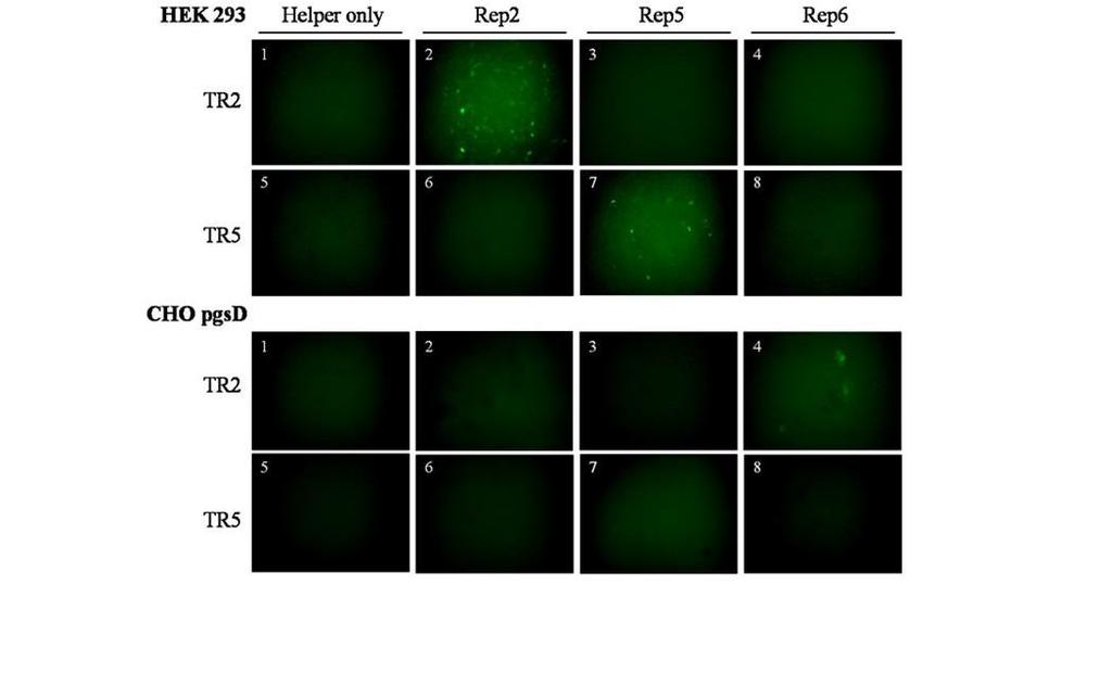
50 2C. Results C1. The AAV5 Origin of Replication is Unique Among Human Serotypes. As previously reported, Rep1-4 and 6 are unable to catalyze replication of ITR5 flanked genomes, while Rep5 is unable to catalyze the replication with vectors flanked by ITR1-4, and 6 (30, 31). Due to high sequence homology, it is likely that AAV serotypes 7-12 are also compatible with ITR2s and not ITR5s (Figure 3 and Table 1). In order to demonstrate the specificity of the Rep5-ITR5 interaction, two GFP vectors were utilized (Figure 7A). These constructs, ITR2-eGFP and ITR5-eGFP, are flanked by either ITR2s or ITR5s and expressing GFP from a CMV promoter. The raav vectors were transfected into 293 cells along with Ad helper plasmid (pxx680), and either Rep2Cap2, Rep5Cap2, or Rep6Cap6. Rep6 was included to confirm the cross-compatibility of AAV2 and AAV6 replication, as well as to underscore the ability of other naturally occurring serotypes to replicate ITR2 vectors. After 48 hours, cells were harvested for Hirt DNA and crude lysate. Hirt DNA (41) was analyzed by Southern blot with a probe for the GFP orf. A DpnI digestion was performed to remove transfected methylated plasmid DNA, but not unmethylated genomes which had been replicated in the cell. The results validated Rep-ITR specificity for the serotypes used, with Rep2 and Rep6 driving replication of only ITR2 vectors, and Rep5 driving replication of only the ITR5 vector. (Figure 7B). Crude lysate from these cells was used to transduce HEK 293 cells (highly transducible by Cap2) and CHO pgsd cells (transducible by Cap6) (Figure 7C). Specificity in the production of raav vectors followed the same pattern as replication, with Rep2 and Rep6 each able to produce infectious ITR2 raav particles and Rep5 able to produce only infectious ITR5 raav particles. 36

51 37
52 Figure 7. The Rep5-ITR5 interaction is unique among the fully characterized AAV serotypes. (A) The vector constructs used in this study. GFP expression was driven by a CMV promoter and SV40 poly(a) element. The Neomycin cassette included the thymidine kinase promoter and the bovine growth hormone poly(a) element. TR2s or TR5s flanked the vectors. ptr5- egfp contained an additional 500bp ahead of the 3 TR. (B) Southern blot of Hirt DNA comparing the ability of Rep2Cap2, Rep5Cap2 and Rep6Cap6 to replicate TR2s or TR5 flanked vector genomes. Hirt DNA was isolated 48 hours after transfection. DpnI cuts only the input plasmid, not the newly replicated AAV genomes. The two major replicative forms of AAV are indicated (m-double stranded monomer, d-double stranded dimer). Higher-order replicative forms are also visible. (C) Transduction of HEK 293 cells (transducible by Cap2) or CHO pgsd cells (transducible by Cap6 or Cap2) with crude lysate from cells harvested 48 hours after transfection of Adhelper plasmid only or triple-transfection of Ad-helper plasmid, TR2 or TR5 GFP, and either Rep2Cap2, Rep5Cap2, or Rep6Cap6. The numbers shown correspond to the lane of the gel in figure 2B. 38
53 C2. Creation and Characterization of Rep5 Helper Constructs for Cap1-5. As the prevalence of AAV5 in the human population is lower than AAV2 or AAV6, and considering the unique specificity between Rep5 and ITR5, we decided to create a raav ITR5 production system for transcapsidation into Cap1-5 similar to previous described system for type 2 (71). In order to confirm the efficacy of ITR5 vectors with respect to existing ITR2 vectors, Rep5 helper constructs were created (Figure 8A). This new system for producing virus vectors utilizes triple transfection with AAV helper plasmids containing the AAV5 Rep gene and one of the AAV serotype 1-5 Cap genes (prep5cap1-5), a reporter transgene plasmid with GFP flanked by AAV5 ITRs, and an Ad helper plasmid (XX680). HEK 293 cells were transfected with prep5cap1-5, Ad helper plasmid, and ITR2 or ITR5 egfp. Analyses of Hirt DNA extracted from these cells showed that Rep5 functioned properly, generating the expected DpnI-resistant AAV monomer and dimer replication intermediates when delivered with the ITR5-eGFP but not the ITR2-eGFP vector (Figure 8B). Additionally, cell lysate harvested from each transfection was tested for infectivity to ascertain the system s ability to produce functional recombinant virus. HEK 293 or Cos1 cells were exposed to lysate from the ITR5 transfections and assayed for GFP expression at 24 hours post-infection. Lysate carrying capsid-specific sequences (types 1-5) all produced GFP-positive cells when ITR5 was complemented with Rep5 expression plasmids during vector production (Figure 8C, panels 1-5). Cells exposed to lysate from ITR2 transfections in the presence of Rep5 proteins were negative for transgene expression (Figure 3C, panel 6). 39

54 40
55 Figure 8. Creation and characterization of Rep5 helper constructs for Cap1-5. (A) The AAV5 Rep and AAV1-4 Cap genes were subcloned into pxr2, a non-itrcontaining plasmid (see Methods for details). These new helper plasmids were used to package TR5 vectors into Cap1-5. (B) Southern blot using a GFP specific probe of Hirt DNA extracted from HEK 293 cells transfected with prep5cap1-5, Ad helper plasmid, and TR5-GFP or TR2-GFP. The two replicative forms of the vector are indicated. (C) Cell lysate from triple transfections described above were used to infect naïve HEK 293 cells. Cells infected with lysate from TR5-eGFP vector transfection of capsid serotypes 1-5 were positive for GFP (panels 1-5 corresponding to AAV1-5). HEK 293 cells infected with lysate from TR2-eGFP vector transfection of capsid serotypes 1-5 did not express GFP (panel 6, AAV1; representative of AAV2-5). (D) Graph comparing the relative titers achieved in the production of TR2 vs. TR5 raav. Samples were titered in duplicate by Q-PCR. Standard error is indicated. (E) Graph comparing the transducing units per vector genome of raav TR2 vs. TR5 vectors. Note that values on the y-axis are multiplied by 1x10⁻⁷. Virus was serially diluted and used to infect cells. GFP positive cells were quantitated and transducing units per microliter was calculated before conversion to transducing units per vector genome. Samples were measured in duplicate and standard error is indicated. 41
56 Having developed a functional AAV capsid production system using ITR5s, we sought to compare it to current Rep2-ITR2 production yields. The protocol designed by our lab (71) for production of transcapsidated raav2 (i.e., using helper plasmids that contain AAV Rep2 and serotypes 1-5 capsid genes, a reporter transgene (GFP) in an ITR2 vector cassette, and Ad helper plasmid) was used as a comparison to evaluate the new Rep5-ITR5 system. Cell lysate was harvested from each transfection and assayed for virus production by Q-PCR titering to determine virus particle number per unit volume of lysate (Figure 8D). The measurements obtained from the dot blots are comparable for ITR2 and ITR5 vectors when packaged in AAV serotype 1-4 capsids. Interestingly, ITR2 vector titers were noticeably reduced when packaged in an AAV5 capsid, possibly due to the evolutionary divergence of AAV5 with respect to the other serotypes (Figure 3 and Table 1). Both ITR5 and ITR2 vector production systems were also assayed for infectivity as measured by the number of transducing viral units per vector genome (Figure 8E). 1 x 10 5 HEK 293 cells were exposed to serial dilutions of cell lysate from each transfection, and the number of resulting transgene-positive cells was used to calculate the transducing units per microliter (TU/µl) of cell lysate. This was then divided by the viral titer (vg/ul) to yield the transducing units per vector genome (TU/vg). ITR5 vectors displayed a minor drop in transduction efficiency compared to ITR2 vectors, ranging from 5-10 fold in Cap1-3, while ITR5 vectors performed better in Cap5. This data demonstrates the transduction potential of each individual vector genome, again suggesting that due to evolutionary divergence, ITR5 vectors may have slightly better transduction potential when encapsidated in an AAV5 capsid. Transduction of the HEK 293 cells by Cap4 was below the detection threshold of the assay. The results indicate that while the yields of ITR5 raav vector production is equivalent to 42
57 the widely utilized ITR2 system, there may be a capsid specific effect on the ability of these vectors to transduce cells efficiently. C3. ITR2 but not ITR5 Vectors can be Mobilized by a wt AAV2 Provirus. AAV2 has been consistently demonstrated to be the most prevalent natural AAV serotype in the human population (33, 40, 59, 91); thus there is a strong likelihood that a large percentage of human individuals harbor a latent AAV2 infection (65). For that reason, we obtained Detroit 5 (D5) cells demonstrated to contain a latent wt AAV2 infection in order to model the potential for ITR2 or ITR5 vector mobilization upon raav and Ad infection. The D5 cell line contains the wt AAV2 genome which is stably integrated at chromosome 19 and is rescuable upon infection by helper virus (14, 80). The parental line, Detroit 6 (D6), is negative for wt AAV and was used as a control. Cell lysate containing ITR2 or ITR5- flanked raav GFP genomes encapsidated into Cap2 were harvested from triple-plasmid transfections of HEK 293 cells. Each type of lysate was used to infect D5 and D6 cells. After 24 hour incubation, cells were washed and co-infected with Ad helper virus. Control plates were exposed to lysate containing either AAV2 ITR2-eGFP only or Ad only. Transgene GFP expression was observed in all cells receiving the original cell lysate, confirming the infectivity of AAV2 ITR2-eGFP and AAV2 ITR5-eGFP in D5 and D6 cells (data not shown). Hirt DNA analysis from infected cells revealed rescue of latent AAV2 genes, in the form of AAV2 replication intermediates, in D5 cells infected with Ad (Figure 9A). As expected, D6 cells without latent AAV or D5 cells without Ad did not show AAV2 replication intermediates (Figure 9A). In addition, rescued latent wt AAV2 genomes were able to complement raav vector genomes when assayed by Southern blot analysis. For example, replication intermediates were observed in D5 cells exposed to the ITR2 vector 43
58 while no vector replication was observed in the cells exposed to the ITR5 vector. (Figure 9B). Longer exposure revealed minor ITR5 vector signal in D5 cells, comparable to background levels of ITR5 and ITR2 vector signal found in D6 Hirt DNA, indicating a lack of replication in the presence of Ad. 44
) or Detroit 5 (D5, latent AAV2 (+)) cell lines were infected with raav2 from crude lysate containing TR2 or TR5-eGFP in the presence or absence of adenovirus.")
59 Figure 9. TR2 but not TR5 vectors are mobilized by latent wt AAV2 and Ad coinfection. Detroit 6 (D6, Latent AAV2 (-)) or Detroit 5 (D5, latent AAV2 (+)) cell lines were infected with raav2 from crude lysate containing TR2 or TR5-eGFP in the presence or absence of adenovirus. Hirt DNA was isolated and analyzed by Southern blot 48 hours post-infection using either an AAV2 (A) or GFP (B) probe. The two major replicated forms of AAV DNA are indicated. Larger replicative forms are also visible. Size marker is denoted. (C) GFP expression was visualized in HEK 293 cells after crude lysate was added from either D5 or D6 cells infected with the vectors shown above the panels. The numbers refer to the gel lane in figure 5B. 45
60 Lysate taken from ITR2 or ITR5 egfp vector-infected D5 and D6 cells was used to infect naïve HEK 293 cells (Figure 9C). HEK 293 cells given lysate taken from D5 cells exposed to the AAV2/TR2-eGFP vector and Ad were positive for GFP (Figure 9C, panel 6), demonstrating that latent wt AAV2 was able to provide Rep and Cap in trans to mobilize the ITR2-flanked GFP vector. In contrast, HEK 293 cells given lysate from D5 cells infected with the ITR5-eGFP vector did not express GFP (Figure 9C, panel 7), demonstrating that the latent AAV2 was not able to mobilize the ITR5-flanked GFP vector. As expected, control lysate from D6 cells did not produce infectious GFP vectors using either ITR, and infection of D5 cells without Ad helper or without ITR2-eGFP vector did not produce infectious GFP vectors (Figure 9C, panels 1-5). C4. Persisting AAV Vector Genomes can be Mobilized. During infection, AAV genomes not degraded have two fates: episomal formation or chromosomal integration (17, 63). While wt AAV2 has been shown in tissue culture cells to integrate into the human chromosome in a site specific fashion (14, 80), it has been demonstrated that Rep is required for this form of latency (4). Ideally, raav vectors should be delivered in the absence of Rep, wherein numerous studies have determined that raav genomes remain episomal, typically circularizing or forming into concatemers as a mechanism of vector persistence. (18, 83, 102) Thus, the infrequent event of integration by raav genomes is not site specific (63). Regardless of the method of molecular persistence, AAV genomes are able to excise themselves from the chromosome or episome upon Ad superinfection to enter the lytic phase of the AAV lifecycle, suggesting wt AAV persists in a conservative manner, keeping at least one ITR sequence intact. (80) 46
61 To test whether persisting raav vectors could be rescued after infection in the absence of site-specific integration, we infected HEK 293 cells with 10,000 vector genomes/cell of either ITR2 or ITR5 GFP virus as determined by dot blot. After 18 days, GFP cells were sorted and pooled to approximate a population of cells infected by the vector. While we did not confirm these vectors had integrated into the host chromosome, we did confirm that after sorting, GFP persistence remained in 100% of the cells for greater than two months. Rep2Cap2, Rep5Cap2, or Rep6Cap6 as well as Ad helper plasmid were transfected into the mock, ITR2, or ITR5 containing cell lines and both Hirt DNA and crude lysate were isolated. Figure 10A reveals both ITR2 and ITR5 genomes were capable of being rescued and replicated (lanes 6, 8, 11). Specificity remained consistent for these vectors, with both Rep2 and Rep6 able to rescue and replicate ITR2s and only Rep5 able to replicate ITR5s. The ptr5-egfp panel was exposed longer than the ITR2 or mock due to the small amount of replicated vector DNA isolated from these cells. 47
 293 cells were infected with either ITR2 or ITR5 egfp vectors and passaged 18 days before cells still expressing GFP were sorted and pooled.")
62 Figure 10. AAV genomes conferring long-term transgene expression in cultured cells can be rescued, replicated, and packaged. (A) 293 cells were infected with either ITR2 or ITR5 egfp vectors and passaged 18 days before cells still expressing GFP were sorted and pooled. AAV helper plasmids (Rep2Cap2, Rep5Cap2, Rep6Cap6) were then added to assay for the ability of persistent AAV genomes to be rescued and undergo replication in the presence or absence of Ad helper plasmid. Hirt DNA was isolated and assayed via Southern blot with a probe for the GFP ORF. The two major replicative forms of the vector genomes are indicated. Larger replicative forms are also visible. The TR5 (+) helper plasmid panel of the blot was subjected to longer exposure in order to visualize the replicating vector genomes. (B) Mobilized genomes were assayed for infectivity by transducing HEK 293 or CHO pgsd cells with crude lysate from the cells described in figure 10A (control, TR2, or TR5 persisting vector genomes transfected with the helper plasmids described) 48 hours after addition of helper plasmids (All transfections for lysate used in figure 10B included Ad helper plasmid). 48
63 To determine if these rescued, replicating genomes could be encapsidated and mobilized to naïve cells, lysate was added to HEK 293 or CHO pgsd cells. Figure 10B shows that rescued genomes were encapsidated and that persisting raav genomes can be mobilized into previously non-transduced cells (HEK 293 panels 6 and 11 and CHO pgsd panel 8). Predictably, the transduction profile of the mobilized ITR2 or ITR5 vector genomes was dependent on the capsid into which they were packaged (Cap2 or Cap6), highlighting the potential danger of ITR2 vector mobilization being driven by a range of wt AAV serotypes. 2D. Discussion This aim suggests that AAV5 based vectors are significantly less likely to be mobilized after administration than the AAV2 based vectors currently used in clinical trials. The two most prevalent human AAV serotypes (AAV2 and AAV6) both have the ability to replicate the ITR2 flanked vectors currently used in AAV clinical trials (30). A less widespread AAV serotype, AAV5, has a unique Rep-ITR interaction making it the only human serotype able to replicate ITR5 flanked vectors (16, 30, 52). This replicative specificity, as well as the relative abundance of these serotypes in the population, (AAV5 over four-fold less abundant that AAV2 (33, 40, 59, 91) led us to hypothesize that ITR5 flanked vector genomes have a significantly reduced risk of vector mobilization. To test this hypothesis, we created an ITR5 based vector production system similar to the ITR2 based system currently used to produce raav (Figure 8A). This system worked well, exclusively packaging ITR5 flanked vectors into Cap1-5 while yielding comparable viral titers and transduction efficiency with respect to the current ITR2 vector production system. While our ITR5 vectors may have shown a minor inherent decrease in transduction efficiency (potentially in a capsid specific manner) optimization of our system may eliminate 49
64 this disparity. A similar comparison between ITR2 and ITR5 vectors in vivo showed no such bias, (30) suggesting the differential may be due to the sensitivity of our in vitro system or otherwise restricted to our assay. In order to confirm that Rep-ITR replicative specificity extended to vector mobilization we adopted two cell culture assays. While the transformed cells used for these assays had the potential to behave differently from the primary cell types AAV vectors would encounter in vivo, we reasoned that Rep-ITR specificity would remain consistent regardless of cell type. That said, demonstrating vector mobilization in primary cells remains an important step in establishing the potential danger to future gene therapy candidates. First, we showed that in cells with latent wt AAV2 infection, introduction of an ITR2 vector and subsequent superinfection by Ad resulted in replication of the wt genome and the ITR2-flanked transgene, (Figure 9A and 9B) and led to the production of infectious raav particles (Figure 9C). These results demonstrated that latent wt AAV2 plus Ad reconstituted the replication-deficient ITR2 vector system, allowing for mobilization of transgene vectors. Once again, AAV2 was unable to replicate or mobilize an ITR5 flanked genome (Figure 9B, lane 7), underlining the potential of our ITR5 based system to decrease AAV vector mobilization due to relative AAV2, AAV5, and AAV6 prevalence in the population. While inclusion of a cell line harboring a latent AAV5 genome would have been ideal for this study, there are no reports of AAV5 integrating site-specifically into the human chromosome. Thus, any cell line harboring an AA5 genome should be recapitulated by our mobilization system in figure 4 where a persisting ITR5 flanked genome is rescued and replicated. Next, cell lines were first created containing stably persisting ITR2 or ITR5 flanked vector genomes. While we did not determine whether these genomes were integrated into the 50
65 host chromosome, the persistence of GFP signal in 100% of these cells two months after sorting suggests chromosomal integration. However, the possibility that they are persisting in some other manner only lends credence to the mobilization assay we have developed, as such genomes may recapitulate any number of modes of persistence in vivo. We next demonstrated that these persisting raav genomes could be rescued and replicated upon the transfection of AAV helper plasmids (Figure 10A). These genomes were also encapsidated and able to transduce naïve cells (Figure 10B). Predictably, the cell/tissue tropism of these mobilized genomes was dependent on the capsid into which they were mobilized. 51
. Cells are infected or transfected with Ad and any AAV serotype in order to mobilize genomes (3).")
66 Figure 11. AAV Vector Mobilization Assay. Cells infected with a raav GFP reporter flanked by the ITR of any serotype are passaged for days, typically resulting in 1-4% of cells GFP positive (1). GFP positive cells are sorted and pooled (2). Cells are infected or transfected with Ad and any AAV serotype in order to mobilize genomes (3). Mobilization is determined by Southern blot of replicating genomes as well as infection of naïve cells with lysate. 52
67 Slightly different levels of replication were detected for ptr2-egfp vectors in the presence of Rep2 or Rep6. (Figure 7B and 10A). While this may suggest Rep6 replicates ITR2s with higher fidelity than Rep2, it is more likely that more Rep6 protein was produced by the plasmid constructs. Western blots were not performed due to the lack of a suitable Rep6 antibody. Interestingly, our ITR5 vector was rescued with lower fidelity from 293 cells than our ITR2 vector (Figure 10A). We have confirmed that our Rep2 and Rep5 constructs produce equivalent amounts of protein by western blot (data not shown) and that they drive comparable amounts of vector genome replication (Figure 7B). As such, the decreased rescue of ITR5 genomes is most likely due to the inability of a subset of the ITR5-flanked GFP vectors in this population to be rescued due to deletions of the integrated or concatamerized ITRs as seen with AAV 2 latent genomes (106). It is possible, however, that persisting ITR5 genomes may be somewhat refractory to rescue and further experiments may be required to definitively answer this question. It is impossible to quantify the degree of safety ITR5 based vectors would add to AAV clinical applications. Based on the exclusivity of the Rep5-ITR5 interaction, as well as the small degree of AAV5 in the population compared to AAV2 and AAV6, we can only postulate that ITR5 vectors possess a significantly lower risk of spreading after raav administration. While ITR5 based vectors may be markedly safer, they are not a solution. More importantly, this work provides an assay to test the ability of any AAV vector to be mobilized by any wt AAV serotype. While the ability to test for mobilization of a vector by an AAV2 provirus is critical, such an approach would require the creation of new cell lines containing a provirus of every AAV serotype so that each ITR vector could be screened for mobilization. Instead, the ability to infect with a vector harboring any ITR and 53
68 then mobilize with the Rep and Cap of any serotype will allow efficient screening of any novel or non-human ITR. 54
69 Chapter 3: Mapping Protein/DNA Specificity of the AAV Origin of Replication
70 3A. Introduction Having developed assays to screen vectors for mobilization against an AAV2 provirus as well as to screen any vector against mobilization by any AAV serotype, the next step towards preventing vector mobilization was the creation of a novel Rep-ITR interaction. To do so, however, required an understanding of the elements of the AAV origin of replication that govern replicative specificity. Identifying these elements was necessary to aid the rational design that would be required to engineer a new and unique Rep-ITR interaction. Initial mapping of the Rep protein has revealed that only the unique N-terminus of the large Rep proteins possesses the ability to bind site-specifically and to nick the ITR, specifically the N-terminal 208aa (107). As such, chimeric Rep proteins have been created which carry the N-terminal 200 residues of one serotype and the C-terminus of the other. These chimeric Reps specifically replicate the ITR corresponding to its N-terminus (107). Previously, AAV replicative specificity was postulated to be driven by the trs sequence (16). Rep2 can nick the ITR2 trs (AGT/TGG) and the AAVS1 trs of human chromosome 19 (GGT/TGG; 98). Rep5 nicks only the ITR5 trs (AGTG/TGG). However, alignment of the ITR2 and ITR5 sequences revealed several significant sequence and structural differences outside the trs sequence (Figure 4). The spacing between the putative RBE and the nicking stem was significantly different; three nt for ITR2 and 15 nt for ITR5. Additionally, while the trs sequence is not tightly conserved between ITR2 and ITR5, neither is the height or overall length of the putative nicking stem. In order to address these concerns the ITR was synthesized and amplified in halves (Figure 12). Assembly of the halves required the inclusion of a SfiI site in one of the hairpin arms of the ITR. SfiI allowed the conservation of the RBE sequence (10). Cloning the ITR in 56
71 a DD format required only one ITR per plasmid for replication (103). The three core Rep functions necessary for AAV replication (Rep binding, helicase, and nicking) were analyzed by the presence or absence of intracellular replication of the plasmid. This assay provided the ability to quantitate Rep-ITR function in a physiological setting, removing the concern that highly purified Rep protein might take on aberrant function in vitro. This system also avoided concerns that previous in vitro assays used only a fragment of the ITR or that oligos used to recapitulate the ITR might not fold correctly. Identification of the elements involved in Rep-ITR specificity stands to increase the understanding of viral and cellular DNA binding and endonucleolytic proteins. It is likely that similar interactions take place in a wide range of viral and cellular replication and repair pathways. Localization of these elements may also facilitate the identification of other unique Parvovirus origins of replication. Here, we demonstrate two unique mechanisms at the DNA and protein level to achieve Rep-ITR specificity and utilize these factors to create a novel AAV origin of replication. 57
72 Figure 12. Diagram of ITR synthesis. (A) The ITR was synthesized as oligonucleotides in two pieces (dark blue and light blue) overlapping across one hairpin stem containing the SfiI site (orange). (B) Each half was amplified via PCR prior to digestion and cloning. (C) Proper triple-ligation with puc18-cmv GFP produced an ITR in DD format. 58
73 3B. Materials and Methods B1. ITR Cloning. ITRs were cloned into a puc-18 plasmid with a GFP cassette (CMV promoter, SV40 polya) cloned between the KpnI and EcoRI restriction sites. The ITRs were synthesized in two halves as 4nmol Ultramer DNA oligos (Integrated DNA Technologies). SfiI restriction sites were incorporated into one hairpin arm the ITR for cloning (Figure 12). Due to inconsistencies of the reported sequence at the tip of the ITR5 hairpins between Chiorini et al., 1999, the published genbank sequence (genbank accession number NC_006152), and restriction mapping (data not shown), an ITR2 hairpin was utilized for the ITR5 construct (Figure 12). 200pg of each oligo was amplified in a PCR reaction using the ITR primers listed in supplemental table U of PfuTurbo DNA Polymerase (Stratagene) was used to amplify each half of the ITR as follows: 1 cycle at 94 o C for 4 minutes, 35 cycles of 45 seconds at 94 o C, 30 seconds at 50 o C, and 30 seconds at 72 o C, 1 cycle of 10 minutes at 72 o C. PCR reactions were purified and subject to digestion by KpnI and SfiI or HindIII and SfiI (NEB). A triple ligation with the puc-18 GFP plasmid and each half of the ITR was performed with T4 DNA Ligase (Invitrogen) for 1.5 hours at room temperature. All constructs were verified by DNA sequencing at the UNC-CH Genome Analysis Facility after linearization of the plasmid and ablation of the ITR secondary structure by SfiI digestion. B2. Rep Cloning. pxr2 (Rep2Cap2) and prep5cap2 AAV helper plasmids served as templates for Rep cloning. The primer sequences used are indicated in Table 3. Two cloning strategies were used. Existing restriction sites were incorporated into primers for PCR (PCR-RD in Table 3) utilizing either pxr out fw or pxr out rev primers. PfuTurbo DNA Polymerase (Stratagene) was used at the manufacturer s recommendations for all PCR 59
74 reactions. PCR-RD products were digested with the enzymes indicated in Table 3 (NEB) prior to ligation with T4 DNA Ligase (Invitrogen) according to manufacturer s instructions. Alternately, an overlap-extension mediated PCR (OE-PCR) approach was used to produce Rep chimeras (39). The Rep2 and Rep5 junction was incorporated into forward and reverse primers which were used in separate PCR reactions with the pxr out fw and rev primers (Table 3, only fw oligos indicated, rev oligos complimentary to fw). These overlapping PCR products were combined into a single PCR reaction and cycled as follows: 1 cycle at 94 o C for 30 seconds, 18 cycles of 30 seconds at 94 o C, 30 seconds at 65 o C, and 4 minutes at 72 o C, 1 cycle of 10 minutes at 72 o C. 1ul of this reaction was used as template for a nested PCR with the pxr in fw and rev primers. Chimeras with the N-terminus of Rep2 and C-terminus of Rep5 were cloned into the Rep25aa166 construct between the PpuMI and MfeI sites. Chimeras with the N-terminus of Rep5 and C-terminus of Rep2 were cloned into the 52aa160 construct between the PpuMI and BstBI sites. All constructs were verified by DNA sequencing at the UNC-CH Genome Analysis Facility. 60
75 Clone/ Cloning Method Orientation Sequence Primer pxr out fw Forward 5 CGAAAAGTGCCACCTGACGTCTAAGAAACC pxr in fw Forward 5 TCGAATTCGACGGCCAGTGAATTGTAATACGACTC pxr out rev Reverse 5 CCATGATTACGCCAAGCTCGGAATTAACCGCATGCG A pxr in rev Reverse 5 CCATGGCCGGGCCCGGATTCACC Rep52aa84 PCR-RD AleI Reverse 5 TTCACCCCGGTGGTTTCCACGAGCACGTGCATGTGG AAGTAGCTCTCTCCCTTTTCAAACTGCACAAAG Rep52aa110 PCR-RD EagI Forward 5 CCTCGGCCGCTACGTGAGTCAGATTCGCGAAAAACT GATTCAGAG Rep52aa126 OE PCR Forward 5 GTGGTCTTCCAGGGAATTGAACCCACTTTGCCAAAC TGGTTCGCGGTC Rep52aa138 OE PCR Forward 5 CTGGGTCGCCATCACCAAGGTAAAGAAGGGAGGCG GGAACAAGGTGGTGGATGAG Rep52aa146 OE PCR Forward 5 GCGGAGCCAATAAGGTGGTGGATGAGTGCTACATCC CCAATTACTTGCTC Rep52aa160 PCR-RD Bpu10I Reverse 5 ACTGGAGCTCAGGTTGGACCTTCGGCAGCAGGTAG Rep52aa175 OE PCR Forward 5 CGTGGACAAACCTGGACGAGTATAAATTGGCCTGTT TGAATCTCACGGAGCGTAAAC Rep52aa187 OE PCR Forward 5 CTGAATCTGGAGGAGCGCAAACGGTTGGTGGCGCAG CATCTGACGCAC Rep52aa207 PCR-RD SgrAI Reverse 5 GATCACCGGCGCATCCGAGAACTCACGCTGCGAAGC Rep25aa77 OE PCR Forward 5 TAAGGCCCCGGAGGCCCTTTTCTTTGTGCAGTTTGAA AAGGGATCTG Rep25aa97 OE PCR Forward 5 CCACATGCACGTGCTCGTGGAAACCTCCGGCATCTC TTCCATGGTCCTCG Rep25aa116 PCR-RD NruI Forward 5 TCAGATTCGCGAAAAACTGGTGAAAGTGGTCTTCCA GG Rep25aa125 OE PCR Forward 5 GAATTTACCGCGGGATCGAGCCGCAGATCAACGACT GGGTCGCCATC Rep25aa141 OE PCR Forward 5 GGTCACAAAGACCAGAAATGGCGCCGGCGGAGCCA ATAAGGTGGTGGATTCTGG Rep25aa149 OE PCR Forward 5 GAGGCGGGAACAAGGTGGTGGATTCTGGGTATATTC CCGCCTACCTGC Rep25aa166 PCR-RD Bpu10I Forward 5 CCAGCCTGAGCTCCAGTGGGCGTGGACAAACCTG Rep25aa187 OE PCR Forward 5 GTTTGAATCTCACGGAGCGTAAACGGCTCGTCGCGC AGTTTCTGGCAG Rep25aa216 PCR-RD SgrAI Forward 5 ATGCGCCGGTGATCAAAAGCAAGACTTCCCAGAAAT ACATGG ITR2 Half1 Kpn Forward 5 ATTATAGGTACCAGGAACCCCTAGTGATG ITR2 Half 1 Sfi Reverse 5 TAATAGGGCCCAAAGGGCCGGG ITR2 Half2 Sfi Forward 5 TTAATAGGCCCTTTGGGCCGGG ITR2 Half2 Hind Reverse 5 TATAATAAGCTTAGGAACCCCTAGTGATGGAG ITR5 Half1 Kpn Forward 5 ATTATAGGTACCTACAAAACCTCCTTGCTTGAG ITR5 Half1 Sfi Reverse 5 TTAATAGGCCCTTTGGGCCGTCGC ITR5 Half2 Sfi Forward 5 TTAATAGGCCCAAAGGGCCGTCGTC ITR5 Half2 Hind Reverse 5 TATAATAAGCTTTACAAAACCTCCTTGCTTGAGAG Table 3. Oligonucleotides Utilized for Chimeric Rep Cloning. OE PCR indicates the oligo was used for the overlap extension method of cloning. PCR-RD indicates that PCR was performed and a restriction digest of the product was used for cloning. 61
76 B3. Western Blot Analysis. Samples for Western blot analysis were harvested hours after transfection of Ad-helper plasmid and the appropriate AAV helper construct. Cells were washed and resuspended in 100ul PBS prior to addition of 100ul 2x Laemmli Sample Buffer (100mM Tris ph 6.8, 4% SDS, 200mM DTT, 20% glycerol, 0.1% Bromophenol Blue). Samples were briefly sonicated and boiled for 10 minutes. Samples were run on NUPAGE 4-12% Bis-Tris gels (Invitrogen) at 160 volts for 90 minutes. Protein was transferred to a Nitrocellulose membrane (Invitrogen) via a wet transfer for 60 minutes at 30 volts. Gels were blocked overnight in 10% nonfat dry milk in 1x PBS/Tween (0.05%). Detection of both Rep2 and Rep5 proteins (all four sizes) was achieved with a monoclonal Anti-Adeno-Associated Virus Rep Protein antibody (clone 259.5, American Research Products) at a 1:20 dilution in PBS/Tween for 60 minutes at room temperature. After washing, a secondary HRP antimouse antibody was added at a 1:5,000 dilution in PBS/Tween for one hour at room temperature. After washing, SuperSignal West Femto Maximum Sensitivity Substrate (Pierce) was added and blots were exposed to X-ray film (Kodak). B4. Densitometry. Densitometry was performed using the public domain NIH Image program (developed at the U.S. National Institutes of Health and available on the Internet at Agarose gels stained with Ethidium Bromide were imaged and a DpnI-resistant cellular DNA was used as a loading control. Values from densitometric analysis vector replication of Southern blots were divided by values obtained from the loading control. The lowest value (absence of any vector replication) was then subtracted from all values as background. In order to gauge relative replication efficiency, values for replicating TR2 vectors were divided by the value obtained from the Rep2-ITR2 control. Replicating TR5 vectors were similarly compared to the Rep5-ITR5 control. 62
77 B5. Molecular Modeling. Molecular models were generated using Swiss-Model ( The published crystal structure of the amino-terminus of Rep5 complexed with the RBE (PDB accession #1rz9) was used as a template for all models. Visualization of protein structure rendering of images were performed with PyMOL ( B6. Plasmid Construction. The N-terminus of Rep8 was synthesized by Gene Art AG (Regensburg, Germany). A PpuMI site was incorporated into the Rep8 sequence 31 bp from the transcriptional start site corresponding to the same site in Rep2. Synthesis ended at the BamHI site at residue 244 common to both serotypes. Rep8 sequence was cloned into the pxr2 (Rep2Cap2) backbone used for AAV helper plasmids to create the plasmid prep82aa244. This plasmid was then used as a template for a PCR reaction with pxr out fw (5 CGAAAAGTGCCACCTGACGTCTAAGAAACC) and Rep282 Bpu10I rev (5 ACTGGAGCTCAGGCTGAGTCTTGGGCAGGAGGTAG). The PCR product was then digested with SfiI (common to Rep8 and Rep2) as well as Bpu10I (incorporated onto the Rep8 sequence using the primer) and cloned into the pxr2 backbone to create the clone Rep282aa C. Results C1. Characterization of Chimeric ITRs. An alignment of ITR2 and ITR5 revealed several divergent elements which might infer Rep specificity (Figure 4). The RBE and hairpins seemed unlikely to impact specificity as Rep2 and Rep5 have been reported to bind ITR2 and ITR5 interchangeably (16). Additionally, no evidence has been presented for Rep interactions with the portion of the D-element outside the nicking stem. Therefore, the spacer and nicking stem elements appeared to be the most likely candidates for unique interactions 63
78 with their cognate Rep protein. This hypothesis was supported by low homology of these elements between AAV2 and AAV5. Wt ITRs containing the SfiI site functioned as expected with Rep2 specific to ITR2 and Rep5 specific to ITR5 (Figure 13B). Rep2-ITR2 replicated approximately 2-fold more efficiently than Rep5-ITR5. This is potentially due to the lower folding energy of ITR5 resulting in reduced plasmid stability prior to replication. Due to this minor difference in replicative fidelity, all ITRs replicated with Rep2 were normalized to Rep2-ITR2, while ITRs replicated with Rep5 were normalized to Rep5-ITR5 (Figure 13B). In order to confirm that the RBE and hairpin arms played no role in Rep specificity, we generated a chimeric ITR with ITR5 binding elements and an ITR2 spacer and nicking stem (ITR5+2SNS). Only Rep2 replicated this ITR, confirming the determinants of replicative specificity lie in the spacer/nicking stem elements (Figure 13B). While ITR5+2SNS replication was not as efficient as ITR2-Rep2, it was replicated at ITR5-Rep5 levels. Conversely, Rep5 specifically replicated an ITR comprised of ITR2 hairpins and hairpin spacer and the ITR5 spacer and nicking stem (ITR2+5SNS, Figure 13B). Rep5 replicated this ITR at wt levels. This data demonstrated that Rep-ITR specificity lies outside of the ITR binding regions. Next, we explored whether the nicking stem or the spacing between the RBE and nicking stem harbored unique interactions with the Rep protein by creating chimeric ITRs which divided these individual elements. An ITR with the ITR5 binding elements and spacer and the ITR2 nicking stem could not be replicated by either Rep2 or Rep5 (ITR5+2NS, Figure 13B). The corresponding chimeric ITR (ITR2 binding elements and spacer with an ITR5 nicking stem) was replicated by both Rep2 and Rep5 (ITR2+5NS, Figure 13B). This 64
79 disparity suggested that the spacer and nicking stem play different roles in Rep-ITR specificity between AAV2 and AAV5. 65
80 66
81 Figure 13. Cloning and Characterization of Chimeric ITRs. (A) Sequence and structure of ITR2 (black) and ITR5 (blue) shown with incorporation of SfiI sites for cloning (green). Length in nt of ITR elements indicated above brackets. RBE is boxed. RBE is indicated by a hatched circle. Nicking stem is extruded with arrow indicating the nicking site and hatched box indicating the trs. The four initial chimeric ITRs generated are shown (right). (B) Replication assay and quantitation of chimeric Reps. Replication products from the indicated ITR and either Rep2 or Rep5 were analyzed by Southern blot. Monomeric (m) and dimeric (d) replicating species are indicated. The level of replication of each sample was measured by densitometric analysis and compared to wt replication. 67
82 C2. Nicking Stem Sequence and Height are Critical for ITR5 specificity. ITR2+5NS established that Rep2 is capable of nicking an ITR with an ITR5 nicking stem and that Rep-ITR specificity is not driven exclusively by the trs sequence (Figure 13B). In order to determine the flexibility of Rep2 toward mutant nicking stems, we generated ITR2s containing altered forms of the hairpin (Figure 14A). Rep2 is able to replicate an ITR with an ITR5 nicking stem even though the ITR5 nicking stem contains a different trs sequence, is one bp shorter, and has two fewer unpaired nucleotides at its tip (Figure 14A). The substitution of the ITR5 nicking stem into ITR2 also allowed replication by Rep5. To determine which element of the ITR2 nicking stem prevented Rep5 activity, we altered specific portions of the ITR2 stem. First, one bp at the top of the putative ITR2 nicking stem was removed to lower the height to that of ITR5 (ITR2-TA). Removing the T-A bp also resulted in a trs resembling ITR5, nicking between G/T opposed to T/T. Rep2 continued to function on this ITR as did Rep5, demonstrating that Rep5 can tolerate five unpaired nucleotides at the tip of the stem as long as the stem height and nt sequence are correct. A similar deletion from the base of the ITR2 nicking stem reduced the height to that of ITR5 while retaining the ITR2 nicking site (ITR2-GC). Rep2 continued to function efficiently on this ITR while Rep5 activity was ablated. This data suggested that the inability of Rep5 to function on ITR2 is primarily the sequence of the trs, specifically the requirement for a nick to be generated between G/T. 68


83 69
84 Figure 14. Relation of Nicking Stem Height and Sequence to Rep-ITR Specificity. (A) Sequence of nicking stem in an otherwise ITR2 context. Arrow indicates trs site. Brackets indicate height of putative stems in nt from the base of the stem to the putative nicking site. Predicted ΔG values for the hairpins are below. Southern blot analysis of the ITRs replicated by Rep2 or Rep5 is shown below. (B) Quantitation of the Southern blots relative to wt replication from (A). (C) Same as (A), except nicking stems indicated were used in an ITR5 context. (D) Quantitation of the Southern blots relative to wt replication from (C). 70
85 To determine the extent of Rep2 flexibility for different nicking stems, we created three additional ITR2 mutants. Extending the nicking stem by one bp at the base had no effect on replication by Rep2 (ITR2 9 nt). However, a three bp extension was sufficient to ablate Rep2 function on the ITR (ITR2 11 nt). Surprisingly, Rep2 was able to tolerate a three bp deletion from the base of the stem, underlining the flexibility of Rep2 with respect to nicking stem substrates (ITR2 5 nt). In order to explore the level of flexibility Rep5 possessed toward non-wt nicking stems, we created a panel of mutant ITR5s harboring altered nicking stems (Figure 14C). Curiously, Rep2 replicated none of these ITRs, suggesting an element outside the ITR5 nicking stem is responsible for preventing Rep2 function. As in Figure 13B, replacement of the ITR5 nicking stem with that of ITR2 resulted in the ablation of replication by Rep5, attributable to the incompatible trs sequence. The addition of one bp at the top of the ITR5 nicking stem severely decreased the ability of Rep5 to replicate the ITR (ITR5 +TA, Figure 14D). This insertion disrupted the ITR5 trs sequence and increased the size of the stem one bp. However, the low level of replication by Rep5 on ITR5 +TA suggests that the entire trs site of ITR2 is necessary to confer Rep2 specificity, not just the presence of a T/T nick site. The addition of one bp to the base of the ITR5 nicking stem, preserving the ITR5 trs at the tip, nearly eliminated replication by Rep5 (ITR5 +GC). Likewise, the removal of one bp from the base of the ITR5 nicking stem strongly decreased replication by Rep5 (ITR5 6nt, Figure 14D). This data suggests that Rep5 is sensitive both to the height of the nicking stem as well as to the sequence of the trs. Thus, Rep5 is unable to replicate ITR2 because the ITR2 nicking stem is one bp too tall and has an incompatible trs sequence. 71
86 C3. Spacer Length is Critical for ITR2, not ITR5. While Rep2 can replicate a vector with an ITR5 nicking stem, it can not replicate wt ITR5 (Figure 13B). The only difference between ITR5+2SNS (which Rep2 can replicate) and ITR5+2NS (which Rep2 can not) is the ITR5 spacer (Figure 13B). The wt Rep2 spacer is three nt long while the wt Rep5 spacer is 15 nt long. Thus, we hypothesized that Rep2 may be sensitive to spacer length. Previous in vitro data supported this conclusion as insertions into the ITR2 spacer prevented nicking by Rep2 (10). To explore the effect of spacer length on ITR2 and ITR5, we generated a series of mutant ITR2s and ITR5s with differing spacer lengths (Figure 15A and 15C). An insertion extending the ITR2 spacer to 10 nt ablated replication by Rep2 (ITR2 10nt, Figure 3B). Similarly, substitution of the ITR2 spacer with the 15 nt spacer of ITR5 also ablated replication by Rep2 (ITR2 15nt, Figure 15B). Rep5 was unable to replicate any of these vectors due to the presence of the ITR2 stem loop. 72
87 73
88 Figure 15. Effect of RBE-Nicking Stem Spacing on Rep-ITR Specificity. (A) ITR2 mutants were synthesized with the indicated spacing between the RBE and nicking stem. (B) Southern blot analysis of the ITRs depicted in (A) replicated by either Rep2 or Rep5 (Left). Quantitation of Southern blots relative to wt replication (Right). (C) ITR5 mutants synthesized as in (A). (D) Southern blot analysis and quantitation of (C). 74
89 Rep5 displayed greater flexibility toward spacer elements of differing lengths. Replacing the 15 nt ITR5 spacer with that of ITR2 resulted in an ITR which Rep5 retained the ability to replicate at a reduced level (ITR5 3nt, Figure 15D). Additionally, the presence of the three nt spacer allowed Rep2 to function on this ITR. The addition of six nt to the ITR5 spacer (for a total spacer length of 21nt) resulted in an ITR capable of being replicated by Rep5 at an efficient level (ITR5 21nt, Figure 15D). Replication by Rep5 was effectively abolished only after the insertion of 15 nt into the spacer (ITR5 30nt, Figure 15D). This panel of mutant ITR5s demonstrates the requirement for a three nt spacer element for Rep2 function. This data confirmed that the length of the ITR5 spacer was critical to block Rep2 function. Even small insertions into the ITR2 spacer were not tolerated by Rep2. Meanwhile, Rep5 is flexible in regard to spacer length, demonstrating the ability to function on ITRs with spacers from 3-21 nt. C4. The ITR5 Spacer Acts as a RBE for Rep5. The inability of Rep2 to function on ITRs with spacers longer than three nt led to the question of why Rep5 was so flexible in this regard. We hypothesized that Rep5 might specifically bind the ITR5 spacer just as it binds the RBE. The inability of Rep2 to bind this sequence would preclude its function on ITR5. Supporting this hypothesis was a moderately conserved GAGY Rep binding motif extending throughout the ITR5 spacer (Figure 16A). Additionally, as Rep monomers bind every four nt, the binding of three Rep5 monomers to the 15 nt spacer element would result in a three nt spacer, similar to that of ITR2 (38). If Rep5 does bind the loosely conserved GAGY motif, the removal of that motif from the spacer should abolish Rep5 function. Indeed, the ITR5 No GAGY mutant could not be 75
90 replicated by Rep2 or Rep5 (Figure 16B). This suggested that the specific sequence of the ITR5 spacer plays an active role in the Rep5-ITR5 interaction. Conversely, a spacer with a pure GAGY repeat should not disrupt the ability of Rep5 to function on the ITR. Indeed, Rep5 was able to replicate this ITR at wt levels (ITR5 GAGY, Figure 16B). Rep2 was also able to replicate this ITR efficiently, suggesting the poorly conserved nature of the GAGY repeat within the ITR5 spacer prevents a critical DNA-protein interaction with Rep2 necessary for replication. To explore how the ITR5 spacer functioned as an RBE, we removed three GAGY repeats from the hairpin side of the RBE (ITR5 Spacer RBE, Figure 16A). This essentially shifted the 16 nt RBE 12 nt closer to the nicking stem. Rep5 replicated this ITR efficiently, confirming the ITR5 spacer acts as a RBE (ITR5 Spacer RBE, Figure 16B). The slight reduction in replication fidelity of this ITR with respect to wt ITR5 may signal the inability Rep to properly interact with the RBE (10). Rep2 was again unable to replicate ITR5 Spacer RBE due to its inability to interact with the ITR5 spacer. 76
91 77
92 Figure 16. The ITR5 Spacer Acts as a RBE for Rep5. (A) ITR5 mutants were synthesized with the indicated RBE and spacer sequence. Brackets indicate individual tetranucleotide repeats bound by Rep monomers. Both strands of the wt ITR5 sequence are shown to illustrate conservation with the GAGY motif (indicated by *). Only one strand shown on others. (B) Southern blot analysis of the ITRs depicted in (A) replicated by either Rep2 or Rep5 (Left). Quantitation of Southern blots relative to wt replication (Right). (C) ITR2 mutants were generated with the RBE and spacer sequences indicated. (D) Southern blot analysis and quantitation for (C). 78
93 Next, we sought to extend the ITR2 spacer element to function as an extended RBE (Figure 16C). The seven nt insertion attempted in Figure 15A possessed essentially no GAGY homology (ITR2 +7, Figure 16C). As a result, Rep2 could not replicate this ITR (Figure 16D). Eight nt (two four nt GAGY repeats) inserted into the ITR2 spacer between the RBE and the existing spacer prevented replication by Rep2, demonstrating that the ITR2 RBE can not be extended. Similar to ITR5 Spacer RBE, we retained the eight nt GAGY insertion into ITR2 while removing eight nt of GAGY from the hairpin side of the RBE (ITR2 +8-8, Figure 16C). This shifted the RBE eight nt closer to the nicking stem. Rep2 replicated this ITR very inefficiently at a level below the detection threshold of densitometric analysis (Figure 16D, Southern). C5. Identification of Regions of Rep Responsible for ITR Specificity. Identifying the two elements of the ITR responsible for Rep specificity allowed us to map the regions of Rep2 and Rep5 involved in ITR specificity. We focused exclusively on the N-terminal 208 aa of the large Rep proteins as this region encompasses the DNA binding and endonucleolytic activity of the protein (107). This region displays approximately 60% sequence conservation evenly distributed across the protein sequence (Figure 17A). Residues involved in the active site of the protein are 100% conserved between Rep2 and Rep5 (37). Residues implicated in binding the RBE are highly conserved (38). Residues which bind the RBE display nearly perfect conservation except for two conservative substitutions near aa 140. In order to map the regions of Rep involved in ITR specificity, we generated a panel of chimeric Reps derived from Rep2 and Rep5 (Figure 17B). The ability of each chimeric 79
94 Rep to replicate an ITR2- or ITR5-flanked vector in HEK 293 cells was determined by Southern blot (Figure 17B and 17D). Each Rep in the panel was verified by DNA sequencing and Western blot analysis (Figure 17C). Every chimeric Rep showed similar protein expression profiles compared to wt. Densitometric analysis provided a comparison of the replication efficiency of each chimeric Rep with that of wt Rep2 or Rep5 (Figure 17E). Chimeric Reps were named according to the aa location of the swap between serotypes; for instance, Rep25aa77 possesses the N-terminal 76 aa of Rep2 and the C-terminus of Rep5. 80

95 81
96 Figure 17. Cloning and Characterization of Chimeric Reps. (A) An alignment of the N-termini of Rep2 and Rep5. (*) represents conserved amino acids. (: and.) indicates conservative substitutions. Blue indicates residues implicated in RBE binding interactions. Pink indicates residues which participate in the endonucleolytic active site. Green indicates residues implicated in RBE binding. (B) Chimeric Reps created and their ability to replicate ITR2 or ITR5 flanked vectors. Numbers indicate the aa position of the switch from one Rep to the other. (+) indicates the presence of replication, ( ) indicates the absence. (C) Western blot for expression of the chimeric Reps. (D) Southern blot demonstrating replication of an ITR2 or an ITR5 vector by the chimeric Reps. Note that the ITR5 vector is 500bp larger than the ITR2 vector. (E) Level of replication of the chimeric Reps relative to wt Rep2 or Rep5. 82
97 In the case of Rep5, replacement of the N-terminal 77 or 97 aa with Rep2 had no effect on ITR specificity nor a significant impact on replicative fidelity (Figure 17D and 17E). Larger pieces of Rep2 substituted onto the N-terminus of Rep5 were sufficient to prevent efficient replication of ITR5s (Rep25aa116, Rep25aa125, and Rep25aa141). This suggested that these chimeras possessed interruptions of a critical region of Rep5 for ITR5 specificity. Rep2-based chimeras were unable to replicate ITR5s without the inclusion of the N- terminal 146 aa of Rep5 (Rep52aa146, Figure 17D). Rep52aa146 replicated ITR5 at wt levels, as did the three chimeras with larger portions of Rep5 on the N-terminus (Rep52aa160, Rep52aa175, Rep52aa207). This mapping reveals that the critical region for ITR specificity in Rep5 lies between aa Surprisingly, the Rep52aa146 clone also functioned efficiently on ITR2, constituting a Rep capable of replicating ITR2 and ITR5. This suggested that ITR specificity existed in two different regions of Rep. For Rep2, the N-terminal 83 or 109 aa of Rep5 could be substituted with no effect on ITR specificity or major influence on replicative fidelity (Rep52aa84 and Rep52aa110, Figure 17D and 17E). Chimeras including slightly larger portions of Rep5 were unable to replicate either ITR, again suggesting the interruption of a domain critical for ITR specificity (Rep52aa126 and Rep52aa138). Rep5-based chimeras were unable to replicate ITR2s without the inclusion of the N-terminal 149 aa of Rep2. However, ITR2 replication was inefficient (Figure 52aa149, Figure 17D and 17E). The inclusion of larger portions of Rep2 allowed replication of ITR2s to increase to wt levels (Rep25aa166, Rep25aa216). This data maps the Rep2 region involved in ITR specificity to aa However, unlike Rep5, this was not the only region which played a role in ITR specificity. The ability of the Rep52aa146 chimera to replicate ITR2 and ITR5 vectors demonstrated a second region of Rep2 between 83
98 aa sufficient to allow replication of ITR2s even when the other critical region (aa ) was Rep5. The isolation of two different Rep regions involved in ITR specificity was consistent with the discovery of two independent elements governing specificity within the ITR. C6. Characterization of Rep Regions Involved in ITR Specificity. To characterize the Rep domains identified in Figure 5, we created chimeric Rep proteins which specifically exchanged the regions implicated in ITR specificity (Figure 18A). Region 1 existed in Rep2 from aa and in Rep5 from aa Region 2 lay within Rep2 from aa and Rep5 from aa As in Figure 5, all chimeras were verified by DNA sequencing and Western blot analysis (Figure 18B). Chimeras were then assayed for the ability to replicate ITR2- or ITR5-flanked vectors (Figure 18C). 84


99 85
100 Figure 18. Characterization of Rep Regions Critical for ITR Specificity. (A) Chimeric Reps and their ability to replicate ITR2 or ITR5 flanked vectors. Numbers indicate the aa position of the switch from one Rep to the other. (+) indicates the presence of replication, ( ) indicates the absence. Region 1 and 2 involved in Rep-ITR specificity are indicated. (B) Western blot for expression of chimeric Reps. (C) Southern blot demonstrating replication of an ITR2 or ITR5 vector by the chimeric Reps. Note that the ITR5 vector is 500bp larger than the ITR2 vector. (D) Structural model illustrating the two Rep regions. Rep2 structure is blue, Rep5 is purple. The nucleophilic tyrosine is indicated. Black hatched circle indicates the predicted structural difference of region 1 in the major groove of the ITR. (E) Structural model as in (D). The nucleophilic tyrosine is indicated. (F) Detailed structural view of region 1. The side-chains of non-conserved residues from Rep5 (purple) and Rep2 (blue) are shown. Three Rep5 residues implicated in RBE binding are indicated. (G) Detailed structural view of region 2. Side chains of active site residues are shown in black. Side chains of non-conserved residues in this region are shown for Rep2 (blue) and Rep5 (purple). The nucleophilic tyrosine is indicated, as is the adjacent Rep2 Asn
101 Replacing Rep5 region 1 with Rep2 yielded a clone unable to replicate either vector, suggesting the chimera lacked the ability to bind the ITR5 spacer or nick the ITR2 nicking stem (Rep525aa , Figure 18C). Replacing Rep5 region 2 with that of Rep2 allowed this chimera to replicate an ITR2 vector, suggesting region 2 of Rep2 was critical to nick the ITR2 nicking stem (Rep525aa ). The inability of this chimera to recognize ITR5 is harder to explain as Rep52aa146 could replicate ITR2 and ITR5 efficiently (Figure 17B). This result suggests that Rep2 region 2 makes specific contacts within Rep2 aa which are necessary in order to function on the ITR5 nicking stem. Replacing regions 1 and 2 of Rep5 with Rep2 resulted in a Rep chimera which replicated only ITR2s (Rep525aa ). Replacing Rep2 region 1 with Rep5 resulted in replication of only ITR2s, again demonstrating a connection between Rep2 region 2 and the ITR2 nicking stem (Rep252aa97-146). The lack of ITR5 replication by Rep252aa is difficult to explain based on the Rep52aa146 chimera which replicates ITR2s and ITR5s efficiently (Figure 17B). This result suggests that Rep5 region 1 makes specific contacts within the preceding 96 aa of Rep5 which are necessary in order to replicate ITR5. Replacing Rep2 region 2 with Rep5 resulted in a chimera unable to replicate either ITR (Rep252aa ). This chimeric Rep possesses neither Rep2 region 2 (required to nick the ITR2 nicking stem) nor Rep5 region 1 which appears necessary to interact with the ITR5 spacer. Finally, replacing both Rep2 regions 1 and 2 with Rep5 resulted in a chimera capable of replicating only ITR5 vectors (Rep252aa97-187). The crystal structure of the N-terminal 193 aa of Rep5 complexed to the RBE allowed the location of these two critical regions to be modeled (38). The structure of the N-terminus 87
102 of Rep2 was modeled with Swiss-Model software using Rep5 as a template. The location of region 1 supports its involvement with the spacer/rbe (Figure 18D). This region interacts with the major groove of the ITR where one of the most apparent structural differences between Rep2 and Rep5 is predicted (Figure 18D, hatched circle). Rep2 contains a two aa insertion in this loop with respect to Rep5. This insertion and other non-conservative substitutions are likely responsible for the inability of Rep2 to interact with the ITR5 spacer. Viewing Rep along the length of the ITR illustrates that region 1 constitutes much of the base of the protein (Figure 18E). Both Reps are predicted to participate in a β-sheet motif in the center of this region, while areas of reduced homology exist toward either side (the loop interacting with the major groove of the ITR on one side, RBE interactions on the other). A more detailed look at region 1 reveals the greatest disparity between Rep2 and Rep5 occurs at the RBE binding interface in the major groove of the ITR (Figure 18F). There is very little predicted structural difference between region 2 of Rep2 and Rep5 (Figure 18D and 18E). In an effort to dissect this region, we created two additional clones: Rep52aa147 and Rep52aa151 (Figure 18A). Like Rep52aa146, both of these Reps were able to replicate ITR2 and ITR5 vectors (Figure 18C). Rep52aa146 and Rep52 aa147 replicated ITR2 and ITR5 vectors with equivalent efficiency, suggesting E147 of Rep2 is not involved in ITR specificity. Rep52aa151 did display a modest reduction in ITR2 replication compared to Rep52aa146, suggesting that C151 of Rep2 plays a role in ITR2 specificity. Because Rep52aa160 can not replicate ITR2, this leaves only two other non-conserved residues between Rep2 and Rep5 in this region (N155 and T161). Both of these residues lie near the active site and are likely to interact with the nicking stem or active site. N155 lies directly 88
103 adjacent to Y156, the nucleophilic tyrosine, and may play a major role in ITR2 specificity (Figure 18G). C7. Comparison of Rep Region 1 across Serotypes. Finding two relatively small regions of the Rep protein important for ITR specificity allowed those regions in the other human/primate AAV serotypes to be analyzed in silico. An alignment of the two Rep regions in AAV serotypes 1-11 was performed by ClustalW analysis. Only Rep5 possessed a significant lack of conservation in Rep region 2 with respect to the other serotypes (data not shown). This suggests that all other AAV serotypes may have flexibility similar to AAV2 toward the size and sequence of the nicking stem of the ITR. For region 1, however, there were three distinct groups (Figure 19A). Every serotype except AAV8 and AAV5 was highly conserved with AAV2. This supports previous reports of the inter-compatibility of serotypes 1-4 and 6 in their Rep-ITR interaction (30, 31). Rep5 was significantly divergent from Rep2, accounting for the inability of Rep2 to bind the extended ITR5 RBE. Surprisingly, AAV8 was poorly conserved in relation to both AAV2 and AAV5. Both the RBE and RBE binding interfaces of region 1 of Rep8 were poorly conserved, despite extremely high homology between the remainder of the N-terminus of Rep8 with Rep2 (Figure 19B). The C-terminus of the protein was also well conserved, suggesting that the differences in region 1 may contribute to a unique origin of replication for AAV8. Additionally, the co-crystal structure of Rep5 bound to its RBE allowed us to model the N-terminus of Rep8. These models suggested that Rep8 possessed unique structural confirmations in both the RBE and RBE interacting domains of Rep region 1 with respect to Rep2 and Rep5. This altered confirmation likely plays a critical role in ITR specificity (Figure 19C and D). 89

104 90
105 Figure 19. Alignment of Rep Region 1. (A) The majority of characterized AAV serotypes are highly conserved with AAV2 in Rep region 1. However, Rep5 and Rep8 display poor conservation to all other human/primate AAV serotypes in this region. (B) Homology between Rep2 and Rep8 along the full length of the Rep78 protein. Region 1 of Rep8 (aa ) is poorly conserved, sharing only 42% homology to Rep2 (cyan). The remainder of the N-terminus is highly conserved (yellow and green). The C-terminus is also well conserved (purple). (C and D) Structural models of the N-terminus of Rep2, Rep5, and Rep8. Rep8 (orange) is predicted to possess a significantly altered structure at the RBE binding interface of region 1 (white arrow in C) as well as the RBE binding interface (white arrow in D). 91
106 Although the Rep8 sequence has been published, the ITR8 sequence has not. As we were unable to obtain the Rep8 construct, Gene Art AG (Regensburg, Germany) synthesized the N-terminal 244 aa. Rep chimeras were created with the N-terminal 244 aa of Rep8 and the C-terminus of Rep2 as well as a 50 aa section of Rep8 replacing Rep2 region 1 (Figure 20A). The N-terminal 244 aa of Rep8 was also used with the C-terminus of Rep5 (data not shown). None of these chimeric Reps were able to replicate an ITR2 or ITR5 vector, suggesting that this region of Rep8 may confer specificity unique specificity to its cognate origin of replication with respect to all other AAV serotypes (Figure 20B). 92
 indicates the absence of replication on the indicated ITR. Numbers indicate the aa boundary of the chimera.")
107 Figure 20. Chimeric Rep8 proteins are not compatible with ITR2 or ITR5. (A) The Rep8-Rep2 chimeric proteins generated. (-) indicates the absence of replication on the indicated ITR. Numbers indicate the aa boundary of the chimera. (B) Southern blot showing replication of ITR2 and ITR5 vectors by Rep2 and the two Rep8 chimeric constructs from (A). 93
108 C8. Structure-Function Model of Rep-ITR Specificity. In order to unify the ITR and Rep elements involved in specificity into a single model, we utilized the chimeric Reps separating region 1 and region 2 along with the chimeric ITRs separating the nicking stem and spacer. Rep2, Rep5, Rep52aa146 (which divides region 1 and 2 of Rep and can replicate ITR2 and ITR5), and Rep25aa149 (essentially no ITR2 or ITR5 replication) were selected. These Reps were tested for their ability to replicate ITR2, ITR5, ITR2+5NS (which is replicated by both Rep2 and Rep5), and ITR5+2NS (which is replicated by neither Rep2 or Rep5). Only Rep2 and Rep52aa146 efficiently replicated ITR2 (Figure 21A and 21B). Only Rep5 and Rep52aa146 replicated ITR5. As in Figure 1, Rep2 and Rep5 replicated ITR2+5NS. Additionally, Rep25aa149 and Rep52aa146 replicated ITR2+5NS. This ITR is universally replicated by every Rep due to the exclusion of DNA elements involved in protein specificity. The three nt ITR2 spacer functions with the DNA binding region 1 of Rep2 or Rep5. The seven bp tall ITR5 nicking stem functions with region 2 of Rep2 or Rep5. Thus, any combination of these regions constitutes a Rep protein capable of replicating ITR2+5NS. Finally, neither Rep2 nor Rep5 replicated ITR5+2NS. Rep2 is unable to interact properly with the 15 nt ITR5. Rep5 is unable to function on the ITR2 nicking stem. For these reasons, Rep25aa149 was also unable to catalyze replication. However, Rep52aa149 was able to replicate this ITR due to the proper combination of Rep regions (Figure 21C). Rep52aa149 possesses Rep5 region 1 which is necessary to interact with the 15 nt ITR5 spacer. This chimera also possesses Rep2 region 2, essential for function on the ITR2 nicking stem. This recombinant DNA-protein interaction is unique from either AAV2 or AAV5 and constitutes a novel Parvovirus origin of replication. 94

109 95
110 Figure 21. Model of Rep-ITR specificity. (A) Southern blot of Hirt DNA demonstrating replication of the indicated ITR vector by the indicated Rep. (B) Table indicating the presence (+) or absence ( ) of replication of the gel from (A). (C) Model of a novel AAV origin of replication. The chimeric ITR can be replicated only by a chimeric Rep protein. Rep5 sequence in region 1 (blue) is required for the extended RBE of ITR5 (purple). Rep2 sequence in region 2 (yellow) is required to function on an ITR2 nicking stem (cyan). 96
111 3D. Discussion Taken as a whole, this work illustrates two specific mechanisms of DNA-protein specificity at the Parvovirus origin of replication. Chimeric ITRs narrowed the DNA elements involved in specificity to the spacer and nicking stem sequences (Figure 13B). These results contradicted previous assertions that Rep-ITR specificity were driven solely by the nicking sequence as Rep2 efficiently nicked an ITR harboring the ITR5 nicking stem (16). Rep2 is highly flexible in the sequence and height of its nicking stem while Rep5 is highly specific to its cognate stem (Figure 14). Three residues of Rep2 are necessary to cleave the ITR2 nicking stem (Figure 17 and 18). Residues C151, N155, and T161 all lie in the active site of the protein in a predicted alpha helix along with the nucleophilic tyrosine Y156. How these residues (termed Rep region 2) grant Rep2 flexibility toward mutant nicking stems remains unclear. The corresponding Rep5 residues (G148, A152, and V158) may participate in highly specific interactions which require specific height and sequence considerations for the ITR5 nicking stem. AAV5 Rep-ITR specificity is mediated by the ITR5 spacer. Replacement of the three nt ITR2 spacer with the 15 nt ITR5 spacer ablated replication by Rep2 (Figure 14B). A poorly conserved Rep binding element allows Rep5 to interact with the elongated ITR5 spacer (Figure 16B). Mutating the spacer to include a strong Rep binding element allowed Rep2 and Rep5 to replicate the ITR. However, insertion of a Rep binding element into the ITR2 spacer still largely decreased Rep2 function. While this data might suggest that additional Rep5 molecules bind to ITR5, previous in vitro experiments have not come to this 97
112 conclusion, although those studies were performed in the absence of hairpins on the ITRs (16). A 49 aa region of Rep5 interacts with the ITR5 spacer (aa , Figure 17 and 18). The crystal structure of the N-terminus of Rep5 reveals that this region (region 1) possesses residues which specifically bind to the RBE and RBE of the ITR. Major structural differences in the Rep5 loop which binds the major groove of the RBE likely account for the majority of ITR5 spacer specificity. While Figure 1B predicts RBE binding should not play a role in Rep-ITR specificity, it is possible that RBE contacts alter the secondary structure of region 1 as it interacts with the RBE. Because the regions of Rep critical for ITR specificity were separate (region 1 of Rep5 from aa and region 2 of Rep2 from aa ), a chimeric Rep possessing both regions was able to efficiently replicate ITR2 and ITR5. An ITR which could be replicated by any wt or chimeric Rep was constructed by excluding the DNA elements required for specificity; the ITR5 spacer and the ITR2 nicking stem. Most significantly, a novel origin of replication was generated. This ITR contained both of the critical elements for Rep specificity; the ITR5 spacer and the ITR2 nicking stem. As a result, only a chimeric Rep protein made up of Rep5 region 1 and Rep2 region 2 was able to replicate the ITR. The creation of a unique origin of replication highlights the power of studying the DNA-protein interactions of a viral origin of replication. The creation of a unique DNA-protein interaction was possible because of the separation of the specific Rep-ITR interactions in AAV2 and AAV5. How and why these two different DNA-protein interactions evolved is unclear. It is likely due to evolutionary divergence in the ITR sequence which may have occurred in different hosts (AAV2 is related 98
113 to other primate AAVs, AAV5 is related to non-primate AAVs such as goat and bovine). This model of replicative specificity can likely be extended to other parvoviruses such as snake AAV which has a highly conserved T-shaped ITR structure but different spacer and nicking stem lengths (24). Similar DNA-protein interactions likely occur in distantly related viruses such as the autonomous human Parvovirus B19 which can have ITRs as long as 400 bp (22). Less conserved Parvovirus origins of replication might also employ additional DNAprotein interactions outside of the nicking stem and spacer sequences. Additionally, this work may provide further insight into why AAV2 is the only known animal virus capable of integrating site-specifically into the human chromosome (80). Integration occurs due to the specific cleavage of the AAVS1 site on chromosome 19 by Rep2. Rep2 is highly flexible in its nicking substrates, functioning on nicking stems from five bp to nine bp in height and on poorly conserved trs sequences. Thus, the only requirement for Rep2 to nick the human chromosome would be a functional nicking stem within three nt of a consensus RBE. As there are an estimated 2x10 5 consensus RBEs in the human chromosome, the likelihood of such an occurrence is high (108). This may also explain why an integration locus for AAV5 has not been identified. Rep5 is highly specific to both the height and sequence of the ITR5 nicking stem. There is likely no ITR5 nicking stem homolog in the human chromosome within range of a consensus RBE to allow nicking by Rep5. It is possible that other hosts infected by AAV5-related serotypes might possess chromosomal integration sites for AAV5. These results also stand to improve the safety of future AAV therapeutic vectors. The danger of AAV vector mobilization by wt AAV could be averted if therapeutic vectors harbored ITRs which no wt Rep could replicate (36). The mechanisms of Rep-ITR specificity 99
114 described here might extend to cellular elements. For instance, the C. elegans mobile element Tc1 contains terminal repeats which are specifically bound and endonucleolytically cleaved by its transposase, Tc1A (95). Biology at the related SV40 T antigen and papillomavirus E1 origins of replication likely possess conserved interactions (37). Bacteriophage φx174 and plant geminiviruses, as well as other viruses which employ rolling circle mechanisms of replication also possess homology to the AAV origins of replication (43). In this way, dissection of specificity at the AAV origin of replication provides a broad platform to investigate other DNA-protein interactions. 100
115 Chapter 4: Methods to Prevent Vector Mobilization
116 4A. Introduction The creation of a novel AAV origin of replication resulted in three different vectors to test for anti-mobilization capabilities. While ITR5 flanked vectors should be at decreased risk of vector mobilization, they do not eliminate the underlying problem (36). The three remaining possibilities for mobilization resistant vectors are: Non-homologous ITR vectors, ITRs from non-human AAV serotypes, and a novel, lab-derived AAV origin of replication. While not complete in the prevention of vector mobilization, mixed ITR or nonhomologous ITR vectors should effectively remove all risk. Mobilization of a standard vector requires a cell to be triple-infected by a raav vector, a corresponding wt AAV serotype, and a helper virus. The inclusion of one ITR from a different serotype should require quadruple infection by the raav vector, two independent AAV serotypes, and a helper virus. Decreasing the risk further would be the natural tropism of the wt AAV serotypes (in this case, AAV2 and AAV5) which is unlikely to infect the same tissue as AAV2 and AAV5 were isolated from completely separate cell types and use different cell receptors. If such a vector behaved as hypothesized, it would decrease the minor risk of vector mobilization to irrelevant levels. Better still is the promise of using unique AAV origins of replication from serotypes which do not infect humans. If such an ITR could be used to produce vector with their cognate Rep in human cells and package vector into the capsids from human/primate AAV serotypes, the risk of mobilization would be effectively eliminated. The only risk would be the unlikely occurrence of the non-human serotype jumping into and spreading through the human population. 102
117 While Chapter 3 provided evidence that the AAV8 origin of replication may be unique within the human/primate AAV serotypes, the ITRs are not readily available for the creation of new vector. Additionally, the isolation of AAV8 from primates would suggest the virus could jump into humans relatively efficiently (25). Therefore, we looked toward distantly related AAV serotypes, discovering that the complete genomic sequence of SAAV had been characterized (24). The snake ITR (sitr) varied substantially from the ITR of AAV2 and AAV5, as did the N-terminus of the snake Rep (srep) protein. Thus, we chose the SAAV origin of replication for study. Finally, the findings in Chapter 3 resulted in the creation of a novel chimeric ITR which could be replicated only by a chimeric Rep protein made up of Rep2 and Rep5. Mobilization of this vector would require the quadruple infection of a cell with a raav vector, a helper virus, wt AAV2, wt AAV5, and a recombination event between AAV2 and AAV5 in a 42 nt region of the Rep protein. Therefore, if this novel origin can be adapted to an efficient vector production system, it will effectively negate any risk of vector mobilization by wt AAV. 4B. Materials and Methods B1. Plasmid Construction. Non-homologous ITR plasmids were constructed from the ptr2 or ptr5-egfp constructs from Chapter 2. Each plasmid was double digested with AgeI and PciI which resulted in two fragments, each carrying a single ITR. The corresponding fragments were then ligated to create the plasmids ptr2-5 or ptr5-2 egfp depending whether the 5 ITR with respect to the GFP transgene was ITR5 or ITR2. The SAAV construct was a gift of Dr. Peter Tijssen. The srepcap2 vector was created by overlap extension PCR. srep was amplified using the primers 5 ATATATGATGCGAGCAATGACGTC 103
118 AGCGGACATGTC and CTGGAAGATAACCATCGGCAGCCATATCTGTTATCAGTAAGTTTATTGTTC CTTGTCCAT. Cap2 was amplified with the primers 5 ATGGACAAGGAACAATAAACTTACTGA TAACAGATATGGCTGCCGATGGTTATCTTCCAG and 5 CCATGATTACGCCAAGCTCGGAATTA ACCGCATGCGA. After overlap extension PCR, the srepcap2 fragment was amplified by the nested primers 5 ATATATCATATGAGCTCCAGC GGACATGTCTGGACATGTCTTTG and 5 CCATGGCCGGGCCCGGATTCACC and inserted into pxr2 utilizing SacI and SacII. B2. Transduction Assay. raav harboring ITR2, ITR5, or ITR5+2NS genomes were purified by a single CsCl gradient were titered by qpcr as in Chapter 2B. 5x10 4 HEK 293 cells were infected by each virus at 10,000, 1,000, and 100 vg/cell. 48 hours post-infection cells were assayed visually to determine the extent of transduction. B3. qpcr Titering for Mobilization. qpcr titering was performed as outlined in Chapter 2B. Cells harboring a persisting GFP encoding vector flanked by ITR2s, ITR5s, or ITR5+2NSs, were transfected with Ad-helper plasmid and either pxr2 (Rep2Cap2), prep5cap2, or prep52aa149 (chimeric Rep specific to ITR5+2NS, Cap2). 48 hours later Hirt DNA was isolated and 10 ul was used in a qpcr reaction. 4C. Results C1. Non-Homologous ITR Vectors. Vectors flanked by one ITR2 and one ITR5 should require co-infection of wt AAV2 and AAV5 to be mobilized. Such vectors have been used to aid directional concatemerization of raav genomes; however, no account of their replicative efficiency or mobilization potential have been reported (104). In order to remain consistent with the results from Chapter 2, we utilized ptr2- and ptr5-egfp to create two non-homologous ITR vectors (Figure 22A). These constructs differed by serotype of the ITR at the 5 end of the GFP reporter gene. 104
119 105
120 Figure 22. Replication of non-homologous ITR vectors. (A) Two non-homologous ITR vectors were created with either ITR2 or ITR5 at the 5 end of the GFP reporter gene. (B) Southern blot analysis of replicating non-homologous ITR vectors. The indicated ITR construct was co-transfected with Ad helper plasmid and the AAV helper plasmids listed. Hirt DNA was isolated and DpnI digested prior to Southern blot probed with labeled fragment corresponding to the GFP orf. Monomer (m) and dimer (d) replication intermediates are indicated. (C) Densitometery analysis of replication levels of TR5-2 vectors replicated by the Reps indicated. Replication was normalized to the Rep2-only replication of the vector. Analysis was performed in duplicate and standard error is indicated. 106
121 As Rep-ITR specificity appears consistent for replication and mobilization, the nonhomologous ITR vectors were first assayed for replication in the presence of Rep2, Rep5, both Rep2 and Rep5, as well as the chimeric Rep52aa149 capable of replicating ITR2 and ITR5 (Figure 22B). Forty-eight hours after transfection, Hirt DNA was isolated and assayed by Southern blot. Replicating monomer genomes were quantitated by densitometry analysis as in Chapter 3 (Figure 22C). These results verified that Rep2 or Rep5 alone was able to catalyze replication of the genome, though at a lower level than Rep2 and Rep5 in combination. In fact, the inclusion of both Reps increased the number of replicating genomes less than 1.5-fold. Rep52aa149 increased the number of replicated genomes by nearly 2-fold. That this Rep was slightly more efficient than Rep2 and Rep5 in combination may suggest a slight inhibitory effect of one Rep on the other. This may also be due to the decreased transfection efficiency of four plasmids (Ad helper, vector, Rep2 helper, Rep5 helper) into a single cell compared to three (Ad helper, vector, chimeric Rep). Despite the ability of a single Rep to efficiently replicate these vectors, we examined their utility as anti-mobilization tools. To do so, non-homologous ITR vector virus was used in the mobilization assay developed in Chapter 2. Twenty days after infection, GFP positive cells were sorted and pooled and persisting vectors were mobilized (data not shown). Not surprisingly, Rep2 or Rep5 alone were able to mobilize and replicate these genomes. While this data has implications for several aspects of AAV biology, it removes nonhomologous ITR vectors from consideration as anti-mobilization vectors. The ability of Rep2 or Rep5 to replicate these vectors demonstrates that they are no different for antimobilization purposes than the ITR2 or ITR5 flanked vectors tested in Chapter 2. Further, due to the incompatibility of the Rep-ITR interaction between AAV2 and AAV5, the ability 107
122 of these vectors to replicate efficiently in the presence of a single Rep suggests a gene correction event resulting in an ITR2 or ITR5 flanked genome. C2. Snake AAV. Using the ITRs of non-human/primate AAV serotypes for AAV therapeutic vectors holds great promise in preventing AAV vector mobilization in humans. Due to almost perfect conservation with AAV5, the goat AAV origin of replication would be ineffective in the prevention of vector mobilization as Rep5 could likely rescue and replicate a goat-itr vector. Alternately, the ITRs of the autonomous parvovirus B19 and several avian AAVs range from bp in length and do not possess the T-shaped structure of human AAV (23). Such ITRs would further shrink the packaging capacity of AAV and may be too divergent to be encapsidated into human AAV capsids. The snake AAV origin of replication overcame most of these limitations. SAAV, isolated from Python regius and Boa constrictor snakes, has been fully sequenced (24). The snake Rep proteins (srep) are only 37% conserved with Rep2 and 34% conserved with Rep5. The snake ITR (sitr) has a highly conserved secondary structure to ITR2 and ITR5; however, there is significant divergence in the nt sequence and the size/length of critical elements for Rep specificity (Figure 23A). The RBE and RBE sequences are reasonably conserved between serotypes, as is the hairpin spacer which separates them. This suggests that Rep2 and Rep5 may be able to bind sitr. However, there are major differences between all three serotypes in the ITR regions critical for specificity identified in Chapter 3. sitr contains an intermediate spacer length (12 nt) compared to ITR2 and ITR5. However, while Rep5 requires a partial GAGY consensus motif in its spacer, sitr has essentially no GAGY conservation in this element. This lack of a strong RBE in the spacer is likely to preclude Rep2 function on sitr. The sitr putative nicking stem is also significantly different from 108
123 that of ITR2 or ITR5, at only four nt in height and 12 nt in total length. This is likely to preclude Rep5 function which has specific requirements for both the height and sequence of the nicking stem. 109


124 110
125 Figure 23. Snake AAV possesses a unique origin of replication with respect to characterized human/primate AAV serotypes. (A) Comparison of sitr with ITR2 and ITR5. The putative RBE is boxed. The putative nicking stem is shown extruded, and the putative nicking site is indicated by an arrow. The length of each segment is indicated above. (B) Schematic of sitr-gfp. (C) Southern blot of replication of sitr-gfp by the srepcap2, Rep2Cap2, Rep5Cap2, and Rep6Cap6 helper plasmids. Replication of a replication deficient snake vector. Monomer (m) and dimer (d) replication intermediates are indicated. (D) srepcap2 is unable to produce infectious vector as observed by a lack of transduction of HEK 293 cells (top). The addition of Rep2Cap2 rescues packaging of sitr-gfp producing vector which can efficiently transduce cells. 111
126 Thus, we created a GFP vector flanked by sitrs (Figure 23B). To determine whether srep was able to encapsidate a sitr vector into a human capsid, we created a helper plasmid comprised of srep and Cap2. Surprisingly, srep was able to efficiently replicate the sitrflanked vector in HEK 293 cells with the human Ad-helper plasmid (Figure 23C). As predicted, Rep2, Rep5, and Rep6 were all unable to replicate the sitr vector. The inability of Rep2 and Rep5 to replicate sitrs confirmed their potential for preventing vector mobilization. However, efficient replication of the sitr vector by srep was only the first step in vector production. The next step was to determine whether srep could encapsidate the vector into Cap2. Infecting naïve 293 cells with lysate from cells transfected with sitr-gfp, srepcap2, and Ad helper resulted in no GFP positive cells, suggesting vector was not produced (Figure 23D, top). Q-PCR results supported the conclusion that srep failed to encapsidate the sitr vector into Cap2, as well as scap (data not shown). A Western blot confirmed that srepcap2 produced Cap2 as expected (data not shown). In an attempt to rescue encapsidation of the sitr vector, we added Rep2Cap2 along with srepcap2. Lysate from this transfection contained infectious particles, effectively transducing naïve 293 cells. This results demonstrate that a chimeric SAAV-AAV2 sitr vector production system can be developed and should effectively prevent AAV vector mobilization. C3. Novel Chimeric ITR as a raav Vector. The novel AAV origin of replication created in Chapter 3 has the potential to prevent AAV vector mobilization. Neither parent serotype (AAV2 and AAV5) can replicate the chimeric ITR. Thus, mobilization of this ITR would require a recombination even between Rep2 and Rep5 in order to reconstitute the chimeric Rep necessary to replicate the novel origin. First, the ability of the novel ITR 112
127 needed to be tested in a vector setting to assure functionality comparable to current vector production methods. For this purpose, this ITR5+2NS was compared to the DD forms of ITR2 and ITR5 to determine whether it could be used for vector systems. The vector was prepared by the triple transfection of Ad-helper plasmid and either Rep2Cap2 with ITR2, Rep5Cap2 with ITR5, or Rep52aa149Cap2 with ITR5+2NS into five 15cm plates. A CsCl gradient was used to purify the vector and the titer was determined by qpcr (28). The ITR2 and ITR5 vectors were both produced with equivalent titers as previously reported (36). The ITR5+2NS vector was consistently one to two orders of magnitude lower in titer than the wt ITRs (Figure 24A). This was consistent with the data from Chapter 3 suggesting the origin may replicate less efficiently than wt. In order to determine whether the transduction efficiency of the novel ITR was also lower than wt, we infected HEK 293 cells with equivalent viral titers of the three vectors. Cells were infected with 10,000 vg/cell, 1,000 vg/cell, and 100vg/cell of each and the relative transduction efficiency of each was assessed (Figure 24B). All three ITRs demonstrated equivalent transduction potential by this assay, verifying that ITR5+2NS vectors are as infectious as ITR2 and ITR5 vectors. This suggests that ITR5+2NS vectors are stable and their low replication efficiency is a result of the Rep-ITR interaction, not the ITR itself. 113
 Viral vector was diluted in order to infect HEK 293 cells with the titer indicated. The vectors were flanked by the ITR and replicated by the Rep indicated.")
128 Figure 24. Vector yields and transduction potential of a novel ITR. (A) Virus was purified via CsCl gradient and viral titer was measured by qpcr. (B) Viral vector was diluted in order to infect HEK 293 cells with the titer indicated. The vectors were flanked by the ITR and replicated by the Rep indicated. All were encapsidated into Cap2. 114
129 C4. Prevention of AAV Vector Mobilization by a Novel Chimeric ITR. While the vector production efficiency of the novel chimeric ITR might preclude it from widespread therapeutic use, its anti-mobilization properties could make it critical to the future of gene therapy. Additionally, while the vector yields are lower, the transduction efficiency is equivalent to the current ITR2 vectors that are widely used. In order to determine whether vectors flanked by this ITR are truly at a reduced risk of vector mobilization, we used the mobilization assay developed in Chapter 2 (Figure 26). HEK 293 cells were infected with 10,000 vg/cell of ITR2, ITR5, and ITR5+2NS flanked virus and passaged for 20 days. GFP positive cells were then sorted and pooled. The cell populations were grown out and remained 100% GFP positive. Each cell population was then transfected with Ad-helper plasmid and either no AAV helper, Rep2Cap2, Rep5Cap2, or Rep52aa149Cap2. Hirt DNA was isolated 48 hours later and 10 ul was used for qpcr with a primer set corresponding to the GFP coding sequence. Additionally, crude lysate was used to infect naïve 293 cells. 115
 qpcr analysis of mobilized ITR2, ITR5, and ITR5+2NS GFP genomes from Hirt DNA isolated from cells")
 Lysate taken from cells containing mobilized genomes from (A) was used to infect naïve 293 cells.")
130 Figure 25. Specificity of the novel recombinant origin of replication extends to vector mobilization. (A) qpcr analysis of mobilized ITR2, ITR5, and ITR5+2NS GFP genomes from Hirt DNA isolated from cells mobilized with Ad-helper plasmid and the AAV helper indicated. (B) Lysate taken from cells containing mobilized genomes from (A) was used to infect naïve 293 cells. Pictures were taken 48 hours after infection. 116
Quality Control Assays
 QUALITY CONTROL An integral part of the Penn Vector Core is its robust quality control program which is carried out by a separate quality control group. Quality control assays have been developed and optimized
AAVpro Titration Kit (for Real Time PCR) Ver.2
 Ver.2") Cat. # 6233 For Research Use AAVpro Titration Kit (for Real Time PCR) Ver.2 Product Manual Table of Contents I. Description... 4 II. Components... 6 III. Storage... 6 IV. Materials Required but not Provided...
Cat. # 6233 For Research Use AAVpro Titration Kit (for Real Time PCR) Ver.2 Product Manual Table of Contents I. Description... 4 II. Components... 6 III. Storage... 6 IV. Materials Required but not Provided...
VECTOR SAFETY INFORMATION. AAV Vectors: Material Information
 VECTOR SAFETY INFORMATION AAV Vectors: Material Information AAV vectors contain recombinant transgene sequences (e.g. encoding reporter or therapeutic genes) flanked by the AAV inverted terminal repeats
Adeno-Associated Viral (AAV) Vectors. Biosafety office
 Vectors. Biosafety office") Adeno-Associated Viral (AAV) Vectors Biosafety office April 2014 Copyright 2014 University of Cincinnati, All Rights Reserved. All rights reserved. No part of this publication may be reproduced, distributed,
Adeno-Associated Viral (AAV) Vectors Biosafety office April 2014 Copyright 2014 University of Cincinnati, All Rights Reserved. All rights reserved. No part of this publication may be reproduced, distributed,
AAVpro Helper Free System
 For Research Use Cat. # 6230, 6650-6657, 6668, 6669, 6673 AAVpro Helper Free System Product Manual For China/Korea/India/Europe Table of Contents I. Description... 4 II. Components... 7 III. IV. Storage...13
Lecture 7 Adeno-associated viral vectors
 Lecture 7 Adeno-associated viral vectors Viral vectors, continued 19th November 2012 AAV adenovirus 2 Infectious cycle of AAV 3 Adeno-associated viruses AAV Small, non-pathogenic single stranded DNA viruses.
Initial recombinant AAV production system
 AAV vectors Initial recombinant AAV production system Construction of plasmids encoding raav genomes in which wild-type sequences necessary for genome replication and packaging (i.e., the AAV ITRs) frame
Selected Techniques Part I
 1 Selected Techniques Part I Gel Electrophoresis Can be both qualitative and quantitative Qualitative About what size is the fragment? How many fragments are present? Is there in insert or not? Quantitative
Issues in production of viral gene transfer vectors. Stefan Kochanek Department of Gene Therapy Ulm University
 Issues in production of viral gene transfer vectors Stefan Kochanek Department of Gene Therapy Ulm University Only few positive results in gene therapy so far - many early phase, few late phase clinical
Manufacturing Viral Gene Therapy Vectors: General Approaches and Challenges John T. Gray
 Manufacturing Viral Gene Therapy s: 1 Dr. Vice President, Research & Development Audentes Therapeutics, Inc. San Francisco, USA Natural virus life cycle Late Early Viral Virally infected cell Naïve cell
AAVpro Helper Free System
 For Research Use Cat. # 6230, 6652, 6655 AAVpro Helper Free System Product Manual For USA Table of Contents I. Description... 4 II. Components... 6 III. Storage... 9 IV. Materials Required but not Provided...
Pre-made expression Adenovirus product manual
 Pre-made expression Adenovirus product manual Catalog# Product Name Amounts AVP001 RFP adenovirus AVP002 AVP004 AVP005 AVP011 AVP012 AVP017 AVP010 AVP013 AVP014 AVP015 AVP016 AVP-Null AVP001-PBS AVP002-PBS
Creating a Novel Origin of Replication through Modulating DNA-Protein Interfaces
 Creating a Novel Origin of Replication through Modulating DNA-Protein Interfaces F. Curtis Hewitt 1,2, R. Jude Samulski 1,2 * 1 Gene Therapy Center, University of North Carolina at Chapel Hill, Chapel
Design. Construction. Characterization
 Design Construction Characterization DNA mrna (messenger) A C C transcription translation C A C protein His A T G C T A C G Plasmids replicon copy number incompatibility selection marker origin of replication
3. Translation. 2. Transcription. 1. Replication. and functioning through their expression in. Genes are units perpetuating themselves
 Central Dogma Genes are units perpetuating themselves and functioning through their expression in the form of proteins 1 DNA RNA Protein 2 3 1. Replication 2. Transcription 3. Translation Spring 2002 21
The 5 end problem. 3 5 Now what? RNA primers. DNA template. elongate. excise primers, elongate, ligate
 1 2 3 DNA Replication Viruses must replicate their genomes to make new progeny This always requires expression of at least one virus protein, sometimes many (hence always delayed after infection) DNA is
Motivation From Protein to Gene
 MOLECULAR BIOLOGY 2003-4 Topic B Recombinant DNA -principles and tools Construct a library - what for, how Major techniques +principles Bioinformatics - in brief Chapter 7 (MCB) 1 Motivation From Protein
Site directed mutagenesis, Insertional and Deletion Mutagenesis. Mitesh Shrestha
 Site directed mutagenesis, Insertional and Deletion Mutagenesis Mitesh Shrestha Mutagenesis Mutagenesis (the creation or formation of a mutation) can be used as a powerful genetic tool. By inducing mutations
Replication of DNA Virus Genomes. Lecture 7 Virology W3310/4310 Spring 2013
 Replication of DNA Virus Genomes Lecture 7 Virology W3310/4310 Spring 2013 It s all about Initiation Problems faced by DNA replication machinery Viruses must replicate their genomes to make new progeny
The Molecular Virology Support Core: Adenoviral Vectors and Beyond
 The : Adenoviral Vectors and Beyond Christoph A. Kahl, Ph.D. Viral Vector Workshop, May 5th, 2011 Outline 1) Overview of the MVSC 2) Adenoviral Vectors 3) Expertise and Services Overview What does the
XactEdit Cas9 Nuclease with NLS User Manual
 XactEdit Cas9 Nuclease with NLS User Manual An RNA-guided recombinant endonuclease for efficient targeted DNA cleavage Catalog Numbers CE1000-50K, CE1000-50, CE1000-250, CE1001-250, CE1001-1000 Table of
Contents... vii. List of Figures... xii. List of Tables... xiv. Abbreviatons... xv. Summary... xvii. 1. Introduction In vitro evolution...
 vii Contents Contents... vii List of Figures... xii List of Tables... xiv Abbreviatons... xv Summary... xvii 1. Introduction...1 1.1 In vitro evolution... 1 1.2 Phage Display Technology... 3 1.3 Cell surface
Chapter 20 Recombinant DNA Technology. Copyright 2009 Pearson Education, Inc.
 Chapter 20 Recombinant DNA Technology Copyright 2009 Pearson Education, Inc. 20.1 Recombinant DNA Technology Began with Two Key Tools: Restriction Enzymes and DNA Cloning Vectors Recombinant DNA refers
3 Designing Primers for Site-Directed Mutagenesis
 3 Designing Primers for Site-Directed Mutagenesis 3.1 Learning Objectives During the next two labs you will learn the basics of site-directed mutagenesis: you will design primers for the mutants you designed
Recitation CHAPTER 9 DNA Technologies
 Recitation CHAPTER 9 DNA Technologies DNA Cloning: General Scheme A cloning vector and eukaryotic chromosomes are separately cleaved with the same restriction endonuclease. (A single chromosome is shown
7.012 Problem Set 5. Question 1
 Name Section 7.012 Problem Set 5 Question 1 While studying the problem of infertility, you attempt to isolate a hypothetical rabbit gene that accounts for the prolific reproduction of rabbits. After much
Mutagenesis and Expression of Mammalian Clotting Factor IX. Mark McCleland The Children s Hospital of Philadelphia and Lycoming College
 Mutagenesis and Expression of Mammalian Clotting Factor IX Mark McCleland The Children s Hospital of Philadelphia and Lycoming College Hemophilia B X-linked blood clotting disorder characterized by a deficiency
Adenoviral Expression Systems. Lentivirus is not the only choice for gene delivery. Adeno-X
 Adenoviral Expression Systems Lentivirus is not the only choice for gene delivery 3 Adeno-X Why choose adenoviral gene delivery? Table I: Adenoviral vs. Lentiviral Gene Delivery Lentivirus Adenovirus Infects
RNA oligonucleotides and 2 -O-methylated oligonucleotides were synthesized by. 5 AGACACAAACACCAUUGUCACACUCCACAGC; Rand-2 OMe,
 Materials and methods Oligonucleotides and DNA constructs RNA oligonucleotides and 2 -O-methylated oligonucleotides were synthesized by Dharmacon Inc. (Lafayette, CO). The sequences were: 122-2 OMe, 5
Custom AAV Vector Production Request Form
 Oregon National Primate Research Center ONPRC Request Date: Mail Code L584-505 N.W. 185th Avenue, Beaverton, OR 97006 Lab Tel: 503-629-4042, Web: goo.gl/3kyai Custom AAV Vector Production Request Form
2054, Chap. 14, page 1
 2054, Chap. 14, page 1 I. Recombinant DNA technology (Chapter 14) A. recombinant DNA technology = collection of methods used to perform genetic engineering 1. genetic engineering = deliberate modification
Bi 8 Lecture 10. Ellen Rothenberg 4 February 2016
 Bi 8 Lecture 10 Bacterial regulation, II Ellen Rothenberg 4 February 2016 Not all bacterial promoters use the same σ factors, and this provides added regulation capability Most sigma factors are related
VECTOR SYSTEMS. Vector Systems
 VECTOR SYSTEMS Vector Systems The vector systems currently available through Penn Vector Core are those derived from adenoassociated virus (AAV), adenovirus and lentivirus. The Core offers AAV-based vectors
Supplementary data. sienigma. F-Enigma F-EnigmaSM. a-p53
 Supplementary data Supplemental Figure 1 A sienigma #2 sienigma sicontrol a-enigma - + ++ - - - - - - + ++ - - - - - - ++ B sienigma F-Enigma F-EnigmaSM a-flag HLK3 cells - - - + ++ + ++ - + - + + - -
Lecture Four. Molecular Approaches I: Nucleic Acids
 Lecture Four. Molecular Approaches I: Nucleic Acids I. Recombinant DNA and Gene Cloning Recombinant DNA is DNA that has been created artificially. DNA from two or more sources is incorporated into a single
Antibiotic Resistance: Ampicillin and Gentamicin Bacterial Backbone: pfastbac (Invitrogen)
") G01066 pfbaavmcswtiresmcherrybghpa Plasmid Features: Coordinates Feature 194-348 Tn7L 377-617 SV40pA Complementary 678-818 AAV2 ITR (141bp) 860-955 mcs 956-1542 wtires 1543-2253 mcherry 2268-2481 BgHpA
G01066 pfbaavmcswtiresmcherrybghpa Plasmid Features: Coordinates Feature 194-348 Tn7L 377-617 SV40pA Complementary 678-818 AAV2 ITR (141bp) 860-955 mcs 956-1542 wtires 1543-2253 mcherry 2268-2481 BgHpA
Molecular Cell Biology - Problem Drill 11: Recombinant DNA
 Molecular Cell Biology - Problem Drill 11: Recombinant DNA Question No. 1 of 10 1. Which of the following statements about the sources of DNA used for molecular cloning is correct? Question #1 (A) cdna
AAVpro Extraction Solution
 Cat. # 6235 For Research Use AAVpro Extraction Solution Product Manual Table of Contents I. Description... 4 II. Components... 4 III. Storage... 4 IV. Materials Required but not Provided... 4 V. Protocol...
Multiple cloning site between CAG and wild type IRES:
 G0747 pfbaavcagmcs wtiresegfpbghpa Plasmid Features: Coordinates Feature 194-348 Tn7L 376-511 SV40pA Complementary 678-818 AAV2 ITR (141bp) 938-2607 CAG promoter 872-955 mcs 2687-3273 wtires (11th ATG
pdsipher and pdsipher -GFP shrna Vector User s Guide
 pdsipher and pdsipher -GFP shrna Vector User s Guide NOTE: PLEASE READ THE ENTIRE PROTOCOL CAREFULLY BEFORE USE Page 1. Introduction... 1 2. Vector Overview... 1 3. Vector Maps 2 4. Materials Provided...
Telomerase Activity Quantification qpcr Assay Kit (TAQ) Catalog # reactions
 Catalog # reactions") Telomerase Activity Quantification qpcr Assay Kit (TAQ) Catalog #8928 100 reactions Product Description Telomeres are repetitive nucleotide elements at the ends of chromosomes that protect chromosomes
Telomerase Activity Quantification qpcr Assay Kit (TAQ) Catalog #8928 100 reactions Product Description Telomeres are repetitive nucleotide elements at the ends of chromosomes that protect chromosomes
Antibiotic Resistance: Ampicillin and Gentamicin Bacterial Backbone: pfastbac (Invitrogen)
") G01067 pfbaavcagmcswtiresmcherrybghpa Plasmid Features: Coordinates Feature 194-348 Tn7L 377-617 SV40pA Complementary 678-818 AAV2 ITR (141bp) 938-2607 CAG 2600-2685 mcs 2687-3273 wtires 3274-3984 mcherry
G01067 pfbaavcagmcswtiresmcherrybghpa Plasmid Features: Coordinates Feature 194-348 Tn7L 377-617 SV40pA Complementary 678-818 AAV2 ITR (141bp) 938-2607 CAG 2600-2685 mcs 2687-3273 wtires 3274-3984 mcherry
Bi 8 Lecture 4. Ellen Rothenberg 14 January Reading: from Alberts Ch. 8
 Bi 8 Lecture 4 DNA approaches: How we know what we know Ellen Rothenberg 14 January 2016 Reading: from Alberts Ch. 8 Central concept: DNA or RNA polymer length as an identifying feature RNA has intrinsically
Understanding the Cellular Mechanism of the Excess Microsporocytes I (EMSI) Gene. Andrew ElBardissi, The Pennsylvania State University
 Gene. Andrew ElBardissi, The Pennsylvania State University") Understanding the Cellular Mechanism of the Excess Microsporocytes I (EMSI) Gene Andrew ElBardissi, The Pennsylvania State University Abstract: Hong Ma, The Pennsylvania State University The Excess Microsporocytes
Understanding the Cellular Mechanism of the Excess Microsporocytes I (EMSI) Gene Andrew ElBardissi, The Pennsylvania State University Abstract: Hong Ma, The Pennsylvania State University The Excess Microsporocytes
Bi 8 Lecture 5. Ellen Rothenberg 19 January 2016
 Bi 8 Lecture 5 MORE ON HOW WE KNOW WHAT WE KNOW and intro to the protein code Ellen Rothenberg 19 January 2016 SIZE AND PURIFICATION BY SYNTHESIS: BASIS OF EARLY SEQUENCING complex mixture of aborted DNA
All-in-One mirna qpcr Primer
 All-in-One mirna qpcr Primer Catalog number: HmiRQP0316 User Manual and Primer Validation Report GeneCopoeia, Inc. 9620 Medical Center Drive, #101 Rockville, MD 20850 USA 301-762-0888 866-360-9531 inquiry@genecopoeia.com
Amplification of Cre- expressing Adenovirus in 293T cells Jointly prepared by Lauren, Kazu, and Bingnan July 2011
 Amplification of Cre- expressing Adenovirus in 9T cells Jointly prepared by Lauren, Kazu, and Bingnan July Background on AdenoCre IRE EGFP system (Vector Biolabs, Ad- Cre- IRE- GFP, cat# 7), titer is x
d. reading a DNA strand and making a complementary messenger RNA
 Biol/ MBios 301 (General Genetics) Spring 2003 Second Midterm Examination A (100 points possible) Key April 1, 2003 10 Multiple Choice Questions-4 pts. each (Choose the best answer) 1. Transcription involves:
Chapter 10 Genetic Engineering: A Revolution in Molecular Biology
 Chapter 10 Genetic Engineering: A Revolution in Molecular Biology Genetic Engineering Direct, deliberate modification of an organism s genome bioengineering Biotechnology use of an organism s biochemical
Recombinant Adeno- Associated Virus (raav) Delivered shrna. - A Technical Guide
 Delivered shrna. - A Technical Guide") Efficient Gene Knockdown via Recombinant Adeno- Associated Virus (raav) Delivered shrna - A Technical Guide Revision 201602.01 ViGene Biosciences 2016 RESEARCH USE ONLY. Not for use in diagnostic procedures
Efficient Gene Knockdown via Recombinant Adeno- Associated Virus (raav) Delivered shrna - A Technical Guide Revision 201602.01 ViGene Biosciences 2016 RESEARCH USE ONLY. Not for use in diagnostic procedures
AAVpro Purification Kit (AAV2)
") Cat. # 6232 For Research Use AAVpro Purification Kit (AAV2) Product Manual Table of Contents I. Description... 4 II. Components... 6 III. Storage... 6 IV. Materials Required but not Provided... 6 V. Protocol...
Cat. # 6232 For Research Use AAVpro Purification Kit (AAV2) Product Manual Table of Contents I. Description... 4 II. Components... 6 III. Storage... 6 IV. Materials Required but not Provided... 6 V. Protocol...
Recombinant DNA Technology. The Role of Recombinant DNA Technology in Biotechnology. yeast. Biotechnology. Recombinant DNA technology.
 PowerPoint Lecture Presentations prepared by Mindy Miller-Kittrell, North Carolina State University C H A P T E R 8 Recombinant DNA Technology The Role of Recombinant DNA Technology in Biotechnology Biotechnology?
2014 Pearson Education, Inc. CH 8: Recombinant DNA Technology
 CH 8: Recombinant DNA Technology Biotechnology the use of microorganisms to make practical products Recombinant DNA = DNA from 2 different sources What is Recombinant DNA Technology? modifying genomes
DNA REPLICATION & BIOTECHNOLOGY Biology Study Review
 DNA REPLICATION & BIOTECHNOLOGY Biology Study Review DNA DNA is found in, in the nucleus. It controls cellular activity by regulating the production of, which includes It is a very long molecule made up
PLA2 domain in parvoviruses The enzymatic features of viral PLA2
 PLA2 domain in parvoviruses Sequence analysis of 21 Pavovirinae members revealed that they contain an 80 amino acid conserved region in their VP1up. 20 amino acids out of the 80 were fully conserved and
Recombinant DNA Technology
 Recombinant DNA Technology Common General Cloning Strategy Target DNA from donor organism extracted, cut with restriction endonuclease and ligated into a cloning vector cut with compatible restriction
CRISPR/Cas9 Gene Editing Tools
 CRISPR/Cas9 Gene Editing Tools - Guide-it Products for Successful CRISPR/Cas9 Gene Editing - Why choose Guide-it products? Optimized methods designed for speed and ease of use Complete kits that don t
Cell-Free Protein Expression Kit
 Cell-Free Protein Expression Kit Handbook Version v.01, January 2018 Cat# 507024 (Sigma 70 Master Mix Kit, 24 Rxns) Cat# 507096 (Sigma 70 Master Mix Kit, 96 Rxns) Please refer to this product in your publication
Technical tips Session 4
 Technical tips Session 4 Biotinylation assay: Streptavidin is a small bacterial protein that binds with high affinity to the vitamin biotin. This streptavidin-biotin combination can be used to link molecules
Genetics and Genomics in Medicine Chapter 3. Questions & Answers
 Genetics and Genomics in Medicine Chapter 3 Multiple Choice Questions Questions & Answers Question 3.1 Which of the following statements, if any, is false? a) Amplifying DNA means making many identical
Enzyme that uses RNA as a template to synthesize a complementary DNA
 Biology 105: Introduction to Genetics PRACTICE FINAL EXAM 2006 Part I: Definitions Homology: Comparison of two or more protein or DNA sequence to ascertain similarities in sequences. If two genes have
CH 8: Recombinant DNA Technology
 CH 8: Recombinant DNA Technology Biotechnology the use of microorganisms to make practical products Recombinant DNA = DNA from 2 different sources What is Recombinant DNA Technology? modifying genomes
Color-Switch CRE recombinase stable cell line
 Color-Switch CRE recombinase stable cell line Catalog Number Product Name / Description Amount SC018-Bsd CRE reporter cell line (Bsd): HEK293-loxP-GFP- RFP (Bsd). RFP" cassette with blasticidin antibiotic
BIOLOGY 205 Midterm II - 19 February Each of the following statements are correct regarding Eukaryotic genes and genomes EXCEPT?
 BIOLOGY 205 Midterm II - 19 February 1999 Name Multiple choice questions 4 points each (Best 12 out of 13). 1. Each of the following statements are correct regarding Eukaryotic genes and genomes EXCEPT?
GeneCopoeia TM. All-in-One qpcr Mix For universal quantitative real-time PCR. User Manual
 GeneCopoeia TM Expressway to Discovery All-in-One qpcr Mix For universal quantitative real-time PCR Cat. No. AOPR-0200 (200 qpcr reactions) Cat. No. AOPR-0600 (600 qpcr reactions) Cat. No. AOPR-1000 (1000
Multiple choice questions (numbers in brackets indicate the number of correct answers)
") 1 Multiple choice questions (numbers in brackets indicate the number of correct answers) February 1, 2013 1. Ribose is found in Nucleic acids Proteins Lipids RNA DNA (2) 2. Most RNA in cells is transfer
1 Multiple choice questions (numbers in brackets indicate the number of correct answers) February 1, 2013 1. Ribose is found in Nucleic acids Proteins Lipids RNA DNA (2) 2. Most RNA in cells is transfer
Guide-it sgrna In Vitro Transcription and Screening Systems User Manual
 Clontech Laboratories, Inc. Guide-it sgrna In Vitro Transcription and Screening Systems User Manual Cat. Nos. 631438, 631439 & 631440 (042114) Clontech Laboratories, Inc. A Takara Bio Company 1290 Terra
Viral DNA replication
 Viral DNA replication Lecture 8 Biology 3310/4310 Virology Spring 2017 The more the merrier --ANONYMOUS Viral DNA genomes must be replicated to make new progeny Parvovirus Retrovirus Poliovirus VII Hepatitis
GENETICS - CLUTCH CH.15 GENOMES AND GENOMICS.
 !! www.clutchprep.com CONCEPT: OVERVIEW OF GENOMICS Genomics is the study of genomes in their entirety Bioinformatics is the analysis of the information content of genomes - Genes, regulatory sequences,
Virus- infectious particle consisting of nucleic acid packaged in a protein coat.
 Chapter 19 Virus- infectious particle consisting of nucleic acid packaged in a protein coat. Most scientists consider viruses non-living because they cannot reproduce or carry out metabolic activities
NUCLEIC ACID PURIFICATION KITS FAST SIMPLE QUALITY CONVENIENT REPRODUCIBLE
 NUCLEIC ACID PURIFICATION KITS FAST SIMPLE QUALITY CONVENIENT REPRODUCIBLE COMPANY PROFILE Since its founding in 1998,, Inc. has been at the forefront of nucleic acid purification by offering products
Phosphate buffered saline (PBS) for washing the cells TE buffer (nuclease-free) ph 7.5 for resuspending the SingleShot RNA control template
 for washing the cells TE buffer (nuclease-free) ph 7.5 for resuspending the SingleShot RNA control template") Catalog # Description 172-5085 SingleShot SYBR Green Kit, 100 x 50 µl reactions For research purposes only. Introduction The SingleShot SYBR Green Kit prepares genomic DNA (gdna) free RNA directly from
Catalog # Description 172-5085 SingleShot SYBR Green Kit, 100 x 50 µl reactions For research purposes only. Introduction The SingleShot SYBR Green Kit prepares genomic DNA (gdna) free RNA directly from
ZR-96 Genomic DNA Clean & Concentrator -5 Catalog Nos. D4066 & D4067
 INSTRUCTION MANUAL ZR-96 Genomic DNA Clean & Concentrator -5 Catalog Nos. D4066 & D4067 Highlights 96-well plate recovery of large-sized DNA (e.g., genomic, mitochondrial, plasmid (BAC/PAC), viral, phage,
All-in-One qpcr Mix. User Manual. For universal quantitative real-time PCR
 All-in-One qpcr Mix For universal quantitative real-time PCR Cat. No. AOPR-0200 (200 qpcr reactions) Cat. No. AOPR-0600 (600 qpcr reactions) Cat. No. AOPR-1000 (1000 qpcr reactions) Cat. No. AOPR-1200
BACTERIOPHAGES: STRUCTURE AND PROPERTIES OF BACTERIAL VIRUSES
 BACTERIOPHAGES: STRUCTURE AND PROPERTIES OF BACTERIAL VIRUSES Bacteriophage (phage) are obligate intracellular parasites that multiply inside bacteria by making use of some or all of the host biosynthetic
Chapter 6 - Molecular Genetic Techniques
 Chapter 6 - Molecular Genetic Techniques Two objects of molecular & genetic technologies For analysis For generation Molecular genetic technologies! For analysis DNA gel electrophoresis Southern blotting
HELINI Hepatitis B virus [HBV] Real-time PCR Kit (Genotype A to H)
![HELINI Hepatitis B virus [HBV] Real-time PCR Kit (Genotype A to H)](/thumbs/87/95094490.jpg "HELINI Hepatitis B virus [HBV] Real-time PCR Kit (Genotype A to H)") HELINI Hepatitis B virus [HBV] Real-time PCR Kit (Genotype A to H) Quantitative In vitro diagnostics Instruction manual Cat. No: 8001-25/50/100 tests Compatible with: Agilent, Bio-Rad, Applied Bio systems
HELINI Hepatitis B virus [HBV] Real-time PCR Kit (Genotype A to H) Quantitative In vitro diagnostics Instruction manual Cat. No: 8001-25/50/100 tests Compatible with: Agilent, Bio-Rad, Applied Bio systems
DNA Cloning with Cloning Vectors
 Cloning Vectors A M I R A A. T. A L - H O S A R Y L E C T U R E R O F I N F E C T I O U S D I S E A S E S F A C U L T Y O F V E T. M E D I C I N E A S S I U T U N I V E R S I T Y - E G Y P T DNA Cloning
Lac Operon contains three structural genes and is controlled by the lac repressor: (1) LacY protein transports lactose into the cell.
 LacY protein transports lactose into the cell.") Regulation of gene expression a. Expression of most genes can be turned off and on, usually by controlling the initiation of transcription. b. Lactose degradation in E. coli (Negative Control) Lac Operon
Regulation of gene expression a. Expression of most genes can be turned off and on, usually by controlling the initiation of transcription. b. Lactose degradation in E. coli (Negative Control) Lac Operon
J. Fraser Wright, Ph.D.
 Towards Better Characterization of Recombinant AAV Gene Transfer Vectors: Case Studies Illustrating Challenges with Dose Determining Vector Concentration methods J. Fraser Wright, Ph.D. Well Characterized
CRISPR/Cas9 Gene Editing Tools
 CRISPR/Cas9 Gene Editing Tools - Separations Simply Spectacular INDELS Identify indels Determine if one or both copies of your gene have indels The Guide-it Genotype Confirmation Kit: Simple detection
G0202 pfbaavmcsbghpa MCS. Red type indicates unique restriction site. Plasmid Features:
 G0202 pfbaavmcsbghpa Plasmid Features: Coordinates Feature 183-348 Tn7L 377-617 SV40pA Complementary 678-818 AAV2 ITR (141bp) 820-975 MCS 976-1189 BgHpA 1243-1372 AAV2 ITR (130bp) 1929-2462 Gentamicin
Bootcamp: Molecular Biology Techniques and Interpretation
 Bootcamp: Molecular Biology Techniques and Interpretation Bi8 Winter 2016 Today s outline Detecting and quantifying nucleic acids and proteins: Basic nucleic acid properties Hybridization PCR and Designing
Chapter 13A: Viral Basics
 Chapter 13A: Viral Basics 1. Viral Structure 2. The Viral Life Cycle 3. Bacteriophages 1. Viral Structure What exactly is a Virus? Viruses are extremely small entities that are obligate intracellular parasites
Red type indicates unique restriction site. Antibiotic Resistance: Ampicillin and Gentamicin Bacterial Backbone: pfastbac (Invitrogen)
") G0619 pfbaavmcscmvegfp SV40pA Plasmid Features: Coordinates Feature 194-348 Tn7L 376-511 SV40pA (complementary-remnant of pfb cloning) 678-818 AAV2 ITR (141bp) 877-969 MCS 970-1549 CMV Promoter 1569-2288
G0619 pfbaavmcscmvegfp SV40pA Plasmid Features: Coordinates Feature 194-348 Tn7L 376-511 SV40pA (complementary-remnant of pfb cloning) 678-818 AAV2 ITR (141bp) 877-969 MCS 970-1549 CMV Promoter 1569-2288
Regulation of enzyme synthesis
 Regulation of enzyme synthesis The lac operon is an example of an inducible operon - it is normally off, but when a molecule called an inducer is present, the operon turns on. The trp operon is an example
7.1 Techniques for Producing and Analyzing DNA. SBI4U Ms. Ho-Lau
 7.1 Techniques for Producing and Analyzing DNA SBI4U Ms. Ho-Lau What is Biotechnology? From Merriam-Webster: the manipulation of living organisms or their components to produce useful usually commercial
Chapter 8: Recombinant DNA. Ways this technology touches us. Overview. Genetic Engineering
 Chapter 8 Recombinant DNA and Genetic Engineering Genetic manipulation Ways this technology touches us Criminal justice The Justice Project, started by law students to advocate for DNA testing of Death
Lecture Series 10 The Genetics of Viruses and Prokaryotes
 Lecture Series 10 The Genetics of Viruses and Prokaryotes The Genetics of Viruses and Prokaryotes A. Using Prokaryotes and Viruses for Genetic Experiments B. Viruses: Reproduction and Recombination C.
Experimental genetics - I
 Experimental genetics - I Examples of diseases with genetic-links Hemophilia (complete loss or altered form of factor VIII): bleeding disorder Duchenne muscular dystrophy (altered form of dystrophin) muscle
CRISPR/Cas9 Genome Editing: Transfection Methods
 CRISPR/ Genome Editing: Transfection Methods For over 20 years Mirus Bio has developed and manufactured high performance transfection products and technologies. That expertise is now being applied to the
MMG 301, Lec. 25 Mutations and Bacteriophage
 MMG 301, Lec. 25 Mutations and Bacteriophage Questions for today: 1. What are mutations and how do they form? 2. How are mutant bacteria used in research? 3. What are the general properties of bacteriophage
Molecular Biology Midterm Exam 2
 Molecular Biology Midterm Exam 2 1. The experiments by Frank Stahl and Matthew Messelson demonstrated that DNA strands separate during DNA replication. They showed that DNA replication is what kind of
EPIGENTEK. EpiQuik Chromatin Immunoprecipitation Kit. Base Catalog # P-2002 PLEASE READ THIS ENTIRE USER GUIDE BEFORE USE
 EpiQuik Chromatin Immunoprecipitation Kit Base Catalog # P-2002 PLEASE READ THIS ENTIRE USER GUIDE BEFORE USE The EpiQuik Chromatin Immunoprecipitation Kit is suitable for combining the specificity of
Supplementary Information
 Supplementary Information Deletion of the B-B and C-C regions of inverted terminal repeats reduces raav productivity but increases transgene expression Qingzhang Zhou 1, Wenhong Tian 2, Chunguo Liu 3,
Fatchiyah
 Fatchiyah Email: fatchiya@yahoo.co.id RNAs: mrna trna rrna RNAi DNAs: Protein: genome DNA cdna mikro-makro mono-poly single-multi Analysis: Identification human and animal disease Finger printing Sexing
AAVanced Concentration Reagent
 AAVanced Concentration Reagent Catalog # AAV100A-1, AAV110A-1 A limited-use label license covers this product. By use of this product, you accept the terms and conditions outlined in the Licensing and
Guide-it sgrna In Vitro Transcription and Screening Systems User Manual
 Guide-it sgrna In Vitro Transcription and Screening Systems User Manual Cat. Nos. 632638, 632639, 632635, 632636, 632637 (040618) 1290 Terra Bella Avenue, Mountain View, CA 94043, USA U.S. Technical Support:
Datasheet for HEK293/EGFP- AAVS1- Puro Stable Cell Line
 Datasheet for HEK293/EGFP- AAVS1- Puro Stable Cell Line Catalog number: Product: Description: SL573 HEK293 cell line stably expressing EGFP from AAVS1 locus. This product is a cell line stably expressing
Supporting Information
 Supporting Information SI Materials and Methods RT-qPCR The 25 µl qrt-pcr reaction mixture included 1 µl of cdna or DNA, 12.5 µl of 2X SYBER Green Master Mix (Applied Biosystems ), 5 µm of primers and