Redacted for privacy Donald E. Mattson
|
|
|
- Megan Verity Sanders
- 6 years ago
- Views:
Transcription
1 AN ABSTRACT OF THE THESIS OF PATRICIA L. PETERSON for the MASTER OF SCIENCE (Name) (Degree) in GENERAL SCIENCE presented on December 13, 1973 (Major) (Date) Title: PATHOGENESIS OF BOVINE ADENOVIRAL INFECTIONS IN NEWBORN CALVE Abstract approved: Redacted for privacy Donald E. Mattson Three newborn, colostrum deprived calves were inoculated by the intranasal, intratracheal, conjunctival and oral routes with a subgroup II bovine adenovirus strain 7T. An initial viremia and febrile response occurred in all calves with widespread dissemination of the virus through the body. Clinical signs consisted of pyrexia, soft feces and diarrhea. Virus isolation from serum samples indicated pyrexia was associated with a viremia of short duration. Virus was isolated from the feces beginning one day following the onset of viremia. At necropsy, virus was recovered from many organs sampled including those of the lower respiratory tract, gastrointestinal tract, genital tract, and central nervous sytem. The virus was rarely isolated from the upper respiratory tract.
2 Parenteral, serum-virus neutralizing antibody was not present in two calves necropsied seven days post inoculation but was present in low concentration in one calf 10 days post inoculation. Fluorescent antibody staining of tissues indicated virus replication throughout the respiratory and enteric tracts as well as other tissues of all calves. Major characteristics of pathogenesis, parameters for further study, and criteria for viral isolation and disease detection and diagnosis were discussed.
3 Pathogenesis of Bovine Adenoviral Infections in Newborn Calves by Patricia L. Peterson A THESIS submitted to Oregon State University in partial fulfillment of the requirements for the degree of Master of Science June 1974
4 APPROVED: Redacted for privacy Associate Professor of Veterinary Medicine in charge of major Redacted for privacy Chairman of Department of General Science Dean of Graduate School Redacted for privacy Date thesis is presented December 13, 1973 Typed by Mary Jo Stratton for Patricia L. Peterson
5 ACKNOWLEDGEMENTS I owe most sincere gratitude to Dr. Mattson for his invaluable time, energy, guidance and encouragement through all phases of this study, and to Tom Kuwahara for his assistance. I am also grateful to my parents for their constant assistance and encouragement and to Michael for incentive and understanding through both the good and bad.
6 TABLE OF CONTENTS Page INTRODUCTION 1 LITERATURE REVIEW Adenoviral Classification Bovine Adenoviruses MATERIALS AND METHODS RESULTS Cell Cultures Virus Test Animals Virus Isolation Antiserum Serology Fluorescent Antibody Technique Photography Clinical Response Virus Isolation Serum-Virus Neutralization Electron Microscopy Fluorescent Antibody Technique DISCUSSION BIBLIOGRAPHY
7 LIST OF TABLES Table I II III IV V Virus Isolation Results During the Trial Period of Calves Experimentally Infected with Bovine Adenovirus Strain 7T. Virus Isolation Results from Samples taken at Necropsy from Calves Experimentally Infected with Bovine Adenovirus Strain 7T. Fluorescent Antibody Test Results from Samples taken at Necropsy from Calves Experimentally Infected with Bovine Adenovirus Strain 7T. Virus Isolation Results of Titered Serum from Calves Experimentally Infected with Bovine Adenovirus Strain 7T. Virus Isolation Results of Titered Feces from Calves Experimentally Infected with Bovine Adenovirus Strain 7T. Page
8 LIST OF FIGURES Figure Page 1 Photomicrograph of confluent monolayer of uninfected bovine testicular cells three days post inoculation Photomicrograph of bovine testicular cell culture infected with strain 7T bovine adenovirus showing rounding, clumping and increased refractibility of 100% complete CPE of the epithelial cells Daily temperatures of calves experimentally infected with strain 7T bovine adenovirus Leucocyte count of calves experimentally infected with bovine adenovirus strain 7T Leucocyte differential of calf experimentally inoculated with bovine adenovirus strain 7T. 6 Leucocyte differential of calf experimentally inoculated with bovine adenovirus strain 7T. 7 Leucocyte differential of calf experimentally inoculated with bovine adenovirus strain 7T. 8 Thin section electron photomicrograph of the nucleus of a bovine testicular cell infected with bovine adenovirus strain 7T, showing virus particles singly and in groups in nuclear vacuoles. 9 Thin section electron photomicrograph of the nucleus of a bovine testicular cell infected with bovine adenovirus strain 7T showing virus particles aligned in a crystalline array
9 Figure 10 Thin section electron photomicrograph of the nucleus of a bovine testicular cell infected with bovine adenovirus strain 7T showing virus particles in a vacuole. 11 Photomicrograph section of ileum (calf 42773) showing greenish-yellow virus infected cells in the lamina propria. 12 Photomicrograph section of jejunum (calf 42773) showing greenish-yellow cells in the lamina propria. Page
10 PATHOGENESIS OF BOVINE ADENOVIRAL INFECTIONS IN NEWBORN CALVES INTRODUCTION The study of adenoviruses has drawn considerable attention since their first isolation from humans in the early 1950's. These agents have been shown to produce a wide variety of syndromes in man relating to infection of the gastrointestinal and upper and lower respiratory tracts. Disease is usually more severe in the young or when one first becomes exposed to the virus. Bovine adenoviruses were first isolated in 1959 and have since been implicated in a wide variety of disease syndromes including enteritis, respiratory dis ease, conjunctivitis, keratoconjunctivitis and febrile disease. One syndrome, pneumoenteritis of calves, has a major world-wide economic significance. Adenoviral infection of this type has been shown to be the major cause of this calfhood disease in central Europe. However, the pathogenesis of this and other bovine diseases caused by adenovirus infection has not been extensively studied. This is especially true for members of the subgroup II bovine adenoviruses where complete pathogensis studies have not been reported. A subgroup II bovine adenovirus (strain 7T) has been isolated in Oregon from a calf with fatal pneumoenteritis. Three newborn,
11 colostrum deprived calves were inoculated by the intratracheal, intranasal, conjunctival and oral routes to insure maximum exposure by these natural routes of infection. A complete pathogensis study was then conducted. Although the number of inoculated calves was limited, the goals of the study which follow were achieved: (1) characterize the major pathogensis features of the disease, (2) to define parameters for further pathogenesis studies, and (3) to establish criteria for viral isolation, and disease detection and diagnosis. 2
12 3 LITERATURE REVIEW Adenoviral Classification Huebner first proposed a definition of the adenoviral group in 1954 (36). Five group-defining characteristics for inclusion within the group were established from the six original human isolates of the virus. These characteristics were: (1) cytopathogenicity primarily in human tissue culture with predilection for epithelial cells, (2) ether resistance and heat stability to 56 C for 30 minutes, (3) lack of pathogenicity for laboratory animals, (4) possession of a soluble complement-fixing (CF) antigen, and (5) possession of a group reactive CF antigen. With few modifications these criteria are still valid for most adenoviruses (36, 63). These viruses have subsequently been studied in some detail and much additional knowledge has been accumulated concerning their physical, chemical and biological properties. The adenovirus particle consists solely of nucleic acid and protein. The nucleic acid is double-stranded deoxyribonucleic acid (DNA). The adenovirus capsid is icosehedral with 5:3:2 cubic symmetry. The capsid is composed of 252. hollow, cylindrical capsomeres. Each of the 12 vertex capsomeres (pentons) has a projecting fiber tipped with a knob. The remaining 240 capsomeres on
13 4 the two- or three-fold symmetry axis have six surrounding capsomeres (27, 33, 34, p , 79, p ). The nucleocapsid has a diameter of mil excluding the fibers. There is no envelope surrounding the nucleocapsid. Adenoviruses are resistant to the action of lipid solvents such as ether, chloroform, fluorocarbons, and deoxycholate (34, p. 864, 83). This finding indicates the viruses are devoid of essential lipid. Adenoviruses are more stable at acid than at alkaline ph (34, p. 864). Adenoviruses in general replicate in the epithelial or epitheliallike cells of cell cultures (15, 24, 34, p. 871). They invoke an enhanced utilization of glucose in these cells leading to an increase in organic acids. For this reason adenovirus infected cell cultures become more acid than noninfected cells (23, 34, p. 871). Replication within the nucleus of infected cells has been studied by fluorescent antibody techniques and by electron microscopy (9, 42, 78). Intranuclear packing of particles into crystalline aggregates is a distinctive property of the adenovirus group (42). Intranuclear inclusions formed by various types of avian and mammalian adenoviruses may differ and both type A and B intranuclear inclusions may be observed (46, 57, 71). Mature virus particles do not readily dissociate from the infected cells and less than one percent are detectable in cell culture fluids (34, p ).
14 5 Assembly of viral subunits is an inefficient process with only 10-15% of the viral DNA and protein incorporated into viral particles; the remainder is found in the nucleus as part of the intranuclear inclusions (26, 30, 34, p. 870). After an initial acute infection, adenoviruses have been found to remain latent in various tissues including tonsillar and adenoidal tissue of children and in bovine testicular tissue and the kidneys of dogs (7, 11, 61, 79, p. 125). Several types of adenoviruses are oncogenic in newborn experimentally infected hamsters causing undifferentiated sarcomas (34, p. 872). Human types 12 and 18, as well as bovine adenovirus type 3, simian, and avian adenoviruses, have been shown to cause such tumors (16, 35, 37, 67). Adenovirus infection in cell cultures produces a typical cytopathogenic effect (CPE) characterized by cell rounding, clumping and increased refractility (32, 57, 63, 65). A variety of antigens are produced by adenoviruses. The major antigen is the soluble cross-reacting antigen that is immunologically and morphologically identical to the non-vertex capsomere (27, 54, 66, 78). This antigen is identified by CF or gel diffusion and is the most definite criterion for inclusion of viruses in the adenovirus group (25, 58). The type specific antigen is also found in the viral capsid (74).
15 Adenoviruses also possess a "cell detaching factor" which causes cultured cells to round, clump and detach from the glass surface (22, 56, 63, 64). This "toxin-like" factor reacts as a group antigen but has not been identified as a viral subunit (22). The presence of this substance probably plays an important role in the production of CPE in virus infected cell cultures (22). Adenoviruses naturally infect several species of animals. These hosts include man, non-human primates, sheep, cattle, horses, pigs, rodents, chickens and dogs (63, 79, p ). Experimentally inoculated laboratory animals such as guinea pigs, rabbits, and hamsters have been infected but clinical disease signs (with the exception of sarcomas in newborn hamsters) are seldom observed (16, 35, 63, 67). Adenoviruses infect a wide variety of different tissues in their respective natural hosts. They are important inhabitants of the intestinal tract in humans (34, p. 860). Ocular infections have been described for humans, monkeys, dogs, horses and cattle (11, 18, 34, p. 878, 50, 77). Lymphoid tissue is a common sight of viral replication for humans and cattle while the liver and kidney are frequently infected in dogs, chickens, pigs, monkeys and mice (11, 17, 18, 38, 58, 69, 70, 71, 79, p ). Virus has also been isolated from the central nervous system in humans, monkeys, cattle, pigs, and the fox (37, p. 876, 41, 55, 58, 62, p. 97, 72), Tissue of 6
16 the respiratory tract supports viral replication in most animal species including man (1, 7, 18, 20, 21, 34, p , 50, 51, 69, 76, 77, 80). 7 Bovine Adenoviruses In the late 1950!s certain animal sera including bovine sera were shown to inhibit the replication of human adenoviruses when such sera were used in growth media (28). Klein and coworkers postulated that the inhibition was a result of antibodies in bovine serum to human adenoviruses or related viruses. It was during the search for the virus or viruses which induced these antibodies that Klein et al. in 1959 isolated the first bovine adenovirus (43). Two isolates were recovered from the feces of apparently healthy bovines (43, 44). These two strains were antigenically distinct from each other but related to human adenovirus by the group-specific antigen. Isolates #10 and #19 have now been designated bovine adenoviruses type 1 and 2 respectively (43, 44). Several additional strains of each virus type have been isolated and studied by inoculation into newborn calves (39, 52). All strains have been shown to produce the same clinical response. Bovine adenovirus type 1 (strain 10088) produced mild respiratory illness when inoculated by the intratracheal and intranasal routes into seronegative 6-12 week old calves (52). Inoculation by the conjunctival
17 route, however, failed to produce a clinical response. Ide et al. inoculated 6-15 week old calves intratracheally or intravenously with a Canadian strain of bovine adenovirus type 1 (39). No clinical response was observed but calves did develop an immune response. Darbyshire et al studied the pathogenesis of both type 1 bovine adenovirus (strain #10) and type 2 bovine adenovirus (strain #19) in newborn, colostrum deprived calves inoculated by the intranasal and intratracheal routes. A mild clinical response consisting of ocular and nasal discharge and mild diarrhea was noted (19). One calf, inoculated with type 2 bovine adenovirus, developed acute fatal pneumonia and enteritis. Gross pathological changes were limited to the lungs (19). Virus was recovered from the nasal cavity, conjunctiva, lungs, intestinal tract, feces and various organs including the central nervous system of these calves. Type 1 bovine adenovirus was recovered more consistently than type 2 (19). A third bovine adenovirus serotype (strain WBR 1) was isolated in 1965 by Darbyshire et al. in Great Britain from the conjunctiva of a healthy adult bovine (18). The isolate exhibited no serological relationship to type 1 or 2 bovine adenovirus or any human adenovirus serotype. However, it possessed properties of adenoviruses including the group reactive antigen and production of typical CPE in bovine cell cultures (18). 8
18 Darbyshire and coworkers inoculated newborn, colostrum, deprived calves intranasally and intratracheally with the WBR 1 strain (18). Calves demonstrated more severe signs than those inoculated with type 1 and 2. Pyrexia and mucoid nasal or conjunctival discharges were seen and five of eight calves developed diarrhea. Virus was recovered from the feces for up to 10 days and nasal and conjunctival secretions for up to 11 days following inoculation. Gross pathologic changes at necropsy were limited to the respiratory tract which showed varying degrees of consolidation, collapse and emphysema depending on the time lapse following inoculation. Lymphadentis of the bronchial and mediastinal lymph nodes was also noted. The most prominent histologic finding was bronchiolitis. Typical intranuclear inclusions were present in the cells of the lungs, tracheal mucosa, and retropharyngeal, mediastinal and bronchial lymph nodes. Immunodiffusion tests indicated that a group reactive precipitating antibody developed in three to four weeks post inoculation and persisted for about two months (18). No viremia was associated with infection in any of the eight calves. Virus was isolated from the conjunctiva, nasal mucosa, tonsil, larynx, tracheal mucosa, bronchial lymph node, mediastinal lymph nodes and lungs of these calves. Only one isolation was made from the enteric tract (18). Bartha and Aldasy in 1966 reported the isolation of an additional adenovirus serotype represented by three identical strains (THT, THB
19 10 and TKO) from calves with virus diarrhea (4). The THT strain was isolated from the lung of a calf that had died as a result of diarrhea and dehydration. Strain THB was isolated from the intestinal contents of one calf and strain TKO from the nasal secretions of another. All strains showed typical characteristics of the adenovirus group and had the common CF antigen (1), Laboratory animals (rabbits, guinea pigs, albino rats, mice and 10-day-old embryonated eggs) did not demonstrate a clinical response when inoculated with the virus. All of the strains produced CPE in epithelial but not fibroblastic cells of bovine testicular cell cultures. No CPE was seen in bovine fetal kidney cell cultures. No clinical response was observed when the virus was inoculated by the intravenous or intranasal routes into six-week-old calves. However, these calves were not tested prior to inoculation to determine whether they were seronegative to the adenovirus (3). The THT isolate was proposed as the prototype strain for type 4 bovine adenovirus (4). Bartha and Aldasy recovered 78 more adenovirus isolates from calves with pneumoenteritis (3). They inoculated several of these isolates by the intravenous, oral, intranasal and intratracheal routes into newborn, colostrum deprived calves. All calves elicited a febrile response. Varying degrees of dyspnea, nasal and ocular discharge, cough and diarrhea were also seen. Virus was recovered from intestinal contents and mucosal scrapings of the lung, trachea
20 11 and other organs of these calves (1). Strain B4/65 was proposed as the prototype strain for type 5 bovine adenovirus since it was not neutralized by antiserum to types 1-4 (4). Rondhuis reported the isolation of another serologically distinct bovine adenovirus recovered as a latent virus in primary veal testicular cell cultures (61). Like types 4 and 5, this virus (strain ) did not replicate or produce CPE in bovine fetal kidney (BFK) cell cultures. The CPE spread slowly in veal testical cell cultures and the titer rarely exceeded TCID 50 log 10/ml. The isolate is now considered to be the prototype strain of type 6 bovine adenovirus (61). The type 7 bovine adenovirus was isolated by Inaba and co workers in Japan in 1967 (40). The prototype strain, Fukoroi, was recovered from the blood of an adult bovine with a febrile disease with signs of watery diarrhea and nasal and conjunctival discharges. The isolate was recovered in veal testicle cell cultures and failed to replicate or produce CPE in BFK cell cultures (40, 49). Bartha in 1970 reported the isolation of yet another bovine adenovirus serotype, strain Misk/67 from calves with pneumoenteritis. This strain showed a partial serological relationship to bovine adenovirus types 4, 5 and 6. The Misk/67 strain was proposed as the prototype for the type 8 bovine adenovirus (6). The most recently recognized bovine adenovirus type was recovered from the kidneys of apparently healthy calves in Bulgaria
21 by Guenov (29). The prototype strain, Sofia 4/67, did not show a relationship to type 1-8 bovine adenoviruses in serum-virus neutralization tests and was thus proposed as type 9 bovine adenovirus (29). Bovine adenoviruses may tentatively be divided into two subgroups on the basis of certain physical and biological characteristics (2). Bovine adenoviruses in subgroup I are represented by types 1, 2, 3 and possibly 9. They have the following characteristics: (1) possess the soluble antigen common with human adenoviruses, (2) are heat inactivated at 56 C for 30 minutes, (3) replicate well in BFK cell cultures, (4) form irregular shaped intranuclear inclusion bodies in infected cells, and (5) may be readily isolated in the first passage in cell cultures. Bovine adenovirus types 4, 5, 6 and possibly 7 and 8 have been included in subgroup II. The properties of this subgroup are: (1) possess only trace amounts of the common soluble antigen of human adenoviruses, (2) are partially inactivated at 56 C for 30 minutes, (3) do not replicate well in bovine fetal kidney cell cultures, (4) form regular multiple intranuclear inclusion bodies, and (5) are isolated in bovine testicular cells after several blind passages (2). 12
22 13 MATERIALS AND METHODS Cell culture growth medium consisted of Ear les Balanced Salt Solution containing 0. 5% lactalbumin hydrolysate (EBSS-LAH)1 supplemented with 10% bovine serum inactivated at 60 oc for 30 minutes. Antibiotics (streptomycin 100 µg /ml and Penicillin G 100 U/ ml) were added to control bacterial growth. EBSS-LAH medium devoid of bovine serum was used to rinse cell monolayers prior to inoculation. Mono layers were rinsed three times with 1.0 ml each time. Maintenance medium consisted of a mixture of equal volumes Eagles Minimum Essential Medium (MEM) and 199 medium (E-199) supplemented with % fetal bovine serum which was heat inactivated at 60 C for 30 minutes. Medium used for first passage of samples for virus isolation contained 50 µg /ml Neomycin sulfate. 2 Phosphate buffered saline (PBS ph 7. 25) was used as a diluent for serum neutralization tests and for suspension of virus for calf inoculation. Virus isolation diluent (VID) consisted of 1X PBS, 1. 0% peptone, 0.25% gelatin and 50 /m1 Neomycin sulfate. This diluent was used to suspend nasal, throat and conjunctival swabs, feces 1 DIFCO Laboratories, Detroit, Michigan. z Neomycin Solution 10, 000 mcg /ml, GIBCO, Grand Island, New York.
23 14 samples and organ preparations for virus isolation. Cell Cultures Primary bovine testicular (BT) cell cultures were used throughout this study. Testicles were obtained asceptically from 2-12 week old calves. The intact testicle was placed in 100 C water for five seconds and removed immediately into sterile water at 4 C and held for two minutes. The tunica and epididymis were dissected from the parenchyma and the latter minced into pieces 2-4 mm2 in size. The minced tissue was then processed by the trypsin digestion method using 0. 15% trypsin and 0. 20% ethylenedinitrilotetraacetic acid. Cells were differentially centrifuged and resuspended in growth media at a concentration of 1 x 10⁶ cells /ml. Screw cap culture tubes (16 x 125 mm) were used for routine virus isolation, titration, identification and serum-virus neutralization. Plastic screw cap tissue culture flasks 25 cm 2 (30 ml) were used for preparation of virus stock. Leighton tubes containing coverslips were employed for production of cells used for fluorescent antibody controls. Screw cap tubes were inoculated with 1.0 ml of cell culture suspension while the plastic flasks and Leighton tubes contained 6. 0 ml and 2.0 ml respectively. All tubes and flasks were incubated at 37 C in a stationary position. Cultures were inoculated in 2-3 days when a confluent monolayer had formed (Figure 1, p. 36).
24 15 Virus The prototype virus (strain 7T) was isolated from the tonsillar fossa of a five-day-old calf experiencing marked mucoid diarrhea and mild upper respiratory disease (48). The calf succumbed to the disease five days after samples were taken. The virus was isolated in the fifth blind passage in BT cell cultures and showed no evidence of replication (CPE) in bovine fetal kidney cell cultures. This prototype virus was shown to be chloroform resistant, produced single to multiple, regular intranuclear inclusion bodies in infected cells and contained DNA as the nucleic acid type. It was tentatively placed in the subgroup II bovine adenovirus classification (2, 48). Stock virus for serum-virus neutralization and rabbit and calf inoculation was prepared by inoculating BT cell cultures in flasks with the 7T virus. Cells were rinsed three times with 5.0 ml of EBSS-LAH prior to inoculation. After rinsing, 4.0 ml of the virus was introduced on the monolayer. The inoculum was absorbed to the cells for minutes at 37 C. Five milliliters of maintenance media were then added to each flask and flasks were incubated at 37 C in a stationary position. When the CPE of the epithelial cells was % complete, approximately five to seven days post inoculation, the flasks were freeze-thawed once (Figure 2, p. 36). Maintenance medium and partially lysed cells were then pooled and subjected to ultrasonic
25 disruption 3 using a high power probe on a scale setting of 40 for seconds. After sonication the suspension was centrifuged at 2, 150 G for 20 minutes and the supernatant fluid placed in vials and stored at -70 C. Prior to test animal inoculation, the above stock virus was 16 thawed and ultracentrifuged 120 minutes at 105, 700 G. The pellet was then resuspended in PBS to the volume indicated under calf inoculation. Virus titration was conducted on an aliquot of the virus suspension to determine the amount of virus present in the inoculum. Virus titrations were conducted using serial 1/2 log 10 dilutions. Three to four tubes were inoculated per dilution. Cell cultures were examined for CPE until the 14th day post inoculation (PI). Virus titers were calculated by the method of Reed and Muench (59). Stock virus for electron microscopy were prepared from virus infected cell layers in screw cap tubes. When CPE of the epithelial cells was % complete, cells were scraped from the glass and centrifuged at 2, 000 G for 20 minutes. The pellet was placed in gluteraldehyde, fixed according to the method of Bennet and Luft, and embedded in "Bojax, " a modified Mollenhauer embedding mixture (8, 53). Thin sections were cut approximately X in thickness and stained with Reynold's lead citrate (60). Sections were then 3 Biosonik IV cell disrupter, Bronwill Scientific Inc., Rochester, New York.
26 17 examined by electron microscopy with a Philips EM 300 transmission electron microscope and photographs were taken. Test Animals Test animals were Holstein-Fresian male calves obtained at birth from a local dairy. Prior to delivery the dams were washed to prevent contamination of the calf with fecal material. Calves were then delivered with assistance and immediately placed in a sanitized box and transported to a sanitized pen in an isolation building. Pens were 4. 5 x 9 feet in size. The temperature was maintained above 15 C with the aid of electric heat lamps and heaters. Calves were initially fed canned condensed milk and a culture of Lactobacillus acidophilus4. They were also initially fed 70 ml and injected intravenously (IV) with 30 ml of normal bovine serum free of antibodies to the 7T virus. Calves subsequently were fed an equal mixture of reconstituted canned condensed milk and a commercial milk replacer. Each animal received the above mentioned milk mixture in a volume equal to 5. 0% of the total body weight on the first day of life, 8. 0% on the second and third days and 10. 0% thereafter. Calf was inoculated when one day old with ml (950 TCID 50) of the prototype virus 7T strain. Ten milliliters were inoculated intratracheally, 5. 0 ml orally, and 4. 0 ml in the conjunctival sac. Calf was also inoculated when one day old. The 4 Lactonoc, Norden Laboratories, Lincoln, Nebraska.
27 total inoculum was ml (900 TCID 50 ) 18 Ten milliliters were given intratracheally and 8. 0 ml orally, Calf was inoculated at 12 days of age with a total inoculum of 17.0 ml (850 TCID50). Ten milliliters were given intratracheally, 5.0 ml intranasally and 1. 0 ml in each conjunctival sac. During the course of the trial period each calf was force exercised on a daily basis. This was accomplished by forcing the animal to walk backward around the pen for approximately 100 feet. Calves were sampled once a day prior to morning or evening feeding. Blood was taken in the morning for the leucocyte count, differential, and virus isolation and serological studies. Blood for serological and virus isolation studies was maintained at 25 C for 8-12 hours then centrifuged at 1, 400 G for 20 minutes. The serum was removed and either stored at -70 C or inoculated directly into cell cultures. Samples for virus isolation from the conjunctiva, nasal cavity and throat (tonsillar fossa) were taken with the aid of a cotton or dacron tipped applicator stick. After sampling, the tip of the applicator stick was broken off and placed in a tube containing 4.0 ml of VID. The tube was frozen at -70 C and stored until processed for virus isolation. Prior to inoculation of cell cultures, the material was thawed and fluid expressed from the swabs. The fluid was then centrifuged at 2, 250 G for 20 minutes and the supernatant fluid was removed and inoculated onto BT cell cultures.
28 19 Temperature of the calves was taken twice daily prior to the morning and evening feedings and prior to force exercise. Feces were collected from each bowel movement and a 30% suspension was made with VID. This suspension was mixed 30 minutes at 4 oc with the aid of a magnetic stirrer. Fluid was then centrifuged for 30 minutes at 2, 250 G. The supernatant fluid was removed and recentrifuged at 12, 100 G for 30 minutes. The remaining fluid was inoculated into cell cultures or stored at -70 C until it was inoculated. The trial period for each calf varied. Calf was held until eight days old, seven days PI; calf until 11 days old, 10 days PI, and calf days old, seven days PI. At the end of each trial period calves were euthanized with sodium barbitol and necropsied. Testicle and lung or kidney were collected asceptically immediately after euthanasia and processed for production of organ cell cultures. Ten to 20 g. of each organ to be tested for virus isolation were collected and frozen at -70 C. Samples for fluorescent antibody studies were placed in isopentane and immediately frozen at -70 C. Ten percent formalin was used to fix samples for histological examination. White blood cells were cultured for virus isolation by collecting 40 ml of blood in Alseverb solution at necropsy. The blood was centrifuged at 1, 400 G for 20 minutes, the buffy coat was removed
29 and washed three times in Alsever's solution. The white blood cells were then removed and inoculated onto cell cultures. Organs collected for organ cultures were dissected into small pieces and trypsinized with a trypsin solution using the same method as used for cell culture production. Cells were dispensed into screw cap tubes at a concentration of 1 x 10⁶ cells per tube and incubated at 37 C in stationary racks. Organ samples taken for virus isolation were thawed and ground 20 with sterile sand to a 20% suspension using VID. The suspension was then centrifuged at 2, 000 G for 20 minutes and the supernatant fluid removed. Heavily contaminated samples (samples from the digestive tract) were recentrifuged at 12, 100 G for 30 minutes. The supernatant fluid was then used for cell cultures inoculation. Virus Isolation Each specimen for virus isolation was inoculated onto three culture tubes using a 0.2 ml inoculum volume. Inoculums were allowed to absorb to the cell monolayers for minutes at 37 C. First passage feces samples were rinsed one time with 1. 0 ml E-199 prior to the addition of the maintenance medium. All cultures were incubated at 37 o. C stationary racks. At least 10% of the total number of tubes inoculated was left uninoculated as cell controls.
30 Cell cultures were examined for CPE at 7-14 days post inoculation, those showing % CPE of the epithelial cells were harvested by freeze-thawing two times. The material from each sample was then pooled and tested for virus identification. Cultures showing no CPE by the 14th day PI were blind passaged four times for a total time in cultures of 56 days before being designated negative. Serial 10-fold dilutions were made of the original serum and fecal samples and each blind passage was inoculated tube-for-tube so an accurate titer of the virus in these specimens could be determined. Antiserum 21 After sonication and ultracentrifugation, stock virus was partially purified with trifluorotrichloroethane 5. This virus preparation was used to inoculate five-to six-week-old New Zealand white rabbits. Rabbits were injected initially with 2.0 ml of the virus preparation in an ear vein. Two milliliters of an equal volume of virus and Fruend's adjuvant were also injected intramuscularly in each hind leg. One milliliter of the virus preparation was then injected IV into each rabbit at weekly intervals for three weeks. Nineteen days following the final injection the rabbits were exsanguinated. Serum extracted from the whole blood was filter sterilized and stored at -700C. 5 Allied Chemical & Dye Corp., New York, New York.
31 22 Serology Virus strains isolated from infected calves were identified by the serum-virus neutralization test using hyperimmune rabbit serum to the 7T virus strain. The antiserum was diluted to contain two antibody units (one antibody unit was defined as the last two-fold dilution series which completely neutralized 50 TCID50 of the 7T virus strain). Virus isolates were considered identical to the prototype virus if all tubes containing the serum-virus mixture showed no CPE while all virus control tubes showed % CPE of the epithelial cells by 14 days PI. Serum-virus neutralization tests were performed to determine the antibody titer of serum collected from calves. An equal amount of serial two-fold dilutions of test serum was added to 50 TCID 50 of the 7T virus strain. The serum-virus mixture was allowed to react for 60 minutes at 37 C and then was inoculated onto each of three cell culture tubes. The cultures were examined for CPE at 10 days PI. The last dilution which showed no CPE was considered the antibody titer of the serum. Fluorescent Antibody Technique Gamma globulin was separated from hyperimmune rabbit serum by the method of Levy and Sober (45). The protein content of the
32 23 gamma globulin was adjusted to mg/ml. The solution was reacted with spectrophotometrically purified fluorescein isothiocyanate (FITC) 6 by the method of Clarke and Sheppard (12). Unreacted FITC was separated from the conjugate on a Sephadex G50 modified dextran column. Thimersol solution 1 :1, 000 was then added to the labelled gamma globulin for a final concentration of 1:10, 000. The conjugated gamma globulin was then stored frozen at -20 C. Coverslips containing infected and non-infected testicular cell monolayers were removed from Leighton tubes when CPE was % complete. They were placed in acetone at 24 C for at least 30 minutes and then stored at -20 C in acetone. Covers lips were removed from acetone and the cell monolayers covered with 1: 30 rhodamine B counterstain in 1/15 M PBS (ph 7. 7). The coverslips were then placed in a prewarmed, humidified chamber for minutes, removed and rinsed three times in 1/15 M PBS (ph 7. 7). The cell layer was then stained with the FITC labelled conjugate for 20 minutes at 37 C in a prewarmed, humidified chamber, rinsed three times in 1/15 M PBS (ph 7.7), and finally rinsed once in distilled water. Covers lips were air dried and mounted on glass microscope slides with a xylol miscible permanent mounting resin 7. After 6 Fluorescein isothiocyanate, BBL Division of BioQuest, Cockeysville, Maryland. 7 Fluoromount, Edward Gurr Ltd., London, England.
33 mounting, slides were stored at 4 C in the dark until examined, usually within 12 hours. Slides were examined with a Zeiss universal microscope with a dark-field condenser, a HB 200 mercury light source (with BG 12/21 exciting filter and 50 barrier filter). Tissue sections for fluorescent antibody studies were removed from -20 C isopentane and immediately mounted in a water soluble tissue embedding mechum 8. The tissue was cut approximately 8 p. thick with a cryostat at -20 C to -25 C. Sections were placed on alcohol cleaned, glass microscope slides, sections per slide. Slides were air dried and placed in acetone at -20 C for at least 15 minutes. Slides with sections were then removed from acetone and stained by the method described above for coverslip staining. 24 Photography Photomicrographs of fluorescent antibody stained tissue sections were taken using a Zeiss camera on the Zeiss microscope described above with Kodachrome X film (ASA 64) with second exposure times. Photomicrographs of cell monolayers were taken using the Zeiss microscope with bright field illumination with Kodachrome X film (ASA 64) and exposure time of 1/50 second. 80. T. C. Compound, Ames Co., Division of Miles Laboratories Inc., Elkard, Indiana.
34 Electron photomicrographs were taken using the Philips EM 300 electron microscope with Kodak electron image plates with exposure time of two seconds. 25
35 26 R ESULTS Clinical Response Calf elicited the most severe clinical disease signs of the three experimentally inoculated calves. The febrile response of this calf is shown in Figure 3. The temperature rose to C on day 2 PI and remained elevated until the calf was necropsied six days later (PI day 7). On PI day 4 blood was observed in the feces of this calf but the feces consistency remained firm. The feces were soft in consistency on PI day 5 and more blood and some mucus were observed. Diarrhea began on PI day 6 when the feces contained a moderate amount of mucus and reddish brown blood. Diarrhea with blood and mucus continued until PI day 7 when the animal was necrop-- sied. The calf remained normal in general appearance until day 6 PI. At this time the calf appeared dull and showed some dyspnea when force exercised. Weakness was also observed. The calf was dull in appearance, showed moderate weakness, and breathed heavily when force exercised on PI day 7. Also on this day, partial anorexia developed and this condition continued until the animal was necropsied. Leucocyte count and differential are shown in Figures 4 and 5 respectively. Calf elicited mild clinical disease signs throughout the
36 27 trial period. Febrile response was first detected on PI day 2 (Figure 3). Pyrexia continued until the calf was necropsied, eight days later. Fecal consistency became soft and feces contained a slight amount of blood on PI days 6 and 7. The feces continued soft in consistency with a slight to moderate amount of mucus until the calf was necropsied on PI day 10. Frank blood was observed in the feces after PI day 7. Leucocyte count and differential are shown in Figures 4 and 6 respectively. Calf showed fewer and less severe clinical disease signs than the other two calves. Febrile response began on day 2 PI and fluctuated above normal for the length of the trial period (Figure 3). Feces became soft on day 2 PI with mucus first appearing on day 4. A trace of blood was seen in the feces only on day 5 PL The feces remained soft and slightly mucoid until necropsy on day 7 PI. Leucocyte count and differential are shown in Figures 4 and 7 respectively. Virus Isolation Virus was isolated from the serum of all three calves (Table I). Virus titers were low in all calves and reached a level of greater than or equal to 100 TCID50 only in calf (Table IV). Virus was isolated from the feces of calves and (Table I). Virus titer reached a peak of 9500 TCID 50 per gram of feces on day 4 in calf (Table V). The maximum titer in calf reached
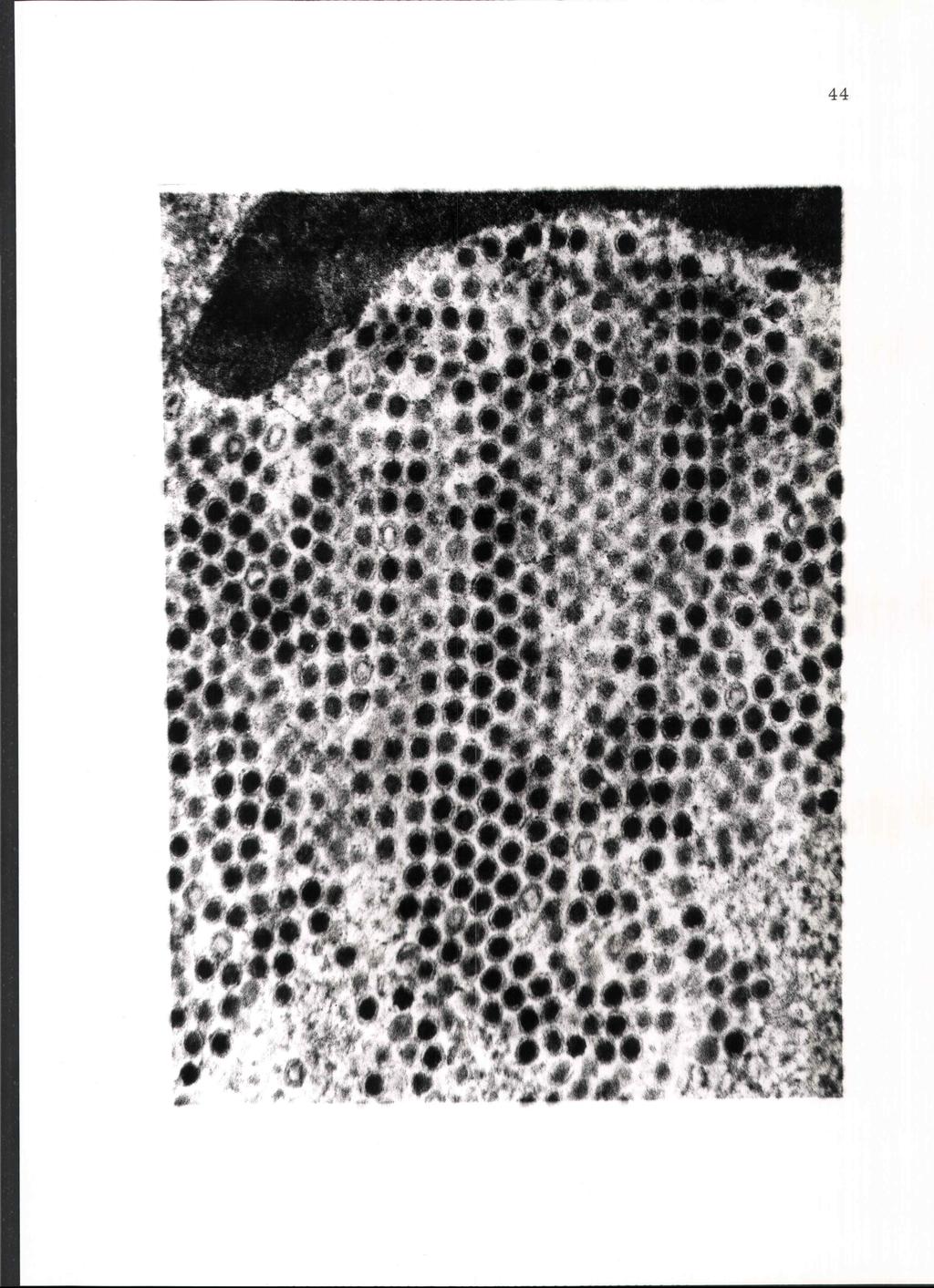
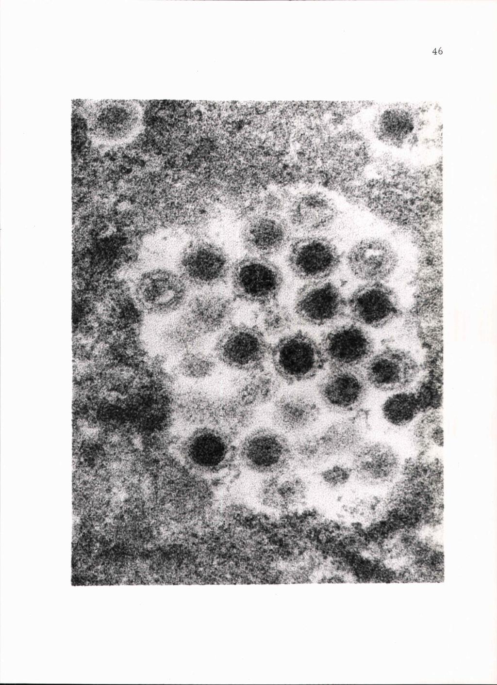
37 TCID 50 per gram of feces on days 5 and 6 PI (Table V). Virus isolation results from organs sampled at necropsy from all three calves are shown in Table II. Virus isolation results from clinical samples (nasal, throat and conjunctival swabs, serum and feces) are shown in Table I. Serum-Virus Neutralization Production of serum neutralizing antibodies was not detected in calves or during the trial periods. An antibody titer of 1:4 was first detected in calf on PI day 7. On PI day 8 the titer reached a level of 1:16. Electron Microscopy Electron photomicrographs revealed both full and empty virus particles in the nuclei of infected testicular cells (Figure 8). Particles were found singly, in groups or in crystalline arrays (Figures 8 and 9). Particles appeared to have a dense inner core and a less dense outer shell and measured mil in diameter (Figures 8, 9 and 10). Fluorescent Antibody Technique Table III. Results of fluorescent antibody stained tissues are shown in In the intestinal tract where the tissue surface could be
38 29 observed in detail, fluorescent cells were consistently noted in the intestinal lamina propria of all three calves (Figures 8 and 9). Massive autofluorescence prevented interpretation of results in heart valve and brain sections from calf
39 Table I, Calf Virus Isolation Results During the Trial Period of Calves Experimentally Infected with Bovine Adenovirus Strain 7T. Day Post Inoculation S S S S S T T T C N NEC T T S S S S NEC F = feces S = serum T = throat C = conjunctiva N = nasal NEC = day of necropsy S S S NEC
40 31 Table II. Virus Isolation Results from Samples taken at Necropsy from Calves Experimentally Infected with Bovine Adenovirus Strain 7T. +b Area Sample a Head & neck Tonsil + Suprapharyngeal LINc Parotid LN + NT d NT Nasal mucosa NT NT + Brain + + Respiratory tract Gastrointestinal tract Other a + = positive viral isolation b = no viral isolation cln = lymph node dnt = sample not tested Lung: apical lobe + + diaphragmatic + + cardiac + + Bronchial LN + NT Mediastinal LN Abomasum Duodenum Jejunum Ileum Colon Mesenteric LN: duodenum jejunum ileum NT NT + NT Heart NT NT Spleen Kidney NT - Adrenal Gland NT + NT Liver + Testicle + + +
41 32 Table III. Fluorescent Antibody Test Results from Samples taken at Necropsy from Calves Experimentally Infected with Bovine Adenovirus Strain 7T. Area Sample Head & neck Tonsil + a b Suprapharyngeal LNc + + Nasal mucosa NT d NT Res piratory tract Lung: apical lobe diaphramatic cardiac Bronchial LN Mediastinal LN Gastrointestinal tract Abomasum NT NT + Duodenum + + Jejunum Ileum Colon NT + Mesenteric LN: duodenum + NT NT jejunum NT + + ileum Other Heart NT NT AUTO Spleen NT NT NT Kidney NT + - Adrenal gland NT - NT Liver NT + Testicle NT NT + Brain NT NT AUTO a + = positive viral isolation b c = no viral isolation NT LN = lymph node dn T = sample not tested e AUTO = autofluorescence - could not be interpreted
42 33 Table IV. Virus Isolation Results of Titered Serum from Calves Experimentally Infected with Bovine Adenovirus Strain 7T. Day Post Calf Calf Calf Inoculation b NEC NEC NEC a +=Undiluted serum positive for virus isolation but serum not titered b - =Negative
43 34 Table V. Day Post Inoculation Virus Isolation Results of Titered Feces from Calves Experimentally Infected with Bovine Adenovirus Strain 7T. Calf Calf Calf `^. -b N Cc NEC NEC NEC a Titer expressed in tissue culture infective doses/50 ml. b - = Negative c NC = no feces collected
44 35 Figure 1. Photomicrograph of confluent monolayer of uninfected bovine testicular cells three days post inoculation. Magnification 83 X. Figure Z. Photomicrograph of bovine testicular cell culture infected with strain 7T bovine adenovirus showing rounding, clumping and increased refractibility of 100% complete CPE of the epithelial cells. Magnification 83 X.

45 . - 'Sr,"*-77-4 A h. el * %!,',,t4t 4* - -,.** (.3*- 'owṫi:;:"..;11::1: I Air ri (7;.41 s Tt,
46 Calf Calf A Calf O cv ;-I cd... 2 ll,. a) f4iiiibil / (1) H - N or ma i Upper Temp. Range I li I I I I I i I I I I I I I I I. am pm am pm am pm am pm am pm am pm am pm am pm am pm am pm Days Post Inoculation Figure 3. Daily temperatures of calves experimentally infected with strain 7T bovine adenovirus.
47 cd ---' U a) Uj o 13 H Normal Upper Leucocyte Range Calf Calf A Calf i i i Day Post Inoculation Figure 4. Leucocyte count of calves experimentally infected with bovine adenovirus strain 7T.
48 coo lymphocyte s polymorphonuclear leucocytes (PMN) o monocytes 4-) U 60 O f: Day Post Inoculation Day Post Inoculation Figure 5. Leucocyte differential of calf Figure 6. Leucocyte differential of calf experimentally inoculated with bovine experimentally inoculated with bovine adenovirus strain 7T. adenovirus strain 7T. AN 10
49 40 Cr) lymphocytes O PMN monocytes kf' Or a e...o----.<>---o---. o.--o----o---- to i Days Post Inoculation Figure 7. Leucocyte differential of calf experimentally inoculated with bovine adenovirus strain 7T.
50 41 Figure 8. Thin section electron photomicrograph of the nucleus of a bovine testicular cell infected with bovine adenovirus strain 7T, showing virus particles singly and in groups in nuclear vacuoles. Magnification 75, 225 X.
51 42
52 43 Figure 9. Thin section electron photomicrograph of the nucleus of a bovine testicular cell infected with bovine adenovirus strain 7T showing virus particles aligned in a crystalline array. Magnification 90, 860X.
53 44
54 45 Figure 10. Thin section electron photomicrograph of the nucleus of a bovine testicular cell infected with bovine adenovirus strain 7T showing virus particles in a vacuole. Magnification 225, 000X.
55 46
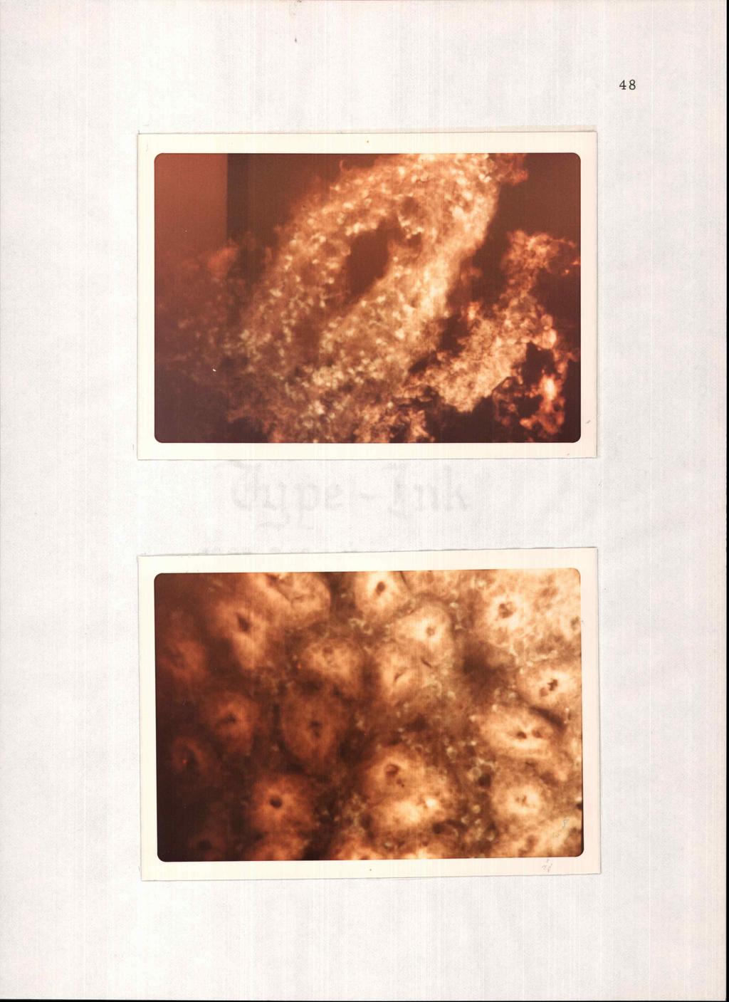
56 47 Figure 11. Photomicrograph section of ileum (calf 42773) showing greenish-yellow virus infected cells in the lamina propria. Fluorescene isothiocyanate conjugated anti-7t virus gamma globulin, rhodamine B bovine albumin counterstain, dark field illumination. Magnification 118X. Figure 12. Photomicrograph section of jejunum (calf 42773) showing greenish-yellow cells in the lamina propria. Fluorescene isothiocyanate conjugate anti-7t virus gamma globulin, rhodamine B bovine albumin counterstain, dark field illumination. Magnification 1I8X.
57 kto
58 49 DISCUSSION Comparatively mild disease signs were observed in experimentally infected calves in this study. Clinical signs of pyrexia, anorexia, diarrhea and increased respiratory rate were all noted. In general these signs were milder than those described in the natural disease (1, 10, 13, 40, 79, p. 126). Calves in this study were maintained in an ideal environment and were not subjected to stress. They were not in contact with other calves and there was no opportunity for the virus to gain virulence by rapid transmission from one animal to another. The navel of each calf was disinfected at birth. Ideal nutritional conditions were maintained. These practices which minimized stress possibly resulted in the reduced severity of the disease. Clinical disease signs were similar to those reported in other calf inoculation studies with subgroup II bovine adenoviruses. In these studies the most consistent clinical finding was a febrile response (1, 10, 1 4, 62, 75). Signs of respiratory embarrassment including dyspnea and hyperpnea have also been reported (1, 10, 16). Calves in this trial did show an abnormally high respiration rate but only when force exercised. The presence of diarrhea or abnormally soft feces appears to be a common sign of infection as seen in this study and as noted by others (1, 10, 75).
59 The leucocyte differential of the normal newborn calf changes considerably with age. The lymphocyte percentage at birth is usually less than 50% (range 30-50%). The neutrophile count usually exceeds the 50% level. These percentages change erratically during the first week of life showing near normal values of 45-75% lymphocytes and 15-45% neutrophiles by one week of age (68, p ). Some viral diseases are associated with an early lymphopenia, eosinophilia and neutrophilia. The extent of lymphopenia varies with the virus and disease severity. A marked neutrophilia is usually associated with secondary bacterial infection (68, p ). Since only three calves were used in the present study, it is not possible to predict any consistent change in the leucocyte count or differential. There was an abnormally high absolute lymphocyte count in calves and (Figure 5). Adenoviruses serve as good antigens and perhaps this is a reflection of the fact that there is a good lymphocyte response prior to antibody production. However, Cole and Tanaka et al. reported leukopenia in calves inoculated with type 4 and 6 bovine adenoviruses (14, 75). Viremia experienced by calves in this study was generally of low titer. Calf had the highest titer viremia which was greater than or equal to 100 TCID 50 per milliliter and was maintained for four days. It is of interest to note that this calf also showed more severe disease signs than the other two calves. The viremia titer in 50
60 51 calves and reached a maximum of 16.0 TCID 50 per milliliter of serum on PI days 6 and 7. The febrile response usually began on PI day 2, approximately one day prior to the detection of viremia. Pyrexia continued throughout the trial with temperatures of calves tending to fluctuate. Morning temperatures were generally lower than evening temperatures. Normal physiological fluctuation, changes in the ambient temperature and pyrexia induced by infection account for this observation. Accordingly, a temperature of oc or greater was considered abnormally elevated. Tanaka et al. also reported viremia of short duration and low titer in calves experimentally infected with type 4 bovine adenovirus (75). Virus isolation from the feces of two calves indicated the presence of virus in the intestinal tract. Virus isolation from feces of calves and began shortly after the onset of viremia (PI day 4) and continued until necropsy. The virus titers in the feces remained comparatively low (between 53 and TCID50 per gram feces). Virus was not isolated from the feces of calf In this calf, virus titer may have been too low to detect virus by isolation methods. Fluorescent antibody staining of several sections of intestinal tissues revealed the presence of virus infected cells. These data indicate the virus was replicating in the intestinal tract and not merely being shed from more anterior organs. It is likely, therefore,
Methods for the Detection of Viruses in Bovine Serum
 JOURNAL OF CLINICAL MICROBIOLOGY, Feb. 1975, p. 212-218 Copyright 1975 American Societv for Microbiology Vol. 1, No. 2 Printed in U.S.A. Methods for the Detection of Viruses in Bovine Serum N. S. SWACK,
THE TECHNIC OF THE KOLMER COMPLEMENT FIXATION TESTS FOR SYPHILIS EMPLOYING ONE-FIFTH AMOUNTS OF REAGENTS JOHN A. KOLMER
 THE TECHNIC OF THE KOLMER COMPLEMENT FIXATION TESTS FOR SYPHILIS EMPLOYING ONE-FIFTH AMOUNTS OF REAGENTS JOHN A. KOLMER WITH THE TECHNICAL ASSISTANCE OP ELSA R. LYNCH Department of Bacteriology and Immunology
AN ELISA FOR THE DETECTION OF ANTIBODIES AGAINST NEWCASTLE DISEASE VIRUS IN AFRICAN VILLAGE POULTRY
 AN ELISA FOR THE DETECTION OF ANTIBODIES AGAINST NEWCASTLE DISEASE VIRUS IN AFRICAN VILLAGE POULTRY J.G. BELL, M. LELENTA Animal Production Unit, Food and Agriculture International Atomic Energy Agency,
Cells and Tissue DNA Isolation Kit (Magnetic Bead System) 50 Preps Product # 59100
 50 Preps Product # 59100") 3430 Schmon Parkway Thorold, ON, Canada L2V 4Y6 Phone: 866-667-4362 (905) 227-8848 Fax: (905) 227-1061 Email: techsupport@norgenbiotek.com Cells and Tissue DNA Isolation Kit (Magnetic Bead System) 50 Preps
3430 Schmon Parkway Thorold, ON, Canada L2V 4Y6 Phone: 866-667-4362 (905) 227-8848 Fax: (905) 227-1061 Email: techsupport@norgenbiotek.com Cells and Tissue DNA Isolation Kit (Magnetic Bead System) 50 Preps
ProductInformation. Genomic DNA Isolation Kit. Product No. GDI-3 Technical Bulletin No. MB-275 May 2000 TECHNICAL BULLETIN
 Genomic DNA Isolation Kit Product No. GDI-3 Technical Bulletin No. MB-275 May 2000 TECHNICAL BULLETIN ProductInformation Product Description Sigma s Genomic DNA Isolation Kit isolates genomic DNA from
THE EFFECTS OF THE INTRATRACHEAL ADMINISTRA- TION OF FOREIGN SERUM.
 THE EFFECTS OF THE INTRATRACHEAL ADMINISTRA- TION OF FOREIGN SERUM. BY F. S. JONES, V.M.D. (From the Deportment of Animal Pathology of The Rockefeller Institute for Medical Research, Princeton, N. J.)
PREPARATION OF HISTOLOGICAL SPECIMENS
 PREPARATION OF HISTOLOGICAL SPECIMENS Histo-techniques Preparation of tissue for microscopic examination Series of processes Ultimate aim to make tissue visible as it is Pathology Vs Anatomy Steps vary
SEROLOGICAL AND PATHOGENICITY STUDIES WITH SOME UNCLASSIFIED PORCINE ADENOVIRUSES
 J. COMP. PATH. 1975. VOL. 85. 437 SEROLOGICAL AND PATHOGENICITY STUDIES WITH SOME UNCLASSIFIED PORCINE ADENOVIRUSES J. B. D ERBYSHIRE,* M. C. CLARKE and A. P. COLLINS BY Agricdttiral Research Council,
TITLE: LIVE/DEAD VIABILITY FOR CLINICAL SAMPLES
 Paul K. Wallace, Ph.D. Director Roswell Park Cancer Institute Elm & Carlton Streets Voice:(716) 845-8471 Buffalo, NY 14263 Fax:(716) 845-8806 FILE NAME FL-SRP-2090.00 Live/Dead Viability for Clinical Samples
Fluorescent Antibody Studies with Herpes Simplex Virus in Unfixed Preparations of Trypsinized Tissue Cultures
 19 O DEA, J. F. & DINEEN, J. K. (1957). J. gen. Microb-iol. 17, 19-24 Fluorescent Antibody Studies with Herpes Simplex Virus in Unfixed Preparations of Trypsinized Tissue Cultures BY J. F. O DEA AND J.
Yellow Fever Vaccine (Live) is a freeze-dried preparation of the 17D strain of yellow fever virus grown in fertilised hen eggs.
 is a freeze-dried preparation of the 17D strain of yellow fever virus grown in fertilised hen eggs.") YELLOW FEVER VACCINE Yellow Fever Vaccine (Live) is a freeze-dried preparation of the 17D strain of yellow fever virus grown in fertilised hen eggs. Production General provisions The production of vaccine
YELLOW FEVER VACCINE Yellow Fever Vaccine (Live) is a freeze-dried preparation of the 17D strain of yellow fever virus grown in fertilised hen eggs. Production General provisions The production of vaccine
For Research Use Only Ver
 INSTRUCTION MANUAL ZR-Duet DNA/RNA MiniPrep Plus Catalog No. D7003 Highlights Efficient isolation and separation of DNA and RNA from any cells, tissue, blood, and biological fluids. High quality DNA and
Product # 24700, 24750
 3430 Schmon Parkway Thorold, ON, Canada L2V 4Y6 Phone: 866-667-4362 (905) 227-8848 Fax: (905) 227-1061 Email: techsupport@norgenbiotek.com Genomic DNA Isolation Kit Product # 24700, 24750 Product Insert
EZ-10 SPIN COLUMN GENOMIC DNA MINIPREPS KIT HANDBOOK
 EZ-0 SPIN COLUMN GENOMIC DNA MINIPREPS KIT HANDBOOK (Bacteria, Plant, Animal, Blood) Version 8 Rev 05/0/03 EZ-0 Genomic DNA Kit Handbook Table of Contents Introduction Limitations of Use Features Applications
Human Pluripotent Stem Cell Functional Identification Kit
 Human Pluripotent Stem Cell Functional Identification Kit Catalog Number SC027B Reagents for the identification of human pluripotent stem cells by in vitro functional differentiation. This package insert
Corning BioCoat Matrigel Matrix 6-well Plates for Embryonic Stem (ES) Cell Culture. Catalog Number Guidelines for Use
 Cell Culture. Catalog Number Guidelines for Use") Corning BioCoat Matrigel Matrix 6-well Plates for Embryonic Stem (ES) Cell Culture Catalog Number 354671 Guidelines for Use Discovery Labware, Inc., Two Oak Park, Bedford, MA 01730, Tel: 1.978.442.2200
Corning BioCoat Matrigel Matrix 6-well Plates for Embryonic Stem (ES) Cell Culture Catalog Number 354671 Guidelines for Use Discovery Labware, Inc., Two Oak Park, Bedford, MA 01730, Tel: 1.978.442.2200
TransIT -Lenti Transfection Reagent
 Quick Reference Protocol, SDS and Certificate of Analysis available at mirusbio.com/6600 INTRODUCTION Lentivirus is an enveloped, single-stranded RNA virus from the Retroviridae family capable of infecting
Propagation of H7 hesc From: UW (John Stamatoyannopoulos) ENCODE group Date: 12/17/2009 Prepared By: S. Paige/S. Hansen (UW)
 ENCODE group Date: 12/17/2009 Prepared By: S. Paige/S. Hansen (UW)") Propagation of H7 hesc From: UW (John Stamatoyannopoulos) ENCODE group Date: 12/17/2009 Prepared By: S. Paige/S. Hansen (UW) Growth and Harvest Modifications Addendum to: Propagation of H7 hesc from UW
Propagation of H7 hesc From: UW (John Stamatoyannopoulos) ENCODE group Date: 12/17/2009 Prepared By: S. Paige/S. Hansen (UW) Growth and Harvest Modifications Addendum to: Propagation of H7 hesc from UW
1. Cross-linking and cell harvesting
 ChIP is a powerful tool that allows the specific matching of proteins or histone modifications to regions of the genome. Chromatin is isolated and antibodies to the antigen of interest are used to determine
SVANOVIR APV-Ab. Avian Pneumovirus Antibody Test
 Avian Pneumovirus Antibody Test Contents Microtitre plate Microtitre plates (96 wells) coated with non-infectious APV antigen (sealed and stored dry) Conjugate Lyophilised (horseradish peroxidase conjugated
Resolve Panel A A Qualitative Test for the Identification of Unexpected Blood Group Antibodies
 Revised October 2015 Reagent Red Blood Cells Resolve Panel A A Qualitative Test for the Identification of Unexpected Blood Group Antibodies 719510 Rx ONLY SUMMARY AND EXPLANATION When an unexpected antibody
Kit Components Product # (50 samples) Wash Solution A Elution Buffer B
 Wash Solution A Elution Buffer B") 3430 Schmon Parkway Thorold, ON, Canada L2V 4Y6 Phone: 866-667-4362 (905) 227-8848 Fax: (905) 227-1061 Email: techsupport@norgenbiotek.com Cells and Tissue DNA Isolation Kit Product # 53100 Product Insert
3430 Schmon Parkway Thorold, ON, Canada L2V 4Y6 Phone: 866-667-4362 (905) 227-8848 Fax: (905) 227-1061 Email: techsupport@norgenbiotek.com Cells and Tissue DNA Isolation Kit Product # 53100 Product Insert
Cyfra 21-1 IRMA. Product information Information about other products is available at: Userś Manual DE52100
 Product information Information about other products is available at: www.demeditec.com Userś Manual Cyfra 21-1 IRMA The CYFRA 21.1 IRMA system provides a direct in vitro quantitative determination of
Adeno-X Rapid Titer Kit
 User Manual Adeno-X Rapid Titer Kit User Manual United States/Canada 800.662.2566 Asia Pacific +1.650.919.7300 Europe +33.(0)1.3904.6880 Japan +81.(0)77.543.6116 Clontech Laboratories, Inc. A Takara Bio
EZ-DNA. Instructions for Use. Genomic DNA Isolation Reagent. Product Description. Kit Reagent. Reagent Required But Not Supplied.
 EZ-DNA Genomic DNA Isolation Reagent Cat. No.: 20-600-50 Store at: Room Temperature Instructions for Use Protocol for Genomic DNA Isolation Tissue Specific Recommendations for the Use of EZ-DNA Assessing
Manufactured by. Zyagen Barnes Canyon Road San Diego, CA 92121, USA
 Alkaline Phosphatase Immunohistochemistry Detection kits For detection of mouse, rabbit, goat, rat, sheep, chicken, guinea pig, and human primary antibodies Size: 500 Tests Catalog #: AK-011, Mouse Kit
Note: for laboratory research use only. RNA High-purity Total RNA Rapid Extraction Kit (Spin-column) Signalway Biotechnology
 Signalway Biotechnology") Note: for laboratory research use only RNA High-purity Total RNA Rapid Extraction Kit (Spin-column) Cat. #: RP1202 (50preps) Signalway Biotechnology I. Kit Content, Storage Condition and Stability Content
Note: for laboratory research use only RNA High-purity Total RNA Rapid Extraction Kit (Spin-column) Cat. #: RP1202 (50preps) Signalway Biotechnology I. Kit Content, Storage Condition and Stability Content
NutriStem V9 XF Medium
 Stem Cells NutriStem V9 XF Medium A defined, xeno-free (XF), serum-free (SF) culture medium for hpsc using vitronectin Instructions for Use Product Description NutriStem V9 XF medium is a defined, xeno-free,
Growth and Maintenance of the 293A Cell Line
 Growth and Maintenance of the 293A Cell Line USER GUIDE Catalog Number R705-07 Publication Number MAN0000303 Revision A.0 For Research Use Only. Not for use in diagnostic procedures. The information in
Contents. Introduction Methods Appendix Kit Contents and Storage...iv. System Overview... 1 Experimental Outline...
 Genomed User Manual ii Contents Kit Contents and Storage...iv Introduction... 1 System Overview... 1 Experimental Outline... 3 Methods... 4 General Information... 4 Purifying gdna from Blood and Body Fluids
L6 Cell Growth Protocol
 L6 Cell Growth Protocol Background: Parental L6 cells were subcloned for high fusion (1, 2). Materials: 1. α-minimal Essential Medium (α-mem) Life Technologies #12571-063 2. Fetal Bovine Serum (FBS) Life
666 THE JOURNAL OF CELL BIOLOGY' VOLUME 71, 1976" pages
 ph-dependent BINDING OF IMMUNOGLOBULINS TO INTESTINAL CELLS OF THE NEONATAL RAT RICHARD RODEWALD. From the Department of Biology, University of Virginia, Charlottesville, Virginia 22901. Neonatal rats
VDL106.3 LARGE-SCALE AMPLIFICATION AND PURIFICATION OF ADENOVIRAL VECTOR
 1. Purpose 1.1. The purpose of this protocol is to amplify an adenoviral vector from a crude lysate (final product in SOP:VDL105.2 Preparation of Adenoviral Vector Lysate) for production of high titer
antigen." 2 Moreover, when mixed populations of normal and sensitive cells
 DELA YED HYPERSENSITIVITY IN VITRO: ITS MEDIATION BY CELL-FREE SUBSTANCES FORMED BY LYMPHOID CELL-ANTIGEN INTERACTION* BY JOHN R. DAVIDt DEPARTMENT OF MEDICINE, NEW YORK UNIVERSITY SCHOOL OF MEDICINE Communicated
The Biotechnology Education Company. Quantitative ELISA. Storage: See Page 3 for specific storage instructions EXPERIMENT OBJECTIVE:
 The Biotechnology Education Company Revised and Updated Quantitative ELISA Storage: See Page 3 for specific storage instructions EXPERIMENT OBJECTIVE: EDVO-Kit # 278 The objective of this experiment is
Chapter 10: Classification of Microorganisms
 Chapter 10: Classification of Microorganisms 1. The Taxonomic Hierarchy 2. Methods of Identification 1. The Taxonomic Hierarchy Phylogenetic Tree of the 3 Domains Taxonomic Hierarchy 8 successive taxa
OIE Guideline. International Reference Antibody Standards for Antibody Assays. 1. Introduction
 OIE Guideline International Reference Antibody Standards for Antibody Assays 1. Introduction 1.1. Purpose This document provides guidelines for the preparation, validation and distribution of antibodies
Analysis of Corynebacterium vaginale by an Immunodiffusion
 APPLIED MICROBIOLOGY, Mar. 1974, p. 469-474 Copyright 0 1974 American Society for Microbiology Vol. 27, No. 3 Printed in U.S.A. Analysis of Corynebacterium vaginale by an Immunodiffusion Technique MARY
Separation and Properties of a Red Cell Sensitizing
 JOURNAL OF BACTERIOLOGY, June, 1966 Copyright @ 1966 American Society for Microbiology Vol. 91, No. 6 Printed in U.S.A. Separation and Properties of a Red Cell Sensitizing Substance from Streptococci MERWIN
SKIN INFECTION OF RABBITS WITH HEMOLYTIC STREP- TOCOCCI ISOLATED FROM A PATIENT WITH ERYSIPELAS.
 SKIN INFECTION OF RABBITS WITH HEMOLYTIC STREP- TOCOCCI ISOLATED FROM A PATIENT WITH ERYSIPELAS. I. METHOD OF DEMONSTRATING PROTECTIVE ACTION OF IMMUNE SERA. BY THOMAS M. RIVERS, M.D. (From the Hospital
CytoPainter Golgi Staining Kit Green Fluorescence
 ab139483 CytoPainter Golgi Staining Kit Green Fluorescence Instructions for Use Designed for the detection of Golgi bodies by microscopy This product is for research use only and is not intended for diagnostic
EZ-10 SPIN COLUMN GENOMIC DNA MINIPREPS KIT HANDBOOK
 EZ-0 SPIN COLUMN GENOMIC DNA MINIPREPS KIT HANDBOOK (Bacteria, Plant, Animal, Blood) Version 5.0 Rev 03/25/205 Table of Contents Introduction 2 Limitations of Use 2 Features 2 Applications 2 Storage 2
Human IL-6 ELISA Set
 Human IL-6 ELISA Set Catalog No. CDK082B Quantity: 10 x 96 tests PRODUCT SPECIFICATIONS : Specificity: Recognizes both natural and recombinant human IL-6 Range: 6.25 pg / ml - 200 pg / ml Sensitivity:
BIOCHEMICAL AND BIOPHYSICAL RESEARCH COMMUNICATIONS
 A PREPARATIVE METHOD FOR OBTAINING ENUCLEATED MAMMALIAN CELLS Michael H. Wigler and I. Bernard Weinstein Institute of Cancer Research and Departments of Medicine and Microbiology, College of Physicians
Product Information. Before you begin. Component A 1 vial of 30 ul vial of 300 ul each Glycerol. Tris
 Glowing Products for Science Mix-n-Stain Antibody Labeling Kits Size: 1 labeling per kit Storage: -20 o C Stability: Stable for at least 1 year from date of receipt when stored as recommended. Components:
MyBioSource.com. Human Total Bilirubin (TBB) Elisa kit. (Competitive ELISA)
 Elisa kit. (Competitive ELISA)") Human Total Bilirubin (TBB) Elisa kit (Competitive ELISA) 96 Tests Catalog Number: MBS756198 Store all reagents at 2-8 C Valid Period: six months For samples: Serum, plasma, cell culture supernatants,
Human Total Bilirubin (TBB) Elisa kit (Competitive ELISA) 96 Tests Catalog Number: MBS756198 Store all reagents at 2-8 C Valid Period: six months For samples: Serum, plasma, cell culture supernatants,
Diagnostic Virology. Diagnostic Virology
 Diagnostic Virology Diagnostic Virology The purpose is to diagnose viral diseases in the patient This can be achieved by: Directly detecting the virus or viral products Indirectly detecting an immunological
T ECHNICAL MANUAL. Culture of Human Mesenchymal Stem Cells Using MesenCult -XF Medium
 T ECHNICAL MANUAL Culture of Human Mesenchymal Stem Cells Using MesenCult -XF Medium i Table of Contents 1.0 Materials... 1 1.1 MesenCult -XF Medium and Required Products... 1 1.2 Additional Required
High Pure PCR Template Preparation Kit for preparation of 100 nucleic acid samples Cat. No
 for preparation of 100 nucleic acid samples Cat. No. 1 796 88 Principle Cells are lysed during a short incubation with Proteinase K in the presence of a chaotropic salt (guanidine HCl), which immediately
QIAGEN Supplementary Protocol: Isolation of genomic DNA from tissue using the QIAGEN-tip Storage of tissue samples.
 QIAGEN Supplementary Protocol: Isolation of genomic DNA from tissue using the QIAGEN-tip 2500 This protocol is designed for the rapid, easy, and non-toxic preparation of up to 2 mg genomic DNA from not
QIAfilter Plasmid Midi Kit (Cat #: 12243)
") QIAfilter Plasmid Midi Kit (Cat #: 12243) Things to do before starting Add the provided RNase A solution to Buffer P1 before use. Use one vial of RNase A (centrifuge briefly before use) per bottle of Buffer
QIAfilter Plasmid Midi Kit (Cat #: 12243) Things to do before starting Add the provided RNase A solution to Buffer P1 before use. Use one vial of RNase A (centrifuge briefly before use) per bottle of Buffer
An indirect haemagglutination test to detect serum antibodies to Giardia lamblia
 J. Biosci., Vol. 10, Number 4, December 1986, pp. 475-480. Printed in India. An indirect haemagglutination test to detect serum antibodies to Giardia lamblia K. N. JALAN, TUSHER MAITRA and RITA DAS Kothari
Part 1 Johne s Disease Overview A concise summary of the latest facts about Johne s disease and recommended methods for diagnosis and control.
 Johne s Disease: Practical Solutions and Problem Solving Michael T. Collins, DVM, PhD, DACVM, Professor School of Veterinary Medicine University of Wisconsin Madison, WI 53706 Part 1 Johne s Disease Overview
Procedure No.: CLSI Effective Date: 09/09 Supersedes Revision/Date: 11/01 Revision: 09/09 Date Adopted:
 Page 1 of 6 Procedure: Doc#: ASI VDRL ANTIGEN TEST 6004-950 CLSI Effective Date: 09/09 Supersedes Revision/Date: 11/01 Revision: 09/09 Supersedes Procedure # Prepared by: Date Adopted: Review Date: Revision
OPPF-UK Standard Protocols: Mammalian Expression
 OPPF-UK Standard Protocols: Mammalian Expression Joanne Nettleship joanne@strubi.ox.ac.uk Table of Contents 1. Materials... 3 2. Cell Maintenance... 4 3. 24-Well Transient Expression Screen... 5 4. DNA
For in vitro Veterinary Diagnostics only. DNA Extraction and PCR Detection Kit for Pasteurella multocida.
 For in vitro Veterinary Diagnostics only. DNA Extraction and PCR Detection Kit for Pasteurella multocida www.kylt.eu DIRECTION FOR USE Art. No. 31058 / 31059 Kylt Pasteurella multocida DNA Extraction and
Bovine IgG ELISA Kit
 Bovine IgG ELISA Kit Cat. No. E11-118 Components Supplied Bovine IgG Pre-Coated 96-well Strip Plate, 1 each Bovine IgG Standard, 500 ng/vial, 2 each Bovine IgG Detection Antibody, 12 ml 20X Dilution Buffer
SANTA CRUZ BIOTECHNOLOGY, INC.
 TECHNICAL SERVICE GUIDE: Western Blotting 2. What size bands were expected and what size bands were detected? 3. Was the blot blank or was a dark background or non-specific bands seen? 4. Did this same
Clonetics Skeletal Muscle Myoblast Cell Systems HSMM Instructions for Use
 Lonza Walkersville, Inc. Walkersville, MD 21793-0127 USA U.S. Scientific Support: 800 521 0390 scientific.support@lonza.com EU/ROW Scientific Support: +32 87 321 611 scientific.support.eu@lonza.com Document
Standard Operating Procedure. Immunology
 Standard Operating Procedure Immunology SOP Ref No SOP title Category SOP-IU008 Human PBMC isolation, freezing and thawing procedures Laboratory Version 1.1 Page 1 of 12 Isolation, freezing and thawing
Rat Calcitonin Gene Related Peptide (CGRP) Elisa kit
 Elisa kit") Rat Calcitonin Gene Related Peptide (CGRP) Elisa kit 96 Tests Catalogue Number: AMS.E02C0744 Store all reagents at 2-8 C Valid Period: six months For samples: Serum, plasma, cell culture supernatants,
Rat Calcitonin Gene Related Peptide (CGRP) Elisa kit 96 Tests Catalogue Number: AMS.E02C0744 Store all reagents at 2-8 C Valid Period: six months For samples: Serum, plasma, cell culture supernatants,
FURTHER EVIDENCE OF FETAL INFECTION WITH HERPES SIMPLEX VIRUS
 THE AMERICAN JOURNAL OF CLINICAL PATHOLOGY Vol. 37, No. 3, pp. 289-293 March, 1962 Copyright 1902 by The Williams & Wilkins Co. Printed in U.S.A. FURTHER EVIDENCE OF FETAL INFECTION WITH HERPES SIMPLEX
LHCN-M2 cell culture, differentiation treatment, and cross-linking protocol.
 Cell Growth Protocol and Differentiation treatment for the LHCN-M2 Cell Line From: HudsonAlpha/Caltech ENCODE group Date: February 17, 2011 Prepared by: Chun-Hong Zhu and Woodring E. Wright (typed by Brian
Human IgG ELISA Quantitation Set
 Human IgG ELISA Quantitation Set Cat. No. E80-104 Components Supplied Affinity purified Goat anti-human IgG-Fc Coating Antibody A80-104A, 1 ml at 1 mg/ml Human Reference Serum, RS10-110-4, 0.1 ml HRP Conjugated
University Animal Care Committee (UACC) Standard Operating Procedure (SOP) Rodent Health Surveillance Program
 Standard Operating Procedure (SOP) Rodent Health Surveillance Program") Institutional SOP M0: Rodent Health Surveillance University Animal Care Committee (UACC) Standard Operating Procedure (SOP) SOP # M0 Title Rodent Health Surveillance Program. Purpose This Standard Operating
Institutional SOP M0: Rodent Health Surveillance University Animal Care Committee (UACC) Standard Operating Procedure (SOP) SOP # M0 Title Rodent Health Surveillance Program. Purpose This Standard Operating
VZV Replication Assays Samantha J. Griffiths * and Jürgen Haas
 VZV Replication Assays Samantha J. Griffiths * and Jürgen Haas Division of Pathway and Infection Medicine, University of Edinburgh, Edinburgh, UK *For correspondence: samantha.griffiths@ed.ac.uk [Abstract]
LECTURE: 26 SIMPLE SEROLOGICAL LABORATORY TECHNIQUES LEARNING OBJECTIVES:
 LECTURE: 26 Title SIMPLE SEROLOGICAL LABORATORY TECHNIQUES LEARNING OBJECTIVES: The student should be able to: Define the term "simple serological techniques". Describe the benefit of the use of serological
HiYield TM Genomic DNA Extraction Kit
 HiYield TM Genomic DNA Extraction Kit CONTENTS Genomic DNA Extraction Kit...... 1 Blood/Bacteria/Cultured Cells Cat.No. YGB50 // YGB100 // YGBM25 Blood Protocol..... 4 Cultured Cells Protocol...... 5 Bacterial
Materials and Methods Materials Required for Fixing, Embedding and Sectioning. OCT embedding matrix (Thermo Scientific, LAMB/OCT)
") Page 1 Introduction Tissue freezing and sectioning is a rapid method of generating tissue samples (cryosections) for histological analysis, and obviates the need for wax embedding. The method is popular
Page 1 Introduction Tissue freezing and sectioning is a rapid method of generating tissue samples (cryosections) for histological analysis, and obviates the need for wax embedding. The method is popular
A Level. A Level Biology. Cells, Microscopes, Cell Cycle and Immunity Questions. AQA, OCR, Edexcel. Name: Total Marks: Page 1
 AQA, OCR, Edexcel A Level A Level Biology Cells, Microscopes, Cell Cycle and Immunity Questions Name: Total Marks: Page 1 Q1.The diagram shows a eukaryotic cell. (a) Complete the table by giving the letter
PeliClass human IgG subclass ELISA kit Enzyme-linked immunosorbent assay
 PeliClass human IgG subclass ELISA kit Enzyme-linked immunosorbent assay Catalog No: M1551 Size: six pre-coated 8-well strips for each of the four IgG subclasses Test description The PeliClass human subclass
Human Myostatin, ELISA Kit (MSTN)
") Human Myostatin, ELISA Kit (MSTN) 96 Tests Catalog Number: MBS733837 Store all reagents at 2-8 C Valid Period: six months For samples: Cell culture fluid & body fluid & tissue homogenate Serum or blood
Human Myostatin, ELISA Kit (MSTN) 96 Tests Catalog Number: MBS733837 Store all reagents at 2-8 C Valid Period: six months For samples: Cell culture fluid & body fluid & tissue homogenate Serum or blood
In-Cell Western Kits I and II
 Odyssey and Aerius Infrared Imaging Systems In-Cell Western Assay Kits I and II Published November, 2006. The most recent version of this protocol is posted at http://biosupport.licor.com/protocols.jsp
Culture and freezing methods for AICS cell lines
 Allen Institute for Cell Science Culture and freezing methods for AICS cell lines Required Reagent List: Complete mtesr1 culture media, referred to in this protocol as simply mtesr1 : 400 ml basal media
Chikungunya IgM ELISA Kit
 Chikungunya IgM ELISA Kit Cat.No: DEIA2163 Lot. No. (See product label) Intended Use The Chikungunya IgM-μ-capture ELISA is intended for measurement of IgM class antibodies to Chikungunya virus in human
ON THE FORMATION OF SPECIFIC ANTI-BODIES IN THE BLOOD, FOLLOWING UPON TREATMENT WITH THE SERA OF DIFFERENT ANIMALS.
 ON THE FORMATION OF SPECIFIC ANTI-BODIES IN THE BLOOD, FOLLOWING UPON TREATMENT WITH THE SERA OF DIFFERENT ANIMALS. GEORGE 1-1. F. NUT T ALL. DURING the thirteen years which have elapsed since I demonstrated
Product Permission Document (PPD) of Botulinum Toxin Type A for Injection Ph.Eur Purified Neurotoxin Complex
 of Botulinum Toxin Type A for Injection Ph.Eur Purified Neurotoxin Complex") Product Permission Document (PPD) of Botulinum Toxin Type A for Injection Ph.Eur Purified Neurotoxin Complex Brand Name : BOTO GENIE 1. Introduction : BOTO GENIE (Botulinum Toxin Type A for Injection Ph.Eur)
Product Permission Document (PPD) of Botulinum Toxin Type A for Injection Ph.Eur Purified Neurotoxin Complex Brand Name : BOTO GENIE 1. Introduction : BOTO GENIE (Botulinum Toxin Type A for Injection Ph.Eur)
Immunoassay Kit Catalog # KCA0021. Canine. C-Reactive Protein
 Immunoassay Kit Catalog # KCA0021 Canine C-Reactive Protein BioSource International, Inc. 542 Flynn Road Camarillo, California 93012 USA Tel: 805-987-0086 800-242-0607 FAX: 805-987-3385 email: tech.support@biosource.com
Antigenic Relationships among Autonomous Parvoviruses
 J. gen. Virol. (1986), 67, 2839-2844. Printed in Great Britain 2839 Key words: antigenic relationships/autonomous parvoviruses Antigenic Relationships among Autonomous Parvoviruses ByW. L. MENGELING, l*
The turnover and shedding of epithelial cells
 Gut, 1961, 2, 110 The turnover and shedding of epithelial cells Part I The turnover in the gastro-intestinal tract B. CREAMER, R. G. SHORTER, AND JOHN BAMFORTH From St. Thomas's Hospital Medical School,
TECHNICAL BULLETIN NUCLEI EZ PREP NUCLEI ISOLATION KIT. Product Number NUC-101 Store at 2-8 C
 NUCLEI EZ PREP NUCLEI ISOLATION KIT Product Number NUC-101 Store at 2-8 C TECHNICAL BULLETIN Product Description Sigma s Nuclei EZ Prep Kit is designed for the rapid isolation of nuclei from mammalian
Human Peptide YY EIA
 KAMIYA BIOMEDICAL COMPANY Human Peptide YY EIA For the quantitative determination of Peptide YY in human serum and plasma. Cat. No. KT-378 For Research Use Only. 1 Rev. 9822378 PRODUCT INFORMATION Human
Rat TAR DNA Binding Protein 43 (TDP-43) ELISA
 ELISA") KAMIYA BIOMEDICAL COMPANY Rat TAR DNA Binding Protein 43 (TDP-43) ELISA For the quantitative determination of rat TDP-43 in serum, plasma, cell culture supernatants, body fluid and tissue homogenate Cat.
KAMIYA BIOMEDICAL COMPANY Rat TAR DNA Binding Protein 43 (TDP-43) ELISA For the quantitative determination of rat TDP-43 in serum, plasma, cell culture supernatants, body fluid and tissue homogenate Cat.
Purification of cytoplasmic RNA from animal cells using the RNeasy Mini Kit
 QIAGEN Supplementary Protocol: Purification of cytoplasmic RNA from animal cells using the RNeasy Mini Kit This protocol requires the RNeasy Mini Kit. IMPORTANT: Please consult the Safety Information and
Adeno Associated Virus (AAV) Purification ViraKit
 Purification ViraKit") Catalog Number 003061 003063 Adeno Associated Virus (AAV) Purification ViraKit Protocol FOR RESEARCH USE ONLY. NOT FOR USE IN DIAGNOSTIC OR HUMAN CLINICAL PROCEDURES. Please read through the entire kit
Catalog Number 003061 003063 Adeno Associated Virus (AAV) Purification ViraKit Protocol FOR RESEARCH USE ONLY. NOT FOR USE IN DIAGNOSTIC OR HUMAN CLINICAL PROCEDURES. Please read through the entire kit
This Document Contains:
 This Document Contains: 1. In-Cell Western Protocol II. Cell Seeding and Stimulation Supplemental Protocol III. Complete Assay Example: Detailing the Seeding, Stimulation and Detection of the A431 Cellular
Human Bordetella Pertussis IgG ELISA kit
 Human Bordetella Pertussis IgG ELISA kit Catalog number: NR-R10157 (96 wells) The kit is designed to qualitatively detect Bordetella Pertussis IgG in Human serum or plasma. FOR RESEARCH USE ONLY. NOT FOR
TransIT -Lenti Transfection Reagent
 Quick Reference Protocol, SDS and Certificate of Analysis available at mirusbio.com/6600 INTRODUCTION Lentivirus is an enveloped, single-stranded RNA virus from the Retroviridae family capable of infecting
Porcine IgM (Immunoglobulin M) ELISA Kit
 ELISA Kit") Porcine IgM (Immunoglobulin M) ELISA Kit Catalogue No: EP0085 Size: 48T/96T Reactivity: Porcine Detection Range: 0.156-10ng/ml Sensitivity:
Porcine IgM (Immunoglobulin M) ELISA Kit Catalogue No: EP0085 Size: 48T/96T Reactivity: Porcine Detection Range: 0.156-10ng/ml Sensitivity:
For Research Use Only Ver
 INSTRUCTION MANUAL Quick-RNA Miniprep Plus Kit Catalog Nos. R1057T, R1057, & R1058 Highlights Purify high-quality total RNA (including small/micro RNAs) from any cells, tissues, blood and biological fluids.
E. coli O26 Latex Test Kit
 Importance of STEC Determination Shiga toxin-producing Escherichia coli strains (non-o157 STEC) have become an increasing public health concern. Some of the non-o157 STEC possess the same range of virulence
Alt-R CRISPR-Cpf1 System:
 user guide Alt-R CRISPR-Cpf1 System: Delivery of ribonucleoprotein complexes in HEK-293 cells using the Amaxa Nucleofector System See what more we can do for you at www.idtdna.com. For Research Use Only
SAMPLE PROCEDURE , 11/07
 SAMPLE PROCEDURE This Sample Procedure is not intended as a substitute for your facility s Procedure Manual or reagent labeling, but rather as a model for your use in customizing for your laboratory s
Compatible with commonly used anticoagulants (i.e., EDTA, heparin, citrate).
.") INSTRUCTION MANUAL Quick-DNA Microprep Kit Catalog Nos. D3020 & D3021 Highlights Quick purification of high quality DNA from whole blood, plasma, serum, body fluids, buffy coat, lymphocytes, swabs or cultured
INSTRUCTION MANUAL Quick-DNA Microprep Kit Catalog Nos. D3020 & D3021 Highlights Quick purification of high quality DNA from whole blood, plasma, serum, body fluids, buffy coat, lymphocytes, swabs or cultured
Human IgG Antigen ELISA Kit
 Human IgG Antigen ELISA Kit Catalog No: IHUIGGKT Lot No: SAMPLE INTENDED USE This human immunoglobulin G antigen assay is intended for the quantitative determination of total human IgG antigen in serum,
CHOgro Expression System
 SDS and Certificate of Analysis available at mirusbio.com/6260 INTRODUCTION The CHOgro Expression System is an optimized platform for transient, high titer protein production in suspension CHO derived
BIOPHARMACEUTICAL PROCESS EVALUATED FOR VIRAL CLEARANCE
 The purpose of Viral Clearance evaluation is to assess the capability of a manufacturing production process to inactivate and/or remove potential viral contaminants. Experience and knowledge in selecting
Canine Symmetric dimethylarginine ELISA Kit
 Distribuito in ITALIA da Li StarFish S.r.l. Via Cavour, 35 20063 Cernusco S/N (MI) telefono 02-92150794 fax 02-92157285 info@listarfish.it www.listarfish.it Optimize Your Research Canine Symmetric dimethylarginine
Human IL-1 alpha ELISA Kit
 Human IL-1 alpha ELISA Kit Catalog No: CDK025B Quantity: 2 x 96 tests SPECIFICITY : RANGE : SENSITIVITY : INCUBATION : SAMPLE TYPES : Recognizes both natural and recombinant human IL-1 alpha 31.2 pg /