Cell Proliferation and Death
|
|
|
- Gertrude Jacobs
- 6 years ago
- Views:
Transcription
1 Cell Proliferation and Death Derek Davies, Cancer Research UK
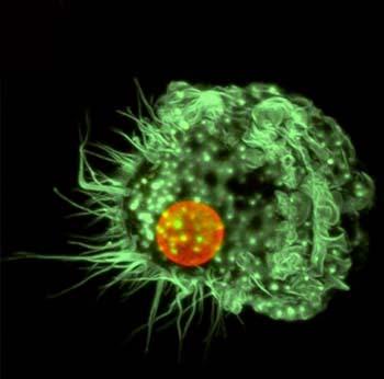


2 Proliferation A cell Apoptosis Cell death Proliferation signals Senescence
 Diamino-2-phenylindole (DAPI) DRAQ5,")
3 DNA analysis Propidium Iodide Ethidium Bromide Hoechst dyes Cyanine dyes eg TO-PRO-3, SYTO/SYTOX dyes Acridine Orange Pyronin Y Styryl Dyes eg LDS-751 Mithramycin, Chromomycin 7 Aminoactinomycin D (7AAD) Diamino-2-phenylindole (DAPI) DRAQ5, DRAQ7
4 DNA analysis We can use DNA dyes in two ways: As viability dyes Exclusion of dead cells during analysis and sorting Identification and quantification of apoptotic cells To measure DNA content and monitor cell cycle and its regulation

5 Dead cell discrimination Dead Dead Propidium Iodide Note log scale! Forward Scatter
6 DNA analysis We can use DNA dyes in two ways: As viability dyes Exclusion of dead cells during analysis and sorting Identification and quantification of apoptotic cells To measure DNA content and monitor cell cycle and its regulation
7 The mammalian cell cycle G 1 G 0 S G1: Gap 1 S: Synthetic G2: Gap 2 M: Mitosis G0: cells that cease division M G 2


8 The mammalian cell cycle RB Phosphorylation Cdk4/Cyclin D Cdk6/Cyclin D Cdk2/cyclin E Cdk2/Cyclin A G 1 INK4, p21, p27, p57 P21, p27, p57 G 0 S Cdk2/Cyclin A M G 2 Cdc2/Cyclin A Cdc2/Cyclin B RB Dephosphorylation
9 Cell cycle analysis by flow cytometry Cells must be permeable - can use detergent or fixation (ethanol is best) DNA in cells can be stained with a fluorescent dye DNA probes like PI are stochiometric and increase fluorescence on binding Dyes either intercalate or bind specific base pairs So we can measure how much DNA is in a cell Basic protocol - fix, wash twice, remove RNA and stain with DNA-binding dye
10 In an ideal world. # of Events Increase in Fluorescence Intensity
11 In the real world. # of Events CV: SD/mean x 100. Increase in Fluorescence Intensity
12 DNA stained with propidium iodide 1000 S Phase 800 G1 Cell count G2/M Propidium Iodide DNA Content Note linear scale!
13 DNA stained with propidium iodide Cell count Propidium Iodide DNA Content We can quantitate the percentage of cells in each phase of the cell cycle and monitor the effect of treatments

14 Example 1: Compare cycles Example 2: S phase block Example 3: M block G1 S G2 G1 S G2 G1 S G2
15 DNA analysis in a clinical situation Many tumours show altered DNA content Diploid index may have prognostic significance Many tumours show increased proliferation S phase fraction may have prognostic significance Diploid G1 Aneuploid G1 DI= Cell count Cell count Aneuploid G2 Cell count DNA Index = Propidium Iodide - Area Propidium Iodide - Area Propidium Iodide - Area
16 Analysis of DNA histograms - pitfalls and a better approach G1 54% S 27% G2 18% The use of markers gives a good indication but is only an estimate! # Cells Propidium Iodide # Cells G1 43% S 45% G2 10% Mathematical modeling is a better approach but still not ideal! Propidium Iodide
17 DNA analysis by a single fluorochrome can only take us so far! G 0 -G 1 Cell Number S G 2 -M Fluorescence Intensity
18 Cell cycle analysis - Bromodeoxyuridine (BrdU) method Thymidine analog Taken up by cycling cells Use for comparative growth rates, length of cell cycle, pulse labelling Staining procedure involves unwinding DNA Combine with Propidium iodide
19 Typical dual parameter plot Anti-BrdU FITC BrdU-FITC S Phase 10 G1 G2/M Propidium Iodide Propidium Iodide

20 Compare comparative growth rates MCF10A Breast cancer cell line Control Drug-treated BrdU FITC % 33% BrdU FITC Propidium iodide Propidium iodide
21 Measuring Proliferation by dye dilution Dye must be taken up by live cells Dye must have low toxicity Dye must be compatible with flow cytometric set-up Dye must be equally apportioned between daughter cells Lipophilic dyes that label cell membrane Succinimidyl dyes that label intracellular proteins
22 Measuring Proliferation by dye dilution Divisions: # Cells /40 Violet-A

23 Measuring Proliferation by dye dilution Serum free + Serum Overlay # Cells # Cells % of Max /40 Violet-A /40 Violet-A /40 Violet-A

24 Measuring Proliferation by dye dilution B-Cell Marker CFSE

25 The other side of the coin

,")
26 apoptosis Falling off Distinct from necrosis and oncosis Programmed cell death Kerr, Wyllie and Currie BJC (1972), 26:239


27 Normal development Normal tissue turnover Negative selection in immune system T cell killing Exposure to certain conditions Where is apoptosis seen?


28 Where is apoptosis seen? Normal development Normal tissue turnover Negative selection in immune system T cell killing Exposure to certain conditions Alzheimer s Disease Parkinson s Disease Autoimmune disorders Neurodegenerative disease Cancer
29 Necrosis Apoptosis Affects groups of cells Affects individual cells Non-physiological induction Physiological induction (viral, poison, ischemia) (lack of signals, changes) Phagocytosis by macrophages Phagocytosis by macrophages or other cells Inflammatory response No inflammatory response
 Loss of cell surface structures Phosphatidylserine externalisation Apoptotic bodies Lamin B proteolysis Cell detachment DNA")
30 Apoptosis Morphological Functional Cell Shrinkage Free Ca2+ rise Cell shape change bcl2/bax interaction Condensation of cytoplasm Cell dehydration Nuclear envelope changes Loss of mitochondrial membrane potential Nuclear fragmentation Enzyme activation (caspases) Loss of cell surface structures Phosphatidylserine externalisation Apoptotic bodies Lamin B proteolysis Cell detachment DNA denaturation Phagocytosis of remains kb cleavage Intra-nucleosomal cleavage Protein cross-linking

31 Why is apoptosis important? Self-sufficiency in growth signals Insensitivity to anti-growth signals Evading apoptosis Cancer Limitless replicative potential Sustained angiogenesis Tissue invasion and metastasis Hanahan, D. and Weinberg, R.A Cell. 100:57.

32 Put simply. reviews/poster/apoptosis
33 Major Apoptotic Pathways in Mammalian Cells Death Receptor Pathway Mitochondrial Pathway DISC Fas/Apo1 /CD95 D D D D D Caspase 8 FasL FADD Procaspase 8 Cellular targets BID DNA damage Procaspase 3 Caspase 3 oxidants ceramide others Apaf-1 Procaspase 9 datp Apaf -1 Caspase 9 apoptosome Bcl-2 datp Cytochrome c

34 The road to commitment


35 How can apoptosis be detected? DNA Laddering Comet assay Electron microscopy Flow cytometry
36 Apoptosis detection by Flow Cytometry Light scattering/cell permeability (PI, DAPI, To-Pro-3) Untreated Treated Dead 10 3 Dead Propidium Iodide 10 2 Propidium Iodide Live 10 1 Live Forward Scatter Forward Scatter
37 Apoptosis detection by Flow Cytometry Changes to the mitochondria (TMRE, CMX dyes, JC-1) Untreated Treated Dead Dead TO-PRO TO-PRO Live 10 1 Live 10 0 Apoptotic CMXRos 10 0 Apoptotic CMXRos
38 Apoptosis detection by Flow Cytometry Changes to the cell membrane (Annexin binding) 10 4 Dead Untreated 10 4 Dead Treated Propidium Iodide 10 2 Live Propidium Iodide 10 2 Live 10 1 Apop 10 1 Apop Annexin V-FITC Annexin V-FITC
39 Apoptosis detection by Flow Cytometry Changes in enzyme expression (Caspases 3, 8 and 9) Untreated Treated Cell count 400 Cell count 400 Cell count Cleaved Caspase-3 FITC Cleaved Caspase-3 FITC Cleaved Caspase-3 FITC
40 Apoptosis detection by Flow Cytometry Changes in cellular DNA (Fragmentation and strand breaks) Untreated Treated Counts 400 Counts Propidium Iodide Propidium Iodide
41 Integration of apoptosis methods TMRE, Annexin, 7-AAD, Hoechst AAD 10 2 TMRE Annexin V-FITC Hoechst TMRE 10 2 TMRE Hoechst Hoechst 33342

42 Which method should I use to assess proliferation and death? What is the question? Cell type? Cultured cells? Suspension or adherent? Primary cells?. What has happened to the cells? Treatment? Time course? What other information is being sought e.g. concurrent phenotyping. Are there any technical restrictions e.g. lasers. Cost, simplicity and number of samples. Expertise available
Technical Bulletin. Multiple Methods for Detecting Apoptosis on the BD Accuri C6 Flow Cytometer. Introduction
 March 212 Multiple Methods for Detecting Apoptosis on the BD Accuri C6 Flow Cytometer Contents 1 Introduction 2 Annexin V 4 JC-1 5 Caspase-3 6 APO-BrdU and APO-Direct Introduction Apoptosis (programmed
Choose the right assay to fit your experimental needs:
 The analysis of cell viability, cytotoxicity, cell cycle state, cell proliferation, and cell death are critical to most cell-based studies. Use this guide to understand your options when you need to assay
Flow Cytometry. Marta Argenti, PhD student. Department of Biomedical Sciences Padua
 Flow Cytometry Marta Argenti, PhD student Department of Biomedical Sciences Padua 14.12.12 Flow ~ cells in motion Cyto ~ cell Metry ~ measure Physical properties: Flow Cytometry is the measurement of cells
Apoptosis detection. Apoptosis assays for the Attune Acoustic Focusing Cytometer. APOPTOSIS DETECTION Attune Acoustic Focusing Cytometer
 POPTOSIS ETECTION ttune coustic Focusing Cytometer poptosis detection poptosis assays for the ttune coustic Focusing Cytometer poptosis is a carefully regulated process of cell death that occurs as a normal
Multiplex Fluorescence Assays for Adherence Cells without Trypsinization
 Multiplex Fluorescence Assays for Adherence Cells without Trypsinization The combination of a bright field and three fluorescent channels allows the Celigo to perform many multiplexed assays. A gating
Early Apoptosis Detection Assay Kit
 Early Apoptosis Detection Assay Kit Item No. 601360 www.caymanchem.com Customer Service 800.364.9897 Technical Support 888.526.5351 1180 E. Ellsworth Rd Ann Arbor, MI USA TABLE OF CONTENTS GENERAL INFORMATION
Cellometer Vision CBA
 Features of the Vision CBA Image Cytometry System All-in-One System Basic cell counting, primary cell viability, and cellbased assays. See for Yourself Why the Top Ten Pharmaceutical Companies Trust Cellometer
Investigating Apoptosis. For more information:
 Investigating Apoptosis For more information: www.invitrogen.com/flowcytometry/ Flow Cytometry: It s in our DNA October 2, 2009 2009 Invitrogen Corporation. All Rights Reserved. Proprietary and Confidential.
Cellometer. Vision CBA. Image Cytometry System for 20µl Cell-Based Assays
 Cellometer Vision CBA Image Cytometry System for µl Cell-Based Apoptosis Cell Cycle and Others Features of the Vision CBA Image Cytometry System All-in-One System Basic cell counting, primary cell viability,
CRC Flow Cytometry Core Facility Knut och Alice Wallenbergs Stiftelse
 CRC Flow Cytometry Core Facility CRC Flow Cytometry Core Facility Knut och Alice Wallenbergs Stiftelse CRC Flow Cytometry Core facility Instruments: 1. FACSCalibur: analyser, 6 parameters, 4 colors (BD),
Flow Cytometry Support Reagents
 Excite and inspire Flow Cytometry Support Reagents Introduction Miltenyi Biotec is a leading supplier of flow cytometry products, offering one of the broadest ranges of antibodies, kits, assays, and support
Cellometer. Vision CBA. Image Cytometry System for 20µl Cell-Based Assays
 Cellometer Vision CBA Image Cytometry System for 2µl Cell-Based Assays Apoptosis Autophagy Cell Cycle Proliferation Transfection Viability and Others Features of the Vision CBA Image Cytometry System All-in-One
EdU Flow Cytometry Kit. User Manual
 User Manual Ordering information: (for detailed kit content see Table 2) EdU Flow Cytometry Kits for 50 assays: Product number EdU Used fluorescent dye BCK-FC488-50 10 mg 6-FAM Azide BCK-FC555-50 10 mg
Nucleic Acid Staining. Fluorophores & Applica6ons
 Nucleic Acid Staining Fluorophores & Applica6ons Types of Nucleic Acid Stains Intercala)ng dyes- - ethidium bromide and propidium iodide Minor- groove binders- - DAPI and the Hoechst dyes Miscellaneous-
ab CFSE Fluorescent Cell Labeling Kit
 ab113853 CFSE Fluorescent Cell Labeling Kit Instructions for Use For the durable fluorescent labeling of live cells for fluorescent microscopy and flow cytometry, population growth studies and within sample
ApoTrack Cytochrome c Apoptosis ICC Antibody
 ab110417 ApoTrack Cytochrome c Apoptosis ICC Antibody Instructions for Use For the Immunocytochemistry analysis of cytochrome c and a mitochondrial marker (Complex Vα) in apoptotic cells and nonapoptotic
NucleoCounter NC-3000
 Application note No. 3002. Rev. 1.5 NucleoCounter NC-3000 Cell cycle analysis of fixed cells Product description The NucleoCounter NC-3000 system enables the user to perform automated cell counting and
APOPTOSIS. Monitor programmed cell death from membrane to nucleus
 APOPTOSIS Monitor programmed cell death from membrane to nucleus INTRINSIC. EXTRINSIC. SIMPLISTIC. Convenient Solutions for Complete Analysis of Apoptosis and Cell Fate Of the three major established programmed
Agarikon.1 and Agarikon Plus Affect Cell Cycle and Induce Apoptosis in Human Tumor Cell Lines
 Agarikon.1 and Agarikon Plus Affect Cell Cycle and Induce Apoptosis in Human Tumor Cell Lines Boris Jakopovich, Ivan Jakopovich, Neven Jakopovich Dr Myko San Health from Mushrooms Miramarska c. 109, Zagreb,
Use of Phase Contrast Imaging to Track Morphological Cellular Changes due to Apoptotic Activity
 A p p l i c a t i o n N o t e Use of Phase Contrast Imaging to Track Morphological Cellular Changes due to Apoptotic Activity Brad Larson and Peter Banks, Applications Department, BioTek Instruments, Inc.,
Xfect Protein Transfection Reagent
 Xfect Protein Transfection Reagent Mammalian Expression Systems Rapid, high-efficiency, low-toxicity protein transfection Transfect a large amount of active protein Virtually no cytotoxicity, unlike lipofection
ab Apoptosis/Necrosis Detection Kit (blue, red, green)
") ab176750 Apoptosis/Necrosis Detection Kit (blue, red, green) Instructions for use: For detection of apoptosis and necrosis in adherent or suspension cells. This product is for research use only and is
ab176750 Apoptosis/Necrosis Detection Kit (blue, red, green) Instructions for use: For detection of apoptosis and necrosis in adherent or suspension cells. This product is for research use only and is
Return to Web Version
 Return to Web Version PKH Linker Kits BioFiles 2007, 2.5, 22. PKH Linker Kits for Fluorescent Cell Labeling Features Achieve stable, uniform, intense, and reproducible fluorescent labeling of live cells
LI-COR, Odyssey, Aerius, IRDye and In-Cell Western are registered trademarks or trademarks of LI-COR Biosciences Inc.
 PROTOCOL PARP-1 (cleaved) In-Cell ELISA Kit (IR) 185 Millrace Drive, Suite 3A Eugene, Oregon 9743 MSA43 Rev. DESCRIPTION An In-Cell ELISA kit for measuring cleaved PARP-1 (89kDa fragment) in human adherent
Cytomics in Action: Cytokine Network Cytometry
 Cytomics in Action: Cytokine Network Cytometry Jonni S. Moore, Ph.D. Director, Clinical and Research Flow Cytometry and PathBioResource Associate Professor of Pathology & Laboratory Medicine University
Direct Cell Counting Assays for Immuno Therapy
 Direct Cell Counting Assays for Immuno Therapy Cytotoxicity assays play a central role in studying the function of immune effector cells such as cytolytic T lymphocytes (CTL) and natural killer (NK) cells.
Lecturer : Khalil Abou-El-Ardat and Michaël Beck
 Module n : 406 Title : Apoptosis and DNA Repair Lecturer : Khalil Abou-El-Ardat and Michaël Beck Molecular Biology and Cytometry Course, May 7-8 2009, Mol Workshop Session C: 406 Apoptosis and DNA repair
ab TMRE Mitochondrial Membrane Potential Assay Kit
 ab113852 TMRE Mitochondrial Membrane Potential Assay Kit Instructions for Use For the measurement of mitochondrial membrane potential by flow cytometry, fluorescence plate reader and fluorescence microscopy.
Basic Principles in Flow Cytometry. Flow Cytometry
 Basic Principles in Flow Cytometry Flow Cytometry» Flow Cytometry is the technological process that allows for the individual measurements of cell fluorescence and light scattering. This process is performed
Engineering Nanomedical Systems. Assessing nanotoxicity at the single-cell level
 BME 695 Engineering Nanomedical Systems Lecture 11 Assessing nanotoxicity at the single-cell level James F. Leary, Ph.D. SVM Endowed Professor of Nanomedicine Professor of Basic Medical Sciences and Biomedical
DETECTION OF APOPTOSIS BY 7-AMINO-ACTINOMYCIN D STAINING
 MATERIALS: DETECTION OF APOPTOSIS BY 7-AMINO-ACTINOMYCIN D STAINING 1. 1 X PBS (PBSAz, 1 X PBS, e.g., Irvine Scientific, CA, containing 2% newborn calf serum and 0.1% sodium azide) 2. 7-Amino-actinomycin
DNA Laddering Assay Kit
 DNA Laddering Assay Kit Item No. 660990 Customer Service 800.364.9897 * Technical Support 888.526.5351 www.caymanchem.com TABLE OF CONTENTS GENERAL INFORMATION 3 Materials Supplied 3 Precautions 4 If You
Electronic Supplementary Material (ESI) for Journal of Materials Chemistry This journal is The Royal Society of Chemistry 2011.
 for Journal of Materials Chemistry This journal is The Royal Society of Chemistry 2011.") Experimental: MTT assay: To determine cell viability the colorimetric MTT metabolic activity assay was used. Hela cells (1 10 4 cells/well) were cultured in a 96-well plate at 37 C, and exposed to varying
Experimental: MTT assay: To determine cell viability the colorimetric MTT metabolic activity assay was used. Hela cells (1 10 4 cells/well) were cultured in a 96-well plate at 37 C, and exposed to varying
For detecting mitochondrial membrane potential changes in cells using our proprietary fluorescence probe.
 ab112133 JC-10 Mitochondrial Membrane Potential Assay Kit Flow Cytometry Instructions for Use For detecting mitochondrial membrane potential changes in cells using our proprietary fluorescence probe. This
Products for Flow Cytometry
 Products for Flow Cytometry CF Dyes for flow cytometer laser lines... p. 2 Secondary antibodies and bioconjugates... p. 3 Antibody labeling kits... p. 4 Cellular assays Cell viability... p. 5 Cell proliferation
Nuclear Condensation Assay Kit Green Fluorescence
 ab139479 Nuclear Condensation Assay Kit Green Fluorescence Instructions for Use Designed to assay chromatin condensation in live cells using an intercalating dye which is excitable with a standard 488nm
Immunological Techniques in Research and Clinical Medicine. Philip L. Cohen, M.D. Chief of Rheumatology, LKSOM 10 March 2016
 Immunological Techniques in Research and Clinical Medicine Philip L. Cohen, M.D. Chief of Rheumatology, LKSOM 10 March 2016 Antibodies Remarkable Tools for Research and Diagnosis You can make an antibody
Products for Flow Cytometry
 Products for Flow Cytometry CF Dyes for flow cytometer laser lines... p. 2 Secondary antibodies and bioconjugates... p. 3 Antibody labeling kits... p. 4 Cellular assays Cell viability... p. 5 Cell proliferation
Celigo Assays.
 Celigo Assays April 2017 www.nexcelom.com/celigo Nexcelom's team of Field Applications Scientists, R&D Specialists and Product Managers are in frequent contact with researchers in the field, developing
Flow Cytometry - The Essentials
 Flow Cytometry - The Essentials Pocket Guide to Flow Cytometry: 1. Know your Cytometer 2. Understanding Fluorescence and Fluorophores 3. Gating Process 4. Controls 5. Optimization 6. Panel Building 7.
ab Hypoxic Response Human Flow Cytometry Kit
 ab126585 Hypoxic Response Human Flow Cytometry Kit Instructions for Use For measuring protein levels by flow cytometry: hypoxia-inducible factor 1-alpha (HIF1A) and BCL2/adenovirus E1B 19 kda proteininteracting
APO-BrdU Kit. A Complete TUNEL Kit for Measuring Apoptosis by Dual Color Flow Cytometry. Cat. No. TNB-6671-KIT. Apoptotic Cells. Gate.
 APO-BrdU Kit Cat. No. TNB-6671-KIT A Complete TUNEL Kit for Measuring Apoptosis by Dual Color Flow Cytometry DNA Width Gate S-phase Apoptosis G1 G2 DNA Area DNA Content Apoptotic Cells S G2 DNA Content
Fundamentals and Applications of Biofilms Analysis, Structure and Physiology of Bacterial Biofilms Ching-Tsan Huang ( 黃慶璨 ) Office: Agronomy
 Office: Agronomy") 1 Fundamentals and Applications of Biofilms Analysis, Structure and Physiology of Bacterial Biofilms Ching-Tsan Huang ( 黃慶璨 ) Office: Agronomy Building, Room 111 Tel: (02) 33664454 E-mail: cthuang@ntu.edu.tw
1 Fundamentals and Applications of Biofilms Analysis, Structure and Physiology of Bacterial Biofilms Ching-Tsan Huang ( 黃慶璨 ) Office: Agronomy Building, Room 111 Tel: (02) 33664454 E-mail: cthuang@ntu.edu.tw
Violet Ratiometric Membrane Asymmetry Probe/Dead Cell Apoptosis Kit
 Violet Ratiometric Membrane symmetry Probe/ead Cell poptosis Kit Catalog no. 35137 Table 1. Contents and storage information. Material mount Storage Stability F2N12S (Component ) 1 μ 2 C essicate SYTOX
Assessing Cell Health and Viability Ian Clements. Molecular Probes Life Technologies Inc.
 Assessing Cell Health and Viability Ian Clements Molecular Probes Life Technologies Inc. Cell Health, Viability and Vitality Cell Health by Cell Counting Viability Determination Live vs Dead Vitality Determination
ab Cell Viability Assay Kit Fluorometric Dual Green/Red
 ab112121 Cell Viability Assay Kit Fluorometric Dual Green/Red Instructions for Use For detecting cell viability in suspension and adherent cells by using dual proprietary green and red fluorescence probes.
ab CytoPainter Mitochondrial Staining Kit NIR Fluorescence
 ab176747 CytoPainter Mitochondrial Staining Kit NIR Fluorescence Instructions for Use For staining Mitochondria in live cells with our proprietary NIR probe. This product is for research use only and is
Pharmacologyonline 3: (2009) Newsletter Jagani et al. Flow Cytometry: Basic Principle and Applications in Biotechnology and Pharmacy
 Newsletter Jagani et al. Flow Cytometry: Basic Principle and Applications in Biotechnology and Pharmacy") Flow Cytometry: Basic Principle and Applications in Biotechnology and Pharmacy 1 Hitesh Jagani, 2 Amit Kumar, 1 Sagar S. Gang, 1 Karteek Hebbar, 3 Sahil Talwar. 1 Department of Pharmaceutical Biotechnology,
Flow Cytometry: Basic Principle and Applications in Biotechnology and Pharmacy 1 Hitesh Jagani, 2 Amit Kumar, 1 Sagar S. Gang, 1 Karteek Hebbar, 3 Sahil Talwar. 1 Department of Pharmaceutical Biotechnology,
Cell type-specific delivery of sirnas with aptamer-sirna chimeras
 Cell type-specific delivery of sirnas with aptamer-sirna chimeras Sullenger, B. A. et al Duke Center for Translational Research, Duke University Nature Biotechnology, 2006, 24, 1005 Julia Vargas November
Strategies for Assessment of Immunotoxicology in Preclinical Drug Development
 Strategies for Assessment of Immunotoxicology in Preclinical Drug Development Rebecca Brunette, PhD Scientist, Analytical Biology SNBL USA Preclinical Immunotoxicology The study of evaluating adverse effects
Cell Synchrony Determination Using Microscopic Imaging
 A p p l i c a t i o n N o t e Cell Synchrony Determination Using Microscopic Imaging Use of Nuclear Staining to Assess Cell Cycle Stage by Nuclear DNA Content Paul Held Ph. D., Laboratory Manager, Applications
PARP-1 (cleaved) Human In-Cell ELISA Kit (IR)
 Human In-Cell ELISA Kit (IR)") ab110215 PARP-1 (cleaved) Human In-Cell ELISA Kit (IR) Instructions for Use For the quantitative measurement of Human PARP-1 (cleaved) concentrations in cultured adherent and suspension cells. This product
ab110215 PARP-1 (cleaved) Human In-Cell ELISA Kit (IR) Instructions for Use For the quantitative measurement of Human PARP-1 (cleaved) concentrations in cultured adherent and suspension cells. This product
WST-1 Cell Proliferation Assay Kit
 WST-1 Cell Proliferation Assay Kit Item No. 10008883 Customer Service 800.364.9897 * Technical Support 888.526.5351 www.caymanchem.com TABLE OF CONTENTS GENERAL INFORMATION 3 Materials Supplied 4 Precautions
Phagocytosis Assay Kit (IgG FITC)
") Phagocytosis Assay Kit (IgG FITC) Item No. 500290 Customer Service 800.364.9897 * Technical Support 888.526.5351 www.caymanchem.com TABLE OF CONTENTS GENERAL INFORMATION 3 Materials Supplied 4 Precautions
Phagocytosis Assay Kit (IgG FITC) Item No. 500290 Customer Service 800.364.9897 * Technical Support 888.526.5351 www.caymanchem.com TABLE OF CONTENTS GENERAL INFORMATION 3 Materials Supplied 4 Precautions
TREVIGEN Instructions For Research Use Only. Not For Use In Diagnostic Procedures.
 TREVIGEN Instructions For Research Use Only. Not For Use In Diagnostic Procedures. TACS TM XTT Cell Proliferation Assay Catalog # 4891-025-K, 2500 Tests The product accompanying this document is intended
Cell Viability and Cell Death
 Cell Viability and Cell Death Apoptosis Reagents NucView caspase-3 substrates... p. 2 Other caspase substrates... p. 3 Apoptosis inducers... p. 3 Annexin V conjugates... p. 4 Tunel assays... p. 5 Mitochondrial
Identification of red and white blood cells from whole blood samples using the Agilent 2100 bioanalyzer. Application Note
 Identification of red and white blood cells from whole blood samples using the Agilent 2100 bioanalyzer Application Note Sylvie Veriac Valérie Perrone Madeleine Avon Abstract Agilent Equipment: 2100 bioanalyzer
SureSilencing sirna Array Technology Overview
 SureSilencing sirna Array Technology Overview Pathway-Focused sirna-based RNA Interference Topics to be Covered Who is SuperArray? Brief Introduction to RNA Interference Challenges Facing RNA Interference
Measuring Physiological and Metabolic Characteristics of Yeast Health for Beer Fermentation Samples using the Cellometer Vision
 Measuring Physiological and Metabolic Characteristics of Yeast Health for Beer Fermentation Samples using the Cellometer Vision Nexcelom Bioscience LLC. 360 Merrimack Street, Building 9 Lawrence, MA 01843
Analysis of Nucleic Acids using Flow Cytometry.
 Analysis of Nucleic Acids using Flow Cytometry. I) Introduction. Huw S. Kruger Gray, Ph.D. I.i) What is Flow Cytometry? Flow Cytometry (FCM) can be defined as: the measurement of physical and/or chemical
Titration of Fluorochrome-Conjugated Antibodies for Labeling Cell Surface Markers on Live Cells
 Titration of Fluorochrome-Conjugated Antibodies for Labeling Cell Surface Markers on Live Cells Ruud Hulspas 1 UNIT 6.29 1 Cytonome/ST, Boston, Massachusetts ABSTRACT Nonspecific antibody binding is best
Application Note. Yeast Analysis with a life - dead staining kit (Yeast control - viability)
") Application Note Yeast Analysis with a life - dead staining kit (Yeast control - viability) (CyFlow Cube 8 and CyFlow Cube 6 with CyView TM version 1.5 CyFlow Space) Introduction The Partec Yeast control
Application Note Yeast Analysis with a life - dead staining kit (Yeast control - viability) (CyFlow Cube 8 and CyFlow Cube 6 with CyView TM version 1.5 CyFlow Space) Introduction The Partec Yeast control
MicroRNA Analysis Paired with Novel Cell Health Assays: A Complete Workflow
 MicroRNA Analysis Paired with Novel Cell Health Assays: A Complete Workflow Brad Hook, Ph.D Manager, NA Scientific Applications 2016 Presentation Outline From cells to RNA a seemingly easy, yet complex
DNA Microarray Technology
 CHAPTER 1 DNA Microarray Technology All living organisms are composed of cells. As a functional unit, each cell can make copies of itself, and this process depends on a proper replication of the genetic
Mitochondrial Membrane Potential Detection Kit
 Mitochondrial Membrane Potential Detection Kit Instruction Manual Catalog #280002 Mitochondrial Membrane Potential Detection Kit Revision B Research Use Only. Not for Use in Diagnostic Procedures. 280002-12
RayBio Apoptotic DNA Ladder Extraction Kit
 RayBio Apoptotic DNA Ladder Extraction Kit User Manual Version 1.1 March 1, 2016 RayBio Apoptotic DNA Ladder Extraction (Cat#: 68SO-DNAL-S50) RayBiotech, Inc. We Provide You With Excellent Support And
Supplemental Information Inventory
 Cell Stem Cell, Volume 6 Supplemental Information Distinct Hematopoietic Stem Cell Subtypes Are Differentially Regulated by TGF-β1 Grant A. Challen, Nathan C. Boles, Stuart M. Chambers, and Margaret A.
Flow Cytometry of Apoptosis
 Flow Cytometry of Apoptosis UNIT 18.8 This unit describes the most common methods applicable to flow cytometry that make it possible to: (1) identify and quantify dead or dying cells, (2) reveal a mode
BD ApoAlert Caspase Fluorescent Assay Kits User Manual
 BD ApoAlert Caspase Fluorescent Assay Kits User Manual Cat. No. K2015-1, -2 K2026-1, -2 K2028-1, -2 PT3191-1 (PR26395) Published 08/22/2002 Table of Contents I. Introduction 3 II. List of Components 5
SANTA CRUZ BIOTECHNOLOGY, INC.
 TECHNICAL SERVICE GUIDE: Western Blotting 2. What size bands were expected and what size bands were detected? 3. Was the blot blank or was a dark background or non-specific bands seen? 4. Did this same
leading the way in research & development
 leading the way in research & development people. passion. possibilities. ABBVIE 2 immunology AbbVie Immunology has a demonstrated record of success in identifying and developing both small molecule and
Cell Health and Viability Assays Real-time automated measurements of cell health and viability inside your incubator
 INCUCYTE LIVE-CELL ANALYSIS SYSTEM Cell Health and Viability Assays Real-time automated measurements of cell health and viability inside your incubator See what your cells are doing and when they do it
Title: Combination of a third generation bisphosphonate and replication-competent adenoviruses augments the cytotoxicity on mesothelioma
 Author s response to reviews Title: Combination of a third generation bisphosphonate and replication-competent adenoviruses augments the cytotoxicity on mesothelioma Authors: Masatoshi Tagawa (mtagawa@chiba-cc.jp)
JC-1 Mitochondrial Membrane Potential Detection Kit
 JC-1 Mitochondrial Membrane Potential Detection Kit Catalog Number: 30001 (100 assays) Contact Information Address: Biotium, Inc. 3159 Corporate Place Hayward, CA 94545 USA Telephone: (510) 265-1027 Fax:
Confocal immunofluorescence microscopy
 Confocal immunofluorescence microscopy HL-6 and cells were cultured and cytospun onto glass slides. The cells were double immunofluorescence stained for Mt NPM1 and fibrillarin (nucleolar marker). Briefly,
Different Potential of Extracellular Vesicles to Support Thrombin Generation: Contributions of Phosphatidylserine, Tissue Factor, and Cellular Origin
 Different Potential of Extracellular Vesicles to Support Thrombin Generation: Contributions of Phosphatidylserine, Tissue Factor, and Cellular Origin Carla Tripisciano 1, René Weiss 1, Tanja Eichhorn 1,
Assessment of Cell Viability
 Assessment of Cell Viability Simon Johnson, 1 Vy Nguyen, 1 and David Coder 2 UNIT 9.2 1 University of Washington School of Medicine, Seattle, Washington 2 Baybioscience, Irvine, California ABSTRACT Cell
Tips for Multiplexing Cell-Based Assays:
 Tips for Multiplexing Cell-Based Assays: Plan for success Fall 2010 Click this icon to view speakers notes for each slide. 2010, Promega Corporation. Considerations for Successful Cell-Based Assays Assay
CytoSelect LDH Cytotoxicity Assay Kit
 Product Manual CytoSelect LDH Cytotoxicity Assay Kit Catalog Number CBA- 241 960 assays FOR RESEARCH USE ONLY Not for use in diagnostic procedures Introduction The measurement and monitoring of cell cytotoxicity
Supplementary Figure 1 Characterization of sirna-onv stability. (a) Fluorescence recovery curves of SQ-siRNA-ONV and SQ-ds-siRNA in 1 TAMg buffer
 Fluorescence recovery curves of SQ-siRNA-ONV and SQ-ds-siRNA in 1 TAMg buffer") Supplementary Figure 1 Characterization of sirna-onv stability. (a) Fluorescence recovery curves of SQ-siRNA-ONV and SQ-ds-siRNA in 1 TAMg buffer containing 10% serum The data error bars indicate means
Supplementary Figure 1 Characterization of sirna-onv stability. (a) Fluorescence recovery curves of SQ-siRNA-ONV and SQ-ds-siRNA in 1 TAMg buffer containing 10% serum The data error bars indicate means
EarlyTox Caspase-3/7 NucView 488 Assay Kits
 EarlyTox Caspase-3/7 NucView 488 Assay Kits The EarlyTox Caspase-3/7 NucView 488 Assay Kit and the EarlyTox Caspase-3/7-D NucView 488 Assay Kit enable detection of apoptosis in intact cell populations
Assays for studying mitochondrial health and function
 APPLICATION NOTE Fluorescence labeling and detection Assays for studying mitochondrial health and function Introduction Mitochondria play a critical role in maintaining normal cellular activities. Mitochondria
Technical Review. Real time PCR
 Technical Review Real time PCR Normal PCR: Analyze with agarose gel Normal PCR vs Real time PCR Real-time PCR, also known as quantitative PCR (qpcr) or kinetic PCR Key feature: Used to amplify and simultaneously
BIO 315 Lab Exam I. Section #: Name:
 Section #: Name: Also provide this information on the computer grid sheet given to you. (Section # in special code box) BIO 315 Lab Exam I 1. In labeling the parts of a standard compound light microscope
BD Pharmingen. Apoptosis, DNA Damage and Cell Proliferation Kit. Technical Data Sheet. Product Information. Description
 Technical Data Sheet Apoptosis, DNA Damage and Cell Proliferation Kit Product Information Material Number: Size: Component: Description: 562253 50 Tests 51-9007685AK Apoptosis, DNA Damage and Cell Proliferation
cell and tissue imaging by fluorescence microscopy
 cell and tissue imaging by fluorescence microscopy Steven NEDELLEC Plateforme Micropicell SFR Santé François Bonamy Nantes 1 A matter of size Limit of resolution 0.15mm aims: building the image of an object
CytoPainter Golgi Staining Kit Green Fluorescence
 ab139483 CytoPainter Golgi Staining Kit Green Fluorescence Instructions for Use Designed for the detection of Golgi bodies by microscopy This product is for research use only and is not intended for diagnostic
Supplementary material to Alterations in the properties of the cell membrane due to glycosphingolipid accumulation in a model of Gaucher disease
 Supplementary material to Alterations in the properties of the cell membrane due to glycosphingolipid accumulation in a model of Gaucher disease Gyula Batta, Lilla Soltész, Tamás Kovács, Tamás Bozó, Zoltán
High accuracy label-free classification of single-cell kinetic states from
 High accuracy label-free classification of single-cell kinetic states from holographic cytometry of human melanoma cells Miroslav Hejna, Aparna Jorapur, Jun S. Song, Robert L. Judson Supplemental Material
Proteomics And Cancer Biomarker Discovery. Dr. Zahid Khan Institute of chemical Sciences (ICS) University of Peshawar. Overview. Cancer.
 University of Peshawar. Overview. Cancer.") Proteomics And Cancer Biomarker Discovery Dr. Zahid Khan Institute of chemical Sciences (ICS) University of Peshawar Overview Proteomics Cancer Aims Tools Data Base search Challenges Summary 1 Overview
Proteomics And Cancer Biomarker Discovery Dr. Zahid Khan Institute of chemical Sciences (ICS) University of Peshawar Overview Proteomics Cancer Aims Tools Data Base search Challenges Summary 1 Overview
Orflo Application Brief 3 /2017 GFP Transfection Efficiency Monitoring with Orflo s Moxi GO Next Generation Flow Cytometer. Introduction/Background
 Introduction/Background Cell transfection and transduction refer to an array of techniques used to introduce foreign genetic material, or cloning vectors, into cell genomes. The application of these methods
American Association of Immunology Heidi Anderson: Lesson Plan for Cell Signaling and Immune System
 B cell Modeling of Cell Communication and Signal Transduction Pathways: How can students visualize cellular discussions? Heidi M. Anderson 1 and Dr. Subbarao Bondada 2 1 Paul Laurence Dunbar High School,
Hey! Have you heard that ebioscience has launched PerCP-Cy5.5 conjugates?
 The Standard of Excellence -- Rely on ebioscience to Enhance Your Rate of Discovery Hey! Have you heard that ebioscience has launched PerCP-Cy5.5 conjugates? Really?! Finally, another choice. Data Comparison
Lecture Four. Molecular Approaches I: Nucleic Acids
 Lecture Four. Molecular Approaches I: Nucleic Acids I. Recombinant DNA and Gene Cloning Recombinant DNA is DNA that has been created artificially. DNA from two or more sources is incorporated into a single
Reagents and cell culture Mcl-1 gene expression: real-time quantitative RT-PCR In vitro PP2A phosphatase assay Detection of Mcl-1 in vivo
 Reagents and cell culture Antibodies specific for caspase 3, PARP and GAPDH were purchased from Cell Signaling Technology Inc. (Beverly, MA). Caspase inhibitor z-vad-fmk and ROS scavenger N-acetyl-Lcysteine
LDH Cytotoxicity Assay Kit. Item No
 LDH Cytotoxicity Assay Kit Item No. 10008882 TABLE OF CONTENTS GENERAL INFORMATION 3 Materials Supplied 4 Precautions 4 If You Have Problems 4 Storage and Stability 4 Materials Needed but Not Supplied
Violet Annexin V/Dead Cell Apoptosis Kit with Pacific Blue Annexin V/SYTOX AADvanced for Flow Cytometry
 Violet nnexin V/Dead Cell poptosis Kit with Pacific Blue nnexin V/SYTOX Dvanced for Flow Cytometry Catalog no. 35136 Table 1. Contents and storage information. Material mount Storage Stability Pacific
ab Cellular ROS/Superoxide Detection Assay Kit
 ab139476 Cellular ROS/Superoxide Detection Assay Kit Instructions for Use Designed to detect ROS/Superoxide production in live cells using Fluorescence Microscopy, Flow Cytometry and microplate assay This
Fluorescent in-situ Hybridization
 Fluorescent in-situ Hybridization Presented for: Presented by: Date: 2 Definition In situ hybridization is the method of localizing/ detecting specific nucleotide sequences in morphologically preserved
Your Research, Revolutionized
 Your Research, Revolutionized Drive Your Research Forward Your research needs are evolving and with the CytoFLEX flow cytometer you ll see just how far your data can take you. CytoFLEX has the advanced