Recent clinical advances and applications for medical image segmentation
|
|
|
- Melina Neal
- 5 years ago
- Views:
Transcription


1 Recent clinical advances and applications for medical image segmentation Prof. Leo Joskowicz Lab website: L. Joskowicz, 2011


2 Key trends in clinical radiology Film/light table Digital images/screen -- early 80 2D X-rays / US 2.5D CT / MRI -- mid D CT / MRI 3D visualization -- mid 00 3D visualization 3D anatomical modeling -- now! 3D modeling Advanced modeling coming soon L. Joskowicz, 2011


3 What is a patient-specific 3D model? Centerline Surface model and lumen Polyp Abdominal CT scan L. Joskowicz, 2011 Colon and intestine surface
4 What is the difference between 3D visualization and 3D modeling? Visualization Model rendering You do the interpretation! You fill/omit missing info L. Joskowicz, 2011 Computer interprets! Explicit delineation
5 Why patient-specific modeling? 3D visualization is great -- but it has limitations no explicit delineation no validation what you see is what is there? limited measurements limited structures discrimination 3D models allow: spatial and volumetric measurements with validation! advanced analysis wide variety of uses in the treatment cycle reduction of radiologist time, easier learning curve L. Joskowicz, 2011

6 Segmentation in commercial systems Copyright L. Joskowicz, 2007 L. Joskowicz, 2011


7 Segmentation in commercial systems Copyright L. Joskowicz, 2007 L. Joskowicz, 2011

8 Segmentation in commercial systems Copyright L. Joskowicz, 2007 L. Joskowicz, 2011

9 3D models in the patient treatment cycle Diagnosis Planning Neurosurgery -- trajectory Orthopaedics -- fixation CAD mammography Virtual colonoscopy Training Learning L. Joskowicz, 2011 Tumor follow-up Implant location MODEL Evaluation Delivery Interventional Radiology Navigation Robotics


10 Diagnosis: computer-aided radiology stenosis thrombus aneurism tumor volume with Prof. J. Sosna L. Joskowicz, 2011
11 Diagnosis: computer-aided radiology 4-phase CT dataset liver contour blood vessels tumors L. Joskowicz, 2011 with Prof. J. Sosna kidneycontour blood vessels urinary vessels with Dr. Y. Mintz


12 Planning: neurosurgery entry point with Dr. Y. Shoshan L. Joskowicz, 2011

13 Planning: neurosurgery L. Joskowicz, 2011 with Dr. Y. Shoshan


14 Planning: neurosurgery Conventional L. Joskowicz, 2011 Our method with Dr. Y. Shoshan
15 Planning: orthopaedics Fracture fixation alternatives L. Joskowicz, 2011 internal fixation external fixation with Dr E. Peleg and Profs. M. Liebergall, R. Mosheiff

16 Planning: orthopaedics L. Joskowicz, 2011 with Dr E. Peleg and Profs. M. Liebergall, R. Mosheiff
17 Planning: orthopaedics L. Joskowicz, 2011 with Dr E. Peleg and Profs. M. Liebergall, R. Mosheiff


18 Delivery: interventional radiology EM real-time tracking US and X-ray imaging add patient-specific models from CT L. Joskowicz, 2011

19 Delivery: intraoperative image guidance augmented continuous X-ray fluoroscopy L. Joskowicz, 2011 with Simbionix and Prof J. Sosna

20 Delivery: intraoperative image guidance L. Joskowicz, 2011 with Simbionix and Prof J. Sosna


21 Evaluation: brain tumor follow-up Tumor internal components solid enhancing cyst Disease progression? T2-weighted T1-weighted Dec 15, 2009 June 9, 2010 L. Joskowicz, 2011 with TA Sourasky and Dana Hospital
22 Key issue: model creation Currently mostly manual delineation slice by slice Desired automatic/nearly automatic a few clicks by physician no technician! accurate and reliable HARD! NO METHOD SUITABLE FOR ALL STRUCTURES L. Joskowicz, 2011

23 Modeling requires segmentation! Identify and delineate anatomical structure contours Very difficult task organ/pathology specific image/scanning protocol anatomical variability intensity values overlap structures proximity
24 Modeling requires segmentation! Identify and delineate anatomical structure contours Very difficult task organ/pathology specific image/scanning protocol anatomical variability intensity values overlap structures proximity

25 Clinical dataset challenges proximity calcifications stenosis
26 Segmentation state of the art Hundreds anatomical segmentation methods for a variety of structures and imaging modalities. Families of techniques: thresholding, region growing, level sets, active contours, and more... Very few, if any, in routine clinical use: huge gap between prototype and clinical use time-consuming, fragile, limited in scope require technical knowledge most have limited validation clinical benefits unproven
27 Observations (1) Segmentation specificity is unavoidable each anatomical structure and pathology has unique characteristics and nearby structures. Organ, structure, and pathology-specific algorithms: heart, liver, long bones, spine,... Very laborious top develop and validate a segmentation algorithm for each trial and error process, many man-months. No universal segmentation method!!
28 Observations (2) Applications have different requirements wrt: Accuracy Robustness User interaction Quality Consider the different requirements of 3D visualization Training simulation FEA simulation Defining application requirements ahead of time is
29 Clinical Goals Automatic or nearly automatic < 5mins user time Can be used by clinician without a technician Produces robust results Technical Requires significantly less time than developing from scratch Takes into account intensity and shape Incorporates shape and intensity priors
30 MIS validation Algorithms without validation are clinically worthless! Validation is with respect to a clinical task Validation requires a ground truth for comparison Physical anatomical models and/or phantoms are typically not available (except sometimes for bones) Ground truth is usually obtained by manual identification and/or segmentation by a user Experts: in most cases, radiologists Extrinsic comparison: compare vs. other methods Copyright L. Joskowicz, 2010
31 MIS validation: issues Large inter- and intra- variability across experts and clinical sites May not be representative of population variability Main quantitative parameters: validation set size number and type of observers intra and inter-observer manual segmentation variability surface-based and volume-based error measures Copyright L. Joskowicz, 2010

32 MIS validation: anatomical variability

33 MIS validation: anatomical variability stenosis narrowing looping
34 MIS validation: metrics Surface-based error measurements Mean surface distance RMS surface distance Maximum surface distance Volume-based error measurements Dice coefficient Volumetric overlap error Copyright L. Joskowicz, 2010
35 MIS validation: metrics Surface-based error measurements Mean surface distance RMS surface distance Maximum surface distance Reference Result Copyright L. Joskowicz, 2010
36 MIS validation: metrics Volume-based error measurements Volumetric overlap error Dice similarity Reference Result Copyright L. Joskowicz, 2010
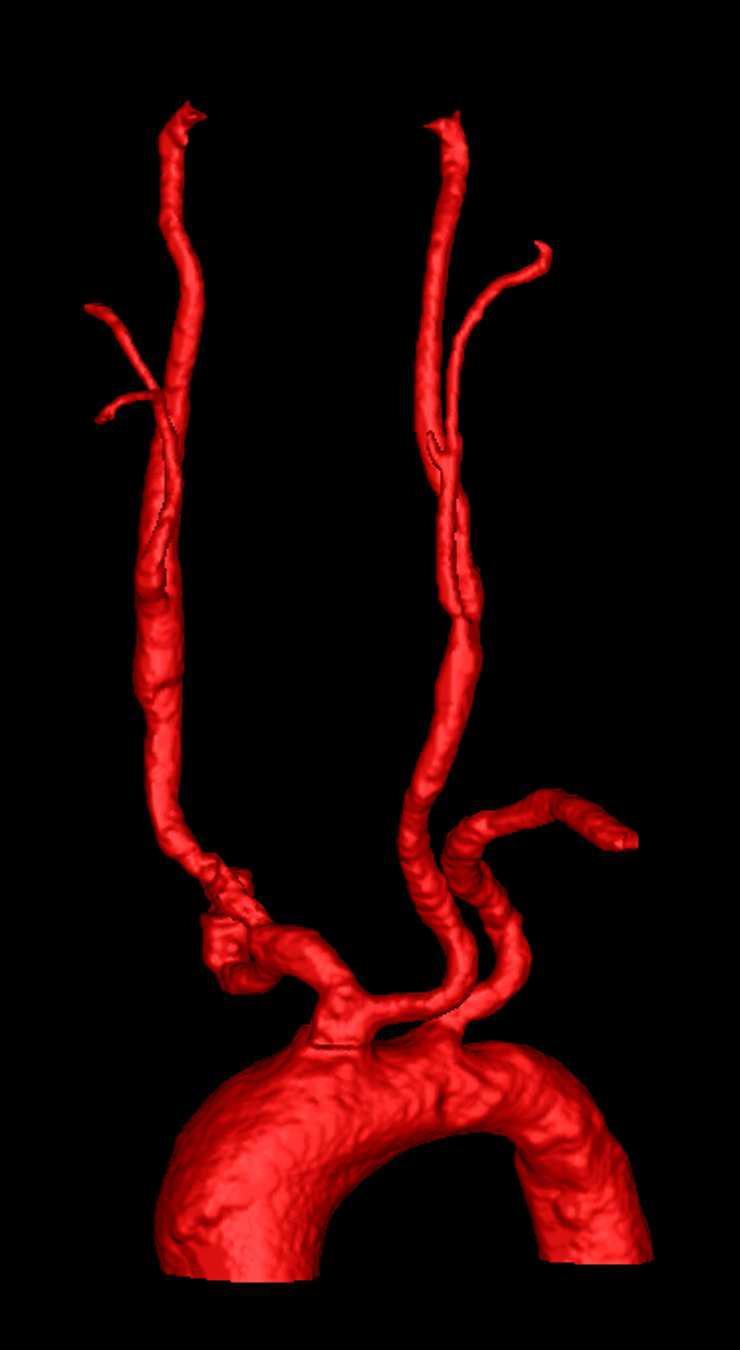
 Int. + Ext Carotids 1.5mm std=1.3mm Common Carotid Surface RMS Bifurcations 0.")
37 All arteries 1.7mm std=0.9mm Subclavian Arteries 1.3mm (std=1.0mm) Aorta 1.6mm (std=0.8mm) Int. + Ext Carotids 1.5mm std=1.3mm Common Carotid Surface RMS Bifurcations 0.7mm std=0.7mm
38 Copyright L. Joskowicz, 2010 MIS validation: observers variability Intra-observer variability: determine how much variation there is when a single observer produced the ground-truth segmentation repeat 5 times the segmentation Inter-observer variability: determine how much variation there is between multiple observers that produced the ground-truth segmentation ask 3 radiologists to do the segmentation Observer expertise and frequency variability
![Study: [Weltens 2001] Axial MRI slice Nine independent observers Inter observer variability](/docs-images/90/101219387/images/39-0.jpg "Repeated delineations Inter/intra observer variability: 30% Key issue: fuzzy tumor")
39 Study: [Weltens 2001] Axial MRI slice Nine independent observers Inter observer variability Repeated delineations Inter/intra observer variability: 30% Key issue: fuzzy tumor boundaries


40 Intra-observer variability 8% 50%


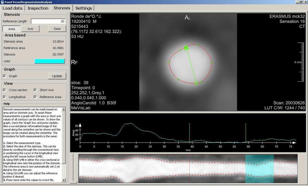
41 Copyright L. Joskowicz, 2010 Carotid Lumen Segmentation and Stenosis Grading Challenge -- The MIDAS Journal


42 Copyright L. Joskowicz, 2010 MIS validation: manual annotation

 Partial")

43 MIS validation: ground-truth generation centerlines (manual) contours (manual) Partial Volume Segmentation Reference standard from 3PVS Copyright L. Joskowicz, 2010
44 Measurements characteristics Ground-truth: not know! Repeatability: intra-observer variability Reliability: Measure: inter observer variability correlation between repeated measurements
45 Inaccuracy vs. Uncertainty Intrinsic uncertainty about the tumor volume. Accuracy for clinical significance is unknown. Inaccuracy > Uncertainty Results can be meaningless Uncertainty may not be improved. Goal: improve accuracy to obtain: Inaccuracy < Uncertainty
46 In summary. Medical image segmentation is on the rise. Basis of many clinical applications Validation is a MUST Many methods and approaches do not re-invent the wheel I expect patient-specific 3D models to be in the clinical mainstream in 3-5 years! Copyright L. Joskowicz, 2011
47 Segmentation challenges Rigorous quantification and evaluation of segmentation algorithms performance Shape priors for pathologies Incorporation of functional information from diffusion/perfusion MRI, fmri, PET into segmentation algorithms. Copyright L. Joskowicz, 2011
48 Summary (1) Patient-specific anatomy model creation is currently a major bottleneck in many clinical applications Automatic anatomical segmentation is essential for model creation Current tools are limited in scope and coverage Clinical use requires the elimination of the technician model generation by the physician!
49 Summary (2) Great opportunity for the development and incorporation of anatomy modeling tools in commercial platforms Growing need and variety of users for anatomical models Training simulators, surgery rehersal Intraoperative guidance Computer Aided Radiology Service providers -- shifting paradigms
Introducing a new take on efficient workflow: Deeply integrated clinical applications
 Clinical Applications Introducing a new take on efficient workflow: Deeply integrated clinical applications The EXPERIENCE of TRUE CLINICAL APPLICATIONS While developments in image acquisition technology
Predictive and patient-specific numerical simulations of endovascular surgery for aortic abdominal aneurysms. Michel Rochette
 Predictive and patient-specific numerical simulations of endovascular surgery for aortic abdominal aneurysms Michel Rochette 1 Our Partners This work has been partially supported by French national research
Low Dose, Lightning Speed, Latest Applications
 RXL EDITION Low Dose, Lightning Speed, Latest Applications The New Standard for Radiology Toshiba Medical Systems is committed to the development of new technologies to minimize radiation dose while maintaining
Extending quality care to more people. Brivo CT315
 GE Healthcare Extending quality care to more people. Brivo CT315 Big impact. Improving the quality of healthcare around the world is a goal we all share. But this can be a daunting challenge, with patients
Quantitative biomarker for clinical decision making
 Deep imaging Quantitative biomarker for clinical decision making Joerg Aumueller English, October 2017 54% of healthcare leaders see an expanding role of in medical decision support.* * The future of healthcare
Clinical Applications. ImagingRite. Interventional Radiology
 Clinical Applications ImagingRite Interventional Radiology ImagingRite, a comprehensive suite of imaging tools offered with Infinix -i angiographic systems, was designed to assist clinicians in optimizing
Interventional Radiology
 Angiography Systems Interventional Radiology *Pictured system is INFX-8000C Unprecedented Flexibility and Integration. Today s radiology interventions require speed, flexibility and high performance. Canon
Computer-Aided Surgical Navigation Coding Guide Neurosurgery. May 1, 2009
 Computer-Aided Surgical Navigation Coding Guide Neurosurgery May 1, 2009 Please direct any questions to: Kim Brew Manager, Reimbursement and Therapy Access Medtronic Surgical Technologies (904) 279-7569
Section: Magnetic Resonance Imaging
 Section: Magnetic Resonance Imaging Available masters project: Cardiac shape modeling for automated wall motion analysis Overview One of the most popular segmentation methods in medical imaging is the
A Novel Segmentation and Navigation Tool for Endovascular Stenting of Aortic Aneurysms
 A Novel Segmentation and Navigation Tool for Endovascular Stenting of Aortic Aneurysms Marco Feuerstein, Konstantinos Filippatos, Oliver Kutter, Eva U. Schirmbeck, Robert Bauernschmitt, Nassir Navab 01
Towards Practical Problems in Deep Learning for Radiology Image Analysis
 Towards Practical Problems in Deep Learning for Radiology Image Analysis Quanzheng Li, Xiang Li, James H.Thrall Center for Clinical Data Science Department of Radiology Massachusetts General Hospital,
Statistical models for supporting medical diagnosis, therapy and training
 Statistical models for supporting medical diagnosis, therapy and training Gábor Székely Computer Vision Laboratory Swiss Federal Institute of Technology Zürich 27 th January 2011 KÉPAF 2011, Szeged 1 of
NIST Medical Imaging Informatics Activities. Medical Imaging
 NIST Medical Imaging Informatics Activities Ram Sriram Mary Brady Alden Dima Medical Imaging Biomarker Testing Improving Change Analysis in Lung Cancer Statistically Valid and Clinically Meaningful Biomarkers
CARESTREAM RIS/PACS SuperPACS Architecture. A Changing Landscape
 CARESTREAM RIS/PACS SuperPACS Architecture A Changing Landscape The field of radiology is in a constant state of acceleration and evolution. Each day, you handle an increasing number of exams and diagnostic
Clinical depth anywhere, anytime
 Clinical depth anywhere, anytime Philips IntelliSpace PACS solution Informed images everywhere it matters Philips is a leader in clinical imaging and in the innovation of clinical imaging informatics.
Your diagnostic partner. Every day. Optima* CT540
 GE Healthcare Your diagnostic partner. Every day. Optima* CT540 At GE Healthcare, the goal was to design a CT system that would provide our clinical partners with the imaging capabilities needed every
Brivo * CT385 Great care by design
 GE Healthcare Brivo * CT385 Great care by design Not for sale or distribution in the United States. Built for total returns. Superb image quality. Advanced features. Compact size. All in one CT system
Integrated planning, navigation and robotic targeting for tumor ablation
 Integrated planning, navigation and robotic targeting for tumor ablation Tumor ablation Current practice Today, clinicians plan their interventional oncology procedures by viewing 2 dimensional CT slices,
Future bound. Philips Ingenuity Core
 Future bound Philips Ingenuity Core High reliability Low-dose, high-quality imaging and coverage, and the ability to personalize image quality* patient by patient. Expect excellence in routine imaging,
American College Of Radiology Data Science Institute. Keith Dreyer DO, PhD, FACR Chief Science Officer ACR Data Science Institute May 30, 2018
 American College Of Radiology Data Science Institute Keith Dreyer DO, PhD, FACR Chief Science Officer ACR Data Science Institute May 30, 2018 ACR Data Science Institute: Participation http://www.acrdsi.org
CQIE MRI PROCEDURES. American College of Radiology Clinical Research Center. Centers for Quantitative Imaging Excellence LEARNING MODULE
 Centers for Quantitative Imaging Excellence LEARNING MODULE CQIE MRI PROCEDURES American College of Radiology Clinical Research Center Imaging Core Laboratory v.2 Centers for Quantitative Imaging Excellence
Your diagnostic partner. Every day. Optima* CT540
 GE Healthcare Your diagnostic partner. Every day. Optima* CT540 At GE Healthcare, the goal was to design a CT system that would provide our clinical partners with the imaging capabilities needed every
Computed tomography. High. performance. no trade-offs. Philips Ingenuity CT family
 Computed tomography High performance no trade-offs Philips Ingenuity CT family Keeping you ahead Until now, CT scanning has too often been about trade-offs. You ve been forced to choose between high image
Advanced visualization for real-time radiology. IntelliSpace Portal
 Advanced visualization for real-time radiology IntelliSpace Portal An oncology patient presents with a suspicious lesion identified on today s CT study. How quickly can you access the patient s prior MR
A Dynamic Data Driven Grid System for Intra-operative Image Guided Neurosurgery
 A Dynamic Data Driven Grid System for Intra-operative Image Guided Neurosurgery A Majumdar 1, A Birnbaum 1, D Choi 1, A Trivedi 2, S. K. Warfield 3, K. Baldridge 1, and Petr Krysl 2 1 S & 2 Structural
CT to Ultrasound Registration: A Porcine Phantom Study. J. Xiang, S. Gill, C. Nguan, P. Abolmaesumi and R. N. Rohling University of British Columbia
 CT to Ultrasound Registration: A Porcine Phantom Study J. Xiang, S. Gill, C. Nguan, P. Abolmaesumi and R. N. Rohling University of British Columbia 1 Motivation Ultrasound to CT registration can improve
Fast enough to stop the Capable of delineating Unprecedented imaging power for the. Virtual endoscopy. The gatewa
 Fast enough to stop the Capable of delineating Unprecedented imaging power for the M U L T I S L I C E Virtual endoscopy The gatewa motion of a beating heart. anatomic structures as small as 0.25mm. earliest,
Vue Radiology Client Module. Simple. Powerful. Efficient. A Single Workspace with the Ideal Tools for Accurate Reading and Insightful Reporting.
 Vue Radiology Client Module Simple. Powerful. Efficient. A Single Workspace with the Ideal Tools for Accurate Reading and Insightful Reporting. Raising the Stature of Radiology. Radiology is facing the
2018 Data Science Summit: The Economics Of Artificial Intelligence In Healthcare
 May 30, 2018 2018 Data Science Summit: The Economics Of Artificial Intelligence In Healthcare Regulation, Payment And The AI Ecosystem Bibb Allen, MD FACR Chief Medical Officer ACR Data Science Institute
C-arm CT with SIREMOBIL Iso-C 3D A New Dimension of Care in the OR
 C-arm CT with SIREMOBIL Iso-C 3D A New Dimension of Care in the OR SIREMOBIL Iso-C 3D C-arm CT brings intra-operative 3D imaging If you are involved in trauma and orthopedic surgery you know the importance
Low-dose and High-resolution Cardiovascular Imaging with Revolution* CT
 GE Healthcare Case study Low-dose and High-resolution Cardiovascular Imaging with Revolution* CT Jean-Louis Sablayrolles, M.D. Laurent Macron, M.D. Jacques Feignoux, M.D. Centre Cardiologique du Nord,
Mirada Case Study. VentureFest, July
 Mirada Case Study VentureFest, July 2015 Synopsis of Mirada Medical Founded in 1999 as a spin-out from the University of Oxford University had 10% equity Headquartered at the OCFI, Oxford Mirada USA based
Automatic Aorta Segmentation and Valve Landmark Detection in C-Arm CT for Transcatheter Aortic Valve Implantation
 Automatic Aorta Segmentation and Valve Landmark Detection in C-Arm CT for Transcatheter Aortic Valve Implantation Yefeng Zheng 1, Matthias John 2, Rui Liao 1, Alois Nöttling 2, Jan Boese 2, Jörg Kempfert
80 Years of Innovation
 3D Synapse 3D History of Innovation 80 Years of Innovation Light, entering through the eyes, is processed and projected as an image in the brain. Image Intelligence performs this same kind of image optimization
Motiva: a TV-based platform for remote patient management
 Technology news New products Motiva: a TV-based platform for remote patient management a simplified user interface, a secure broadband connection and a standard set-top box. A nurse care manager, using
Making the difference with Live Image Guidance
 OncoSuite Interventional X-ray Making the difference with Live Image Guidance Next generation of Image Guided Therapy in Interventional Oncology Live Image Guidance in Interventional Oncology OncoSuite:
Adaptive Iterative Dose Reduction in 3D
 technology history For over 130 years, Toshiba has been a world leader in developing technology to improve the quality of life. Our 50,000 global patents demonstrate a long, rich history of leading innovation.
Clarity CT Technology
 Clarity CT Technology WHITE PAPER January 2013 Using state of the art algorithms Sapheneia Clarity CT allows physicians to lower radiation dose when acquiring CT data while maintaining image quality. The
AAPM Scientific Meeting Imaging Symposium. State of the Art in Quantitative Imaging CT, PET and MRI. Which Imaging Modality is the Most Quantitative
 AAPM Scientific Meeting Imaging Symposium State of the Art in Quantitative Imaging CT, PET and MRI Michael McNitt-Gray, PhD, FAAPM; UCLA Paul Kinahan, PhD, U. Washington Ed Jackson, PhD, FAAPM, UT-MD Anderson
Electrophysiology Systems
 Biplane, Single Plane Electrophysiology Systems *Pictured system is INFX-8000V (BP) State-of-the-art WorkRite technology creates an efficient workflow and provides optimum patient access while allowing
03/08/2017. Advances and Challenges in Contour QA for Adaptive RT. Objective. Disclosures
 Advances and Challenges in Contour QA for Adaptive RT Kristy K Brock, PhD, DABR, FAAPM Professor, Department of Imaging Physics Director, Image Guided Cancer Therapy Program University of Texas MD Anderson
Simulator Features. Sanford-USD Surgical Residency and Cardiology Fellowship Programs IMAGING. C-arm and patient table maneuvering
 ANGIO Mentor TM Sanford-USD Surgical Residency and Cardiology Fellowship Programs Simulator Features IMAGING C-arm and patient table maneuvering Real time fluoroscopy, cineangiography, DSA and roadmapping
This is probably the kind of radiotherapy that you are used to delivering in your country.
 This is probably the kind of radiotherapy that you are used to delivering in your country. This slide shows images two patients, one with a T3 N2c nasopharyngeal CA and the other with a T1/2 N1/2 lung
First Experiences with the Ziehm Vision FD Mobile C-Arm with Flat-Panel Detector
 01 White Paper No. 02/2009 First Experiences with the Ziehm Vision FD Mobile C-Arm with Flat-Panel Detector Leiden University Medical Center (LUMC) in the Netherlands is the first hospital in the world
QIBA RIC Collaboration
 QIBA RIC Collaboration Providing the Radiology Research and Development Community with the Tools toward Quantitative Imaging Methods with which to Detect, Diagnose and Treat Disease Katherine P. Andriole
Review of Biomedical Image Processing
 BOOK REVIEW Open Access Review of Biomedical Image Processing Edward J Ciaccio Correspondence: ciaccio@columbia. edu Department of Medicine, Columbia University, New York, USA Abstract This article is
Optimal landmarks selection and fiducial marker placement for minimal target registration error in image-guided neurosurgery
 Optimal landmarks selection and fiducial marker placement for minimal registration error in image-guided neurosurgery Reuben R Shamir 1*, Leo Joskowicz 1, and Yigal Shoshan 2 1 School of Engineering and
Progress in X-Ray & MR
 Progress in X-Ray & MR Michiel Manuel Analyst Meeting June 15 th, 2005 X-Ray & MR: Agenda Introduction General X-Ray Cardio/Vascular X-Ray Magnetic Resonance China growth opportunity Conclusion 2 X-Ray
Pinnacle 3. Treatment Planning. Flexible, intuitive planning environment
 Pinnacle 3 Treatment Planning Flexible, intuitive planning environment Pinnacle 3 Flexible, intuitive planning environment Planning plays a central role in influencing treatment confidence as well as departmental
Pinnacle 3. Treatment Planning. Flexible, intuitive planning environment
 Pinnacle 3 Treatment Planning Flexible, intuitive planning environment Pinnacle 3 Flexible, intuitive planning environment Planning plays a central role in influencing treatment confidence as well as departmental
Clinical Applications. ImagingRite. Neuro Intervention. 1 ImagingRite
 Clinical Applications ImagingRite Neuro Intervention 1 ImagingRite ImagingRite, a comprehensive suite of imaging tools offered with Infinix -i angiographic systems, was designed to assist clinicians in
3D Imaging in Radiology
 3D Imaging in Radiology Gordon J. Harris, Ph.D. Director, 3D Imaging Service and Radiology Computer Aided Diagnostics Laboratory, Massachusetts General Hospital Director, Tumor Imaging Metrics Core Dana
Declaration of Relevant Financial Interests or Relationships
 Declaration of Relevant Financial Interests or Relationships Speaker Name: Andriy Fedorov I have no relevant financial interest or relationship to disclose with regard to the subject matter of this presentation.
GE Healthcare. Introducing Veo * on Discovery * CT750 HD Great care by design
 GE Healthcare Introducing Veo * on Discovery * CT750 HD Great care by design Radiologists and clinicians demand the ultimate levels of clarity and detail in their images. In making an informed and confident
LIVER EXTRACTION USING HISTOGRAM AND MORPHOLOGY
 LIVER EXTRACTION USING HISTOGRAM AND MORPHOLOGY S. Kiruthika 1, I. Kaspar Raj 2 1 Women Scientist, Computer Centre, Gandhigram Rural Institute-Deemed University, Tamilnadu 1 kiruthikasm09@gmail.com 2 Director
Computer Assisted Surgery Basics of medical imaging
 Computer Assisted Surgery Basics of medical imaging Prof. Leo Joskowicz School of Engineering and Computer Science The Hebrew University of Jerusalem, ISRAEL Medical Image Processing Basics of medical
CT post processing and low dose scanning
 CT post processing and low dose scanning Gabor Szell GE Healthcare CT Modality Manager EE Annual Scientific and Educational Meeting Innovations in Cardiothoracic Imaging 201 13-14 May 2011, Tokuda Hospital
CS5540: Computational Techniques for Analyzing Clinical Data
 CS5540: Computational Techniques for Analyzing Clinical Data Prof. Ramin Zabih (CS) Prof. Ashish Raj (Radiology) Today only: Gary Dorfman, MD (Radiology) What is CS5540 about? The practice of medicine
8/2/2017. Key attributes of scientific excellence: rigor, innovation, and relevance. Medical Physics 3.0. Key Attributes of Scientific Excellence
 Medical Physics 3.0 in Design Key attributes of scientific excellence: rigor, innovation, and relevance Maryellen Giger, Ph.D. A. N. Pritzker Professor of Radiology / Medical Physics The University of
N.MAFFEI, G.GUIDI, C.VECCHI, G.BALDAZZI Physics Department, University of Bologna, via Irnerio Bologna, Italy
 AN ARTIFICIAL NEURAL NETWORK TO PREDICT TIME OF REPLANNING FOR TOMOTHERAPY TREATMENTS N.MAFFEI, G.GUIDI, C.VECCHI, G.BALDAZZI Physics Department, University of Bologna, via Irnerio 40 40138 Bologna, Italy
GE Healthcare. Introducing Discovery MI DISCOVERY MI
 GE Healthcare Introducing Discovery MI DISCOVERY MI Introducing Discovery MI Introducing Discovery MI MEANINGFUL INSIGHTS. FROM YOUR PATIENT TO EVERY PATIENT. Meet Discovery TM MI. A PET/CT system conceptualized
Un Sistema di Navigazione basato su Mixed Reality per Trattamenti HIFU
 La Medicina Incontra la Realtà Virtuale 2011- MIMOS Un Sistema di Navigazione basato su Mixed Reality per Trattamenti HIFU C. Freschi, V. Ferrari*, F. Porcelli, A. Peri, L. Pugliese, L. Morelli, M. Ferrari,
MEDIATE Patient friendly medical intervention. Co-summit 2015, March 10-11, 2015 Berlin, Germany Herman Stegehuis (Philips Healthcare)
") Patient friendly medical intervention Co-summit 2015, March 10-11, 2015 Berlin, Germany Herman Stegehuis (Philips Healthcare) Agenda Trends in healthcare Role of MEDIATE Clinical use case scenarios Results
Patient friendly medical intervention Co-summit 2015, March 10-11, 2015 Berlin, Germany Herman Stegehuis (Philips Healthcare) Agenda Trends in healthcare Role of MEDIATE Clinical use case scenarios Results
Imaging anytime, anywhere.
 Imaging anytime, anywhere. Vitrea Enterprise Suite Vitrea Enterprise Suite is Vital Images premier advanced visualization solution that provides clinical applications and data management systems, backed
SOLUTIONS Express Enterprise SP Enterprise XP Integral Breast Workspace. better images mean better results
 SOLUTIONS Express Enterprise SP Enterprise XP Integral Breast Workspace better images mean better results A Complete Clinical Solution Built Around You. DynaCAD Express Built upon the powerful components
Making Reliable Low-dose CT a Clinical Reality
 Making Reliable Low-dose CT a Clinical Reality By Ramakrishnan RS, MD, DNB, Consultant Radiologist 1 ; George Joseph, MD, DMRD, Consultant Radiologist 2 ; and Sundar RK, BSc, DRT, DAMIT, Clinical Applications
Principles of translational medicine: imaging, biomarker imaging, theranostics
 Principles of translational medicine: imaging, biomarker imaging, theranostics Compiled by: Endre Mikus PhD, CEO Budapest, 21/9/2015 Imaging and imaging biomarkers An imaging biomarker is an anatomic,
Abdominal CT with Single-Energy Metal Artifact Reduction (SEMAR): Initial Experiences
: Initial Experiences") Abdominal CT with Single-Energy Metal Artifact Reduction (SEMAR): Initial Experiences Poster No.: C-0674 Congress: ECR 2014 Type: Scientific Exhibit Authors: K. Sofue 1, T. Yoshikawa 1, N. Negi 1, Y. Ohno
Abdominal CT with Single-Energy Metal Artifact Reduction (SEMAR): Initial Experiences Poster No.: C-0674 Congress: ECR 2014 Type: Scientific Exhibit Authors: K. Sofue 1, T. Yoshikawa 1, N. Negi 1, Y. Ohno
Imaging. Steve Rusckowski CEO Philips Healthcare
 Imaging Steve Rusckowski CEO Philips Healthcare Imaging markets by region and modality 2008 Market by Region 2008, Total Market 14.900 M Market by Modality 2008 LATAM China MCR APAC MCR EMEA Japan Western
background digitalization
 FRAUNHOFER Institute For integrated circuits IIS Microscopy SOFTWARE AND SYSTEMS FOR HISTOLOGY 1 Background Examination and assessment of tissue samples by the pathologist represent an essential element
Data Mining for Genomic- Phenomic Correlations
 Data Mining for Genomic- Phenomic Correlations Joyce C. Niland, Ph.D. Associate Director & Chair, Information Sciences Rebecca Nelson, Ph.D. Lead, Data Mining Section City of Hope National Medical Center
Current Market Dynamics and Future Vision of the Care Cycle
 Current Market Dynamics and Future Vision of the Care Cycle Tim Irish Analysts Meeting June 15 th, 2005 Overview Market and Customer trends The Care Cycle and Molecular Medicine 2 Healthcare is the world
SYNAPSE is a registered trademark of FUJIFILM Medical Systems U.S.A., Inc. in United States, Australia and Canada
 Version 4 FUJIFILM Medical Systems USA, Inc. SYNAPSE is a registered trademark of FUJIFILM Medical Systems U.S.A., Inc. in United States, Australia and Canada Image Intelligence is a registered trademark
Computed Tomography: Optimization of acquisition protocols & Justification of clinical referrals. Koos Geleijns, medical physicist
 Computed Tomography: Optimization of acquisition protocols & Justification of clinical referrals Koos Geleijns, medical physicist CT delivers excellent 3D image quality CT delivers excellent 3D image quality
Registration-Free Laparoscope Augmentation for Intra-Operative Liver Resection Planning
 Registration-Free Laparoscope Augmentation for Intra-Operative Liver Resection Planning Marco Feuerstein, Thomas Mussack*, Sandro M. Heining*, Nassir Navab 20 February 2007 chair for computer aided medical
Interoperability. Radiology without
 Interoperability ADVANCED Solutions to complex ISSUES in Radiology Radiology without Barriers ISSUES IN RADIOLOGY ACHIEVING interoperability in medical imaging Many of the current issues in medical imaging
IMV 2006 CT Market Summary Report Featuring results from 2,565 hospital and non-hospital CT imaging sites in the United States
 Medical Information Division 1400 East Touhy Ave., Suite 250 Des Plaines, Illinois 60018 Phone: 847-297-1404 Fax: 847-297-5010 www.imvinfo.com IMV 2006 CT Market Summary Report Featuring results from 2,565
Registration of 3D Ultrasound to Computed Tomography Images of the Kidney
 Registration of 3D Ultrasound to Computed Tomography Images of the Kidney Jing Xiang Supervisors: Dr. Robert Rohling Dr. Purang Abolmaesumi Electrical and Computer Engineering University of British Columbia
Ingenia MR-OR. MR Systems. Confident decision making with intraoperative insights
 Ingenia MR-OR MR Systems Confident decision making with intraoperative insights Confident decision making with intraoperative insights Ingenia MR-OR intraoperative MRI delivers high-quality images during
Endovascular Simulators Reduce Patient and Staff Exposure
 Endovascular Simulators Reduce Patient and Staff Exposure Gabriel Bartal MD, FCIRSE, FSIR Dept of Diagnostic and Interventional Radiology, Meir MC, Kfar-Saba, Affiliated to Tel-Aviv University, Tel-Aviv,
Visualizing the invisible
 Visualizing the invisible Intraoperative Multi-Modal Imaging in traoperative visualization technology SonoWand Invite allows the surgeon to identify and distinguish lesions from normal brain tissue and
Defining Use Case Standards For Diagnostic Radiology Artificial Intelligence Algorithms
 May 19, 2018 Defining Use Case Standards For Diagnostic Radiology Artificial Intelligence Algorithms Bibb Allen, MD FACR Chief Medical Officer ACR Data Science Institute Keith Dreyer, DO PhD FACR Chief
Simulator Features. Imaging. Device Simulation. Patient Management. Educational Features
 TM ANGIO Mentor Simulator Features Imaging C-arm and patient table maneuvering Real time fluoroscopy, cineangiography, DSA and roadmapping Real time Echocardiography (ICE) and TEE Device Simulation A wide
Generating ROI with Advanced PET and Diagnostic CT Imaging
 USTOMER SPOTLIGHT NERR IMGING GROUP PET/T U S T O M E R S P O T L I G H T Generating ROI with dvanced PET and iagnostic T Imaging Known as the ush apital, anberra is ustralia s largest inland city located
Deep Learned and Practical
 Deep Learned and Practical Implementing Machine Learning Techniques in the Real World Timor Kadir DPhil Mirada Medical CTO Optellum CTO Visiting Fellow University of Oxford 7/31/2017 1 Disclosures Mirada
GE Healthcare. PET/CT + MR Trimodality Imaging
 Unlocking new possibilities with PET/CT + MR 1 PET/CT MR PET/CT + MR The exquisite soft-tissue-contrast of 3.0T MR. The exceptional metabolic insight of PET imaging. The precise anatomical reference of
Ingenia MR-RT. MR Systems. The comprehensive MR-sim solution to fit your planning
 Ingenia MR-RT MR Systems The comprehensive MR-sim solution to fit your planning Table of contents Experience the difference MRI makes 3 A comprehensive MR-sim solution 4 Position with precision 6 See clearly
GE Healthcare. Revolution ACT. Your Aspirations. Realized.
 GE Healthcare Revolution ACT Your Aspirations. Realized. Your Aspirations. Realized. Helping you feel MORE CONFIDENT about moving to the next level, CLINICALLY & ECONOMICALLY that s intelligent innovation.
Quantitative Imaging Biomarker DCE - MRI
 Quantitative Imaging Biomarker DCE - MRI DCE MRI: What is it about? DCE MRI: quantitative analysis of dynamic T1 contrast enhanced images Use cases: Clinical trial related UC1: pharmacodynamic investigations
CQIE PET PROCEDURES. American College of Radiology Clinical Research Center. Centers for Quantitative Imaging Excellence LEARNING MODULE
 Centers for Quantitative Imaging Excellence LEARNING MODULE CQIE PET PROCEDURES American College of Radiology Clinical Research Center Imaging Core Laboratory v2.1 Centers for Quantitative Imaging Excellence
Computational biomechanics of the cardiovascular system
 Modelling in Medicine and Biology VI 381 Computational biomechanics of the cardiovascular system T. Yamaguchi Department of Bioengineering and Robotics, School of Engineering, Tohoku University, Sendai,
Integrated on-board CBCT-US imaging system for soft tissue IGRT and real-time intra-fraction monitoring
 Integrated on-board CBCT-US imaging system for soft tissue IGRT and real-time intra-fraction monitoring John Wong Radiation Oncology and Molecular Radiation Sciences Johns Hopkins University School of
Healthcare Informatics. Oran Muduroglu CEO of Healthcare Informatics May 31 st 2007
 Healthcare Informatics Oran Muduroglu CEO of Healthcare Informatics May 31 st 2007 Agenda Healthcare Informatics Key Clinical Strategy Enabling improved outcomes 2 Update in 2007 continuing the journey
Press. Innovative systems for radiology from Siemens. Healthcare Erlangen, March 3, ECR 2016 in Vienna: Hall X5, Booth 12
 Press Healthcare Erlangen, March 3, 2016 ECR 2016 in Vienna: Hall X5, Booth 12 Innovative systems for radiology from Siemens Faster MRI applications for neurology provide better diagnostic results First
The Orthopaedic Research Laboratory (ORL) performs research in the field of orthopaedics.
 performs research in the field of orthopaedics.") The Orthopaedic Research Laboratory (ORL) performs research in the field of orthopaedics. The biomechanical section focusses on bone, soft tissues and on pre-clinical testing of implants. We are specialized
The Orthopaedic Research Laboratory (ORL) performs research in the field of orthopaedics. The biomechanical section focusses on bone, soft tissues and on pre-clinical testing of implants. We are specialized
Radiology Rounds A Newsletter for Referring Physicians Massachusetts General Hospital Department of Radiology
 Radiology Rounds A Newsletter for Referring Physicians Massachusetts General Hospital Department of Radiology Multi-Energy Computed Tomography - New Opportunities In Imaging the Abdomen Materials can be
Automatic Aorta Segmentation and Valve Landmark Detection in C-Arm CT for Transcatheter Aortic Valve Implantation
 IEEE TRANSACTIONS ON MEDICAL IMAGING, VOL. 31, NO. 12, DECEMBER 2012 2307 Automatic Aorta Segmentation and Valve Landmark Detection in C-Arm CT for Transcatheter Aortic Valve Implantation Yefeng Zheng*,
Pre-op Planning. Dedicated to planning of Endovascular Interventions
 Pre-op Planning Dedicated to planning of Endovascular Interventions Pre-op Planning OPTIMIZE YOUR PRE-OP WORKFLOW What would you say if you were offered a mobile and accessible solution for planning of
Application for Training Centre. BSET Fellowships
 Application for Training Centre BSET Fellowships Institution: St George s Vascular Institute, London, UK Correspondence: Professor Matt Thompson St George s Vascular Institute 4 th Floor St James Wing