BIOMIMETIC HYDROGELS BASED ON NATURAL DEXTRAN AND GELATIN FOR VASCULAR TISSUE ENGINEERING APPLICATION
|
|
|
- Loreen Bradley
- 5 years ago
- Views:
Transcription
1 BIOMIMETIC HYDROGELS BASED ON NATURAL DEXTRAN AND GELATIN FOR VASCULAR TISSUE ENGINEERING APPLICATION LIU YUNXIAO SCHOOL OF CHEMICAL AND BIOMEDICAL ENGINEERING A thesis submitted to the Nanyang Technological University in partial fulfillment of the requirement for the degree of Doctor of Philosophy
2 Acknowledgement The author would like to thank Prof. Mary B. Chan-Park for her excellent standard, patient tutoring, and continuous encouragement throughout the period of this research work. The enthusiasm, sincerity, and earnestness that she has shown have impressed me the most and will benefit me in my future work. Dr. Gong Yihong is thanked for his help and willingness to share his knowledges of material and biology. Mr. Su Kai is greatly appreciated for the help in real-time PCR. Prof. Wang Dongan, Dr. Poon Yin Fun, Dr. Zhou Chuncai are thanked for their valuable advice in the experiment. Special thanks go to Mr. Clement Lim Choon How for his kind help with chemical purchase. This projected was supported by a Singapore Ministry of Education Tier 2 (M ). The author would like to thank Singapore Ministry of Education for providing this research funding. Thanks also give to Nanyang Technological University (NTU) for supporting the author with a NTU PhD scholarship. My family and all of my friends are greatly appreciated for their powerful support. 2
3 Table of contents Acknowledgement... 2 Table of contents... 3 Summary... 8 List of Figures List of Tables Table of abbreviations Chapter 1 Introduction Background Objective and Scope Outline Chapter 2 Literature review Tissue engineering Overview of tissue engineering Cell source in tissue engineering Scaffolds in tissue engineering Vascular tissue engineering Blood vessel structure Vascular smooth muscle cells Vascular disease and replacement Tissue engineering of small-diameter vascular grafts The ideal tissue-engineered vascular grafts Scaffold-based tissue-engineered vascular grafts Cell sheet-based tissue-engineered vascular grafts Summary
4 2.3 Hydrogels for cell encapsulation Gelation mechanism of hydrogels Structure and chemistry of hydrogels Degradation of hydrogels Rational design of biomimetic hydrogels Native ECM Hydrogels that present adhesion ligands Hydrogels that present enzymatic degradability Hydrogels that dynamically present ECM effectors...78 Chapter 3 Hydrogel based on methacrylate- and aldehyde- bifunctionalized dextran and gelatin for 2D endothelial cell and 3D smooth muscle cell cultures Introduction Materials and methods Materials Synthesis of methacrylated dextran (Dex-MA) Synthesis of methacrylated dextran with aldehyde groups (Dex-MA-AD) Hydrogel preparation Characterization of hydrogel Endothelial cell culture Smooth muscle cell encapsulation within hydrogels Results Synthesis of bifunctional dextran Hydrogel sol content Hydrogel water content Characterization of hydrogel mechanical properties
5 Dynamic mechanical properties Static compressive properties Characterization of gelatin release EC growth on the surface of hydrogels SMC encapsulation within hydrogels Discussion Conclusion Chapter 4 Biomimetic hydrogel based on methacrylated dextran-graft-lysine and gelatin for 3D smooth muscle cell culture Introduction Materials and methods Materials Synthesis of dextran derivatives Synthesis of gelatin derivatives Hydrogel formation and characterization SMC encapsulation Cell viability Cell proliferation Statistical analysis Results Synthesis of dextran and gelatin derivatives Hydrogel characterization Spreading of SMCs inside hydrogels Proliferation of SMCs inside hydrogels Discussion Conclusion Chapter 5 Phenotype and collagen type I synthesis of smooth muscle cells in 5
6 3D dextran-based hydrogels Introduction Methods SMC culture and encapsulation within hydrogel Real-time reverse transcription polymerase reaction (real-time RT-PCR) Immunofluorescent staining Results Differentiation of SMCs inside hydrogels Immunofluorescent staining of α-actin and collagen type I Discussion Conclusion Chapter 6 Impact of endothelial cells on smooth muscle cells in 3D culture Introduction Experimental details Co-culture of ECs and SMCs Characterization of SMC spreading and proliferation Gene expression analyse Degradation of hydrogel constructs Results Impact of ECs on SMC spreading and proliferation Impact of ECs on SMC differentiation Impact of ECs on ECM production of SMCs Impact of ECs on hydrogel degradation Discussion Conclusion
7 Chapter 7 Conclusions and recommendations Hydrogels based on Dex-MA-AD and gelatin for 3D SMC and 2D EC cultures (D-G series) Hydrogels based on Dex-MA-Ly and Gel-MA for 3D SMC culture (L-G series) SMC phenotypic modulation inside 3D dextran-based hydrogels Impact of ECs on SMCs in 3D hydrogels Recommendations for future work Degradation of hydrogels Regulation of SMC phenotype Reference: Appendix: Publication
8 Summary Hydrogels are crosslinked polymer networks with high water content. They have the capability to mimic native ECM and thus are highly desirable as 3D scaffolds for cell encapsulation. Synthetic hydrogels for cell encapsulation have hitherto been based on poly(ethylene glycol), which is non-natural, non-biodegradable, and only terminal-functionalizable. Dextran is highly hydrophilic but also biodegradable and pendant-functionalizable. More importantly, it resembles the native glycosaminoglycans. This study aims to fabricate hydrogels based on natural dextran and gelatin, which could promote 3D SMC spreading and proliferation. Two series of hydrogels were fabricated. The first hydrogel series is based on the interpenetrating polymer network (IPN) of gelatin and dextran bifunctionalized with methacrylate (MA) and aldehyde (AD) (Dex-MA-AD). These IPN hydrogels not only supported endothelial cell (EC) adhesion and spreading on the surface, but also allowed encapsulated SMCs to proliferate and spread in the bulk interior of the hydrogel; however, the Schiff base reaction was not easily controllable and while SMC spreading within the hydrogel did occur, it was rather limited. In order to improve SMC spreading, dextran was functionalized with 8
9 methacrylate and lysine (Dex-MA-Ly). A second hydrogel system based on Dex-MA-Ly and methacrylamide modified gelatin (Gel-MA) was developed. The behaviors of SMCs encapsulated within these hydrogels were influenced by the mechanical stiffness. Rapid cell spreading, high SMC proliferation, and extensive cellular network formation occurred within softer hydrogels; while in stiffer hydrogels SMCs maintained a round morphology and their viability declined during culture. In a softer hydrogel, the encapsulated SMCs appear to be relatively more contractile in the initial culture compared to those on tissue culture polystyrene dish due to physical constraint imposed by the hydrogels; but they become more synthetic with time, possibly due to the inability of the cells to reach confluence during the culture period inside these cell-mediated degradable hydrogels. The impact of endothelial cells (ECs) on SMCs was investigated using an EC/SMC co-culture model in which SMCs were encapsulated in Dex-MA-Ly/Gel-MA hydrogels and exposed to a monolayer of sub-confluent ECs during culture. Without EC co-culture, we found that when the seeding density was higher, a softer Dex-MA-Ly/Gel-MA hydrogel increased the contractility of the SMCs somewhat as shown by increased expression of several contractile marker genes (i.e. α-actin, calponin and smooth muscle-myosin heavy chain (SM-MHC)). Co-cultured with ECs, SMC growth 9
10 was promoted and the formation of much denser cellular networks was enhanced. ECs did not influence the general expression trends and the maximum expression levels of contractile α-actin, calponin, and SM-MHC, but delayed the time point to reach these maximum transcriptions. Either with or without ECs, the encapsulated SMCs though showed some contractility but were not fully differentiated. ECs also promoted the synthesis of elastin at transcriptional level. Due to the impressive cellular proliferation and network formation, these new hydrogels combining polysaccharide and protein derivatives appear to be excellent candidates for further development as bioactive scaffolds for vascular tissue engineering. A functional vascular media layer may be developed through the use of these biomimetic hydrogels in combination with EC stimulation. 10
11 List of Figures Fig. 2.1 (A) The layered structure of blood vessels. (B) Elastin distribution within the vessel wall. Left is a muscular artery; right is an elastic artery. These pictures are adopted from references 87,88 with permission Fig. 2.2 (A) Model structure of a self-assembling peptide molecule. Hydrophobic and hydrophilic residues locate alternatively on the backbone. The peptide promotes ß-sheet formation. K: hydrophilic positively charged lysines; D: hydrophilic negatively charged aspartic acids; L: hydrophobic Leucines. (B) A chondrocyte-encapsulated peptide hydrogel construct. (C) Image of chondrocytes encapsulated in the peptide hydrogel. These pixctures are adopted from reference 188 with permission Fig.2.3 Schematic representations of hydrogel networks and their degradation products formed through (a) chain-growth polymerization, (b) step-growth polymerization, and (c) mixed-mode polymerization. These pictures are adopted from reference 205 with permission Fig. 2.4 (A) A selection of reactions which meet the requirements of click chemistry. This picture is adopted from reference 217 with permission. (B) Click reaction between an azide and a di-fluorinated cycloocytne moiety without Cu catalyst. This picture is adopted from reference 216 with permission Fig. 2.5 Synthesis and degradation of photodegradable hydrogel. (A) The base photodegradable monomer. (B) The photodegradable crosslinking macromer 11
12 obtained by attaching carboxylic acid of monomer A to poly(ethylene glycol)-bis-amine. (C) Photolytic cleavage of hydrogel upon irradiation. (D) The degradation of hydrogel can be controlled by irradiation intensity and wavelength. (a) 365 nm at 20 mw/cm 2, (b) 365 nm at 10 mw/cm 2, (c) 405 nm at 23 mw/cm 2. (E) Continuous (a) or periodic (b) irradiation modulates hydrogel degradation. (F) hmscs exhibit a spherical morphology encapsulated within hydrogel. (G) hmscs spreading was promoted through irradiation (480 s, 365nm at 10 mw/cm 2 ). These pictures are adopted from reference 235 with permission Fig. 2.6 HA-MA hydrogels with (a) high crosslinking density or (b) low crosslinking density have different structures and degradation products. These pictures are adopted from reference 180 with permission. 73 Fig. 2.7 (A) Hydrogels formed of synthetic polymers simply function as templates for cells; they lack the capacity of activating cell surface integrins (1) and other receptors (2), through which multiple cell behaviors will be initiated. (B) Hydrogels formed of biologically derived polymers provide multiple biological cues, such as intergin-binding sites (3) and growth factors (4), to direct cell behavior. These pictures are adopted from reference 178 with permission Fig. 3.1 Synthesis of Dex-MA-AD Fig. 3.2 IPNs hydrogel networks formed by photopolymerization of Dex-MA-AD with low (a) and high (b) amounts of gelatin Fig H-NMR spectra of (a) dextran, and (b) Dex-MA-AD dissolved in D 2 O
13 Fig. 3.4 Influence of (a) crosslinking time (at 20 mw/cm 2 ) and (b) UV intensity (with 5 min irradiation) on sol content of hydrogels. 99 Fig. 3.5 Equilibrium swelling ratios of hydrogels (with 5 min irradiation at 20 mw/cm 2 ). Comparisons were made with pairs between D-A and IPN hydrogels using Student s t-test: * denotes significant difference in equilibrium swelling ratio at the level of P<0.05. (n=3) Fig. 3.6 Dynamic mechanical analysis of hydrogels. (a) Storage modulus (E, solid symbols) and loss modulus (E, hollow symbols) versus frequency at 37 C. (b) tan_delta versus frequency Fig. 3.7 Typical stress - strain curves of D-A, D-G-2, D-G-4, and D-G-6 (a, b, c, and d, respectively) hydrogels at room temperature Fig. 3.8 Gelatin release at 37 C. The cumulative released amount of gelatin was normalized to initial gelatin content Fig. 3.9 Phase contrast microscopic images of endothelial cells grown on surface of D-M (A), D-G-2 (B), D-G-4 (C), D-G-6 (D) hydrogels and on TCPS (E) 4 h after seeding. Scale bar represents 100 μm. Cell seeding density: cells/well Fig Fluorescence micrographs of endothelial cells grow on surface of D-M (A), D-G-2 (B), D-G-4 (C), D-G-6 (D) hydrogels, and on TCPS (E) 1 day after seeding. Live cells were stained green, while dead cells red. Scar bar represents 100 μm. Cell seeding density: cells/well Fig Viability of the endothelial cells cultured on the surfaces of hydrogels after 1day. All samples and TCPS control exhibit improved viability compared with D-M hydrogel (at the significance level P<0.05 by Student's t-test as indicated by *). 111 Fig Phase contrast macroscopic images of smooth muscle cells encapsulated in D-G-2, D-G-4, and D-G-6 hydrogels (A, B, and C, respectively) 13
14 after 4 h, 1 day, 3 days, and 6 days. The magnified inset in image C (4 h) is a close-up of the cell highlighted with the circle and arrows. Scale bar represents 50 μm. Cell seeding density: cells/ml Fig Fluorescence micrographs of SMCs encapsulated into D-G-2 (A1, A2, and A3), D-G-4 (B1, B2, and B3), and D-G-6 (C1, C2, and C3) hydrogels after 1 day (A1, B1, and C1), 7 days (A2, B2, and C2), and 14 days (A3, B3, and C3) using Live/Dead assay. The living cells were stained green, and the dead cells red. Scale bar represents 100 μm. Cell seeding density: cells/ml Fig Viability of the encapsulated smooth muscle cells in hydrogels after 7 days, and 14 days. Comparisons were made with pairs with statistical analysis by the Student s t-test: at the level of P<0.05, * significantly different from that of D-G-6 hydrogel after 7 days; # significantly different from that of D-G-6 hydrogel after 14 days. Cell seeding density: cells/ml. (n=3) 119 Fig. 4.1 Synthesis of Dex-MA-Ly (A) and Gel-MA (B) Fig H-NMR spectra of Dex-MA-Ly dissolved in D 2 O Fig. 4.3 (A) UV spectrum of gelatin and its methacrylamide derivatives reacting with TNBS. (B) Gross view of Dex-MA(9)/Gel-MA(16) (left) and Dex-MA(9)-Ly/Gel-MA(16) (right) solutions.139 Fig. 4.4 Properties of hydrogels: (A) swelling ratio, (B) shear storage modulus and (C) sol content. * P<0.05 vs. other hydrogels with Dex-MA-Ly and Gel-MA of the same degrees of substitution; # P<0.05 vs. other hydrogels with the same concentration Fig. 4.5 (A) Live/Dead staining of SMCs encapsulated inside hydrogels after 1 day (left column), 7 days (middle column), and 14 days (right column) of culture. Scale bar equals to 200 µm. Cell seeding density: cells/ml. (B) Cytoskeletal F-actin staining of SMCs in L-G-1C hydrogel after 7 days. 14
15 Confocal stacks of 300 µm depth were taken. The image sections were projected to one composite image (i, left), and a pseudo-colored composite of image sections of SMCs at varying depths (ii, right) was created using LSM image browser. The scale bar is 100 µm Fig. 4.6 Phase contrast microscopic images of smooth muscle cells encapsulated inside L-G-1A (top row), L-G-1B (middle row), and L-G-1C (bottom row) hydrogels after 1day (left column), 2 days (middle column), and 7 days (right column) of culture. Scale bar is 100 µm. Cell seeding density: cells/ml Fig. 4.7 Comparison of proliferative capacity, as assessed by WST-1 assay, of SMCs encapsulated within L-G-1A, L-G-1B, L-G-2A, and L-G-3A hydrogels. *: p<0.05 vs. adjacent time point before (same hydrogel) Fig. 4.8 SMCs showing incorporation of BrdU after encapsulation inside L-G-1A (top row) and L-G-2A (bottom row) hydrogels for 5 days. Scale bar is 50 μm. Ph refers to phase contrast image Fig. 5.1 Expression of SM1/SM2 (A), α-actin (B), SMemb (C), and collagen type I (D) over 20 days of culture in L-G-2A hydrogels and over 7 days of culture on TCPS. The inset in B enlarges the vertical scale for the α-actin expression in L-G-2A hydrogel. Expression levels shown are relative to GAPDH expression in the same sample. *: p<0.05 vs. TCPS (4 th day hydrogel vs. 1 st day TCPS; 9 th day hydrogel vs. 4 th day TCPS, 20 th day hydrogel vs. 7 th day TCPS); #: p<0.05 vs. same sample prior time point Fig. 5.2 Immunofluorescent staining of α-actin afer 4 days (top row), and collagen type I after 9 days (bottom row) in L-G-1A and L-G-2A hydrogels. Scale bars are 100 µm (top row) and 50 µm (bottom row), respectively Fig. 6.1 EC/SMC co-culture model. SMCs were encapsulated in hydrogels and exposed to a monolayer of ECs
16 Fig. 6.2 Phase contrast microscopy images of (A) EC monolayer on TCPS after 2-3 days of culture, (B) SMCs just after encapsulation and (C) cultured for 1 day within L-G-2A hydrogel. Scale bar is 100 µm. SMC seeding density in hydrogel: cells/ml Fig. 6.3 Morphology of SMCs encapsulated within L-G-2A hydrogels and cultured for 2 days (left column), 4 days (middle column), and 7 days (right column) with ECs (top row) and without ECs (bottom row). Scale bar is 100 µm. SMC seeding density: cells/ml Fig. 6.4 Measurement of cell proliferation, using WST-1 assay, of SMCs encapsulated within L-G-2A hydrogels with or without the presence of ECs (EC + and EC - constructs). *: p<0.05, EC + vs. EC - constructs at the same time point; #: p<0.05, vs. prior time point (comparison was performed within EC + and EC - groups respectively, except that the 2 d result of EC + constructs was compared with the 1 d result of EC - constructs) Fig. 6.5 Expression of (A) α-actin, (B) calponin, (C) SM-MHC, (D) smoothelin, and (E) SMemb over 14 days of culture in EC + and EC - constructs. Expression levels shown are relative to GAPDH expression in the same sample. *: significant difference (p<0.05) between EC + and EC - constructs at the same time point; #: significant difference (p<0.05) vs. prior time point (within the same group of constructs except that the 2 d result of EC + constructs was compared with the 1 d result of EC - constructs) Fig. 6.6 Gene expressions of ECM proteins: (A) collagen type I and (B) elastin, over 14 days of culture in EC + and EC - constructs. Expression levels shown are relative to GAPDH expression in the same sample. *: significant difference (p<0.05) between EC + and EC - constructs at the same time point; #: significant difference (p<0.05) vs. prior time point (within the same group of constructs except that the 2 d result of EC + constructs was compared with the 1 d result of 16
17 EC - constructs) Fig. 6.7 Dry weight of cell-laden hydrogel constructs (EC + and EC - ) during 14 days of culture. *: Significant difference (p<0.05) between EC + and EC - constructs at the same time point. (n=6) Fig. 7.1 Proposed induction of SMC alignment within hydrogels via 3D micropattern. Polydimethylsiloxane (PDMS) molds with an array of posts will be fabricated. Hydrogel precursor solution with cells will be injected into the PDMS mold. Upon UV irradiation, SMCs will be encapsulated in the hydrogel which is trapped by the posts. The physical constraint of the pattern may induce the alignment of SMCs List of Tables Table 3.1 Compositions of hydrogels Table 3.2 Compression properties of hydrogels at room temperature Table 4.1 Compositions of hydrogels Table 5.1 Primers used for real-time RT-PCR Table 6.1 Primers used for real-time RT-PCR
18 Table of abbreviations 2D 3D ADCs α-sma ECM ECs ESCs FBS FGF GAGs GAPDH HA hegf IEL ipscs LO two-dimensional three-dimensional anchorage-dependent cells α-smooth muscle actin extracellular matrix endothelial cells embryonic stem cells fetal bovine serum fibroblast growth factor glycosaminoglycans glyceraldehyde-3-phosphate dehydrogenase hyaluronic acid human epidermal growth factor internal elastic lamina induced pluripotent stem cells lysyl oxidase 18
19 MMP MSCs PCL PDGF PEG PEGDM PET PGA PLA PLGA PTFE RGD SIS SMCs SMemb SM-MHC TCPS TEBVs TGF VEGF matrix metalloproteinase mesenchymal stem cells polycarpolactone platelet-derived growth factor poly(ethylene glycol) poly(ethylene glycol) dimethacrylate polyethylene terephthalate polyglycolic acid polylactic acid poly(lactic-co-glycolic acid) polytetrafluoroethylene Arg-Gly-Asp small intestinal submucosa smooth muscle cells nonmuscle-myosin heavy chian B smooth muscle-myosin heavy chain tissue culture polystyrene tissue-engineered blood vessels transforming growth factor vascular endothelial growth factor 19
20 UV VICs ultra-violet valvular interstitial cells 20
21 Chapter 1 Introduction 1.1 Background Vascular diseases, particularly coronary and peripheral vascular atherosclerosis, continue to be the leading causes of mortality and morbidity in developed countries today 1. Synthetic grafts made of polyethylene terephthalate or polytetrafluoroethylene have performed well when used clinically to replace large-diameter (~10 mm or more) blood vessels. However, they have failed in vivo in replacing small-diameter (< 6 mm) vessels for a variety of reasons, including thrombosis, compliance mismatch, and neointima formation 2, 3. It is believed that grafts capable of dilation and contraction and responsive to vasoactive agents are needed for the replacement of vessels with smaller diameters. Tissue engineering has been proposed as a promising approach to generate biologically functional small-diameter vascular grafts. Significant progress has been made in this field over the past two decades. However, despite this, current tissue engineering approaches are inadequate to yield functional tissue-engineered blood vessels (TEBVs) with both the structural and functional complexity comparable to native blood vessels. A blood vessel typically has three concentric layers, i.e. tunica intima, 21
22 media and adventitia. The media is made up of circumferentially oriented smooth muscle cells (SMCs) embedded in extracellular matrix (ECM). It contributes significantly to the vessel s tensile strength and compliance. SMCs in the media respond to vasoactive stimulants and contribute to the contraction and dilation of a blood vessel. In attempting to re-create a native-like biological substitute, many tissue engineering approaches have focused on the regeneration of the blood vessel media layer 4-6. SMCs are an important cellular component of small-diameter blood vessels. However, their mere presence in TEBVs is not sufficient for generating a functional graft. Controlling the phenotype of SMCs is critical for creating a successful blood vessel substitute. The SMC phenotype is plastic and spans a continuum between the two ends of synthetic and contractile with genetic indicia 7, 8. This phenotypic plasticity and diversity allow SMCs to perform multiple functions during normal development and under pathological conditions. For the purpose of tissue regeneration, both synthetic and contractile phenotypes of SMCs are needed. The synthetic SMC phenotype is required for the cellular expansion and deposition of ECM components, such as collagen and elastin, to replace the scaffold as it degrades. Before implantation into the body, a transition to a contractile SMC phenotype is required for the engineered vessel to achieve vasoactivity. 22
23 In tissue engineering approaches, a scaffold is typically needed for organizing the cells before they create their own ECM. The scaffold should provide initial mechanical support and also supply biophysical and biochemical cues to regulate and manipulate cell behaviors, such as spreading, proliferation, and differentiation 9. Hydrogels, which are highly hydrated crosslinked polymer networks, are thought to resemble native tissues and are thus increasingly used as scaffolds for cell encapsulation in tissue engineering 10, 11. However, few cell types have been successfully encapsulated inside hydrogels. Chondrocytes, which do not require extensive cell spreading, have been encapsulated within various hydrogels while still showing good viability Anchorage-dependent cells (ADCs), such as fibroblasts and SMCs, require extensive cell spreading in order to avoid apoptosis and to maintain the natural phenotype 18. However, the initial nanometer-scale mesh size of hydrogels, typically smaller than the cell size 19, 20, has been shown to promote a round cell morphology and to inhibit cell spreading 21. Thus it is desirable that the hydrogel degrades during culture to make the space needed for ADCs to spread and proliferate. Most of the degradable synthetic hydrogels used for cell encapsulation are based on poly(ethylene glycol) (PEG) 19, For example, hydrogels prepared from copolymers of poly(lactic acid) and PEG are hydrolytically degradable
24 Compared with non-degradable PEG hydrogels, these hydrolytically degradable hydrogels improved the spreading, differentiation, as well as ECM synthesis and spatial distribution of the encapsulated cells 22, 23, 27. However, it is difficult to synchronize the rate of hydrolytic degradation with cellular growth and matrix accumulation to match tissue evolution 24, 26. Enzymatically labile hydrogels that are degraded by cell-secreted or exogenously applied enzymes constitute an alternative family of degradable hydrogels which have been shown to support three-dimensional (3D) cell spreading 19, 25, Many of these synthetic hydrogels are also based on PEG. One such series of proteolytically degradable hydrogel is based on photopolymerization of acrylated PEG derivatives containing peptide substrates (for plasmin or matrix metalloproteinases) in their backbone 29, 32. Another PEG-derived protease-degradable hydrogel is based on the Michael addition between reactive thiol groups on cysteine residues of a peptide sequence and vinyl sulfone terminals of end-functionalized PEG 19, 30, 31, 33. Fibroblasts, SMCs and mesenchymal stem cells (MSCs) have been shown to spread, migrate, and establish cell-cell contacts within such hydrogels 19, 39. Proteins have also been modified to make protease-degradable hydrogels. Seliktar s group 25, 28, 36, 40, 41 has reported their findings concerning photopolymerizable hydrogels made from PEGylated collagen and fibrinogen. These biosynthetic hydrogels have 24
25 been shown to support the outgrowth of dorsal root ganglion cells 40, and promote bone formation in a rat segmental bone defect 36. An advantage of a proteolytically degradable hydrogel is the pericellularly localized degradation profile, which enables the tuning of the hydrogel degradation rate to closely match tissue formation rate 35. However, recreating the complexity of native ECM using PEG-based hydrogels is still a challenge as synthetic PEG is inherently bioinert and dissimilar in structure and function to native ECM components. Dextran, a natural biodegradable polysaccharide with abundant hydroxyl groups, is highly hydrophilic and can be modified to make hydrogels. It typically has a high molecular weight so that relatively good mechanical properties can be expected from its hydrogel. More importantly, dextran is chemically similar to glycosaminoglycans (GAGs) which are important constituents of ECM. In this thesis hydrogels based on natural dextran and gelatin were developed for SMC encapsulation. Gelatin is capable of promoting cell adhesion and proteolytic degradation and is relatively inexpensive compared to collagen or fibrinogen that has been used by others 42. We postulate that the combination of polysaccharide and protein in a composite hydrogel should more closely mimic the natural structure and function of ECM, particularly 25
26 proteoglycan, than do PEG-based systems. Detailed studies of the effect of mechanical stiffness of hydrogels on SMC spreading, proliferation, and 3D cellular network formation were carried out. The phenotype of SMCs within the 3D hydrogel environment and in the presence of endothelial cells (ECs) was also investigated. 1.2 Objective and Scope The overall aim of the study was to design and synthesize novel biomimetic hydrogels based on dextran and gelatin, which were to be capable of supporting extensive spreading, proliferation, and phenotype regulation of SMCs encapsulated within them. Specifically, the objectives of this study were as follows: (1) Design functionalized dextran and gelatin to produce ultra-violet (UV) photo-crosslinkable hydrogels which can support extensive spreading and proliferation of SMCs encapsulated inside the 3D environment. (2) Fabricate hydrogels with tunable mechanical stiffness and examine the influence of the mechanical stiffness of the hydrogel on SMC spreading, proliferation and 3D cellular network formation. (3) Investigate the phenotype change of SMCs within 3D hydrogels as the cells reached maximum density and compared with SMCs on two-dimensional 26
27 (2D) culture. (4) Modulate the spreading, proliferation, and phenotype of SMCs in 3D hydrogels via co-culture with ECs. 1.3 Outline This thesis has 7 chapters. In the first chapter, the background, scientific aim, and objectives of this PhD project are described. Chapter 2 is the literature review. In Chapter 3 methacrylate- and aldehyde-bifunctionalized dextran (Dex-MA-AD) was synthesized. The capacity of hydrogels based on Dex-MA-AD and gelatin to support 2D EC attachment and promote 3D SMC spreading and proliferation was investigated. In Chapter 4, the followings are described: the synthesis of methacrylated dextran-graft-lysine (Dex-MA-Ly) and methacrylamide modified gelatin (Gel-MA), and the influence of mechanical stiffness (specifically shear storage modulus which varies from 898 to 6075 Pa) of hydrogels based on Dex-MA-Ly and Gel-MA on SMC spreading, proliferation and 3D cellular network formation. In Chapter 5 the softer hydrogels from Chapter 4 were used for SMC encapsulation. The cellular phenotype was investigated via relevant gene expressions, and compared with those of SMCs cultured on 2D tissue culture polystyrene (TCPS). In Chapter 6, the impact of ECs on SMCs spreading, proliferation and differentiation in 3D 27
28 hydrogels was investigated. The conclusions and recommendations for future works are offered in Chapter 7. 28
29 Chapter 2 Literature review 2.1 Tissue engineering Overview of tissue engineering The term Tissue engineering was coined by Professor Y. C. Fung of the University of Californian at San Diego in Later, a review paper in Science on the great medical potential of tissue engineering by Professors R. Langer and J. Vacanti, accelerated the momentum in this field 44. Tissue engineering was defined as the re-creation of new tissue or organ by applying engineering principles in combination with biological sciences and technologies. Tissue engineering has been driven by the need to find replacements for damaged or failed tissue/organs. Currently, organ transplantation or autologous graft from another part of the patient s own body is applied to replace damaged tissue/organ. However, donor shortage and the risk of complications have limited these procedures. Tissue engineering involves the expansion of harvested cells in temporary scaffolds in the presence of proteins (such as growth factors and adhesive proteins) and other stimulants (such as mechanical stress) to mimic the native microenvironment so as to produce native-like tissue 29
30 substitutes. It offers an alternative solution for creating biological tissue or organ substitutes so that the long waiting time or rejection associated with organ transplant can be eliminated. Early tissue engineering efforts included attempts by various pioneers to regenerate the skin, blood vessel, cartilage and liver 43, Two major approaches are being used in tissue engineering for cell recruitment. The in vitro approach involves seeding cells onto scaffolds outside the human body. Cellular proliferation, phenotype, and spatial organization are controlled so that the cell count increases and extracellular matrix (ECM) production is stimulated to produce a functional graft suitable for transplantation. The other approach is in situ tissue regeneration. Scaffolds, combined with growth factors, drugs, or DNA, are implanted with the capacity to recruit local cells in vivo upon implantation. In both approaches, the interaction and integration of cells with the scaffolds are critical to the success of the engineered tissue Cell source in tissue engineering Cells are an important component in tissue engineering. Through cellular proliferation, differentiation, and secretion of ECM, biodegradable scaffolds are remodeled to generate new functional tissues. Cells used for tissue engineering 30
31 can be autologous, allogeneic, or xenogeneic. For any engineered tissue, it is important to identify the relevant cell types and to obtain sufficient cell numbers. Some cells such as endothelial cells, fibroblasts, chondrocytes, and smooth muscle cells are highly proliferative 51. Isolation of these types of cells from native tissues and expansion in vitro are feasible for the regeneration of related tissues. However, maintaining the differentiated cell phenotype in line with the original during in vitro expansion is generally difficult. For example, repeated expansion of chondrocytes results in the downregulation of proteoglycan synthesis indicating the reversion to a dedifferentiated phenotype 45, 52. Aortic smooth muscle cells undergo phenotypic change and become fibroblast-like cells upon passaging 53. For other cells, such as adult cardiomyocytes, neurons, and hepatocytes, proliferation is slow or stagnant 45, 51, to the degree that obtaining large numbers of these cells is not easy. Due to the limited accessibility of cell sources or the dedifferentiation of mature cells, pluripotent stem cells have been investigated as an alternative cell source for tissue engineering. These include embryonic stem cells (ESCs) 54 and somatic stem cells such as mesenchymal stem cells (MSCs) derived from bone marrow (BM-MSCs) 51 or from umbilical cord (UC-MSCs) 51, 55. Stem cells have the capacity for replication as undifferentiated cells and differentiation into various cell types. For example, BM-MSCs can differentiate into adipocytes, 31
32 osteoblasts, chondrocytes, smooth muscle cells, and skeletal muscle cells Somatic stem cells are more attractive for tissue engineering since there are no ethical and legal concerns such as those associated with the use of ESCs. For stem cells, the purification and regulation of differentiation to the desired cell types are critical in the approaches of tissue engineering. Differentiated somatic cells have been reprogrammed back to totipotency via transfer of nuclear contents into oocytes 58 or fusion with ESCs 59, 60. This indicates that oocytes or ESCs contain certain factors that could induce this reprogramming. The factors essential for the maintenance of ESC identity have been identified 61, 62. This knowledge contributes to the development of induced pluripotent stem cells (ipscs). ipscs are a type of pluripotent stem cell artificially derived by inducing the expression of certain stem cell-associated genes in a non-pluripotent cell, typically an adult somatic cell 61, For example, ipscs have been created from mouse fibroblasts 61, 63, 64, adult human fibroblasts 65, 66, and mouse hepatocytes and gastric epithelial cells 67. ipscs are important in research and therapeutic applications due to its potential to create patient- and disease-specific stem cells without needing to deal with ethical concerns. However, most of the systems used viral vectors to insert the genes into the host cells, thus increasing the risk of tumorigenicity. Therefore, it is necessary to develop new safe gene delivery methods, such as those using 32
33 plasmid Scaffolds in tissue engineering Scaffolds are another important building block used in tissue engineering. They provide mechanical support and guide the 3D organization of the newly forming tissue. Scaffolds can be fabricated from naturally derived materials such as collagen, fibrin, hyaluronic acid (HA) and hydroxyapatite, or from synthetic polymers such as polylactic acid (PLA), polyglycolic acid (PGA) and polycaprolactone (PCL). The chemical, structural, and mechanical properties of scaffolds influence cellular behavior. An ideal 3D scaffold should be biocompatible and capable of being gradually replaced by newly formed tissue after implantation. High porosity and high interconnectivities of scaffolds are required to facilitate cell ingrowth and nutrient diffusion. Porous scaffolds can be fabricated via a variety of techniques such as phase separation, solvent casting combined with porogen leaching, gas foaming, emulsion freeze drying, electrospinning, and 3D micro-printing 55, 69. Scaffolds are fashioned from biomaterials; the use of these has enabled tissue engineering to progress 70. To date, biomaterials have gone through three generations of development 71. The first generation of biomaterials was developed during the 1960s and 1970s. The physical properties of these 33
34 biomaterials were designed to match the replaced tissue, and they were designed to be as inert as possible to minimize degradation or harm to the host 71. The second generation of biomaterials was designed with bioactive components to initiate controlled reactions under physiological conditions, or be resorbable to obtain better integration with the host tissue. Examples of these biomaterials include synthetic hydroxyapatite ceramics to produce bioactive fixation 72, and biodegradable suture composed of PLA and PGA 73. The third generation of biomaterials has been designed to stimulate specific cellular response at the molecular level by combining bioactive and resorbable properties. The incorporation of cell-specific recognition factors, such as RGD (Arg-Gly-Asp) peptide and fibronectin protein, has been attempted to mimic the ECM to encourage cell attachment and induce cell migration 74, 75. Biomaterials have been designed with controlled release of growth factors via either diffusion or degradation of materials in an attempt to mimic natural development or healing Intelligent growth factor release, such as release based on cellular demand accomplished through cell-mediated proteolytic degradation of the matrix, better emulates the mechanism of growth factor release in tissue repair 79, 80. Synthetic materials susceptible to proteases, including plasmin and matrix metalloproteinases (MMPs), have been synthesized to mimic the proteolytic degradation of natural ECM 32, These 34
35 materials are still much simpler than natural ECM. They lack the essential temporal and spatial complexity of natural ECM. However, they have the advantage of promoting 3D cell migration, which is fundamental to the morphogenesis of tissue or organ 86. Biomaterials for tissue engineering are developing rapidly. The current trend is towards designing smart biomaterials which can provide signals for guiding cellular growth and thus tissue regeneration. Further understanding of biology, particularly of tissue morphogenesis and healing, will direct the development of intelligent biomaterials. 2.2 Vascular tissue engineering Blood vessel structure Blood vessels have a concentric layered structure (Fig. 2.1A) 87, 88. The innermost layer is called the tunica intima. It has an endothelium on the internal elastic lamina (IEL). The endothelial layer serves as a tight non-thrombogenic barrier inhibiting platelet activation and preventing thrombosis through the secretion of specific molecules like nitric oxide 89, 90. Endothelial cells (ECs) also provide vasoactive molecular signals, such as prostacyclin and endothelin, to the underlying vascular smooth muscle cells (SMCs), and regulate vascular tone 91. IEL, composed of elastic fibers, is convoluted so that with each dilation 35
36 and contraction, the confluent EC layer is still intact and not torn. Hence, the convoluted IEL structure plays an important role in keeping the integrity of a confluent monolayer of ECs under physiological conditions 92. The tunica media, the muscular layer of artery, is separated from the tunica intima by the IEL. A typical tunica media consists of concentrically oriented SMCs densely packed within a network of ECM such as collagen, elastin and proteoglycans. In elastic arteries, the media is subdivided into layered lamellar units (composed of the elastic lamella and adjacent SMCs) (Fig. 2.1B) 88, 93, 94. The larger vessels which experience more stress have the greater number of lamellar units 94. SMCs in the media response to stimulating signals secreted by ECs or directly from the nervous system to contract or dilate, which leads to the constriction or dilation of the vessel (i.e. vasoactivity) 93. The external layer is the tunica adventitia, consisting of fibroblasts embedded in a loosely packed collagen matrix. The tunica adventitia helps the anchorage of a vessel to its neighboring tissues, and also provides additional structural support to the blood vessel. 36

37 Fig. 2.1 (A) The layered structure of blood vessels. (B) Elastin distribution within the vessel wall. Left is a muscular artery; right is an elastic artery. These pictures are adopted from references 87, 88 with permission. The ECMs surrounding the vascular cells contain primarily collagen, elastin, and some proteoglycans and glycoproteins. The collagen types in blood vessels are mostly fibrillar collagen Type I and III 94. They bear most of the stress forces during the dilation and contraction of blood vessels due to their 37
38 high tensile strength and elastic modulus. Elastin with a low tensile strength serves mainly as an elastic reservoir and helps distributing stress evenly throughout the vessel wall 88, 94, 95. In the media of muscular arteries (with medium diameters), such as coronary and below-knee arteries, elastin molecules are assembled into fibers. In the elastic arteries (with large diameters), such as the aorta and its immediate major branches, the assembled elastin fibers are further arranged into concentrically organized sheets of elastic lamellae around the media (Fig. 2.1B) 88, 96. Proteoglycans, which are highly hydrated, contribute to vessel compressibility, and together with collagen and elastin maintain the structural framework of the vessel under pulsatile blood flow 2. In addition to the above structural matrix, there are also matrix macromolecules which provide regulatory signals to influence cell attachment, polarization, migration, and function. Adhesive glycoproteins, such as fibronectin, laminin, and fibulins, are one type of these matrix molecules Vascular smooth muscle cells SMCs are an essential cellular component in blood vessels. They provide mechanical stability, and regulate the balance of production and degradation of ECM. They also mediate vasoactivity by cellular contraction and relaxation. All these functions are important in vascular development, maintenance, and 38
39 remodeling 7, 8, 97. The different functions of SMCs are associated with a diversity of phenotypes. SMCs have been shown to possess the capacity to shuttle between multiple phenotypes, ranging from contractile to synthetic 7, 8, which is referred to as phenotype plasticity. In normal blood vessels, SMCs are in the quiescent contractile phenotype. They are characterized by spindle-shaped morphology, low rate of proliferation, low activity of ECM production, and expression of contractile protein, ion channels, and signaling molecules required for contraction 98. However, in response to blood vessel disease or injury, such as damage to the IEL, SMCs convert to a synthetic phenotype. This results in cobblestone morphology, vigorous proliferation and migration, increased ECM production, and loss of contractile apparatus 7, 8, 98. A similar phenotype shift occurs when SMCs are isolated from native vessels and cultured in vitro 99, 100. A variety of SMC-specific genes and proteins can be used as markers for SMC phenotype state 7, 8. These include the contractile markers α-smooth muscle actin (α-sma), smooth muscle-myosin heavy chain (SM-MHC), smoothelin, and calponin; many of which are involved in SMC contraction, either as a cytoskeletal component or as a regulator of contraction 8, 19, 98, When SMCs convert towards a more synthetic phenotype, these contractile proteins have been shown to decrease the expression levels 8. However, 39
40 virtually all these contractile protein markers, possibly except SM-MHC and smoothelin, have been detected in non-smcs 7, 8, 104, 105. For example, the most abundant protein α-sma in differentiated SMCs is also detected in fibroblasts, endothelial cells, and cardiomyocytes under certain conditions 7. Nonmuscle-myosin heavy chain B (SMemb) is a suitable synthetic marker whose expression is quickly upregulated in proliferating SMC 106. However, it is also detected in neuronal lines 107. In vivo, the SMC phenotype is dependent on a variety of biochemical and/or biomechanical signals, such as ECM components, growth factors, cell-cell interaction, and mechanical stress 6-8, 108. The degree of phenotypic plasticity of a specific SMC cell line is influenced by the cell origins Vascular disease and replacement A variety of events and processes, such as inflammation, injury, or disease, can weaken the blood vessel wall and result in aneurysm and dissection (Aneurysm refers to a localized balloon-like bulge of vessels which increases the risk of vessel rupture; dissection refers to a tear in the wall of a blood vessel.). But occlusion caused by atherosclerosis, a chronic inflammatory response in the arterial wall, is the most common cause of blood vessel failure. Atherosclerosis is characterized by endothelial dysfunction, vascular 40
41 inflammation, and the buildup of lipids and cellular debris plaque at the intima of the vessel wall. This results in decreased blood vessel luminal space and consequent decreased flow of blood to downstream tissues. It can lead to serious problems including heart attacks, strokes, gangrene, or even death. The prevalence of atherosclerosis is increasing in an ageing society 110 ; cardiac and peripheral atherosclerosis are, today, the leading causes of death in the Western World. Functional vascular grafts are required to replace disease-affected peripheral or coronary vessels. Allogeneic grafts have demonstrated a long-term potency but their clinical use is hampered by their high immunorejection and the consequent need for patients to take immunosuppressant drugs. Autografts, predominantly from saphenous veins or radial arteries, are currently the most widely used benchmarks for bypass grafting 3, 111. However, the limited availability of suitable vessels and the undesirable side effects arising from the removal of healthy vessels from their normal locations have hindered its application. Synthetic conduits made from expanded polytetrafluoroethylene (eptfe, also called Teflon) and polyethylene terephthalate (PET, with the tradename Dacron) have been widely used with great success for larger-diameter (>6 mm) vessel replacements However, the use of synthetic grafts suffers from a high failure rate due to thrombus and plaque 41
42 formation when applied to small-diameter (<6 mm) vascular replacements. Initial attempts were made to solve the high failure rates associated with small-diameter synthetic grafts. These focused on endothelialization (that is seeding ECs onto the lumen wall of synthetic grafts), and the use of biomaterials which improve blood compatibility and inhibit thrombosis The success of endothelialization has been limited due to weak attachment and poor retention of human ECs on the surface of grafts. In addition, compliance mismatch between synthetic grafts and native vessels has led to anastomotic hyperplasia (unusual SMC proliferation), which increases the likelihood of thrombotic stenosis or occlusion 118. These limitations have led to the development of tissue-engineered vascular grafts which offer the possibility of overcoming the limitations associated with recent and current options Tissue engineering of small-diameter vascular grafts Due to substantial clinical needs and the disappointing use of synthetic grafts, tissue engineering has been utilized to produce biological grafts to replace damaged or diseased small-diameter blood vessels 119, 120. By using autologous cells with or without a scaffold, tissue engineering introduces the possibility of creating a substitute with both structural and functional similarities to native vessels. Much progress has been made over the last two 42
43 decades 1, 3, 93, 118, Some approaches have been pursued which can be grouped into two categories: (1) scaffold-based tissue engineering; and (2) non-scaffold-based tissue self-assembly The ideal tissue-engineered vascular grafts An ideal tissue-engineered blood vessel (TEBV) should meet several requirements 6, 112, 121, It must be nonthrombogenic which is associated with a confluent endothelial lining 93. It must have the capability for inducing healing upon implantation and then lead to the integration of the implant into native tissues. It must have the appropriate mechanical properties to sustain hemodynamic stress without rupture. An arterial graft is typically required to withstand normal blood flow in pressures ranging from 80 to 120 mmhg. It should possess a burst strength of more than 1700 mmhg (similar to that of saphenous vein), and a suture retention strength of 3N 127, 128. The compliance (the capacity of a vessel to distend and increase volume with increasing transmural pressure) of a TEBV is also an important parameter. This is because mismatching between the native vessel and a graft has been reported to induce initial hyperplasia and cause graft failure 129. For males aged years at 150 mmhg, the compliance is in the range of MPa Additionally, a TEBV must exhibit vasoactivity (that is vasoconstriction/ 43
44 dilation) upon implantation. Contractile SMCs within a TEBV are required for vasoactivity. However, in vascular tissue engineering, it is required that initially SMCs proliferate to obtain a large population and enough ECM components to build up the tissue. Therefore, the modulation of SMC phenotype is paramount in vascular tissue engineering. Finally, the manufacture of the grafts must be done in a relatively short time and the cost must be not too high. Although tissue engineering of blood vessels has been an active focus of research for many years, currently there is no TEBV that meets all the requirements. In the following sections, the approaches applied towards developing biological vascular grafts will be highlighted, together with the causes of failure Scaffold-based tissue-engineered vascular grafts In a typical vascular tissue engineering approach, cells are combined with 3D scaffolds which initially serve as templates that finally degrade, resulting in a neovessel 130. Scaffolds can be made of either naturally derived or synthetic materials. Natural materials have the advantage of bearing biological features. Synthetic materials, however, eliminate the immunogenic characteristics associated with natural materials and offer a great degree of control over the scaffold properties. Designing scaffolds with the capacity to guide vessel 44
45 regeneration is a challenge in vascular tissue engineering. Protein-based hydrogel scaffolds Fibrillar structural proteins such as collagen and elastin are abundant and important in blood vessels. The reconstruction of protein matrices with embedded cells was one of the earliest approaches that have been applied in vascular tissue engineering 46, 93. Living cells were suspended in a protein solution and the suspension was poured into a mold. The microenvironment conditions, such as the ph level and temperature, could then be modulated and a protein matrix with evenly dispersed cells was reconstructed. Cells can then attach to the proteins, migrate through the matrix and/or remodel the scaffold. Collagen type I is often used in tissue engineering due to its abundance in native tissues and easy process of isolation and reconstruction. In the case of blood vessels, the pioneering work was done by Weinberg and Bell 46. A multilayer tubular structure was made, composed of collagen embedding SMCs and fibroblasts as the medial and adventitial layers respectively. An EC layer seeded on the luminal surface was created that resembled the structure of a native blood vessel. Certain features, the healthy and well differentiated SMCs and functional ECs, made this model attractive. However, the lumen did not have a convoluted elastic surface; it could not, even in principle, maintain a competent and perfectly intact endothelium under biological pressure and 45
46 diameter variations 92, 131. Imperfect endothelialization was also because of the thrombogenicity of the ECM proteins of the medial layer. Further, this model had poor mechanical properties. Dilation and rupture were observed at less than 10 mmhg of intraluminal pressure. Embedding a Dacron mesh into the wall did increase the burst pressure to 40~70 mmhg. The burst pressure was further reinforced to 120~180 mmhg when two Dacron meshes were embedded separately in the collagen matrix 46. However, the improved burst pressure was still significantly lower than that of human saphenous vein (1680 mmhg) or native arteries (>2000 mmhg) 3, 124, 132. In order to improve the mechanical properties of such constructs, a variety of methods have been used Crosslinking of the protein matrix is one way but this should be done in a way that avoids damage to the cellular components. Non-enzymatic crosslinking of proteins by reducing sugars 140, 141, and enzymatic crosslinking using lysyl oxidase (LO) 142 have been explored. Although the results of these studies were encouraging, crosslinking alone has not resulted in the production of protein hydrogels with mechanical properties adequate for implantation. The possible reasons for low mechanical properties of collagen-based TEBVs are suppressed ECM production (especially elastin) and lower SMC density due to poor proliferation 88, 121, Fibrin has also been explored for the fabrication of hydrogels in vascular 46
47 tissue engineering. Fibrinogen is the precursor of fibrin. The enzyme thrombin when added cleaves fibrinogen, leading to insoluble fibrin which assembles into protein fibers with diameters ranging from 40 to 400 nm 121. The technique for creating fibrin-based hydrogel constructs is similar to creating collagen-based hydrogel, except that the fibrinogen solution with SMCs suspended is mixed with thrombin solution before injection into the mold 4, 146. The gel structure can be manipulated by adjusting the concentrations of thrombin 147 and calcium ion 148, as well as the ionic strength. The degradation of fibrin gels can be controlled through the use of fibrinolytic inhibitors such as aprotinin or ε-aminocaproic acid 149. Fibrin binds a variety of ECM proteins (such as fibronectin) and growth factors (for example the fibroblast growth factor (FGF) and the vascular endothelial growth factor (VEGF)) 121. It has been reported that fibrin stimulates collagen and elastin biosynthesis of SMCs 145. Finally, it is possible to get fibrin from a patient s own blood. All these make fibrin an attractive scaffold material for vascular tissue engineering. Decellularized native tissues Decellularized native tissues have been applied to tissue engineering both experimentally and clinically They are made up of natively organized structural and functional ECM proteins obtained by the removal of cells from a native tissue. Decellularized tissues are typically implanted into the body 47
48 without cells, and are assumed to be remodeled by in-migrating host cells following implantation due to their natural compositions. For the tissue engineering of blood vessels, decellularized tissues of either vascular or nonvascular origin can be used. Arteries (for example the carotid and aorta) and veins (such as the jugular) from porcine and canine species are used widely. This has the benefit of maintaining the natural composition and structure of blood vessels. However, the ultimate tensile strength and compliance may be reduced following the process of decellularization 153. Additionally, the use of xenografts resulted in infection, thrombosis, and aneurysm formation 112, 154. Small intestinal submucosa (SIS) is another widely used decellularized tissue for vessel substitutes. SIS contains about 90% collagen (primary type I), fibronectin, glycosaminoglycans, proteoglycans, and glycoproteins 155, as well as various growth factors 156. SIS has been implanted in sheep, and showed progressive remodeling with moderate and regressive neointimal formation and moderately stable endothelialization 157. However, poor understanding of the thrombogenic properties and immune response to exogenic collagen are problems associated with SIS 158. Biodegradable polymer scaffold Several biodegradable polymer scaffolds have been applied to vascular 48
49 tissue engineering. Many properties, including molecular weight, microstructure, mechanical properties, and degradation of the synthetic scaffolds, can be tailored to enhance tissue ingrowth and remodeling 159. However, toxic monomers or catalyst and degradation byproducts may alter the local cellular microenvironment and cellular functions 160, 161. Typically, cell entrapment during scaffold formation is not feasible, since the scaffold fabrication conditions are too harsh for cell survival. Cellularization is generally accomplished by subsequent cell-seeding in vitro or cell-invasion in vivo which may result in suboptimal cell distribution 162. Polyglycolic acid (PGA) is the most widely investigated synthetic scaffold material PGA can be easily fabricated into different shapes. However, its degradation is very rapid which may lead to the breakdown of the scaffold before cells have produced a structurally competent tissue. Furthermore, the accumulation of acidic degradation products would lead to cell toxicity 166. Several approaches have been taken in attempt to improve the mechanical properties as well as the biocompatibility of PGA. These include the copolymerization of glycolic acid (GA) with L-lactic acid (LA) to make poly(lactic-co-glycolic acid) (PLGA) 167 and the blending of PGA with other polymers 168, 169. However, these synthetic grafts are relatively noncompliant compared with native vessels. This may result in intimal hyperplasia and 49
50 decrease the patency of the grafts in the long-term Cell sheet-based tissue-engineered vascular grafts In 1998, L Heureux et al 171 reported a novel approach to develop vascular graft that was exclusively based on the use of cultured human cells. By culturing human vascular SMCs with ascorbic acid (in order to decrease collagen production), a cellular sheet was produced. Wrapping this sheet around a tubular support resulted in a concentric vascular media layer. In the same manner, human fibroblasts were grown to form a sheet which was placed around the constructed media layer to create the adventitia layer. After maturation, a single cohesive layer was formed through fusion of the SMCs and fibroblasts layers. Finally, the tubular support was removed and the luminal surface was seeded with ECs. This sheet-based blood vessel construct had a three-layered structure which was well organized. Numerous ECM proteins, including elastin, were detected within this construct. The complete vessel had burst strength of more than 2000 mmhg which is comparable to human vessels. Furthermore, these grafts were biologically functional and responded to vascular agonists 172, 173. When implanted in a canine model, the graft showed good handling and suturability. However, blood infiltrated intramurally in the wall of the vessel; and a low patency rate (50%, 7 days after implantation) was 50
51 observed 171. This model has the same problem as all constructs with a smooth and unconvoluted luminal surface. This is that pressure variation-induced luminal expansion pulls the ECs away from each other, which can expose the thrombogenic ECM proteins to the humoral clotting cascade. Sheet-based vascular tissue engineering has been improved by eliminating the need for SMCs 174. This tissue-engineered blood vessel consisted of a living adventitia, a decellularized internal membrane, and an endothelium. Both the adventitia and the internal membrane were obtained from autologous fibroblasts sheets. The mechanical properties were comparable to human vessels. Complete tissue integration, formation of vasa vasorum, and regeneration of vascular media were obtained in vivo. However, the time required to produce this autologous tissue-engineered blood vessel (about 28 weeks) was too long for urgent clinical use. In addition, the long-term patency and vasoactivity of this TEBV have yet to be investigated Summary Currently, there is no tissue engineering approach that has yielded vascular grafts meeting all the requirement of an ideal TEBV. For example, high burst strength and compliance match seem to be two conflicting characteristics: compliant grafts often lack high burst strength 141 whereas grafts with high burst 51
52 strength are often noncompliant 171. In native blood vessels, the main determinant of mechanical properties is the composition and orientation of ECM and cells in the vessel wall (mainly the media). The SMCs in the media are responsible for the synthesis of large amount of collagen (which offers tensile stiffness to the vessel 94 ) and elastin (which provides compliance 94 ). However, in current vascular tissue engineering approaches, SMC proliferation and secretion of ECM were unsatisfactory 121. The biological functions of TEBVs are concerns that are beyond mechanical properties. In order for a TEBV to perform vasoactivity, SMCs must be of the contractile phenotype. Therefore, the phenotype modulation of SMCs in vascular tissue engineering is vital. Initially, SMCs should be highly proliferative and capable of synthesizing large amounts of ECM to build up the vessel wall. However, proliferative SMCs do not carry out vasoactivity. They must be induced to possess a contractile phenotype before implantation in order to avoid intimal thickening and to allow the TEBV to function 6, 175. Modulation of SMC phenotype switch to carry out all the required SMC functions at the appropriate stage has not yet been realized in current tissue engineering approaches. SMCs phenotype and function are influenced by the local chemical and mechanical environment, such as growth factors, ECM components, and 52
53 cell-cell interactions. For example, collagen type I, collagen type IV and laminin promote a more contractile phenotype of SMCs, whereas fibronectin has been shown to promote a more synthetic SMC phenotype 8, 176, 177. Compared with collagen gel, fibrin gel promoted elastogenesis 145. Creating a microenvironment which could modulate SMC phenotype and function may lead to the success of TEBVs. 2.3 Hydrogels for cell encapsulation Tissue engineering has benefited greatly from the use of three-dimensional (3D) scaffolds which provide a template for cell adhesion, proliferation, migration, and differentiation. Designing of scaffolds with the capacity to recapitulate the native cellular microenvironment is a current trend 178. Hydrogels created from a variety of hydrophilic polymers, either naturally derived or synthetic, are the most appealing candidates as scaffolds for tissue engineering. They have many advantages: high water content which promotes facile mass transport; tunable gel chemistry; and, most importantly, tissue-like environment suitable for cell survival. All of these features result in their increased use in tissue engineering 11, Hydrogels can be created via crosslinking, either physically or chemically, in the presence of living cells, so that direct cell encapsulation within the 3D 53
54 environment is realized. In a typical cell encapsulation strategy, cells are suspended in a precursor solution which is then gelled to entrap cells within the hydrogel matrix. There are several requirements that one should consider when using hydrogels for cell encapsulation. Firstly, the precursor solution must be non-toxic to the cells. Secondly, the gelation process must be mild and not harmful to the cells. Thirdly, the physical and chemical structure of hydrogels must be suitable for cell survival. In the following sections, important considerations for the rational design of hydrogels for cell encapsulation will be described Gelation mechanism of hydrogels Hydrogels can be formed through various mechanisms. For example, through ionic interaction, hydrophobic interaction, hydrogen bonds or covalent bonds 182. For cell encapsulation, gelation should be conducted under mild conditions so that living cells can be present without suffering damage. Ionic interaction Extracted from seaweeds, alginate consists of guluronic (G) and mannuronic (M) acids organized in various sequences and relative percentages. When divalent ions (for example Ca 2+ ) are present, the interaction with G blocks will lead to the formation of hydrogels 183. It can also be crosslinked by 54
55 positively charged molecules, such as chitosan 184,185 and poly-l-lysine 185. Because of the gentle gelation, alginate hydrogels have been used for cell encapsulation Molecular self-assembly Certain proteins and peptides can self-assemble to form stable hydrogels This self-assembly is driven by non-covalent bonding, which includes four commonly mentioned interactions: ionic bonds, hydrogen bonds, van der Waals forces, and hydrophobic interactions 192. Collagen is one such protein and was applied earliest in cell encapsulation 26, 93. Collagen molecules form a triple helix structure with three helically wound proline-rich polypeptide strands. In each of the three chains, the amino acid sequence Gly-X-Y is repeated multiple times, where X and Y are often proline and hydroxylproline respectively. Glycine amino forms a hydrogen bond with the X-position amide carbonyl on an adjacent strand. The triple helix is stabilized by stereoelectronic effects and water-bridged hydrogen bonds 193. Individual collagen molecules self-assemble to generate fibrils, which are further strengthened by disulfide links between chains 194. As alternatives to natural proteins, synthetic peptides have been created with the capacity to form hydrogels through self-assembly under suitable conditions , 195, 196. Some self-assembling peptides have unique amino acid 55
56 sequences with hydrophobic and hydrophilic side groups located alternatively. The hydrophilic groups include alternating positively and negatively charged ones. In aqueous solutions, they form the ß-sheet structure with one hydrophilic and one hydrophobic surface. Exposure of the self-assembly peptides to a salt solution or physiological media typically accelerates the formation of interweaving nanofiber structures. The diameter of the self-assembled fibers is typically around 10 nm, and pores of the resulting scaffolds range from 5 to 200 nm in diameter 187, 192. The self-assembling peptide scaffolds have a high water content (>99.5%) and, more importantly, possess a structure similar to native ECM. In addition, specific cell-matrix interactions can be accomplished through the designing of peptide sequences 196, 197. These hydrogel scaffolds are potentially useful in 3D cell culture. Bovine chondrocytes encapsulated in peptide hydrogels have shown a differentiated phenotype and abundant ECM production (Fig. 2.2)
 A chondrocyte-encapsulated peptide hydrogel construct.")
57 Fig. 2.2 (A) Model structure of a self-assembling peptide molecule. Hydrophobic and hydrophilic residues locate alternatively on the backbone. The peptide promotes ß-sheet formation. K: hydrophilic positively charged lysines; D: hydrophilic negatively charged aspartic acids; L: hydrophobic Leucines. (B) A chondrocyte-encapsulated peptide hydrogel construct. (C) Image of chondrocytes encapsulated in the peptide hydrogel. These pictures are adopted from reference 188 with permission. Radical chain polymerization Radical chain polymerization is the most widely used crosslinking method for the fabrication of hydrogels for cell encapsulation. In this strategy, functional monomers with two or more vinyl groups are crosslinked in the 57
58 presence of initiators and suitable initiating signals, such as light or heat. Upon exposure to the initiating signals, radicals are generated by the reaction of the initiator, which initiate the kinetic chain propagation. Initiators are generally small molecules with high reactivity but may be cytotoxic. Chain transfer to the membrane proteins or molecules of cells during polymerization can cause deleterious effects 198. By controlling the chemistry and concentration of initiators, as well as the intensity of initiating signal, polymerization can be modulated to result in a fast gelation (seconds to several minutes). This could minimize damage to living cells 180. There are further requirements for the monomers. They must be water soluble. Their molecular weight should not be too low (> 2-3 kd), as molecules with lower molecular weight are more liable to cellular internalization which may lead to cell damage 11, 180. Several initiators are used for cell encapsulation. These include: 2, 2- demethoxy-1-phenylacetophenone (DMPA or Irgacure 651) 32, 198, 2-hydroxy-1-[4-(hydroxyl ethoxy)phenyl]-2-methyl-1-propanone (Irgacure 2959) 198, 199, eosin-y/triethanolamine/n- vinyl-2-pyrrolidinone (NVP) 20, 200, and ammonium persulfate (APS)/N,N,N,N - tetramethylethylenediamine (TEMDA) 13, 201. Irgacure 2959 is the most widely used UV- induced photoinitiator. It is tolerated comfortably by many cell types during cell encapsulation 198, 202. The toxicity of Irgacure 2959 towards cells is dependent 58
59 on its concentration and UV exposure time 198. Besides, cells vary in sensitivity to photoinitiators due to their different expressions of antioxidant enzymes 198. DMPA was reported to be more toxic than Irgacure , 202, though successful encapsulation of rat aortic SMCs has been reported using DMPA in NVP with a short UV exposure (20 s) 32. Instead of UV irradiation, visible light has also been used to initiate crosslinking. The eosin-y/triethanolamine/nvp system is the commonly used visible light initiating system. APS/TEMDA is a redox-initiating system. APS is an oxidizing reagent while TEMDA is a reducing reagent. When the two are mixed, active radicals are formed to initiate polymerization. This system has been explored for the encapsulation of a variety of cells, including chondrocytes 13 and marrow stromal stem cells 203. The cytotoxicity was dependent on initiator concentrations 203. Choosing an appropriate combination of initiating system and reaction condition is the first step towards successful cell encapsulation involving radical chain polymerization. Mixed mode polymerization As an alternative to chain polymerization, mixed mode polymerization, combining radical mediated chain growth and step-growth polymerization mechanisms, has been employed to form hydrogel networks 204, 205. This gelation is based on the copolymerization of multifunctional macromers 59
60 containing thiols and (meth)acrylate functionalities. Two competing reactions are involved: acrylate radical homopolymerization and thiol-acrylate step growth reaction. Since propagation and chain transfer occur simultaneously, the resulting kinetic chains are shorter than in homopolymerization, which is important in tuning the degradation process and the ultimate degradation products (Fig. 2.3) 180, 205. Further, by changing the stoichiometric ratio of thiol to acrylate, the network structure could be easily modified. More importantly, thiol-containing peptides can be introduced into the networks of hydrogels either in a pendant fashion or as crosslinkers, so that biological functionalization of the networks can be realized 204, In addition to the advantages in manipulating network structures, thiol-acrylate mixed mode polymerization could occur without photoinitiators, which resolves the drawbacks associated with photoinitiators, such as toxicity
61 Fig.2.3 Schematic representations of hydrogel networks and their degradation products formed through (a) chain-growth polymerization, (b) step-growth polymerization, and (c) mixed-mode polymerization. These pictures are adopted from reference 205 with permission. 61
62 MSCs have been successfully encapsulated within hydrogels through the mixed mode polymerization of PEG diacrylate and cysteine-containing peptide 204, 207, 208. However, when a higher peptide concentration (15 mm) was used for crosslinking, dramatic cell death was observed. This may be due to the effect of excess thiols on the ph of the system 204. Control of the stoichiometric ratios of thiol to acrylate has been shown to be necessary to create a healthy environment for cells. Chemical reaction Chemical reactions have been employed to fabricate hydrogels for cell encapsulation. For example, N-carboxyethyl chitosan has been crosslinked using oxidized dextran through Schiff base formation at physiological ph and body temperatures 210. Gelation time which could be as short as 80 seconds was dependent on the degree of oxidation of dextran. Mouse fibroblasts encapsulated in these hydrogels showed high cellular viability for at least 3 days of culture. Alternatively, a disulfide crosslinking strategy has been explored for preparing hydrogels Hyaluronic acid was modified with thiol groups, which could be oxidized in air to form disulfide linkages 211. The disulfide crosslinked hydrogel can be formed under physiological conditions without additional crosslinking agents, and no by products were produced during crosslinking. Murine fibroblasts have been encapsulated within 62
63 disulfide-crosslinked hydrogels but the gelation time required was rather long (about 30 minutes). Cells remained viable and proliferated during the 3 days of culture though they maintained a round shape which is different from the normal spindle morphology 211. Hubbell s group has explored the Michael-type addition reaction for cell encapsulation 30, 84, 214. This is a mild and highly self-selective chemical reaction between a thiol and an unsaturated ester. In fact, the deprotonated thiols (thiolates) rather than the thiols are the reactive species, so that gelation process can be modulated by ph and pka of the thiol group. Both of these are made adjustable by the presence of differently charged amino acid residues in close proximity to the thiol 30, 214. Typically, a decrease in the pka value accelerates the gelation. Thiol-containing peptides, proteins or polysaccharides and vinyl sulfone or acrylate terminated PEGs have been used as macromers for cell encapsulation 30, 33, 215. Click chemistry has recently been explored for direct cell encapsulation 216. Click chemistry refers to a group of reactions with high selectivity and orthogonality (Fig. 2.4A) 217, such as the 1,3-dipolar Huisgen cycloaddition between azides and alkynes 218. Click chemistry has been widely used in drug discovery and material science 217. However, the synthetic schemes are often toxic. For example, the Huisgen cycloaddition between azides and alkynes 63
64 requires the use of a copper catalyst which is cytotoxic. The development of an activated alkyne containing a difluorinated cycloocytne moiety (Fig. 2.4B) has overcome this drawback. The ring strain and electron-withdrawing fluorine enable the reaction with azides to occur under Cu-free conditions 219. A difluorinated cycloocytne-containing peptide has been reacted with four-arm PEG tetra-azide to form hydrogels under physiological conditions 216. Gelation occurred in less than 5 minutes, and 3T3 fibroblasts have been successfully encapsulated in the hydrogels and shown a spreading morphology. A 64
65 B Fig. 2.4 (A) A selection of reactions which meet the requirements of click chemistry. This picture is adopted from reference 217 with permission. (B) Click reaction between an azide and a difluorinated cycloocytne moiety without Cu catalyst. This picture is adopted from reference 216 with permission Structure and chemistry of hydrogels Hydrogel-based cell encapsulation involves the entrapment of cells within the 3D networks of hydrogels. The structure and chemistry of hydrogels will largely dictate cell behaviors, such as spreading, proliferation, migration, differentiation and ECM production 11, 35, 181. As one example, the ECM production and distribution of chondrocytes embedded in PEG hydrogels were found to vary, depending on the crosslinking density 220. An increase in collagen type II production was observed in intermediately crosslinked hydrogels. Only a lower crosslinking density facilitated the homogeneous distribution of cell-secreted glycosaminoglycans (GAGs). Hydrogels should have suitable mechanical stiffness to support cell migration, proliferation, and differentiation 221. The initial mechanical 65
66 properties and mesh size are determined by crosslinking density. The mesh size, which is a measure of the distance between crosslinking points, affects the diffusion of nutrients, biological signaling molecules, and cell-secreted molecules. An increased crosslinking density results in increased mechanical stiffness but decreased swelling and mesh size. Higher mechanical stiffness is advantageous in bearing mechanical loads in vivo, but the associated lower swelling and diffusion capabilities may impair cell viability and tissue growth 19. Therefore, the design of hydrogels requires a balance: sufficient mechanical stiffness is needed, but cell viability and macroscopic tissue development should not be impaired. In addition to physical properties, chemical properties are also important design parameters regarding hydrogels for regulating cell functions. Most hydrogels resist protein adsorption due to the hydrophilic nature of the building blocks. In addition, these building blocks are generally bio-inert without cell adhesion receptors, leading to a non-adhesive environment in which cells are constrained to a round morphology 26. This would be advantageous for anchorage-independent cells, such as chondrocytes, in that a spherical morphology is the native state and the outstretching may lead to dedifferentiation 52. Furthermore, spherical cellular morphology in hydrogels is beneficial to the chondrogenic differentiation of stem cells
67 However, many cells are anchorage-dependent. They must attach to a substrate and spread in order to survive and maintain their normal phenotype 223, 224. For example, although agarose hydrogels have been reported to promote chondrogenic differentiation 222, the proliferation as well as the expression of bone markers of osteoblasts in such gels was inhibited 225. hmscs encapsulated in PEG hydrgoels decreased their viability over time 207. In order to overcome this impediment, proteins or peptide sequences containing binding sites for cellular receptors have been incorporated into hydrogels 28, 30, 35, 214. For example, tethered RGD peptide was incorporated into PEG hydrogels which had sustained a high level of hmscs survival 207, 208. Other biological cues, such as growth factors, have also been added into hydrogels in attempt to control cell function and tissue morphogenesis 226, Degradation of hydrogels Generally, the mesh size of hydrogels is in the range of several to hundreds of nanometers, which is much smaller than a typical cell 11. For anchorage-dependent cells encapsulated inside hydrogels, cell spreading not only requires adhesion molecules, but also calls for enough space. The degradation of hydrogels will result in increased mesh size, providing space to facilitate cell spreading, migration and ECM diffusion. The degradability and 67
68 degradation rate influences cell behaviors. For example, MSCs adopted a spherical morphology when encapsulated in PEG hydrogels that present limited degradation. With sufficient degradation, however, migration, formation of cell-cell junction, and more osteoblastic-like phenotype were obtained 228. Understanding and manipulating the degradation profiles of hydrogels are important. The degradation of hydrogels should be tuned to match the rate at which the encapsulated cells are producing ECM. If degradation is too rapid, hydrogels may dissolve before macroscopic tissue formation takes place. On the other hand, slow degradation would delay cell spreading and proliferation, which would be problematic in many clinical applications. Furthermore, slow degradation would limit the diffusion of cell-secreted ECM to pericellular regions, a situation that may also influence cell function 229. Hydrogels formed through physical crosslinking may undergo dissolution reversing the process of gelation due to changes of the environment. For example, new tissue formation can result in the substitution of monovalent cations for divalent cations, causing the breakdown of alginate gels 11. Hydrogels can also be rendered degradable either hydrolytically or enzymatically with the incorporation of functional blocks. For example, PEG has been used to initiate copolymerization with lactide, glycolide and caprolactone to form triblock copolymers. These are then end-capped with 68
69 polymerizable groups so as to form hydrolytically degradable hydrogels 22, 24, 201, 230, 231. The degradation rate is controllable through manipulating the structure and chemistry of the block copolymer, such as increasing the percentage of ester bond 22, 23, 27. The degradation rate was found to influence the differentiation of osteoblasts 22, as well as the distribution of chondrocyte-synthesized ECM in neocartilaginous tissue 23. For hydrolytically degradable hydrogels, degradation occurs immediately after exposure to aqueous solution at a rate predetermined by the macromonomer chemistry. It is difficult to synchronize the degradation rate with cellular growth and ECM deposition, since predicting the exact degradation rate required for a specific cell line is not currently possible. The fact that different cells produce ECM at different rates makes the situation more complicated. Moreover, the released acidic degradation products of hydrogels with ester bonds may influence cell functions. A more robust degradation scheme of hydrogels has been achieved using exogenously added enzymes 232. One example is the hydrogel with exogenous enzyme-triggered degradability that is formed through polymerization of dimethacrylated polycaprolactone-b-poly(ethylene glycol)-b-polycaprolactone. A lipase enzyme can catalyze the cleavage of caprolactone ester bonds 233. Manipulation of the degradation rate to match the rate of ECM deposition by 69
70 the exogenous delivery of lipase is feasible. However there are limitations, such as the poor diffusion of lipase in hydrogels with higher crosslinking density. Hydrogels sensitive to cell-secreted or cell-activated enzymes (such as MMPs, serine protease, and hyaluronidase) make up an alternative family of degradable hydrogels, leading to pericellularly localized degradation 19, 25, 28-39, 41, 234. In these hydrogels, the localized degradation profile, imitating the proteolytic recognition of natural ECM, dynamically matches the ECM deposition pattern 35. This approach is beneficial since the mechanical integrity of hydrogel is sustained during the process of cell-triggered remodeling. These hydrogels are created from natural materials (for example hyaluronic acid which is degradable by cell-secreted hyaluronidase) or synthetic polymers containing blocks susceptible to cell-secreted protease (such as protein or peptide sequence that is cleavable by MMPs) 19, 36, 39, 234. Recently, photolytically degradable hydrogels with dynamically tunable physical and chemical properties have been developed by Anseth 235. By incorporating a photolabile moiety into PEG macromer, photodegradable hydrogel was formed, as shown in Fig Upon exposure to light, photoliable backbone was cleaved resulting in a decrease of the network crosslinking density. The stiffness, water content, and diffusivity of this hydrogel were changed. All this induced a significant morphology change of encapsulated 70
 The base photodegradable monomer. (B) The photodegradable crosslinking macromer obtained by attaching carboxylic acid of monomer A to poly(ethylene 71")
71 hmscs from spherical to spreading (Fig. 2.5F and G). These photodegradable hydrogels enable the dynamic and external control over cell-material interactions. Fig. 2.5 Synthesis and degradation of photodegradable hydrogel. (A) The base photodegradable monomer. (B) The photodegradable crosslinking macromer obtained by attaching carboxylic acid of monomer A to poly(ethylene 71
72 glycol)-bis-amine. (C) Photolytic cleavage of hydrogel upon irradiation. (D) The degradation of hydrogel can be controlled by irradiation intensity and wavelength. (a) 365 nm at 20 mw/cm 2, (b) 365 nm at 10 mw/cm 2, (c) 405 nm at 23 mw/cm 2. (E) Continuous (a) or periodic (b) irradiation modulates hydrogel degradation. (F) hmscs exhibit a spherical morphology encapsulated within hydrogel. (G) hmscs spreading was promoted through irradiation (480 s, 365 nm at 10 mw/cm 2 ). These pictures are adopted from reference 235 with permission. In addition to the degradation profile, degradation products also influence cell function, especially for hydrogels based on biopolymers, such as hyaluronic acid (HA) and chondroitin sulfate. HA is a polysaccharide involved in a variety of biological processes in vivo. During wound healing, HA influences cellular differentiation, proliferation, and migration 236. It also promotes angiogenesis through its degradation products 237. Certain cells like the valvular interstitial cell (VIC) have receptors to internalize HA (that is CD44 and receptor for HA-mediated motility) 238. In one study, the degraded fragments of HA, as well as methacrylate group modified HA (HA-MA), were found stimulating the growth of VICs in a molecular weight-dependent fashion 238. Lower molecular weight HA fragments (< 27 kda) induced greater stimulation of VIC proliferation and synthesis of elastin than did higher molecular weight HA fragments. HA-MA was used to encapsulate VICs through photopolymerization. 72
73 The crosslinking density of the hydrogel is tunable, which influences the molecular weight of the degradation products (Fig. 2.6) 180. A high crosslinking density creates fragments with lower molecular weight than does a low crosslinking density. Therefore, it is possible to control the ECM production of encapsulated VICs through the modulation of the crosslinking structure of HA-MA hydrogel. Fig. 2.6 HA-MA hydrogels with (a) high crosslinking density or (b) low crosslinking density have different structures and degradation products. These pictures are adopted from reference 180 with permission. 73
74 2.3.4 Rational design of biomimetic hydrogels Hydrogels can be made of either synthetic or natural molecules. For tissue engineering applications, hydrogels formed of non-natural synthetic molecules, such as PEG, are highly controllable in both the gelation process and the resulting network structure. But they lack biological cues to promote active cell behaviors, thus act mainly as passive templates (Fig. 2.7A) 178, 228. Biologically derived materials (for example purified protein components, such as collagen and fibrinogen) are advantageous due to their inherent biological functions (Fig. 2.7B) 178. In fact the earliest works in cell encapsulation started with hydrogels prepared from natural collagen or fibrin 11, 93. However, the control of the gelation process, mechanical properties, and degradation of these hydrogels is difficult. To improve the plasticity of the physical and chemical characteristics of hydrogels to enhance de novo tissue formation, many recent studies have focused on recapitulation of the key characteristics of natural ECM in synthetic hydrogels. 74

75 Fig. 2.7 (A) Hydrogels formed of synthetic polymers simply function as templates for cells; they lack the capacity of activating cell surface integrins (1) and other receptors (2), through which multiple cell behaviors will be initiated. (B) Hydrogels formed of biologically derived polymers provide multiple biological cues, such as intergin-binding sites (3) and growth factors (4), to direct cell behavior. These pictures are adopted from reference 178 with permission Native ECM The native ECM may be considered to be complex and bioactive hydrogels. These are composed of various insoluble fibrous proteins (such as elastin, collagen, and laminin) and soluble growth factors and cytokines within a hydrated network of glycosaminoglycans (GAGs). The ECM in combination with interstitial fluid provides a structural support with the fibrous proteins resisting tensile stresses and the hydrated GAGs resisting compressive stresses. It also provides signals (for example adhesion ligands, soluble growth factors and cytokines) to the surrounded cells. These signals direct cellular behaviors, 75
76 such as adhesion, proliferation, migration, differentiation, and ECM production. At the same time cells dynamically remodel the ECM microenvironment by cleaving the matrix through their secretion of enzymes (such as MMPs) and production of new ECM components. Thus the information flow between ECM and cells in vivo is highly bidirectional Hydrogels that present adhesion ligands Native ECM presents adhesion ligands such as collagen, fibronectin, vitronectin and laminin. Cell surface integrins bind to these adhesion ligands, through which a cell s cytoskeletons are connected with its surrounding microenvironment. As a result, specific signal pathways are activated, which regulate cell functions 239, 240. These binding ligands can be simply incorporated into hydrogels by physical entrapment of ECM proteins into the network 241. However, the effects may be confounded by denaturing, aggregation, or the heterogeneous distribution of ECM proteins within hydrogels. With the development of molecular biology, adhesive peptide sequences, such as RGD and laminin-derived IKVAV, have been identified from native proteins. Several synthetic schemes are available for tethering the peptide sequences to hydrogel networks either in a pendant fashion or as crosslinkers without compromising their bioactivity 30, 204, 207, 208, 242. For example, peptide was conjugated with 76
77 PEG acrylate by the reaction of free amino groups on peptide with acryloyl-peg-n-hydroxysuccinimide, and through polymerization it was incorporated into PEG hydrogels 242. Thiol containing peptides have been incorporated into hydrogel networks through Michael-type addition with vinyl groups 30, 214, as well as photoinitiated thiol-acrylate and thiol-ene chemistries 204, 207, 208. The incorporation of RGD into hydrogels has been shown to promote adhesion and to increase the viability of the encapsulated cells 19, 207, Hydrogels that present enzymatic degradability Native tissue is in a highly dynamic state which is regulated temporally and spatially by the coordination of cell-matrix and cell-cell interactions. For example, native ECM is cleaved by proteases secreted or activated by cells, such as MMPs and serine protease, through which the remodeling of the cellular microenvironment is accomplished. At the same time, the proteolytic degradation of ECM stimulates 3D cell migration and invasion 35. Synthetic hydrogels have been developed which imitate the proteolytic recognition of natural ECM. These have been made through the incorporation of thiol-containing MMP-cleavable peptide sequences or protein building blocks by Michael-type addition and photoinitiated thiol-acrylate reactions 32, The MMPs produced by cells allow cells to migrate, deposit their own ECM, and 77
78 remodel the hydrogel environment as it is in vivo. These hydrogel systems enable a dynamic feedback between the surrounding environment and cells Hydrogels that dynamically present ECM effectors In native tissues, ECM effectors, such as binding ligands and growth factors, are regulated to be present spatially and temporally. For example, early in chondrogenesis, differentiating hmscs upregulate fibronectin that contains the RGD binding domain for cell adhesion 243. However, fibronectin is thereafter downregulated through the upregulation of cell-secreted metalloproteinase 13 (MMP-13) which cleaves fibronectin 244, 245. It has been shown that RGD initiates the chondrogenic differentiation of hmscs 204 but the persistence of RGD impairs this effect 246. A hydrogel containing binding domains that were temporarily controllable was made by the incorporation of MMP-13 cleavable RGD peptide sequences into PEG networks 208. This method of cleavable and releasable delivery of RGD initiated extensive chondrogenic differentiation of the encapsulated hmscs whilst maintaining a high level of cellular survival. Natural ECM has the capacity to bind chemokines, store and release these effectors upon demand. Since very tiny amounts of a growth factor will elicit a biological response, controlling the local concentration of growth factors is a 78
79 major focus in the design of hydrogels that can present growth factors. Several strategies to create hydrogels with controlled delivery of growth factors have been attempted. These include physical entrapment in which release is dependent on diffusion and degradation 247, and incorporation of heparin which harbors growth factor in vivo 248. Growth factors have been bound to proteolytically degradable matrices (for example, hydrogels incorporating MMP-cleavable sequences or ECM proteins) and released in a cell-demanded fashion. This imitates the in vivo tissue repair situation in which invading cells release growth factors from their storage 79, 80, 249. In vivo, multiple ECM cues work synergistically or antagonistically to direct tissue development, homeostasis and regeneration. In order to induce tissue regeneration, biomimetic hydrogel networks should not only present multifunctional biomimetic characteristics including ligands for cell adhesion, localized matrix dissolution, and morphogenetic signals, but also be able to control the presentation and organization of these cues dynamically and spatially. The simultaneous achievement of all these is a major ongoing challenge. 79
80 Chapter 3 Hydrogel based on methacrylate- and aldehyde- bifunctionalized dextran and gelatin for 2D endothelial cell and 3D smooth muscle cell cultures The objective of this chapter is to develop biomimetic hydrogels for vascular tissue engineering. Specifically, hydrogels which allow endothelial cells (ECs) to attach and proliferate on the two-dimensional (2D) surface and smooth muscle cells (SMCs) encapsulated within the three-dimensional (3D) environment to spread and proliferate were developed. 3.1 Introduction Encapsulation of anchorage-dependent cells (ADCs) within hydrogels, whilst preserving their viability and controlling their proliferation and phenotype, is a challenge. In vitro, spreading of ADCs is required in order to avoid apoptosis and to maintain the natural phenotype 18. In 2D, cell spreading can be promoted by cell adhesion molecules, such as RGD, collagen and fibronectin. In 3D hydrogel systems, on the other hand, the extent of cell spreading may be hampered even in the presence of adhesion molecules due to the physical obstruction posed by the dense matrix 21. So far, ADCs such as smooth muscle cells, mesenchymal stem cells 80
81 (MSCs) and fibroblasts have been successfully encapsulated within PEG-peptide/protein hybrid hydrogels with spreading 19, 25, 28, 34, 37, 39, 250. These PEG-based hybrid hydrogels, including PEG-conjugated fibrinogen hydrogel 25, 28, 34, 37, 250 and PEG-derived hydrogel crosslinked with matrix metalloproteinase (MMP) degradable peptides 19, 39, provide both cell adhesion and proteolytic degradation-generated space that enable ADCs to spread within the 3D microenvironment. However, synthetic hydrogels that can successfully encapsulate proliferating and spreading ADCs have hitherto been based on PEGylated proteins. Recreation of the complexity of native ECM is still a challenge since synthetic PEG is inherently bioinert without structural or functional similarity to native ECM components. Also, the fabrication of hydrogels using peptides or natural ECM protein has thus far been restricted to small scale. On the other hand, dextran, a natural hydrophilic degradable polysaccharide with high water solubility, is an alternative to PEG for hydrogel formation Although it is resistant to protein adsorption and cell adhesion, dextran possesses abundant pendant hydroxyl functional groups making it easily amenable to chemical modification for adding specific cell-recognizing sites 257, 260. Due to its biocompatibility and susceptibility to enzymatic degradation, dextran has been used as 2D or 3D porous scaffolds in tissue 81
82 engineering and in drug delivery 261. It has also been used in 3D cell encapsulation by Langer s group 255, in which dextran was modified with RGD for encapsulation of aggregates (rather than individual) of human embryonic stem cells (hescs). Although enhanced vascular differentiation was reported, cellular proliferation was not observed in this work. Rather than using adhesive peptide, gelatin has been blended with dextran to form hydrogels 252, 253, 262. Gelatin has good biodegradability and low level of immunogenicity and cytotoxicity, and has also been FDA approved as a clotting agent and exudate-absorbing construct 42. Suitably modified dextran and gelatin can be made into hydrogels which are proteolytically degradable but more biomimetic than PEG-based hydrogels since both dextran and gelatin are naturally derived. This chapter reports the synthesis of a methacrylate- and aldehyde-bifunctionalized dextran (Dex-MA-AD) (Fig. 3.1) and the formation of a new cell-encapsulating hydrogel family based on the interpenetrating networks of Dex-MA-AD and gelatin (Fig. 3.2). The hydrogel is formed by ultraviolet (UV)-crosslinking between pendant methacrylate groups on Dex-MA-AD and Schiff base reaction between Dex-MA-AD and gelatin (Fig. 3.2). The synthesized Dex-MA-AD was characterized by 1 H-NMR. The physical properties (i.e. sol content, swelling ratio and compressive mechanical properties) of the hydrogels were also measured. Adhesion and spreading of 82
83 ECs on 2D culture, and spreading and proliferation SMCs in 3D culture were observed with these hydrogels. Also, this hydrogel possesses relatively high elastic modulus (on the order of 10 4 Pa) making it desirable for vascular tissue engineering. 83
84 HO HO O O O OH HO HO Dextran O OH HO HO O CH 3 H 2 C C C O O O OH O H 2 C O HO HO O Dex-MA O O OH HO HO O O OH HO O O O OH O O NaIO 4 HO HO Dex-MA-AD O OH HO O O CHO O OHC HO O O O OH O Fig. 3.1 Synthesis of Dex-MA-AD. 84
85 (a) (b) Gelatin chains Dex-MA-AD chains Polymethacrylate kinetic chains Schiff base Fig. 3.2 IPNs hydrogel networks formed by photopolymerization of Dex-MA-AD with low (a) and high (b) amounts of gelatin. 85
86 3.2 Materials and methods Materials Dextran (from leuconostoc mesenterioides, average mol. wt. 400,000~500,000 Dalton, M n ), gelatin type B (from bovine skin), dimethyl sulfoxide (DMSO), 4-dimethylaminopyridine (DMAP), deuterium oxide, glycidyl methacrylate (GMA), phosphate buffered saline (PBS) and sodium periodate were obtained from Sigma-Aldrich. 4-(2-hydroxyethoxy) phenyl-(2-hydroxy-2-propyl) ketone (Irgacure 2959) was purchased from Ciba (Singapore). All reagents were used as received. Deionized (DI) water was produced with a Millipore Milli-Q Plus ultra-pure water system. Human umbilical artery endothelial cells (HUAECs, in EGM TM MV, cryopreserved) and human umbilical artery smooth muscle cells (HUASMCs, in SmGM -2, cryopreserved) were purchased from Cambrex Bio ScienceWalkersville Inc. (USA). HUAECs were maintained in 75 cm 2 tissue culture polystyrene flasks in Microvascular Endothelial Cell Medium Bulletkit (Cambrex Bio ScienceWalkersville Inc. (USA), Endothelial Basal Medium supplemented with 2.5% fetal bovine serum (FBS), Bovine Brain Extract, hydrocortisone, human epidermal growth factor (hegf), and gentamicin/amphotericin-b (GA)). HUASMCs were cultured in Smooth 86
87 Muscle Cell Medium Bulletkit (Cambrex Bio ScienceWalkersville Inc., USA). The medium contains FBS (5%), hegf, insulin, human fibroblastic growth factor-basic (hfgf-b), and GA. Cells were passaged by trypsinization with 0.25% (w/v) Trypsin-0.03% (w/v) EDTA solution before reaching confluence. Cells within passage 7 were used in this work Synthesis of methacrylated dextran (Dex-MA) Dex-MA was synthesized by modifying dextran with GMA (Fig. 3.1) as described in the literature Briefly, 2 g dextran was dissolved in 20 ml anhydrous DMSO under nitrogen, followed by addition of 0.4 g of DMAP. GMA (300 μl) was added to the solution. The mixture was stirred at room temperature for 48 h under nitrogen. Then DMAP was neutralized by adding an equimolar amount of concentrated HCl solution. The product was precipitated with a large excess of acetone and redissolved in water. The Dex-MA solution was dialyzed against DI water at 4 C, lyophilized and stored at -20 C. The degree of substitution (i.e. DS, the number of functional groups per 100 glucopyranose residues) of methacrylate was confirmed by proton nuclear magnetic resonance ( 1 H-NMR) according to the literature 264 using a Bruker Avance 300 MHz instrument with D 2 O as the solvent. 87
88 3.2.3 Synthesis of methacrylated dextran with aldehyde groups (Dex-MA-AD) Dex-MA was further oxidized with sodium periodate to introduce aldehyde groups. Briefly, 500 mg Dex-MA was dissolved in 10 ml DI water. 60 mg sodium periodate was added to the solution. The mixture was stirred at room temperature in the dark for 4 hours. An equimolar ethylene glycol was added to quench the reaction. The resulting Dex-MA-AD solution was dialyzed against DI water, lyophilized and stored at -20 C. The aldehyde substitution degree was determined using the hydroxylamine hydrochloride titration assay Hydrogel preparation Hydrogels were fabricated by photopolymerization of precursor solutions. Two mold geometries were used: a cylindrical metal mold (12 mm diameter and 10 mm deep) and a flat glass mold with a 0.25 mm-thick spacer frame. The precursor solutions were prepared with 6 wt% Dex-MA-AD and a range (2, 4, and 6 wt%) of gelatin concentrations in PBS, as outlined in Table 3.1. The solution was vigorously stirred in 40 C water bath for 10 min followed by the addition of Irgacure 2959 (I-2959) solution (in PBS); the final concentration of I-2959 was kept at 0.1 wt%. After homogenizing, the solution was injected into 88
89 the mold, and exposed to UV light (λ=365 nm) under argon atmosphere using a UV exposure system (Honle UV technology) equipped with a mercury lamp. For endothelial cell culture studies, hydrogels fabricated by photopolymerization of 6 wt% Dex-MA solution in PBS without gelatin (labeled as D-M, Table 3.1) were also used. Table 3.1 Compositions of hydrogels. Sample Concentration of (mg/ml) Dex-MA Dex-MA-AD Gelatin Total D-M D-A D-G D-G D-G Characterization of hydrogel Sol content: Hydrogels disks (12 mm in diameter and about 1 mm in thickness) were placed in PBS at 37 C for 24 h immediately after gelation. The swelling medium was changed every 12 h. This was followed by freeze-drying for 24 h. The sol content, S, was determined by S= (W 1 -W 2 )/W 1, with W 1 the 89
90 initial solid weight of the sample, and W 2 the weight of the sample after freeze-drying. Equilibrium swelling ratio: The sol-removed dry gels (weight W 2 ) were immersed in PBS at 37 C in a CERTOMAT H incubator (Sartorius, B. Braun Biotech International) with constant shake. The swollen hydrogel disks were withdrawn on a filter paper after certain time intervals. After removal of the excess superficial water, the weight of the samples in the swollen state (W s ) was measured. After W s reached its equilibrium value W s,eq, the equilibrium swelling ratio was determined by (W s,eq -W 2 )/W 2. All experiments were conducted in triplicate. Dynamic mechanical analysis (DMA): A RSA III Rheometrics System Analyzer (TA instruments) in parallel-plate configuration was used for DMA. Before testing, the samples (typically, of diameter of about 12 mm and thickness about 3 mm) were incubated in PBS overnight. Mechanical spectrometry was carried out using dynamic frequency sweep with frequencies ranging from 0.1 Hz up to 10 Hz at 37 C and with strain amplitude of 5%, which was in the linear region of viscoelasticity. Both auto-tension and auto-strain adjustment were applied. Force ramped from N to 0.2 N (depending on the gel strength), and the maximum allowed strain was set at 10%. The storage modulus (E ) and loss modulus (E ) of the samples were 90
91 measured. The phase angle delta (tan_delta) was computed from tan_delta= E /E. Static compression test: The mechanical properties of the hydrogels were also characterized by compressive stress-strain measurements which were performed on swollen gels using an Instron 5543 Single Column Testing System. The cylindrical gel sample, 12 mm in diameter and 3 mm thick, was put on the lower plate and compressed by the upper plate, which was connected to a load cell, at a strain rate of 0.1 mm/min. The elastic modulus was determined by the average slope of the stress-strain curve over the strain range 0-20%. The fracture stress and fracture strain were also reported. Four parallel samples per measurement were performed, and the obtained values were averaged. Gelatin release test: The release of gelatin from hydrogels was tested using BCA assay (Bicinchoninic Acid Kit for Protein Determination, Sigma-Aldrich). 120 μl of precursor solution was used to fabricate a hydrogel film with dimensions of 1.5 cm 1.3 cm 140 µm. The hydrogel film was immersed into 1 ml PBS supplemented with 0.05% sodium azide, and incubated at 37 C under constant agitation. At various time points, 100 μl supernatant was withdrawn and replaced with 100 μl fresh buffer. The amount of gelatin in the supernatants was determined by BCA assay, and cumulative 91
92 release of gelatin was computed. The standard curve was obtained using gelatin solution in PBS with 0.05% sodium azide with concentrations of 200, 400, 600, 800 and 1000 μg/ml Endothelial cell culture Hydrogel films were cut into small circular pieces and rinsed with PBS solution for 4 days at 37 C and then transferred to 96-well tissue culture polystyrene (TCPS). HUAECs were seeded on the hydrogel surfaces to measure gel cytocompatibility. TCPS well was used as a positive control. Cell attachment and spreading on the surface of the hydrogels were monitored after seeding using an Axiovert 200 Motorized Inverted Microscope System (Carl Zeiss Vision GmbH) and cell morphology was recorded with a digital CCD camera using Axiovision 4.0 software. Cells were also stained by LIVE/DEAD Viability/Cytotoxicity Assay Kit (Molecular Probe), containing calcein AM and ethidium homodimer (EthD-1). Nonfluorescent cell-permeant calcein AM will be enzymatically converted to intense green fluorescent calcein in live cells, while EthD-1 enters cells with damaged membranes, binding to nucleic acids and producing a bright red fluorescence in dead cells. After cell seeding for certain times, the medium was discharged and the culture was washed with PBS, then Live/Dead solution containing 2 µm calcein AM and 92
93 4 mm EthD-1 was added and incubated at 37 C for about 20 min. The stained cultures were viewed using fluorescent microscope. The Cell Proliferation Reagent WST-1 (4-[3-(4-iodophenyl) -2-(4-nitrophenyl)-2H-5-tetrazolio]-1,3-benzene disulfonate, Roche Diagnostics) was used for spectrophotometric quantification of cell viability. Following incubation for various periods, 20 µl of WST-1 reagent was added to each well of the 96-well TCPS with 200 µl of fresh culture medium without FBS, and the plates were again incubated for an additional 4 h at 37 C in a humidified, 5% CO 2 atmosphere. During this period, viable cells could cleave WST-1 to water-soluble formazan dye. The absorbance of the formazan dye solution in culture medium at 440 nm was recorded using a 96-well microplate reader (Biorad). Results of the absorbance values were corrected by subtracting the background reading of the media and WST-1 reagents alone in the corresponding trial. 4 parallels were averaged for each hydrogel sample Smooth muscle cell encapsulation within hydrogels Hydrogels for SMC encapsulation were formulated from Dex-MA-AD macromer solution in PBS, gelatin stock solution (10% w/v in SMC culture medium) and I-2959 photoinitiator solution (0.25% w/v in PBS). The final concentration of Dex-MA-AD was kept at 6% w/v, I-2959 at 0.1% w/v, while 93
94 gelatin concentration varied from 2% to 6% w/v. Cultured monolayers of SMCs were trypsinized, centrifuged and then re-suspended in hydrogels precursor solutions to a final concentration of about cells/ml. 50 µl of cell-laden precursor solution was injected into each well of a 96-well TCPS, and subjected to low intensity UV illumination (365 nm, 20 mw/cm 2 ) for 5 min under argon atmosphere. The encapsulatd cell/hydrogel constructs were supplied with 200 µl SMC culture medium. The medium was refreshed every day. The viability of the encapsulated cells was determined after 1 day, 7 days and 14 days of incubation. The medium was discarded and the constructs were washed twice with PBS. Live and dead cells were determined by the LIVE/DEAD Viability/Cytotoxicity Assay Kit (Molecular Probe). Hydrogel constructs were incubated in Live/Dead solution containing 2 µm calcein AM and 4 mm EthD-1 for about 45 min at 37 C. Fluorescent microscopy was performed using a fluorescein optical filter and a rhodamine optical filter. SMC proliferation was also tested using the Cell Proliferation Reagent WST-1 after 7 and 14 days of incubation, as described above. Typically, after refreshing the cell-loaden hydrogels with 200 µl medium without FBS, 20 µl of WST-1 reagent was added. After incubating for an additional 4 h, hydrogels were smashed to ensure complete dissolving of the formazan dye, and then the absorbance of the medium at 440 nm was recorded. 94
95 3.3 Results Synthesis of bifunctional dextran Dex-MA-AD was synthesized by coupling GMA to dextran followed by oxidation of Dex-MA with sodium periodate, as shown in Fig The 1 H-NMR spectrum of dextran (Fig. 3.3a) confirms its structure 264, 265, 267. The 1 H-NMR spectrum for Dex-MA-AD (Fig. 3.3b) shows, in addition to the distinctive resonance peaks of pristine dextran, the methyl protons from the methacryloyl group (position 7) at about δ 1.85 ppm and the protons from the double bond (position 8) at δ 5.65 ppm and δ 6.14 ppm; there is no detectable aldehyde protons peak (position 9, expected at about δ 8.3 ppm 12 ), and this may be due to the low extent of oxidation. However, when grafting L-lysine onto Dex-MA-AD by reaction with aldehyde groups, peaks assigned to lysine was detected in the 1 H-NMR spectrum (see Fig. 4.2), confirming the existence of aldehyde groups in Dex-MA-AD molecules. Using the integrated areas of the relevant chemical shifts in Fig. 3.3b, the DS of methacrylate in Dex-MA-AD was determined to be 14, and using an alternative technique of hydroxylamine hydrochloride titration assay 12, 266, the DS of aldehyde was determined to be 13.9 ± 1.3, this corresponds to (7.8 ± 0.7) 10-4 mole aldehyde groups per 1 g of Dex-MA-AD molecules. 95
96 O O HO HO O OH HO HO O OH HO HO O O 1&1' O 2 OH HOD ' 5 4 Fig. 3.3a (Cont d) 96
97 O HO HO O OH HO O O O 9 CHO OHC 4 8 HO O 3 7 O 6 5 O 1&1' O 2 OH HOD ' Fig. 3.3b Fig H-NMR spectra of (a) dextran, and (b) Dex-MA-AD dissolved in D 2 O. 97
98 3.3.2 Hydrogel sol content The precise compositions of the UV photopolymerized hydrogels are outlined in Table 3.1. Fig 3.4a shows that for all the hydrogels, the sol content decreased with increasing crosslinking time up to the initial 5 min and plateaued at longer times. Sol content increased monotonically with gelatin concentration; this is attributed to increased viscosity and steric hindrance to methacrylate crosslinking 268. Fig. 3.4b shows the influence of UV intensity on sol content for a 5-min crosslinking time. As expected, for all the hydrogels, the sol contents decreased as the UV intensity increased. With 5 min exposure at 20 mw/cm 2, the D-A hydrogel reached a sol content of 4.6 ± 3 %; the D-G-2, D-G-4 and D-G-6 hydrogels had sol contents of 9.1 ± 2.2 %, 13 ± 2.8 % and 15.7 ± 2.6 %, respectively. 98
99 (a) D-A D-G-2 D-G-4 D-G-6 Sol content (%) Crosslinking time (min) Sol content (%) (b) D-A D-G-2 D-G-4 D-G UV intensity (mw/cm 2 ) Fig. 3.4 Influence of (a) crosslinking time (at 20 mw/cm 2 ) and (b) UV intensity (with 5 min irradiation) on sol content of hydrogels. 99
100 3.3.3 Hydrogel water content All four hydrogels contained significant amounts of water, as reflected in their equilibrium swelling ratios of ~ 5 to 6 (Fig. 3.5). The D-A gel had an equilibrium swelling ratio of 5.56 ± The ratio was significantly decreased to 4.91 ± 0.03 in the D-G-2 hydrogel (20 mg/ml gelatin). With further increase of the gelatin concentration to 40 mg/ml and 60 mg/ml (D-G-4 and D-G-6 hydrogels), the equilibrium swelling ratio increased to 5.36 ± 0.45 and 5.83 ± 0.07 respectively. The equilibrium swelling ratio is generally influenced by the crosslinking density, gel composition, charge density, etc 269. In PBS, the ionic charges on gelatin molecules are shielded by buffer salts, so we postulate that the charge effect on equilibrium swelling ratio can be neglected here. Of the three IPN hydrogels, D-G-6 with the highest sol content due to methacrylate crosslinking steric hindrance also had the highest equilibrium swelling ratio. D-G-2 had higher sol content than D-A, but had lower equilibrium swelling ratio; this may be due to increased crosslinking density in D-G-2, compared to D-A, contributed by the Schiff base reaction with the additional interpenetrating gelatin added. 100
101 6 * Equilibrium swelling ratio * 0 D-A D-G-2 D-G-4 D-G-6 Fig. 3.5 Equilibrium swelling ratios of hydrogels (with 5 min irradiation at 20 mw/cm 2 ). Comparisons were made with pairs between D-A and IPN hydrogels using Student s t-test: * denotes significant difference in equilibrium swelling ratio at the level of P<0.05. (n=3) Characterization of hydrogel mechanical properties Dynamic mechanical properties Fig. 3.6 shows the dynamic storage (E ) and loss (E ) moduli of the hydrogels. All the hydrogel samples have E >E over the entire frequency range (0.1 to 10 Hz) with E on the order of 10 4 Pa, indicating an elastic solid behavior 270. Also, the E and E generally increased as frequency increased, indicative of 101
102 network structures relaxation at higher frequencies, which is the case for classical hydrogels 271. The D-A hydrogel had the lowest E and highest E among all the hydrogels. The tan_delta for the D-A hydrogel was much larger than those of IPN hydrogels and increased dramatically with frequency. The IPN hydrogels showed no obvious change of tan_delta versus frequency and all three compositions had nearly the same tan_delta. The different rheological behaviors between D-A and the IPN hydrogels are due to their different network structures. The D-A hydrogel crosslinking structure was formed only by methacrylate polymerization, while in the IPN hydrogels, both the methacrylate polymerization and Schiff base reaction contribute to the network formation. Our IPNs hydrogels storage moduli values are higher than the reported values of less than 10 4 Pa for Dex-MA hydrogels with similar concentration and methacrylate DS 263, 272 ; this may be due to the incorporation of gelatin in our hydrogels. Generally, the dynamic mechanical properties of a hydrogel depends on the density of polymer chains, the friction between the chains, and the homogeneity of hydrogel intrinsic structure 273, 274. Incorporation of gelatin in hydrogels not only increases the density of polymer chains, but also increases the friction between chains due to Schiff base formation, thus leading to higher storage moduli of IPN hydrogels than the D-A hydrogel. 102
103 D-A D-G-2 D-G-4 D-G-6 (a) E', E'' (Pa) Frequency (Hz) D-A D-G-2 D-G-4 D-G-6 (b) 0.20 tan_delta Frequency (Hz) Fig. 3.6 Dynamic mechanical analysis of hydrogels. (a) Storage modulus (E, solid symbols) and loss modulus (E, hollow symbols) versus frequency at 37 C. (b) tan_delta versus frequency. 103
104 Static compressive properties Fig. 3.7 shows typical compressive stress-strain curves of the hydrogels. The elastic modulus, fracture stress and fracture strain are summarized in Table 3.2. The D-A hydrogel had an elastic modulus of about ± 3.00 kpa, an ultimate compressive stress of about ± 3.36 kpa and an ultimate strain of about ± 6.15 %. The addition of gelatin into the hydrogel increased the elastic moduli to fold (i.e. to ± 5.04 kpa and ± 0.12 kpa), but decreased the fracture strains to about 0.7 fold of the original value. The low-gelatin-content hydrogel (20 mg/ml) had a fracture stress not significantly different (P>0.05) from that of D-A (21.91 ± 3.36 kpa). The D-G-4 and D-G-6 hydrogels, with gelatin concentrations of 40mg/ml and 60mg/ml had higher fracture stresses (36.78 ± 0.16 kpa and ± 0.55 kpa, respectively). 104
105 0.04 c Compression stress (MPa) b d a Compression strain (%) Fig. 3.7 Typical stress-strain curves of D-A, D-G-2, D-G-4, and D-G-6 (a, b, c, and d, respectively) hydrogels at room temperature. Table 3.2 Compression properties of hydrogels at room temperature. Samples Modulus (kpa) Fracture stress (kpa) Fracture strain (%) D-A ± ± ±6.15 D-G ± 5.04 * ± ± 1.64 * D-G ± 0.12 * ± 0.16 *, # ± 0.36 * D-G ± 8.8 * ± 0.55 *, #, & ± 2.54 * 105
106 Comparisons were made with pairs with statistical analysis by the Student s t-test: at the level of P<0.05, * significantly different from that of D-A hydrogel; # significantly different from that of D-G-2 hydrogel; & significantly different from that of D-G-4 hydrogel. (n=4) Characterization of gelatin release As shown in Fig.3.8, most of the gelatin release occurred during the first two days and by the third day the release rate had declined. The amount of gelatin released increased monotonically with the gelatin content in the hydrogel. After 3-day immersion, 13.8 ± 1.6%, 17.2 ± 1.4% and 20.5 ± 2.0% of gelatin originally in the formulation were released from D-G-2, D-G-4 and D-G-6 hydrogels, respectively. In the more gelatin-rich formulations, the declining fraction of initial gelatin retained can be understood in term of lower methacrylate crosslinking efficiency as well as insufficient reaction between aldehyde and amino groups. The released gelatin during the observed time period (3 days) was believed to be the unbonded one instead of hydrolytically cleaved one from the networks, since Schiff base has been reported to be stable in water at ph 7 at 40 o C for at least 2 days
107 Normalized cumulative release of gelatin (%) D-G-2 D-G-4 D-G Time (d) Fig. 3.8 Gelatin release at 37 C. The cumulative released amount of gelatin was normalized to initial gelatin content EC growth on the surface of hydrogels The adhesion and proliferation of vascular endothelial cells on the IPN hydrogels are studied by culturing the cells on thin (approximately 140 μm thick) hydrogel films. TCPS and D-M hydrogel (hydrogels fabricated from 6 wt% Dex-MA solution) served as positive and negative controls respectively. The results are shown in Fig After 4 h of culture, ECs did not spread on D-M hydrogel (Fig. 3.9A). Most of the cells remained round and cell 107
108 aggregations were formed. In contrast, spindle shaped cells and cell spreading could be seen on all IPN hydrogels (Fig. 3.9B-D) and on TCPS (Fig. 3.9E) after 4 h culture. Fig shows ECs stained using Live/Dead assay (live cells are stained green, dead cells red). The ECs spread quite well on all IPN hydrogels (Fig. 3.10B-D), with clearly visible pseudopods (indicated by arrows). There appear to be more cells adhering on the surfaces of the IPN hydrogels than on the TCPS control (Fig. 3.10E). Fig shows the results of a WST-1 assay, after one day of culture. The cells grew much better on IPN hydrogels than on D-M hydrogel. Cells cultured on TCPS appear to have somewhat lower viability than on the IPN hydrogels, but the differences are not statistically significant. There is also a hint of declining viability with increase in gelatin concentration among the IPN hydrogels, but this trend is too weak to be significant given the measurement uncertainties. The difference in cell viability between the IPN hydrogels and the negative control, D-M, is highly significant and establishes that incorporation of gelatin into the hydrogel networks promotes EC adhesion and spreading. 108


, D-G-4 (C),")
 4 h after")

109 A B C D E Fig. 3.9 Phase contrast microscopic images of endothelial cells growing on surface of D-M (A), D-G-2 (B), D-G-4 (C), D-G-6 (D) hydrogels and on TCPS (E) 4 h after seeding. Scale bar represents 100 μm. Cell seeding density: cells/well. 109


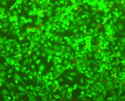

110 A B C D E 110
111 Fig Fluorescence micrographs of endothelial cells growing on surface of D-M (A), D-G-2 (B), D-G-4 (C), D-G-6 (D) hydrogels, and on TCPS (E) 1 day after seeding. Live cells were stained green, while dead cells red. Scar bar represents 100 μm. Cell seeding density: cells/well * * * * WST-1 Abs D-M D-G-2 D-G-4 D-G-6 TCPS Fig Viability of the endothelial cells cultured on the surfaces of hydrogels after 1 day. All samples and TCPS control exhibit improved viability compared with D-M hydrogel (at the significance level P<0.05 by Student's t-test as indicated by *) SMC encapsulation within hydrogels The hydrogel constructs with embedded cells were imaged with optical phase contrast microscope at various durations (4 h, 1 d, 3 d and 6 d) of culture. 111
112 After 4 h, cells inside D-G-2 and D-G-4 hydrogels were almost round (Fig. 3.12A and Fig. 3.12B), while inside D-G-6 hydrogels, some of the cells had started to form extensions (Fig. 3.12C), indicating that the cells were starting to spread. After one day, cells inside D-G-2 hydrogels were also starting to spread, indicated by the protruding pseudopods (Fig. 3.12A), whereas evidence of cell spreading inside D-G-4 hydrogel was not apparent until 3 days (Fig. 3.12B). Moreover, after 3 days, direct cell-cell contact between spreading cells could be seen clearly in the D-G-2 and D-G-6 hydrogels, as indicated by the circles in Fig. 3.12A and Fig. 3.12C. The cell-cell contact became obvious after 6 days in D-G-4 hydrogel (Fig. 3.12B, highlighted by circles). Spindle shaped cells were seen within all hydrogels, indicating that the hydrogels support SMC spreading. 112

")

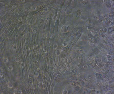
113 4h 1d 3d 6d Fig A (Cont d) 113

")
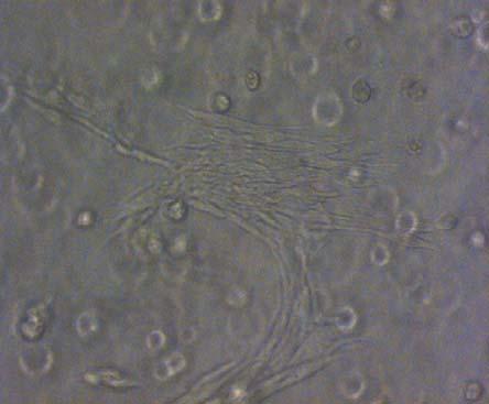
114 4h 1d 3d 6d Fig B (Cont d) 114

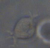

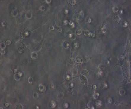

115 4h 1d 3d 6d Fig C 115
116 Fig Phase contrast macroscopic images of smooth muscle cells encapsulated in D-G-2, D-G-4, and D-G-6 hydrogels (A, B, and C, respectively) after 4 h, 1 day, 3 days, and 6 days. The magnified inset in image C (4 h) is a close-up of the cell highlighted with the circle and arrows. Scale bar represents 50 μm. Cell seeding density: cells/ml. To ascertain whether the encapsulated SMCs were alive, the hydrogel/cell constructs were tested with the Live/Dead assay. After 1-day encapsulation, (Fig (A1, B1 and C1)), most of the cells inside the hydrogels were stained green (live) while only a few were stained red (dead). Most of the live cells were round in shape. The large preponderance of live cells indicates that the hydrogels are not cytotoxic and that the UV crosslinking process caused little immediate damage to the encapsulated cells. Cell viability after longer time periods culture was also verified. After 7-day culture, many of cells in the D-G-2 and D-G-6 hydrogels had developed elongated spindle shapes (Fig (A2 and C2)), while in D-G-4 hydrogel (Fig. 3.13B2), most of cells were still round and only a few had assumed spindle shape. There were few dead cells in all the hydrogels after 7-day 3D culture. By 14-day culture, the number of spreading live cells in the D-G-4 hydrogel had visually increased (Fig. 3.13B3) and many live cells persisted in the D-G-2 and D-G-6 constructs at this stage of culture. 116


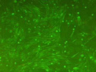
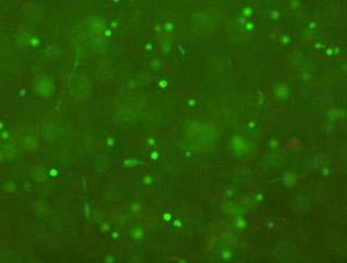

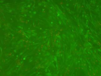

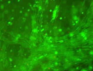
117 A1 B1 C1 A2 B2 C2 A3 B3 C3 117
118 Fig Fluorescence micrographs of SMCs encapsulated into D-G-2 (A1, A2, and A3), D-G-4 (B1, B2, and B3), and D-G-6 (C1, C2, and C3) hydrogels after 1 day (A1, B1, and C1), 7 days (A2, B2, and C2), and 14 days (A3, B3, and C3) using Live/Dead assay. The living cells were stained green, and the dead cells red. Scale bar represents 100 μm. Cell seeding density: cells/ml. Cell viability was also quantified spectrophotometrically using Cell Proliferation Reagent WST-1 (Fig. 3.14). After 7-day in vitro culture, cells in D-G-6 hydrogel had the highest viability. D-G-2 and D-G-4 hydrogels had significantly lower viability than D-G-6 but were statistically indistinguishable from each other. After 14 days of in vitro culture, cell viability in D-G-2 and D-G-4 hydrogels increased. In these two hydrogels with lower gelatin content, cell viability increased significantly during the 7 to 14 days period, while the increase during the same period for D-G-6 hydrogel was not significant. After the longer-term (14-day) culture, there was no significant difference in cell viability between D-G-2 and D-G-6 hydrogels (n.b.; this may be due to the large error bar in the D-G-2 measurement), but D-G-4 hydrogel had statistically lower cell viability than D-G-6 hydrogel. The lower 14-day WST-1 absorbance (i.e. proliferation) in D-G-4 also corroborates with the observation of fewer live cells using fluorescent microscope in Fig B3. Although there are variations in the detailed performance of the IPN hydrogel, all three promote cell proliferation in 7 to 14 days of culture. 118
119 During the course of the cell culture, mechanical strength loss was also observed, with the most obvious in the D-G-6 hydrogel (data not shown)); this may be a consequence of hydrogel degradation d 14d 2.5 # WST-1 Abs * * D-G-2 D-G-4 D-G-6 Fig Viability of the encapsulated smooth muscle cells in hydrogels after 7 days and 14 days. Comparisons were made with pairs with statistical analysis by the Student s t-test: at the level of P<0.05, * significantly different from that of D-G-6 hydrogel after 7 days; # significantly different from that of D-G-6 hydrogel after 14 days. Cell seeding density: cells/ml. (n=3) 3.4 Discussion We have synthesized and characterized an IPN hydrogel series based on methacrylate- and aldehyde-bifunctionalized dextran (Dex-MA-AD) and 119
120 gelatin. The methacrylate groups on Dex-MA-AD were used for UV crosslinking and the aldehyde groups enabled the incorporation of chemically linked gelatin. We chose a low extent of dextran oxidation DS of aldehyde of 13.9 ± 1.3 since increasing the density of aldehyde groups on dextran decreases its solubility but hastens the formation of Schiff base, making the thorough mixing of the two biomacromolecules and hence formation of homogeneous hydrogel network difficult. An optimum time of more than 5 minutes at the higher UV intensity of 20 mw/cm 2 appear to be feasible for cell viability and acceptable sol content 263 and mechanical properties 263, 272. As illustrated in Fig. 3.2, two different kinds of networks contribute to the covalent crosslinking in our system, namely the polymethacrylate kinetic chains produced by carbon-carbon double bond polymerization and Schiff base obtained by reaction between Dex-MA-AD aldehyde and gelatin amino groups. The unique crosslinking mechanism confers superior mechanical properties to the IPN hydrogels. The measured elastic moduli are on the order of 10 4 Pa, which are considerately higher than the reported 10 2 Pa value for PEG-based hydrogels which promote SMC spreading 19, 28, 34. A hydrogel that is mechanically robust would be highly desirable as a scaffold for tissue engineering of blood vessels. Cell spreading is highly important for obtaining cell-cell contacts, and 120
121 hence the outcome of tissue. In 2D systems, cell spreading can be promoted by cell adhesion molecules, such as RGD, collagen, and fibronectin. Due to the incorporation of gelatin, ECs cultured on the surface of our IPN hydrogels spread after seeding for as little as 4 h. In 3D hydrogel systems, cell spreading may be hampered even in the presence of adhesion molecules, due to the physical obstruction posed by the dense matrix 21. It has been proposed that the inclusion of degradable crosslinkers sensitive to either hydrolysis 22, 27 or cell-mediated proteolysis 19, 30, 31, 33, 35, 39 will permit formation of 3D cell extensions, since degradation will generate space for cells to make their way through the matrix, spread, migrate and establish cell-cell contacts. In our study, the incorporation of gelatin serves the dual purpose of providing cell adhesion sequences and enzymatic degradability. IPN hydrogels promoted 3D SMC spreading and proliferation. Cells acquired an elongated, spindle-shaped morphology as they spread within the hydrogels. The time course of spreading and proliferation was related to gelatin concentration (Fig and Fig. 3.14), with D-G-6 hydrogels promoting the quickest spreading and proliferation, followed by D-G-2 and then D-G-4 hydrogels. It has been proposed that cell spreading within hydrogels is influenced by both cell adhesion ligand density and matrix stiffness 19, 21, 34 ; high ligand density and soft matrix are expected to promote cell spreading. In our IPN 121
122 hydrogels, although higher gelatin concentration (60 mg/ml) increased the stiffness of the hydrogels, the high ligand density enabled the quickly spreading of encapsulated cells. Our hydrogel marries the unique properties of polysaccharides and proteins to mimic ECM. Gelatin, like other ECM proteins such as collagen and fibrin, is capable of promoting cell adhesion and proteolytic degradability via its biological domains. However, a major drawback in using these proteins solely is the limited control of the physical and degradation properties that can lead to premature matrix breakdown due to the release of cell-secreted matrix metalloproteinases. Polysaccharides have high hydration capability and are also non-antigenic, non-immunogenic 276 and feasible for easy modification with photochemically or thermally crosslinkable groups to produce hydrogels. The combination of the two in a single hydrogel produces a material with an excellent combination of mechanical and biological properties. However, in a very small percentage of human population, dextran is able to induce anaphylactoid reactions when binding to endogenous dextran-reactive immunoglobulin (Ig) G antibodies 277, and the chemical modification may also lead to immunological reactions. All these should be taken into account before putting dextran into clinical use. 122
123 3.5 Conclusion We have demonstrated a new class of gelatin-bonded dextran-based hydrogel with relatively high modulus that is also suitable for 3D encapsulation of SMCs and 2D culture of ECs. Using bifunctional dextran modified with acrylate and aldehyde groups mixed with gelatin, UV crosslinked hydrogels encapsulating vascular SMCs were fabricated. The Dex-MA-AD component imparted to the hydrogels elastic properties that are superior to commonly reported PEG-based hydrogels. The incorporation of gelatin into the hydrogels provided cell adhesive and enzymatically degradable properties and also significantly increased the compressive modulus and strength through the Schiff base contribution to the crosslinking density. We have shown that these hydrogels promoted adhesion of vascular endothelial cells in 2D culture and supported spreading and proliferation of vascular smooth muscle cells in 3D culture for up to 14 days. The attractive mechanical properties of this new class of hydrogel coupled with 2D and 3D biocompatibility with vascular cells make this a promising material for 3D scaffolds for vascular tissue engineering and regeneration. 123
124 This chapter has been reproduced from the following paper published by the PhD student. Liu YX, Chan-Park MB. Hydrogel based on interpenetrating polymer networks of dextran and gelatin for vascular tissue engineering. Biomaterials 30: ,
125 Chapter 4 Biomimetic hydrogel based on methacrylated dextran-graft-lysine and gelatin for 3D smooth muscle cell culture 4.1 Introduction In the previous chapter, we reported the initial work on hydrogels based on natural gelatin and dextran bifunctionalized with methacrylate and aldehyde. That series of hydrogels allowed adhesion of vascular endothelial cells on 2D culture and supported spreading and proliferation of vascular smooth muscle cells in 3D culture. However, the Schiff base reaction between dextran and gelatin was not easily controllable and while SMC spreading within the hydrogel occurred, it was rather limited. In the current chapter, a novel methacrylate and lysine functionalized dextran (Dex-MA-Ly) was designed and synthesized (Fig. 4.1A). A series of hydrogels based on Dex-MA-Ly and methacrylamide modified gelatin (Gel-MA) (Fig. 4.1B) with varying mechanical stiffness was developed by ultraviolet (UV)-crosslinking. The swelling, sol contents and mechanical properties of these hydrogels were characterized. The viability, proliferation and spreading of human umbilical artery SMCs inside these hydrogels were tracked. 125
126 The crosslinking of this new series of hydrogels based on Dex-MA-Ly and Gel-MA is much more controllable. Further, we found this series to allow the encapsulated SMCs to spread more extensively compared to the Dex-MA-AD series in Chapter
127 O O A HO HO NaIO 4 O O OH HO HO HO O Dextran O OH HO HO O CHO OHC O HO HO O Dex-MA-AD O O OH O O OH HO O O CH 3 H 2 H 2 C C C O C O O OH O Lysine O HO HO O O OH HO HO O O HO CHHC O N N HO HO HC H 2 N COOH CH H 2 N COOH O O OH HO O O O OH O Dex-MA O O OH O HO O O OH O Dex-MA-Ly CH 3 B NH 2 NH 2 NH 2 CH 2 CH 2 O H 3 C CH 3 O O NH 2 O NH CH 2 NH O CH 2 CH 3 NH 2 NH 2 NH NH 2 NH 2 H 2 C O NH 2 CH 3 Gelatin Gel-MA Fig. 4.1 Synthesis of Dex-MA-Ly (A) and Gel-MA (B). 127
128 4.2 Materials and methods Materials The chemicals, reagents and cells described in Section were used in this chapter. Besides, 2,4,6-trinitrobenzene-sulfonic acid (TNBS), methacrylic anhydride, and L-lysine obtained from Sigma-Aldrich were all used as received Synthesis of dextran derivatives Dextran was first modified to introduce methacrylate and aldehyde groups (Dex-MA-AD) (Fig. 4.1A) as described in Section and Briefly, dextran was dissolved in anhydrous DMSO under nitrogen, followed by addition of DMAP. GMA was added to the solution. The mixture was stirred for a certain time under nitrogen to produce dextran with methacrylate groups (Dex-MA). Dex-MA was precipitated with a large excess of acetone, purified by dialysis and freeze-dried. By varying the amount of GMA and reaction time, Dex-MAs with different degrees of substitution (i.e. DS, the number of functional groups per 100 glucopyranose residues) of methacrylate were obtained. The Dex-MAs were further oxidized by reaction with sodium periodate at room temperature in the dark for 4 h to introduce aldehyde groups (Dex-MA-AD). The Dex-MA-AD solutions (2 wt %) were added dropwise to 128
129 L-lysine solution (in 0.1 M NaHCO 3, 2-fold molar excess over aldehyde groups) over 5 h. The reactions were continued overnight. Dextran with methacrylate and lysine functionalities (Dex-MA-Ly) was collected by dialysis and freeze-drying. The degree of substitution of methacrylate was confirmed by proton nuclear magnetic resonance ( 1 H-NMR) spectroscopy using a Bruker Avance 300 MHz instrument with D 2 O as the solvent. The concentration of free primary amino groups, essentially the amino groups of lysine, was determined by the Habeeb method Synthesis of gelatin derivatives Gelatin methacrylamide (Gel-MA) was prepared by reaction with methacrylic anhydride (Fig. 4.1B) following the procedure of reference 279. Briefly, after gelatin was dissolved in phosphate buffer saline (PBS) at 50 o C, methacrylic anhydride was added. The mixture was stirred under nitrogen atmosphere for 1 h, and then dialyzed for 24 h against DI water at 40 o C. The product was collected by freeze-drying. The degree of substitution was defined, using the Habeed method, as the percentage of ε-amino groups that had been modified 278. By changing the amount of methacrylic anhydride, Gel-MAs with a range of degrees of substitution were obtained. 129
130 4.2.4 Hydrogel formation and characterization Hydrogels were fabricated by exposure of precursor solutions to UV light (λ=365 nm) at 20 mw/cm 2 for 5 min under argon atmosphere using a UV exposure system (Honle UV technology). The compositions of the precursor solutions are outlined in Table 4.1. Before UV crosslinking, the precursor solutions (in PBS) with 0.1 wt% of Irgacure 2959 (I-2959) pre-dissolved in PBS (10 mg I-2959 in 6 ml PBS) were vigorously stirred at 37 C for 30 min. 130
131 Table 4.1 Compositions of hydrogels. Sample Concentration of (mg/ml) Dex-MA-Ly Gel-MA Total Degree of methacrylate substitution L-G-1A L-G-1B L-G-1C L-G-2A L-G-2B L-G-3A L-G-3B Dex-MA(9)-Ly Gel-MA(16) Dex-MA(9)-Ly Gel-MA(51) Dex-MA(30)-Ly Gel-MA(16) Dex-MA(9)-Ly and Dex-MA(30)-Ly: with 9 and 30 methacrylate groups per 100 glucopyranose residues respectively; Gel-MA(16) and Gel-MA(51): with 16% and 51% of ε-amino groups been modified. 131
132 For sol content and mass swelling ratio measurements, 250 µl of precursor solution was crosslinked in the well of a 24-well tissue culture polystyrene plate (TCPS). Immediately after gelation, the obtained hydrogel disks were placed in PBS at 37 C for 24 h with swelling medium changed every 12 h, followed by freeze-drying for 24 h. The sol content, S, was determined by S= (W 1 -W 2 )/W 1, with W 1 the initial solid weight of the sample, and W 2 the weight of the sample after freeze-drying. The sol-removed dry gels (weight W 2 ) were re-immersed in PBS at 37 C in a CERTOMAT H incubator (Sartorius, B. Braun Biotech International). At discrete time intervals the swollen hydrogel disks were blotted on a filter paper. After removal of the excess superficial water, the weight of the samples in the swollen state (W s ) was measured. After W s reached its equilibrium value W s,eq, the equilibrium mass swelling ratio, q, was determined by q=(w s,eq -W 2 )/W 2. The volumetric swelling ratio, Q, was calculated based as Q=1+q*ρ 1 /ρ 280 2, while ρ 1 is the density of dry polymer (mixture of Dex-MA-Ly and Gel-MA, obtained by calculation based on the weight ratio) and ρ 2 is the density of the solvent (buffer solution, 1.09 g/ml). All experiments were conducted in triplicate. Shear storage modulus was measured using a Thermo Haake Rheostress 600 rheometer in conjunction with a UV spot curing system (LUMATEC 132
133 SUV-DC-P). Dynamic time sweep in plate-plate geometry was performed at room temperature, at a constant stress of 100 Pa and a constant frequency of 1 Hz. Briefly, 150 µl of precursor solution was deposited onto the lower plate, and the 2 cm diameter upper plate was moved down to obtain a gap of 0.5 mm. After 60 s the solution was exposed to UV light. The storage (G ) moduli were recorded after 5 min of UV exposure. Three parallel samples for each formulation were measured, and the obtained values were averaged SMC encapsulation Cultured monolayers of SMCs were trypsinized after reaching confluence. Hydrogels precursor solutions (in Smooth Muscle Cell Medium Bulletkit ) were added to re-suspend the SMCs. 40 µl of cell-laden precursor solution was deposited onto the surface of polytetrafluoroethylene (PTFE) film, and subjected to low intensity UV illumination (365 nm, 20 mw/cm 2 ) for 5 min under argon atmosphere. The cell/hydrogel constructs were transferred into 24-well Ultralow attachment plates (Corning, Fisher) and supplied with Dulbecco's Modified Eagle's Medium (DMEM) containing 10% FBS. The medium was refreshed every two days. SMCs within hydrogels were monitored during culture using an Axiovert 200 Motorized Inverted Microscope System. F-actin of SMCs was stained 133
134 using Alexa Fluor 488 phalloidin (Invitrogen) solution (1:200 dilute in 1% BSA/PBS buffer), and viewed using an LSM 510 confocal microscope (Carl Zeiss, Germany), after hydrogel constructs were fixed (using 4% paraformaldehyde), permeabilized (using 0.1% Triton X-100) and blocked (using 1% BSA/PBS solution) Cell viability The viability of the encapsulated cells was assessed by LIVE/DEAD Viability/ Cytotoxicity Assay Kit (Invitrogen). The medium was removed and the constructs were washed twice in PBS and incubated in Live/Dead solution containing 2 µm calcein AM and 4 mm EthD-1 for about 45 min at 37 C. Cells were imaged using a Zeiss inverted microscope with fluorescein and rhodamine optical filters Cell proliferation SMC proliferation was quantified using Cell Proliferation Reagent WST-1 (Roche). Typically, after refreshing the cell-laden hydrogels with 200 µl medium without FBS, 20 µl of WST-1 reagent was added. After 4 hours of further incubation, the hydrogels were smashed to ensure complete dissolution of the formazan dye after which the absorbance of the medium at 440 nm was 134
135 recorded. 5-bromo-2 -deoxy-uridine (BrdU, Invitrogen) was used to visualize proliferative cells. After discrete peroids of culture, BrdU was added to the culture medium. After BrdU exposure overnight at 37 o C, cells were fixed (4% paraformaldehyde), permeabilized (0.1% Triton-X 100) and treated with HCl solution to break the DNA structure. Alexa Fluor 488 anti-brdu antibody (Invitrogen) was applied to visualize the proliferative cell nuclei. Cell images were taken using an LSM 510 confocal microscope Statistical analysis Statistical analysis using Student s t-test was performed where appropriate. Data are represented as mean ± standard derivation of at least three measurements. P<0.05 was considered significantly different. 4.3 Results Synthesis of dextran and gelatin derivatives Dextran was modified to incorporate both methacrylate and lysine functionalities (Fig. 4.1A). The structure of Dex-MA-Ly was confirmed by 1 H-NMR spectroscopy (Fig. 4.2). In addition to the peaks of pristine dextran (between δ 4.0 and 3.3 ppm due to protons at positions 2, 3, 4, 5 and 6 of the 135
136 glucose unit, at δ 4.8 ppm due to the anomeric proton (position 1) and at δ 5.1 ppm due to the α-1,3 linkages), additional peaks appear at about δ 1.8 ppm (position 7) due to methyl protons from the methacryloyl group, and at about δ 5.6 ppm and δ 6.1 ppm (position 8) due to the double bond protons. Additional signals due to protons of lysine also appear between δ 1.0 ppm and δ 1.7 ppm for γ, δ, β-protons (positions 11, 10, and 12), and at δ 2.9 ppm for ε-proton (position 9). The peak of α-protons (position 13) overlaps with that of the position 4 proton of the glucose unit. Two versions of Dex-MA-Ly with different degrees of methacrylate substitution (DS) of 9 and 30 were synthesized; these are hereafter denoted Dex-MA(9)-Ly and Dex-MA(30)-Ly. The amino group concentration measured using TNBS was mmole/mg for Dex-MA(9)-Ly, and mmole/mg for Dex-MA(30)-Ly. The measured zeta potentials of Dex-MA-Lys, which are zwitterionic, were negative: ± 1.99 mv for Dex-MA(9)-Ly and ± 1.98 mv for Dex-MA(30)-Ly. Gelatin was modified to introduce methacrylamide functionality (Fig. 4.1B). By changing the amount of methacrylic anhydride, Gel-MA(s) with a range of degrees of substitution (s, where s is 16, 51, 72, or 78) were prepared. As shown in Fig. 4.3A, the absorbance at both 340 nm and 420 nm decreases with increasing degree of substitution measured using the Habeeb method. Due 136
137 to the incorporation of charged carboxyl and amino groups, Dex-MA-Ly has better compatibility with Gel-MA than Dex-MA has. As shown in Fig. 4.3B, the solution of Dex-MA(9)-Ly and Gel-MA(16) is more transparent than that of Dex-MA(9) and Gel-MA(16). 137
138 O HOD HC HO N H 2 N COOH CH O HC N HO HO CH H 2 N COOH O O OH 1&1' HO O O O O OH O &4 5 & 6 8 1' Fig H-NMR spectra of Dex-MA-Ly dissolved in D 2 O. 138
 6: Blank 5 1 6 0 300 350 400 450 500 550 600 Wavelength (nm) Fig. 4.3 A Fig.")
139 Absorbance : Gelatin 2: Gel-MA(16) 3: Gel-MA(51) 4: Gel-MA(72) 5: Gel-MA(78) 6: Blank Wavelength (nm) Fig. 4.3 A Fig. 4.3 B 139
140 Fig. 4.3 (A) UV spectrum of gelatin and its methacrylamide derivatives reacting with TNBS. (B) Gross view of Dex-MA(9)/Gel-MA(16) (left) and Dex-MA(9)-Ly/Gel-MA(16) (right) solutions Hydrogel characterization Three series (with a total of seven distinct formulations) of hydrogels with varying precursor concentrations and degrees of methacrylation of Dex-MA-Ly and Gel-MA were fabricated (Table 4.1); these are labeled L-G-1x, L-G-2x and L-G-3x respectively where x is A, B or C depending on the ratio of Dex-MA-Ly to Gel-MA, which was varied from 80:40 to 80:80. At fixed degree of methacrylation of Dex-MA-Ly and Gel-MA, increasing Gel-MA concentration (e.g. compare L-G-1A with L-G-1C, Fig. 4.4A and B) somewhat reduces swelling ratio, but dramatically increases storage modulus. Similarly, at fixed Dex-MA-Ly to Gel-MA ratio, increase in DS of either Dex-MA-Ly or Gel-MA (e.g. compare L-G-1A with L-G-3A or L-G-1A with L-G-2A) decreases swelling ratio and increases storage modulus (Fig. 4.4A and B). The trend of decreased swelling ratio and increased storage modulus with higher Gel-MA concentration and higher methacrylation is due to increased hydrogel precursor double bond density leading to higher hydrogel crosslinking density. The sol 140
141 content increased slightly with increasing Gel-MA concentration (Fig. 4.4C); no statistically significant trend of sol content with DS is discernible due to the relatively large measurement uncertainties. 28 A 24 # # *, # Mass Volumetric Swelling ratio * *, # # # L-G-1A L-G-1B L-G-1C L-G-2A L-G-2B L-G-3A L-G-3B B *, # Shear storage modulus (Pa) *, # *, # *, # *, # *, # 1000 *, # 0 L-G-1A L-G-1B L-G-1C L-G-2A L-G-2B L-G-3A L-G-3B 141
142 16 C Sol content (%) L-G-1A L-G-1B L-G-1C L-G-2A L-G-2B L-G-3A L-G-3B Fig. 4.4 Properties of hydrogels: (A) swelling ratio, (B) shear storage modulus and (C) sol content. * P<0.05 vs. other hydrogels with Dex-MA-Ly and Gel-MA of the same degrees of substitution; # P<0.05 vs. other hydrogels with the same concentration Spreading of SMCs inside hydrogels Gelatin was incorporated into the hydrogel networks in order to support cell adhesion and promote proteolytic degradation, both of which are needed for cell spreading within a 3D scaffold. It is known that cells are also sensitive to gel mechanics 19, 21, 37, 281, 282, which motivates the study of a range of formulations with diverse mechanical properties (Fig. 4.4B). The green fluorescence in Fig. 4.5A shows that the SMCs encapsulated in the hydrogels 142
143 are mostly alive at different time points. However, extensive SMC spreading and formation of 3D cellular networks were found only in the softer hydrogels (L-G-1A to L-G-1C and L-G-2A, Fig. 4.5A). The stiffer hydrogels (L-G-2B, L-G-3A and L-G-3B, Fig. 4.5A) exhibited high viability as indicated by high green fluorescent density but inhibited SMC spreading and showed no cellular networks formation after 14 days of culture. On the other hand, SMCs within the softer L-G-1A, L-G-1B, and L-G-1C hydrogels started to acquire elongated morphology after as little as 1 day of culture, and exhibited evidence of 3D cellular network formation after 2 days of culture (Fig. 4.6). Phalloidin-staining of the cytoskeleton F-actin shows spread out cells with assembled stress fibres (Fig. 4.5Bi). A composite of confocal image sections pseudo-colored as a function of depth indicates extensive 3D spreading of SMCs within the hydrogel (Fig. 4.5Bii). 143


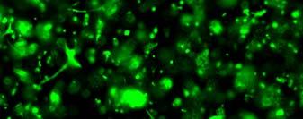
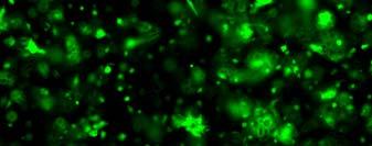
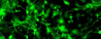
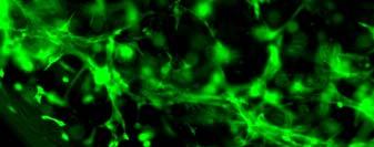
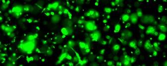


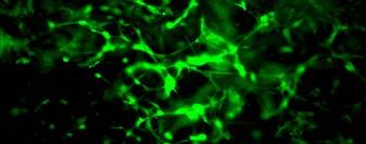
144 1 d 7d 14 d L-G-1A L-G-1B 144

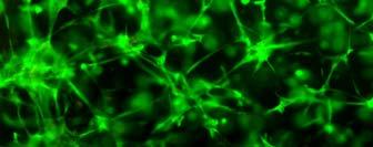


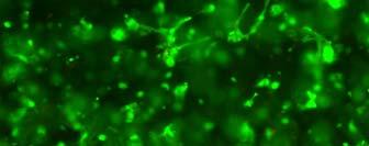
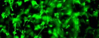
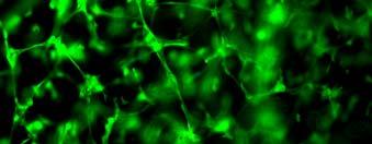
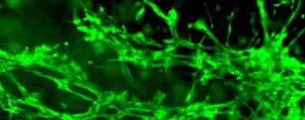
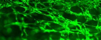
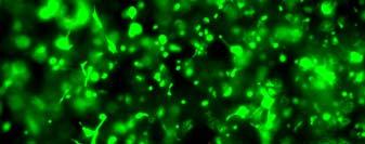

145 L-G-1C L-G-2A 145

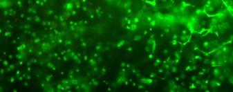
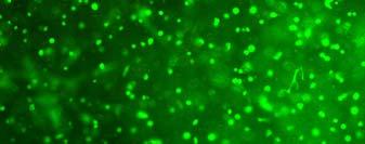
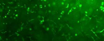

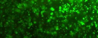
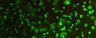
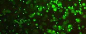


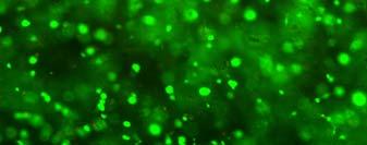
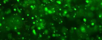
146 L-G-2B L-G-3A 146

 147")


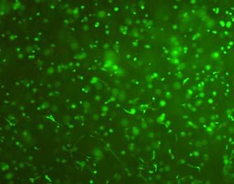
147 L-G-3B Fig. 4.5 A (Cont d) 147


148 i ii L-G-1C, 7days Fig. 4.5 B Fig. 4.5 (A) Live/Dead staining of SMCs encapsulated inside hydrogels after 1 day (left column), 7 days (middle column), and 14 days (right column) of culture. Scale bar equals to 200 µm. Cell seeding density: cells/ml. (B) Cytoskeletal F-actin staining of SMCs in L-G-1C hydrogel after 7 days. Confocal stacks of 300 µm depth were taken. The image sections were projected to one composite image (i, left), and a pseudo-colored composite of image sections of SMCs at varying depths (ii, right) was created using LSM image browser. The scale bar is 100 µm. 148


149 1 day 2 days 7 days L-G-1A L-G-1B L-G-1C 149
150 Fig. 4.6 Phase contrast microscopic images of smooth muscle cells encapsulated inside L-G-1A (top row), L-G-1B (middle row), and L-G-1C (bottom row) hydrogels after 1 day (left column), 2 days (middle column), and 7 days (right column) of culture. Scale bar is 100 µm. Cell seeding density: cells/ml Proliferation of SMCs inside hydrogels To quantify the proliferative capacity of SMCs inside the 3D hydrogels, cell viability during 2 weeks of culture was measured using WST-1 reagent in representative hydrogels (i.e. L-G-1A, L-G-1B, L-G-2A, and L-G-3A). As shown in Fig.4.7, cells in L-G-1A, L-G-1B and L-G-2A hydrogels proliferated over the two-week study period, as indicated by the increase in WST-1 absorbance with culture time. Within L-G-1A hydrogels, cell viability increased ~3 fold during the first two days of culture and less rapidly thereafter. In L-G-1B and L-G-2A hydrogels, cells proliferated rapidly during the first week of culture with viability increasing ~4-5 fold; the viability plateaued or declined somewhat thereafter. In L-G-3A hydrogels, cell viability decreased about 50% during the first two days of culture and then stabilized; this is interpreted to be a result of inhibition of cell spreading. The four hydrogels were created with identical cell densities but cell proliferation did not begin within the first day of culture (determined by the 150
151 absence of BrdU incorporation, discussed below, data not shown), so that the viability at day 1 is proportional to the number of cells encapsulated within the hydrogels (and surviving the first day of culture). The day 1 viability is thus a proxy for the encapsulation efficiency of the hydrogel formulation, i.e. the proportion of the cell inoculum incorporated into the gel network. In the context of the present experiments, the encapsulation efficiency depends on the speed with which the precursor solution gels compared with the speed with which cells settle from the precursor solution. The gelation time depends on the concentration of double bonds in the precursor solution while the cell settling time depends on the solution viscosity. The relatively low encapsulation efficiency implied by day 1 viability in the L-G-1A hydrogel is attributable to the low viscosity (quick cell settling) and low double bond concentration (slow gelation) of this hydrogel precursor solution. The L-G-1B precursor solution has higher viscosity due to higher Gel-MA concentration, while the L-G-2A and L-G-3A precursor solutions have higher concentrations of double bonds, with consequent quicker gelation, so that the cell encapsulation efficiencies of these three hydrogels are higher than that of L-G-1A hydrogel. Cell proliferation was detected using Bromodeoxyuridine (BrdU). BrdU was added to the cell culture medium and the cells were further incubated overnight. During that time, the proliferating cells incorporated BrdU into 151
152 newly synthesized DNA. Fluorescent anti-brdu was used to visualize the incorporated BrdU. Green colored nuclei were seen in L-G-1A and L-G-2A hydrogels after 5 days of culture (Fig. 4.8), which is consistent with the cell viability measurements (Fig. 4.7) showing an increase of cell viability. 152
153 WST-1 absorbance (a.u.) L-G-1A * WST-1 absorbance normalized to day 1 (a.u.) WST-1 absorbance (a.u.) L-G-1B * * * WST-1 absorbance normalized to day 1 (a.u.) Culture time (d) Culture time (d) WST-1 absorbance (a.u.) 2.8 L-G-2A * * 1.6 * Culture time (d) WST-1 absorbance normalized to day 1 (a.u.) WST-1 absorbance (a.u.) L-G-3A * * Culture time (d) WST-1 absorbance normalized to day 1 (a.u.) Fig. 4.7 Comparison of proliferative capacity, as assessed by WST-1 assay, of SMCs encapsulated within L-G-1A, L-G-1B, L-G-2A, and L-G-3A hydrogels. *: p<0.05 vs. adjacent time point before (same hydrogel). 153


154 L-G-1A, 5days BrdU Ph Merge L-G-2A, 5days BrdU Ph Merge Fig. 4.8 SMCs showing incorporation of BrdU after encapsulation inside L-G-1A (top row) and L-G-2A (bottom row) hydrogels for 5 days. Scale bar is 50 μm. Ph refers to phase contrast image. 154
155 4.4 Discussion Hydrogels have been widely explored as substrates for cell encapsulation, and it is hypothesized that cell functions, including adhesion, spreading, proliferation, and differentiation, can be controlled by manipulating the physical and chemical characteristics of hydrogels 31, 35, 38. The aim of this study was to investigate the ability of hydrogels based on natural dextran and gelatin derivatives to support SMC spreading and proliferation and to investigate the effect of mechanical stiffness on 3D SMC spreading and proliferation. Dextran was functionalized with methacrylate and lysine, and gelatin was modified to introduce methacrylamide groups. By varying the concentration of Gel-MA, as well as the DS of methacrylate, hydrogels with a range of storage moduli (from 898 to 6075 Pa) were produced by UV crosslinking. For cell encapsulation, SMCs were suspended in precursor solution and encapsulated during UV polymerization. Within the hydrogels, SMCs invaded the matrix through spreading, which is characterized by the protrusion of cellular lamellipodia. We have found that SMC spreading and proliferation is sensitive to hydrogel mechanics. The softer hydrogels (with shear storage moluli ranging from 898 Pa to 3124 Pa) promoted fast cell spreading, profound proliferation, and extensive 3D cellular network formation, while in stiffer hydrogels (shear 155
156 storage moduli ranging from 4026 Pa to 6075 Pa) SMCs maintained a round morphology and their viability declined by 50% over 2 weeks of culture (Fig. 4.5A and Fig. 4.7). The stiffer hydrogels have correspondingly denser networks that are more obstructive to cell spreading and proliferation. The spreading of SMCs within the hydrogels was clearly accompanied by the assembly of F-actin stress fibers (Fig. 4.5B). This relationship between hydrogel mechanics and SMC spreading agrees with similar studies using PEG-based hydrogels 19, 34, 37, although other reported hydrogels differed in the range of mechanical stiffness. For example, in one study using PEG-conjugated fibrinogen hydrogels, a qualitative spindle index was applied to classify the degree of cell spreading 34 : a spindle index of 4 indicated highly spindled spreading SMCs, while 1 referred to round-shaped SMC with minor lamellipodia. When shear modulus increased from 62 Pa to 497 Pa, the spindle index decreased from 4 to 1 after 24 h in 3D culture. Compared with this study, our hydrogels have higher mechanical stiffness; nevertheless, it is only an approximate comparison, since the parameters regarding characterization of mechanical stiffness of hydrogels differed. However, in our case, even hydrogels with storage modulus as high as 3124 Pa permitted the initiation of cell spreading behavior within 24 h (Fig. 4.5A and Fig. 4.6). In addition to spreading, SMC proliferation and formation of cellular networks in our hydrogels were also extensive, as indicated by the 156
157 high-fold increase in cell viability (Fig. 4.7) and densely and extensively organized 3D cellular networks (Fig. 4.5A). The excellent cellular response (i.e. spreading, proliferation and formation of 3D cellular networks) of our dextran and gelatin-based hydrogels may be due to their closer resemblance to native proteoglycan, which may provide a highly bioactive 3D microenvironment. The Gel-MA content confers both cell adhesion and cell-mediated degradation to our hydrogels. Although pristine dextran is non-adhesive, like the bioinert PEG, it is degradable through the action of dextranases which are present in various tissues of the human body, such as liver, spleen, and kidney 261. In addition, lysine-modified dextran structurally resembles natural glycosaminoglycans (GAGs), which are an important component of connective tissues, and is also negatively charged like GAGs. Our composite hydrogel based on the copolymerization of gelatin and lysine-modified dextran has a degree of molecular structure-similarity to proteoglycans, which are GAGs covalently linked to a protein. It is well known that proteoglycans can affect the activity and stability of proteins and signaling molecules (e.g. growth factors) within ECM matrix, thus influencing cell function 283. So, we hypothesize that the combination of lysine-modified dextran and gelatin is capable of recapitulating some of the intricacies of natural ECM matrix. 157
158 4.5 Conclusion We have developed novel biocompatible and biodegradable hydrogels based on methacrylate and lysine functionalized dextran and methacrylamide modified gelatin. The mechanical stiffness of our hydrogels can be altered over a relatively wide range of moduli by varying the degree of methacrylation of dextran and gelatin, as well as the concentration of precursor solution. Rapid 3D SMC spreading, profound proliferation and extensive cellular network formation were obtained in a subset of these novel hydrogels which had suitably low mechanical stiffness. Our hydrogels resemble native proteoglycans both structurally and functionally. The excellent cell proliferation and network formation indicate that these hybrid hydrogels based on natural polysaccharides and proteins are promising scaffolds for vascular tissue engineering. This chapter has been reproduced from the following paper published by the PhD student. Liu YX, Chan-Park MB. A biomimetic hydrogel based on methacrylated dextran-graft-lysine and gelatin for 3D smooth muscle cell culture. Biomaterials 31: ,
159 Chapter 5 Phenotype and collagen type I synthesis of smooth muscle cells in 3D dextran-based hydrogels 5.1 Introduction SMC phenotype plays a key role in the performance of vasculature and consequently must be regulated along with spreading and proliferation in vascular tissue engineering 6, 7. Differentiated SMCs in normal blood vessels exhibit a quiescent contractile phenotype with relatively low rate of proliferation and ECM synthesis 7, 8, 97. Under a variety of pathological conditions, such as restenosis and atherosclerosis, SMCs dedifferentiate from their normal quiescent contractile phenotype to a synthetic phenotype characterized by migration, proliferation and abundant ECM production 98, 100, 284. The SMC phenotype transition can be regulated via environmental cues, such as mechanical stimuli, soluble signaling factors and cell-cell and cell-substrate interactions 6, 285. For tissue regeneration, a synthetic SMC phenotype in the early stages of tissue culture is desired to facilitate cellular expansion and deposition of ECM components, such as collagen and elastin, to replace the degrading scaffold but ultimately a transition to contractile phenotype is required for a vasoactive vessel 6. This has been achieved in prior 159
160 work by our group on 2D culture of SMCs within microstructured scaffolds 286. In this chapter, the phenotype state of SMCs within a particular grade of Dex-MA-Ly and Gel-MA 3D hydrogels (i.e. L-G-2A hydrogel in Chapter 4) was monitored during 20 days of culture by the gene expressions of contractile proteins (α-smooth muscle actin (α-sma) and smooth muscle-myosin heavy chain (SM-MHC)) and nonmuscle-myosin heavy chain B (SMemb), a synthetic SMC marker 7, 8. Collagen type I production was also monitored indirectly at gene level and directly using immunostaining. 5.2 Methods SMC culture and encapsulation within hydrogel SMCs were cultured and encapsulated in L-G-2A (Table 4.1) hydrogel as described in section For 2D comparison studies, SMCs were cultured on 24-well TCPS Real-time reverse transcription polymerase reaction (real-time RT-PCR) RNA was collected from cell-laden hydrogels using the combination of Trizol (Invitrogen) and RNeasy Mini Plant Kit (Qiagen) following the procedure of reference 287. RNA from 2D TCPS surface was obtained by 160
161 RNeasy Mini Plant Kit (Qiagen) following the manufacturer s instructions. The acquired RNA was reverse-transcribed using Sensiscript RT Kit (Qiagen). Real-time PCR analysis was performed in an iq TM qpcr system (Bio-Rad) using iq SYBR Green reagent (Bio-Rad). The relative gene expression values were calculated with the ΔC T (threshold cycle) methods normalized to the housekeeping gene glyceraldehyde-3-phosphate dehydrogenase (GAPDH). Gene specific primers 111, 288, 289 are listed in Table 5.1. Statistical analysis using Student s t-test was performed as described in section Immunofluorescent staining After discrete periods of culture, hydrogel constructs were rinsed three times with PBS, fixed in 4% paraformaldehyde, permeabilized in 0.1% Triton X-100, and blocked in 1% BSA/PBS solution. For α-actin and collagen type I staining, monoclonal anti-α-smooth muscle actin antibody (Sigma) and anti-collagen type I antibody (Sigma) were applied at 1:100 dilution at 4 o C overnight or for 48 h. After washing in 1% BSA/PBS solution, secondary antibodies (Alexa Fluor 488 goat anti-mouse for α-actin, Alexa Fluor 488 Signal Amplification Kit for collagen type I) were applied at 1:200 dilution at room temperature for 4 h. Images were taken using an LSM 510 confocal microscope (Carl Zeiss, Germany). 161
162 Table 5.1 Primers used for real-time RT-PCR. Gene Primer sequence (5'-3') A.T. ( o C) P.S. (bp) Ref. GAPDH F: ATGGGGAAGGTGAAGGTCG R: GGGGTCATTGATGGCAACAATA Myosin heavy chain11, smooth muscle (SM-MHC or SM1/SM2) Alpha-actin Myosin heavy chain10, non smooth muscle (SMemb) F:TGAACGCACTCAAGAGCAAG R:TCTACGTCCTCCAGACCTTCT F:CATCACCAACTGGGACGA R: GGTGGGATGCTCTTCAGG F:CCCATGAAGAGATTCTGTCAATGC R: ACATTCATCCCAAGAAGATGGC F: ATGTGGCCATCCAGCTGAC Collagen type I R: TCTTGCAGTGGTAGGTGATGTTCT A.T., P.S., and Ref. refer to annealing temperature, product size, and references, respectively
163 5.3 Results Differentiation of SMCs inside hydrogels The expression of mrna of smooth muscle myosin heavy chain 11 (SM1/SM2), α-actin, non-smooth muscle myosin heavy chain 10 (SMemb), and collagen type I were monitored during 20 days of culture in hydrogels (Fig. 5.1). SM1/SM2 and α-actin are contractile markers 7. Collagen type I is an ECM marker whose expression depends somewhat on SMC phenotype. Substantial ECM synthesis is typically associated with proliferative SMC phenotype 291. SMemb is profoundly expressed in proliferating SMCs and is considered to be a marker for phenotype change from a contractile to a proliferative type 292. The gene expression tests were performed on the 3D cultures at three stages, the 4 th, 9 th and 20 th day of culture. Day 9 corresponds to the maximum of cell viability in L-G-2A hydrogel (Fig. 4.7). For comparison with phenotype evolution in 2D culture, mrna was also extracted from monolayers of SMCs on TCPS at days 1, 4, and 7. The cells in 2D culture reached confluence at day 4. The 2D cultures progressed more rapidly than the 3D cultures and, for the purpose of comparison, the relative state of confluence between cells on TCPS and in L-G-2A was estimated to be comparable (though not at the same absolute state of proliferation) at these time points: 4 th day hydrogel vs. 1 st day 163
164 TCPS, 9 th day hydrogel vs. 4 th day TCPS, 20 th day hydrogel vs. 7 th day TCPS. We first discuss the gene expression pattern in the 2D cultures on TCPS. Expression of the contractile phenotype genes SM1/SM2 and α-actin peaked near confluence and declined in longer culture (Fig. 5.1A and B). The expression pattern of the proliferative phenotype gene SMemb reversed that of the contractile phenotype genes, though the amplitude of variation was much lower (Fig. 5.1C). The expression pattern of the gene for ECM protein collagen type I resembled that of the contractile markers (Fig. 5.1D), which seems paradoxical in view of the known tendency of SMCs in the proliferative state to secrete abundant ECM. The pattern of SM1/SM2 and α-actin expression is consistent with the interpretation of cells in the process of switching from a synthetic/proliferative to a contractile phenotype as they crowd themselves near confluence, a pattern that we have observed in SMC culture in 3D constrained microenvironments 286. The decline in expression of SM1/SM2 and α-actin genes at day 7 point is presumably due to cells reaching the completion of their switch from synthetic/proliferative to contractile/quiescent phenotype. Although the expression of SMemb is somewhat higher at day 7 than at day 4, at both days it is lower than at day 1, suggesting an overall trend of decrease in synthetic/proliferative state with passage of time, again consistent with a picture of transition from synthetic/proliferative to quiescent/contractile state. At day 4, 164
165 the collagen type I gene expression is also high, like the contractile SM1/SM2 and α-actin gene expression, and it may be that the cells which are reaching, but have not reached, confluence are still producing significant ECM. There is manifestly a dramatic decline in expression of collagen type I gene at day 7, consistent with the picture of the culture assuming a quiescent/contractile state. 165
166 # 6 B # * 4d * 4d 9d * 20d 1d 4d TCPS # 7d *, # 12 D # * 4d *, # 9d *, # 20d 1d 4d # Collagen type I gene expression (2 -delta CT ) alpha-actin gene expression (2 -delta CT ) L-G-2A 9d 20d alpha-actin gene expression (2 -delta CT ) A C ATTENTION: The Singapore Copyright Act applies to the use of this document. Nanyang Technological University Library * 4d * 9d L-G-2A # 20d 1d 4d TCPS # 7d L-G-2A * 4d *, # 9d 20d 1d # 4d # 7d 7d L-G-2A TCPS L-G-2A TCPS 166 SMemb gene expression (2 -delta CT ) SM1/SM2 gene expression (2 -delta CT )
167 Fig. 5.1 Expression of SM1/SM2 (A), α-actin (B), SMemb (C), and collagen type I (D) over 20 days of culture in L-G-2A hydrogels and over 7 days of culture on TCPS. The inset in B enlarges the vertical scale for the α-actin expression in L-G-2A hydrogel. Expression levels shown are relative to GAPDH expression in the same sample. *: p<0.05 vs. TCPS (4 th day hydrogel vs. 1 st day TCPS; 9 th day hydrogel vs. 4 th day TCPS, 20 th day hydrogel vs. 7 th day TCPS); #: p<0.05 vs. same sample prior time point. In L-G-2A hydrogel, the expression patterns of SM1/SM2, α-actin, SMemb and collagen type I were broadly similar in shape to those on TCPS, but significantly different in absolute level. Here we summarize the details; the implications are discussed in the next section. The initial (4 d) expression level of SM1/SM2 gene is significantly higher than that of day 1 TCPS (p<0.015, Fig. 5.1A). Expression of SM1/SM2 first increased but not significantly (p>0.06, 9 th day vs. 4 th day), then significantly decreased (p<0.015, 20 th day vs. 9 th day). The highest expressed SM1/SM2 gene at day 9 was significantly lower than that with TCPS (p<0.0003). The initial (4 d) expressions of the other contractile gene α-actin (Fig. 5.1B) and synthetic maker SMemb (Fig. 5.1C) were significantly lower in L-G-2A hydrogel than on TCPS (p<0.015 and p< for α-actin and SMemb respectively). Expression of α-actin increased from 4 days to 9 days of culture (p>0.06, not significantly), and decreased from 9 days to 20 days of culture (p>0.3, not significantly). The α-actin gene expression was 167
168 much lower than that of the culture on TCPS at all measured times. Expression of SMemb first significantly decreased (p<0.003, 9 th day vs. 4 th day) then dramatically increased (p< , 20 th day vs. 9 th day); the final stage measurement of SMemb expression in the hydrogel was much higher than that in the 2D culture. Expression of collagen type I first significantly increased (p<0.0006, 9 th day vs. 4 th day) and then decreased significantly (p<0.025, 20 th day vs. 9 th day) Immunofluorescent staining of α-actin and collagen type I The expression of smooth muscle specific marker α-actin was confirmed by immnofluorescent staining (Fig 5.2 top row). SMCs in L-G-1A and L-G-2A hydrogels after 9 days of culture stained positive for collagen type I (Fig. 5.2 bottom row), with pericellular localization, indicating that the secreted proteins had not diffused far from the cells. 168
, and")
 and 50")


169 L-G-1A, 4days L-G-2A, 4days L-G-1A, 9days L-G-2A, 9days Fig. 5.2 Immunofluorescent staining of α-actin afer 4 days (top row), and collagen type I after 9 days (bottom row) in L-G-1A and L-G-2A hydrogels. Scale bars are 100 µm (top row) and 50 µm (bottom row), respectively. 5.4 Discussion We investigated the evolution of SMC phenotype, which is closely related to SMC function, in our 3D hydrogels. We examined the regulation of phenotype-specific proteins at gene level during 20 days of culture in L-G-2A hydrogel, and compared that with cultures on 2D TCPS. It has been reported 169
170 that as SMCs cultured on 2D micropatterned surface approach confluence, their morphology changed toward spindle-like, accompanied by upregulated expression of contractile α-actin, indicating a switch to a more contractile phenotype 286. Here, we also observed tendency towards increased contractility through upregulation of contractile genes α-actin and SM1/SM2, and downregulation of synthetic gene SMemb on 2D TCPS near confluence. The switch of SMC phenotype towards more contractile near confluence may be due to the tensional stress imposed on cells by neighbors within a densely packed monolayer 286, 293, 294. In 3D hydrogels, SMCs proliferated during the first week of culture. Confluence was not observed, perhaps due to the low cell seeding density and limited proteolytic degradation of the hydrogels during the culture period. In L-G-2A hydrogels, the two contractile genes measured exhibited a time pattern of expression similar to that of 2D cultures on TCPS (Fig. 5.1), though with substantially lower absolute levels of expression; the α-actin expression is dramatically lower in the 3D culture than in the 2D culture at all time periods. The SM1/SM2 and α-actin marker evidence for phenotype switch is equivocal in view of their relatively low (relative to 2D culture) expression, particularly that of α-actin. The most puzzling feature of the gene expression data, however, is the radical increase in SMemb expression in the 3D culture at day 20. We do 170
171 not have independent information on the state of the hydrogel network at this stage of culture, but this large increase in a proliferative marker suggests that the network may have degraded to the point that the cells are more nearly able to freely proliferate. In the 3D cultures, the cells are from the outset in contact with the hydrogel network, which initially is quite densely crosslinked. This initial constraint may explain the higher early expression of the contractile marker SM1/SM2 and the lower early expression of the synthetic marker SMemb in the 3D culture than in the 2D culture. However, the switch to contractile phenotype at the measured point of maximum proliferation (as measured by the viability study, Fig 4.7) for 3D encapsulated cells is not significant, presumably because the cells have not reached confluence due to the prior restraint on proliferation imposed by the slow degradation of the hydrogel matrix. Instead, with longer culture (20 d) and further matrix degradation, it appears that the SMCs start to change to a synthetic/proliferative state. This is corroborated by the higher ECM gene expression in longer cultures (9 d and 20 d) compared to the initial value. Our results corroborate with the observation that stress environment, mechanical influence such as mechanical stimulation 6, 285 and physical constraint 286 can promote contractile phenotype or suppress the synthetic phenotype. The pericellular environment in the 3D culture is very different from 171
172 that of the 2D culture at all measured stages, and is dynamic due to proteolytic degradation of the hydrogel network. Cells in the 3D cultures appear to be more synthetic at the latest measured period than in 2D due to their nonconfluent state, coupled with presumed increase in space for spreading due to cell-mediated degradation of the hydrogel matrix. Although various environmental cues, such as mechanical stress, substrate stiffness, and soluble signal factors (e.g. ascorbic acid and TGF-β1), could be applied to influence SMC phenotype 6, 250, 285, regulating SMC phenotype, especially redifferentiating SMCs towards a contractile phenotype, in 3D hydrogels remains challenging compared to 2D culture --- a key question is whether the 3D culture can proliferate to a state resembling 2D confluence with corresponding contractile phenotype. Synchronization of degradation of matrix with active cellular proliferation may be a key factor. The encapsulated SMCs in our 3D hydrogels produced collagen type I at both transcriptional and protein levels (Fig. 5.1D and Fig. 5.2). Synthesis of de novo ECM proteins is beneficial for the remodeling of hydrogel microenvironment; however the diffusion of cell-secreted collagen type I, which may bind to cell surface receptor and influence cell function 229, was limited to the pericellular region (Fig. 5.2). The largely pericellular deposition of collagen type I has also been reported in PEG-protein or peptide hybrid 172
173 hydrogels with encapsulated SMCs 19, 34, 250. This may be due to the limited degradation of the hydrogels, which may inhibit the diffusion of ECM molecules. Regulating the distribution of de novo ECM proteins within 3D hydrogels remains another challenge for cell encapsulation for tissue engineering or tissue regeneration. 5.5 Conclusion SMCs cultured in our 3D hydrogels showed evidence of dynamic phenotype modulation by the gel. The cells initially exhibited a mild degree of contractile gene expression, attributed to the tight mechanical constraint of the highly crosslinked gel. After prolonged culture, synthetic/proliferative gene markers increased, which may be due to decreased mechanical constraint as the hydrogel began to proteolytically degrade. Cells in these 3D hydrogels are generally more synthetic than in 2D. De novo ECM component collagen type I was detected within hydrogels at both transcription and translation levels. Although promising for vascular tissue engineering, further study and perhaps additional modalities for regulation of SMC phenotype control in 3D hydrogels are needed. 173
174 This chapter has been reproduced from the following paper published by the PhD student. Liu YX, Chan-Park MB. A biomimetic hydrogel based on methacrylated dextran-graft-lysine and gelatin for 3D smooth muscle cell culture. Biomaterials 31: ,
175 Chapter 6 Impact of endothelial cells on smooth muscle cells in 3D culture 6.1 Introduction In blood vessels, smooth muscle cells (SMCs) and endothelial cells (ECs) are the two important cellular components for vessel function. Communication between SMCs and ECs through direct physical contact 295, 296 or synthesis and diffusion of specific mediators 91, 297, 298 regulates the behaviors of both ECs and SMCs, and the structural and functional maintenance of vessels is dependent on such communication ECs have marked impact on SMC behaviors. The EC-produced vasoactive molecules, including endothelin, platelet-derived growth factor (PDGF), transforming growth factor (TGF)-β, prostacyclin etc, influence SMC behaviors such as spreading, proliferation, migration, differentiation, and secretion of ECM proteins 91, 296, Much work has been done to determine the regulatory effects of ECs on SMCs and vice versa. These investigations were performed in various ways: direct co-culture in which ECs were seeded directly on top of SMCs 91, bilayer co-culture in which ECs and SMCs were seeded on opposite sides of a porous membrane 296, 302, 303, and SMC culture using 175
176 EC-conditioned medium 296. Although in vitro EC-SMC interactions have been shown to lead to more biomimetic environment, the 2D culture of SMCs reported 91, 296, 302, 303, which is unlike the native situation, may induce behaviors different from that of in vivo. Several co-culture models using 3D cultured SMCs have also been investigated thus far A co-culture model with SMCs sandwiched between two layers of collagen gel and ECs seeded on the top surface has been used to explore the effects of SMCs on ECs 306. Although approximating the vessel wall architecture, this co-culture model has the limitation that SMCs were essentially presented in a monolayer. Bilayered poly(ethylene glycol) diacrylate (PEGDA) hydrogel constructs composed of SMCs and ECs encapsulated in two different layers were developed as a 3D co-culture model 305. However, the spherical morphology of SMCs within PEGDA hydrogels and the 3D encapsulated rather than the 2D monolayer arrangement of ECs are limitations of the system in mimicking the native condition. In another co-culture model, SMCs were embedded in collagen gel, and ECs were cultured directly on the surface of the cell-laden gel ECs did not show significant proliferation on collagen gel, not to mention the formation of monolayer 309. Also the growth of SMCs was suppressed in 3D collagen gels compared to 2D culture 138. In previous chapters, we have reported biomimetic hydrogels which 176
177 promoted 3D SMC spreading and proliferation. The excellent SMC proliferation in these hydrogels offers the possibility to study the effect of ECs on 3D-cultured SMCs. In this chapter, we reported an EC/SMC co-culture system using SMCs encapsulated inside the L-G-2A hydrogel developed in Chapter 4. Our initial results showed that it was difficult to maintain an integrated monolayer of ECs on the surface of hydrogels itself, and during longer time cultures, the ECs detached from the surface, which may be due to hydrogel compaction. So in this co-culture system, ECs were cultured on surface of tissue culture polystyrene (TCPS) and allowed to form a sub-confluent monolayer prior to co-culture with SMCs which were encapsulated in hydrogel (Fig. 6.1). Hydrogel with SMCs encapsulated Culture medium TCPS ECs Fig. 6.1 EC/SMC co-culture model. SMCs were encapsulated in hydrogels and exposed to a monolayer of ECs. Hydrogels with encapsulated SMCs and cultured in the presence of ECs 177
178 are denoted EC + constructs. Cell/hydrogel constructs were also cultured without the EC monolayer as the control group; these are referred to hereafter as EC - constructs. Using this co-culture model, the effect of ECs on SMC spreading and proliferation in a 3D environment was investigated. The transcriptional expressions of SMC-specific smooth muscle (SM) α-actin, calponin, smooth muscle-myosin heavy chain (SM-MHC), smoothelin, and nonmuscle-myosin heavy chain B (SMemb) were measured in both EC + and EC - constructs to investigate the impact of ECs on SMC differentiation. The ECM production of SMCs was also measured at transcriptional level. 6.2 Experimental details Co-culture of ECs and SMCs Human umbilical artery endothelial cells (HUAECs) were cultured on the surface of 24-well TCPS using Endothelial Basal Medium supplemented with 2.5% fetal bovine serum (FBS), Bovine Brain Extract, hydrocortisone, human epidermal growth factor (hegf), and gentamicin/amphotericin-b (GA)), as described in section Depending on the seeding density, ECs were cultured for 2-3 days to sub-confluence and then subjected to co-culture with SMCs as described below. SMCs were encapsulated in L-G-2A (Table 4.1) hydrogel as follows: 178
179 SMCs were suspended in hydrogel precursor solution; unless otherwise indicated, 80 µl of cell-laden precursor solution was deposited onto the wells of 48-well nontreated TCPS (BD Falcon), and subjected to low intensity UV illumination (365 nm, 20 mw/cm 2 ) for 5 min under argon atmosphere. After UV irradiation, the cell/hydrogel constructs were washed with PBS and supplied with DMEM containing 10% FBS. One day after cell encapsulation, the constructs were transferred to the wells of 24-well TCPS which has a monolayer of ECs and supplied with DMEM containing 10% FBS (Fig. 6.1). The medium was refreshed every two days. In order to avoid problems associated with long time culture of ECs, such as detachment, the SMC-laden hydrogel constructs were transferred to other freshly cultured EC monolayers every 3-4 days Characterization of SMC spreading and proliferation SMC spreading in the hydrogels was monitored after seeding using an Axiovert 200 Motorized Inverted Microscope System and recorded with a digital CCD camera. SMC proliferation was tested via quantification of cell viability using Cell Proliferation Reagent WST-1. Following incubation for various periods, EC + and EC - constructs (fabricated from 40 µl of precursor solution) were transferred to the wells of a new 24-well TCPS and supplied 179
180 with 400 µl of fresh DMEM without phenol red or FBS. 40 µl of WST-1 reagent was added to each well. After incubation for an additional 4 h, the absorbance of the formazan dye solution in culture medium at 440 nm was recorded. 6 parallels were averaged for each hydrogel sample, and data are expressed as mean ± standard deviation values Gene expression analyse RNA was extracted from cell/hydrogel constructs using the RNeasy Mini Plant Kit (Qiagen) following the manufacturer s protocols. cdnas were generated from 200 ng of total extracted RNA using Omniscript Reverse Transcription Kit (Qiagen) with oligo-dt primer. Gene expression levels were determined via real-time PCR analysis using iq SYBR Green reagent on an iq TM qpcr system (Bio-rad). Melt curve analysis was performed to ensure a single amplification for each gene. The ΔC T method as normalized to GAPDH was applied to calculate the relative gene expression. Gene specific primers for α-actin, collagen type I, SMemb and GAPDH were the same as those listed in Table 5.1. Primers for smoothelin, calponin, elastin, and SM-MHC (also referred to as SM1/SM2 in Chapter 5) 289, 290 are listed in Table
181 Table 6.1. Primers used for real-time RT-PCR. Gene Primer sequence (5'-3') A.T. ( o C) P.S. (bp) Ref. Smoothelin F: CCCTGGCATCCAAGCGTTT R: CTCCACATCGTTCATGGACTC Calponin1, basic F: GAGTCAACCCAAAATTGGCAC R: GGACTGCACCTGTGTATGGT F: GAGCTTTTGCTGGAATCCCA Elastin R: GGCAGTTTCCCTGTGGTGTAG Myosin heavy chain11, F: CTGCAGCTTGGAAATATCGT smooth R:GAGTGAGGATGGATCTGGTG muscle (SM-MHC) A.T., P.S., and Ref. refer to annealing temperature, product size, and references, respectively Degradation of hydrogel constructs Following culture for certain periods, cell/hydrogel constructs were washed with PBS and freeze-dried. The dry weight of the hydrogel constructs was measured to investigate the degradation of hydrogels during cell culture. 6.3 Results Impact of ECs on SMC spreading and proliferation ECs were cultured on TCPS for 2-3 days (Fig. 6.2A) before the beginning 181


")
182 of co-culture with SMCs. The ECs were sub-confluent at the beginning of co-culture. SMCs were encapsulated in L-G-2A hydrogels. Initially, SMCs had a round morphology (Fig. 6.2B). After 1 day of culture, some of the cells started to spread out (Fig. 6.2C). After 1 day of culture, the SMC/hydrogel constructs were transferred to TCPS with a monolayer of ECs. A B C Fig. 6.2 Phase contrast microscopic images of (A) EC monolayer on TCPS after 2-3 days of culture, (B) SMCs just after encapsulation and (C) cultured for 1 day within L-G-2A hydrogel. Scale bar is 100 µm. SMC seeding density in hydrogel: cells/ml. 182
183 To examine the effect of EC monolayer on SMC spreading, phase contrast images of SMCs within EC + and EC - constructs were taken during 7 days of culture (corresponding to 6 days of exposure of SMCs to EC monolayer in the EC + group). After 4 days of culture, more spreading SMCs and denser cellular network were present in EC + constructs than in EC - constructs (Fig. 6.3). This phenomenon became more obvious after 7 days of culture. 183


184 2d 4d 7d EC + EC - 184
Artificial blood vessels
 Artificial blood vessels S. Swaminathan Director Centre for Nanotechnology & Advanced Biomaterials School of Chemical & Biotechnology SASTRA University Thanjavur 613 401 Tamil Nadu Joint Initiative of
Stem cells and tissue engineering
 Stem cells and tissue engineering S. Swaminathan Director Centre for Nanotechnology & Advanced Biomaterials School of Chemical & Biotechnology SASTRA University Thanjavur 613 401 Tamil Nadu Joint Initiative
Growth factor delivery
 Growth factor delivery S. Swaminathan Director Centre for Nanotechnology & Advanced Biomaterials School of Chemical & Biotechnology SASTRA University Thanjavur 613 401 Tamil Nadu Joint Initiative of IITs
Bioreactors in tissue engineering
 Bioreactors in tissue engineering S. Swaminathan Director Centre for Nanotechnology & Advanced Biomaterials School of Chemical & Biotechnology SASTRA University Thanjavur 613 401 Tamil Nadu Joint Initiative
Hyaluronic Acid Based Hydrogels
 Marie Skłodowska-Curie Actions (MSCA) Innovative Training Networks (ITN) H2020-MSCA-ITN-2014 Hyaluronic Acid Based Hydrogels Evgenia Tsanaktsidou, Engineering Responsive and Biomimetic Hydrogels for Biomedical
Tissue Engineering and Regenerative Medicine
 Tissue Engineering and Regenerative Medicine NIH Center for Engineering Complex Tissues (CECT) June 8, 2018 Bhushan Mahadik, Ph.D. Assistant Director, CECT University of Maryland Regenerative Medicine
Lecture #8: ECM Natural Scaffold Materials
 Lecture #8: ECM Natural Scaffold Materials Extracellular Matrix (ECM) ECM is a complex structural network surrounding and supporting cells Most natural polymers used as biomaterials are constituents of
BEH.462/3.962J Molecular Principles of Biomaterials Spring 2003
 Lecture 6: Biodegradable Polymers for Tissue Engineering Last time: Today: enzymatic degradation of solid polymers Engineering biological recognition of polymers Designing polymers for tissue engineering
Biomaterials in regenerative medicine. Synthetic materials. Dr. Uwe Freudenberg
 Biomaterials in regenerative medicine Synthetic materials Dr. Uwe Freudenberg freudenberg@ipfdd.de . most of the current medical devices do not cure diseases but only treat symptoms. What is missing there?
Prof. Steven S. Saliterman. Department of Biomedical Engineering, University of Minnesota
 Department of Biomedical Engineering, University of Minnesota http://saliterman.umn.edu/ Mimicking the fibrillar structure of the extracellular matrix is important for scaffolds. Clinical trails to date
PRINCIPLES AND PRACTICE OF TISSUE ENGNEERING:
 Harvard-MIT Division of Health Sciences and Technology HST.535: Principles and Practice of Tissue Engineering Instructor: Myron Spector Massachusetts Institute of Technology Harvard Medical School Brigham
Introduction to Cell/ Biomaterial Engineering
 Introduction to Cell/ Biomaterial Engineering Module 3, Lecture 1! 20.109 Spring 2011! Topics for Lecture 1 Introduction to tissue engineering! motivation! basic principles + examples! Introduction to
NPTEL Biotechnology Tissue Engineering. Stem cells
 Stem cells S. Swaminathan Director Centre for Nanotechnology & Advanced Biomaterials School of Chemical & Biotechnology SASTRA University Thanjavur 613 401 Tamil Nadu Joint Initiative of IITs and IISc
A NANOFIBROUS HYDROGEL FOR BONE TISSUE ENGINEERING
 A NANOFIBROUS HYDROGEL FOR BONE TISSUE ENGINEERING Umadevi Kandalam, PhD Assistant Professor Department of Pediatric Dentistry College of Dental Medicine Nova Southeastern University Fort Lauderdale, Florida
UNIT CELL PROCESSES UNDERLYING TISSUE ENGINEERING AND REGENERATIVE MEDICINE
 Massachusetts Institute of Technology Harvard Medical School Brigham and Women s Hospital VA Boston Healthcare System 2.79J/3.96J/20.441/HST522J UNIT CELL PROCESSES UNDERLYING TISSUE ENGINEERING AND REGENERATIVE
Introduction to Cell and Biomaterial Engineering! Module 3, Lecture 1!! Spring 2014!
 Introduction to Cell and Biomaterial Engineering! Module 3, Lecture 1!! 20.109 Spring 2014! Topics for Lecture 1! Introduction to tissue engineering! motivation! basic principles! examples! Introduction
Cross-Linker Modulation to Maintain Phenotype of RGD-Alginate-Embedded Mesenchymal Stem Cells
 Cross-Linker Modulation to Maintain Phenotype of RGD-Alginate-Embedded Mesenchymal Stem Cells Ashley B. Allen, Hazel Y. Stevens, Robert E. Guldberg. Georgia Institute of Technology, Atlanta, GA, USA. Disclosures:
CHALLENGES OF 3D BIOPRINTING IN CLINICAL PRACTICE
 CENTRE DE THÉRAPIE TISSULAIRE & CELLULAIRE CHALLENGES OF 3D BIOPRINTING IN CLINICAL PRACTICE Pr. D. Dufrane MD, PhD 3D-BIOPRINTING: MYTH OR REALITY? 2 REGENERATIVE MEDICINE FOR ORGAN AND TISSUE A LARGE
Biomimetic Strategies in Vascular Tissue Engineering
 JENNIFER L. WEST Departments of Chemical Engineering and Bioengineering Rice University Houston, Texas INTRODUCTION Cardiovascular disease remains the leading cause of death in the United States, claiming
Regenerative Medicine and Stem Cell Therapies
 Regenerative Medicine and Stem Cell Therapies Regenerative Medicine Major component of successful regenerated / tissue engineered organs Scaffolds A critical element is the binding of the repopulating
Lyset BOOST YOUR CELL CULTURE TODAY FOR THE EXPERIMENTS OF TOMORROW
 Lyset BOOST YOUR CELL CULTURE TODAY FOR THE EXPERIMENTS OF TOMORROW Lyset, the human platelet derived supplement for cell culture Among the different alternatives to animal serum, platelet derived preparations
Cell-Extracellular Matrix Interactions
 Cell-Extracellular Matrix Interactions Extracellular Matrix (ECM) Organised network outside of the cell s plasma membrane Between cells Composition varies throughout the ECM Continuous sheet of ECM = Basement
Polymeric hydrogels are of special importance in polymeric biomaterials because of
 POLYMERIC HYDROGELS Polymeric hydrogels are of special importance in polymeric biomaterials because of their favorable biocompatibility. Hydrogels are cross-linked macromolecular networks formed by hydrophilic
Seevix s SVXgro: A Spidersilk Scaffold for Tissue Engineering
 Seevix s gro: A Spidersilk Scaffold for Tissue Engineering Spider dragline silk exhibits extraordinary mechanical properties that combine strength with elasticity, resulting in a toughness exceeding that
Supplementary Figure 1. Soft fibrin gels promote growth and organized mesodermal differentiation. Representative images of single OGTR1 ESCs cultured
 Supplementary Figure 1. Soft fibrin gels promote growth and organized mesodermal differentiation. Representative images of single OGTR1 ESCs cultured in 90-Pa 3D fibrin gels for 5 days in the presence
Pittsburgh Tissue Engineering Initiative Annual Progress Report: 2010 Formula Grant
 Pittsburgh Tissue Engineering Initiative Annual Progress Report: 2010 Formula Grant Reporting Period July 1, 2011 December 31, 2011 Formula Grant Overview The Pittsburgh Tissue Engineering Initiative,
Introduction to Cell- Biomaterial Engineering!
 Introduction to Cell- Biomaterial Engineering! Module 3, Lecture 1! 20.109 Spring 2010! Topics for Lecture 1!! Introduction to tissue engineerin! motivation"! basic principles + examples"! Introduction
Des cellules-souches dans le poumon : pourquoi faire?
 Des cellules-souches dans le poumon : pourquoi faire? Karl-Heinz Krause Dept. of Pathology and Immunology, Medical Faculty Dept. of Genetic and Laboratory Medicine, University Hospitals Geneva, Switzerland
NEXT GENERATION ECM-BASED ALLOGRAFT TECHNOLOGY:
 NEXT GENERATION ECM-BASED ALLOGRAFT TECHNOLOGY: Potent biological scaffolds strategically control stem cell fate and function, allowing our allografts to harness the regenerative potential of patient s
UK +44 (0) CH +41 (0) DE +49 (0) US
 CH +41 (0) DE +49 (0) US") UK +44 (0) 1235 232100- CH +41 (0) 91 604 5522 - DE +49 (0) 69 779099 - US +1 855 267 2464 Featured Product Areas Stem Cell Fate Regulators and Synthetic Retinoid ec23 Recombinant Growth Factor Mimetics
UK +44 (0) 1235 232100- CH +41 (0) 91 604 5522 - DE +49 (0) 69 779099 - US +1 855 267 2464 Featured Product Areas Stem Cell Fate Regulators and Synthetic Retinoid ec23 Recombinant Growth Factor Mimetics
White Paper: Textile Engineered Tissue Scaffolds Offer Advances in Hollow Organ Regenerations. Peter D. Gabriele Director, Emerging Technology
 White Paper: Textile Engineered Tissue Scaffolds Offer Advances in Hollow Organ Regenerations Peter D. Gabriele Director, Emerging Technology Regenerative medicine (RM) holds the potential to address some
1) Determining the best cell sources and scaffold materials for TEHV development.
 Determining the best cell sources and scaffold materials for TEHV development.") Broadly speaking, my primary research interests focus on the development and application of methodologies that can be employed in the basic understanding and optimization of tissue engineered heart valves
Broadly speaking, my primary research interests focus on the development and application of methodologies that can be employed in the basic understanding and optimization of tissue engineered heart valves
3D Transfection System
 3D Transfection System Why 3D Technology? Introduction to Three Dimensional (3D) Cell Culture The goal of three-dimensional (3D) cell culture is to eliminate the stress and artificial responses cells experience
Tissue Engineering: The art of growing body parts. Robby Bowles, Ph.D Cornell University
 Tissue Engineering: The art of growing body parts Robby Bowles, Ph.D Cornell University What is Tissue Engineering? What is Tissue Engineering? TE is an interdisciplinary field that applies the principles
Cell-Environment Interactions. Chieh-Chun Chen
 Cell-Environment Interactions Chieh-Chun Chen Part 1: Soft Lithography in Biology and Biochemistry Chieh-Chun Chen Outlines Introduction Key features of soft lithography Applications In microscopic biochemical
Methods in Bioengineering: 3D Tissue Engineering
 Methods in Bioengineering: 3D Tissue Engineering Berthiaume, Francois ISBN-13: 9781596934580 Table of Contents Preface Chapter 1. Chemical Modification of Porous Scaffolds Using Plasma Polymers 1.1. Introduction
Lab Module 7: Cell Adhesion
 Lab Module 7: Cell Adhesion Tissues are made of cells and materials secreted by cells that occupy the spaces between the individual cells. This material outside of cells is called the Extracellular Matrix
Endothelium conference
 Endothelium conference Epithelium FUNCTIONS: COVER ORGANS, LINE VISCERA AND BLOOD VESSELS, SECRETORY CELLS OF GLANDS DISTINGUISHING FEATURES AND DISTRIBUTION: ALWAYS SIT ON A BASEMENT MEMBRANE, BUT COME
Adipose rabbit mesenchymal stem cells for the treatment of the chronic scar tissue of the vocal cords
 Adipose rabbit mesenchymal stem cells for the treatment of the chronic scar tissue of the vocal cords Dr. Vasiliki E Kalodimou, Head of Flow Cytometry-Research and Regenerative Medicine Department, IASO-Maternity
Stem Cel s Key Words:
 Stem Cells Key Words: Embryonic stem cells, Adult stem cells, ips cells, self-renewal, differentiation, pluripotent, multipotent, Inner cell mass, Nuclear transfer (Therapeutic cloning), Feeder cells,
Cell and Tissue Engineering, Nanotechnology
 Cell and Tissue Engineering, Nanotechnology Cells Tissue Engineering Scaffolds BIOE 506 Muqeem Qayyum Biorectors Signals Why? In-vitro In-vivo Because we can no longer to view a cell as self contained
Contents. The Right Surface for Every Cell Extracellular Matrices and Biologically Coated Surfaces ECM Mimetic and Advanced Surfaces...
 Contents The Right Surface for Every Cell... 1 Extracellular Matrices and Biologically Coated Surfaces... 2 Corning Matrigel Matrix... 2 Corning BioCoat Cultureware... 3 ECM Mimetic and Advanced Surfaces...
3D Hyaluronan Hydrogels as Matrices for Spheroid Formation
 3D Hyaluronan Hydrogels as Matrices for Spheroid Formation Baker, Alexander E.G. (University of Toronto) Tam, Roger Y. (University of Toronto) Muthuswamy, Senthil (University of Toronto) Shoichet, Molly
Hydrogels. Soft materials for tissue repair Contact lenses Slow or smart drug release implants Diaper absorbent Toys (slime) food
 food") Hydrogels A hydrogel is a gel in which water is the dispersed media Hydrogel is typically a 3D network of hydrophilic polymer molecules that attract water molecules Many applications, including Soft materials
Hydrogels A hydrogel is a gel in which water is the dispersed media Hydrogel is typically a 3D network of hydrophilic polymer molecules that attract water molecules Many applications, including Soft materials
The Effects of Scaffold Rigidity on Retinal Pigment Epithelial Cells. Corina White Symposium on Biomaterials Science 24 October 2016
 The Effects of Scaffold Rigidity on Retinal Pigment Epithelial Cells Corina White Symposium on Biomaterials Science 24 October 2016 Background Physiology The retina is the light-responsive tissue layer
RADIATION STERILIZATION OF DEVICES AND SCAFFOLDS FOR TISSUE ENGINEERING. Celina I. Horak National Commission of Atomic Energy Argentina
 RADIATION STERILIZATION OF DEVICES AND SCAFFOLDS FOR TISSUE ENGINEERING Celina I. Horak National Commission of Atomic Energy Argentina Effectively sterilize The selected method for sterilization of scaffolds
ISTINYE UNIVERSITY INSTITUTE OF HEALTH SCIENCES DEPARTMENT OF STEM CELL AND TISSUE ENGINEERING (THESIS) COURSE DESCRIPTIONS
 COURSE DESCRIPTIONS") ISTINYE UNIVERSITY INSTITUTE OF HEALTH SCIENCES DEPARTMENT OF STEM CELL AND TISSUE ENGINEERING (THESIS) COURSE DESCRIPTIONS 1 st SEMESTER Adult Stem Cell Biology 5 ECTS In this course, the characteristics
ISTINYE UNIVERSITY INSTITUTE OF HEALTH SCIENCES DEPARTMENT OF STEM CELL AND TISSUE ENGINEERING (THESIS) COURSE DESCRIPTIONS 1 st SEMESTER Adult Stem Cell Biology 5 ECTS In this course, the characteristics
Contribution and Mobilization of Mesenchymal Stem Cells in a mouse model of carbon tetrachloride-induced liver fibrosis
 Contribution and Mobilization of Mesenchymal Stem Cells in a mouse model of carbon tetrachloride-induced liver fibrosis Yan Liu 1,*, Zhipeng Han 1,*, Yingying Jing 1,*, Xue Yang 1, Shanshan Zhang 1, Chen
Applications in Cardiology Hollow Fiber Membranes and Applications
 Applications in Cardiology Want is meant by the term silicones?; Describe in general terms a typical synthetic scheme for a silicone consisting of half PDMS and half polysiloxane; Describe three cross-linking
Lecture Outline. History. Purpose? Func:on of Bioscaffolds. Extracellular Matrix (ECM) 12/08/15
 12/08/15") Associate Professor Rod Dilley Dr Rob Marano Ear Sciences Centre School of Surgery Harry Perkins Research Building 4 th Floor Lecture Outline History Purpose Functions Properties Approaches to bioscaffold
Associate Professor Rod Dilley Dr Rob Marano Ear Sciences Centre School of Surgery Harry Perkins Research Building 4 th Floor Lecture Outline History Purpose Functions Properties Approaches to bioscaffold
Tissue Engineered Vascular Grafts
 Tissue Engineered Vascular Grafts Jeffrey H. Lawson, M.D., Ph.D. Chief Medical Officer Humacyte Incorporated Professor of Surgery and Pathology Duke University Medical Center Disclosure Information Financial
Bone Tissue Engineering
 Bone Tissue Engineering Miqin Zhang University of Washington Dept of Materials Science and Engineering Funded by UWEB: A National Science Foundation Engineering Research Center (NSF-EEC 9529161) Bone Defects
Course Handbook MSc in Bioengineering Tissue Engineering Specialisation
 Course Handbook 2013-2014 MSc in Bioengineering Tissue Engineering Specialisation 1 Course Objectives & Learning Outcomes This programme aims to give a sound and broad basis in tissue engineering. In particular,
A Thesis Presented to the Faculty of California Polytechnic State University, San Luis Obispo
 PREPARATION AND CHARACTERIZATION OF ELECTROSPUN POLY(D,L-LACTIDE-CO-GLYCOLIDE) SCAFFOLDS FOR VASCULAR TISSUE ENGINEERING AND THE ADVANCEMENT OF AN IN VITRO BLOOD VESSEL MIMIC A Thesis Presented to the
Sabrina Jedlicka. Interactions of Neurons and Materials
 Sabrina Jedlicka Interactions of Neurons and Materials Developmental Cell Biology Some Perspective Stem cells: Undifferentiated cells that are capable of proliferation, self-maintenance and differentiation
Attenuation of MSC Hypertrophy in 3D Culture via Treatment with a Retinoic Acid Receptor Inverse Agonist
 Attenuation of MSC Hypertrophy in 3D Culture via Treatment with a Retinoic Acid Receptor Inverse Agonist Christian G. Pfeifer, M.D. 1,2, Minwook Kim 2, Megan J. Farrell, B.S. 2, Maurizio Pacifici, Ph.D.
10:10-10:22. YIA-1 A study of newly established human peripheral blood monocyte-derived ips cell line used in allergy research 10:22-10:32
 10:10-10:22 YIA-1 A study of newly established human peripheral blood monocyte-derived ips cell line used in allergy research 10:22-10:32 EPA-1 Integration of conventional cell viability assays- recruiting
Fundamental properties of Stem Cells
 Stem cells Learning Goals: Define what a stem cell is and describe its general properties, using hematopoietic stem cells as an example. Describe to a non-scientist the current progress of human stem cell
Introduction to Nanotechnology
 Introduction to Nanotechnology Textbook: Nanophysics and Nanotechnology by: Edward L. Wolf Instructor: H. Hosseinkhani E-mail: hosseinkhani@yahoo.com Classroom: A209 Time: Thursday; 13:40-16:30 PM Office
Chapter 8. Comparison of static vs dynamic culture
 Chapter 8 Comparison of static vs dynamic culture 8.1. Literature Review Articular cartilage is a load-bearing connective tissue in which its functions not only transmitting the compressive joint loads
TISSUE ENGINEERING AND REGENERATION: TECHNOLOGIES AND GLOBAL MARKETS
 TISSUE ENGINEERING AND REGENERATION: TECHNOLOGIES AND GLOBAL MARKETS HLC101B August 2014 Yojana Jeevane Project Analyst ISBN: 1-56965-894-3 BCC Research 49 Walnut Park, Building 2 Wellesley, MA 02481 USA
Cells Culture Techniques Marta Czernik
 Cells Culture Techniques 13.03.2018 Marta Czernik Why we need the cell/tissue culture Research To overcome problems in studying cellular behaviour such as: - confounding effects of the surrounding tissues
CHAPTER 8. orthopaedic and bone tissue engineering applications.
 CHAPTER 8 Study the synergistic effect of collagen type I, hyaluronic acid and chitosan on adhesion, proliferation and osteoblast differentiation of bone marrow derived human mesenchymal stem cells. 8.1
3D Cell Culture Product Intro. Bio-Byblos Biomedical
 3D Cell Culture Product Intro Bio-Byblos Biomedical Rundown Product Intro Cellusponge Series Go Matrix Applications Upcoming Products Degradable series Cellusponge CB series Cell Alignment plate Vivoalign
At the conclusion of this lesson you should be able to:
 Learning Objectives At the conclusion of this lesson you should be able to: Understand the key terms and definitions regarding stem cells Differentiate between the adult and embryonic stem cells Differentiate
Accelerating skin wound healing by M-CSF through generating SSEA-1 and -3 stem cells. in the injured sites
 Accelerating skin wound healing by M-CSF through generating SSEA-1 and -3 stem cells in the injured sites Yunyuan Li, Reza Baradar Jalili, Aziz Ghahary Department of Surgery, University of British Columbia,
Lecture 4: Degradable Materials with Biological Recognition (part II)
") Lecture 4: Degradable Materials with Biological Recognition (part II) Last time: Today: Reading: Biological recognition in vivo Engineering biological recognition of biomaterials: adhesion/migration peptides
Lecture 4: Degradable Materials with Biological Recognition (part II) Last time: Today: Reading: Biological recognition in vivo Engineering biological recognition of biomaterials: adhesion/migration peptides
Subject Index. Antithrombotic drugs See Drugs, antithrombotic
 STP898-EB/Sep. 1986 Subject Index A Absorbable vascular grafts, 197-217 Acellular vascular matrix, 219-235 Aneurysm, 58, 98 See also Failure, vascular grafts Animal experimental models baboon, endothelial
Negatively charged microspheres provide an additional surface for cell attachment leading to proliferation, tissue regeneration and wound healing
 Negatively charged microspheres provide an additional surface for cell attachment leading to proliferation, tissue regeneration and wound healing Authors: Correa LG, Peter R, Clerici G, Ritter V. 2017
Mechano-dependent biosynthetic response of microintegrated cells in elastomeric scaffolds.
 Mechano-dependent biosynthetic response of microintegrated cells in elastomeric scaffolds. Lauren Anderson, Department of Bioengineering, The Pennsylvania State University Dr. Michael Sacks, Mentor, Department
ANNOUNCEMENTS. HW2 is due Thursday 2/4 by 12:00 pm. Office hours: Monday 12:50 1:20 (ECCH 134)
") ANNOUNCEMENTS HW2 is due Thursday 2/4 by 12:00 pm. Office hours: Monday 12:50 1:20 (ECCH 134) Lectures 6 8 Outline Stem Cell Division - symmetric - asymmetric Stem cell lineage potential - pluripotent
ANNOUNCEMENTS HW2 is due Thursday 2/4 by 12:00 pm. Office hours: Monday 12:50 1:20 (ECCH 134) Lectures 6 8 Outline Stem Cell Division - symmetric - asymmetric Stem cell lineage potential - pluripotent
Erectile and voiding dysfunction: will stem cells solve it all?
 Erectile and voiding dysfunction: will stem cells solve it all? Maarten Albersen MD PhD Dept. of Development and Regeneration Leuven University, Belgium Disclosures No conflicts of interest. ED stem cell
DEVELOPMENT OF FUNCTIONAL BIOENGINEERED MUSCLE MODELS AND A NOVEL MICRO-PERFUSION SYSTEM. Louise Hecker
 DEVELOPMENT OF FUNCTIONAL BIOENGINEERED MUSCLE MODELS AND A NOVEL MICRO-PERFUSION SYSTEM by Louise Hecker A dissertation submitted in partial fulfillment of the requirements for the degree of Doctor of
SUPPLEMENTARY INFORMATION
 Cell-mediated fibre recruitment drives extracellular matrix mechanosensing in engineered fibrillar microenvironments Brendon M. Baker 1,2,*+, Britta Trappmann 1,2,*, William Y. Wang 1, Mahmut S. Sakar
cellular interactions
 cellular interactions chapter 20 tissues cellular interaction in some organisms, cells interact to form defined tissues extracellular matrix allows for cellular interaction extremely important in certain
Monteiro, G., Sundararaghavan, H. Fernandes, A. and Shreiber, D. Rutgers, the State University of New Jersey, New Brunswick, NJ, USA
 Monteiro, G., Sundararaghavan, H. Fernandes, A. and Shreiber, D. Rutgers, the State University of New Jersey, New Brunswick, NJ, USA Introduction: Cell migration is a ubiquitous process that is of fundamental
hpsc Growth Medium DXF Dr. Lorna Whyte
 hpsc Growth Medium DXF Dr. Lorna Whyte 27.06.2014 Training from Heidelberg Overview Background: Stem Cells Introduction: Human Pluripotent Stem Cells (hpsc) vs. Adult Stem Cells Promise of PSC Research
Lesson 7A Specialized Cells, Stem Cells & Cellular Differentiation
 Lesson 7A Specialized Cells, Stem Cells & Cellular Differentiation Learning Goals I can explain the concept of cell differentiation and cell specialization. I can explain how the cell structure relates
Cartilage TE: from in vitro and in vivo models to the clinic. Module 3, Lecture 6!! Spring 2014!
 Cartilage TE: from in vitro and in vivo models to the clinic Module 3, Lecture 6!! 20.109 Spring 2014! Lecture 5 review What are some advantages of ELISA as a protein assay?! Compare qpcr and end-point
Arterial Tissue Mimics for Studying Cerebral Aneurysm Formation
 Arterial Tissue Mimics for Studying Cerebral Aneurysm Formation Emily N. Sevcik Faculty Mentor: Patrick W. Alford Undergraduate Research Opportunities Program Project Final Report Spring 2014 Project and
Sabrina Jedlicka. Interactions of Neurons and Materials
 Sabrina Jedlicka Interactions of Neurons and Materials Neurological Disorders Over 600 known neurological disorders ú Diseases of the CNS and PNS Epilepsy Alzheimer s Parkinson s Etc. ú Diseases that attack
Mechanical Characterization and Stimulation Solutions for Biomaterials
 Mechanical Characterization and Stimulation Solutions for Biomaterials BioDynamic Instruments Biomaterials and Tissue Characterization Application Examples Bone Bending Creep Test Clinical Need: Understand
Sabrina Jedlicka 9/24/2012. NEUROENGINEERING: Interactions of Neurons and Materials
 Sabrina Jedlicka 9/24/2012 NEUROENGINEERING: Interactions of Neurons and Materials What is neuroengineering? Neuroengineering combines engineering and computational approaches to problems in basic and
How You Can Use Stem Cells
 How You Can Use Stem Cells Dr. Ralph T Bright Macquarie Cosmetic Medicine 21B Bathurst Street Liverpool NSW 2170 www.cosmeticmedicine.com.au (02) 9824 3044 Stem Cell Transfer Many of the people in this
BNG 331 Cell-Tissue Material Interactions. Wound Healing I
 BNG 331 Cell-Tissue Material Interactions Wound Healing I Course update LBL 4 Friday; paper posted Need a volunteer to switch groups! Much better job on LBL 3 figure summaries BNG spring seminar 3 this
INVESTIGATION OF MSC DIFFERENTIATION ON ELECTROSPUN NANOFIBROUS SCAFFOLDS
 With support of NSF Award no. EEC-0754741 INVESTIGATION OF MSC DIFFERENTIATION ON ELECTROSPUN NANOFIBROUS SCAFFOLDS NSF Summer Undergraduate Fellowship in Sensor Technologies Emily Wible (Bioengineering)
Pittsburgh Tissue Engineering Initiative Annual Progress Report: 2011 Formula Grant
 Pittsburgh Tissue Engineering Initiative Annual Progress Report: 2011 Formula Grant Reporting Period July 1, 2012 December 31, 2012 Formula Grant Overview The Pittsburgh Tissue Engineering Initiative received
Cell and Tissue Culture
 Cell and Tissue Culture S. Swaminathan Director Centre for Nanotechnology & Advanced Biomaterials School of Chemical & Biotechnology SASTRA University Thanjavur 613 401 Tamil Nadu Joint Initiative of IITs
Matrices sourcebook. Your guide to Gibco extracellular matrices products
 Matrices sourcebook Your guide to Gibco extracellular matrices products Introduction Gibco products, including extracellular matrices, 3D scaffolds, and attachment proteins, are essential tools for providing
Active biomaterials/scaffolds for stem cell-based soft tissue engineering (in a nutshell )
") Active biomaterials/scaffolds for stem cell-based soft tissue engineering (in a nutshell ) Emanuele Giordano Responsabile Lab ICM BioEngLab DEI CIRI SdV-TS Università di Bologna emanuele.giordano@unibo.it
Active biomaterials/scaffolds for stem cell-based soft tissue engineering (in a nutshell ) Emanuele Giordano Responsabile Lab ICM BioEngLab DEI CIRI SdV-TS Università di Bologna emanuele.giordano@unibo.it
Time allowed: 2 hours Answer ALL questions in Section A, ALL PARTS of the question in Section B and ONE question from Section C.
 UNIVERSITY OF EAST ANGLIA School of Biological Sciences Main Series UG Examination 2017-18 CELL BIOLOGY BIO-5005B Time allowed: 2 hours Answer ALL questions in Section A, ALL PARTS of the question in Section
me239 mechanics of the cell homework biopolymers - motivation 3.1 biopolymers - motivation 3. biopolymers biopolymers biopolymers
 3. biopolymers the inner life of a cell, viel & lue, harvard [2006] me239 mechanics of the cell 1 2 biopolymers Figure 3.1. Biopolymers. Characteristic length scales on the cellular and sucellular level..
Adult Stem Cells for Chronic Pain. Dr. John Hughes, DO January 24 th, 2018
 Adult Stem Cells for Chronic Pain Dr. John Hughes, DO January 24 th, 2018 Dr. John Hughes, DO Doctor of Osteopathy From Georgia Arizona College of Osteopathic Medicine - 2007 Aspen Integrative Medicine
Characterisation of the osteogenic differentiation of human mesenchymal stem cells using Raman spectroscopy Lindsay L. McManus
 Characterisation of the osteogenic differentiation of human mesenchymal stem cells using Raman spectroscopy Lindsay L. McManus IOM3 Young Persons World Lecture Competition São Paulo, Brazil 29 th September
Tissue Engineering of the Mitral Valve Leaflets and Abdominal Aorta
 Medizinische Hochschhule Hannover Dr Morticelli L Supervisor: Dr Korossis S Niedersächsischen Zentrum für Biomedizintechnik und Implantatforschung The Tissue Engineering (TE) Concept Tissue Engineering
Nanosystems in regenerative medicine. Jöns Hilborn Materials Chemistry The Ångström Laboratory Uppsala University Sweden
 Nanosystems in regenerative medicine Jöns Hilborn Materials Chemistry The Ångström Laboratory Uppsala University Sweden Outline Motivation for tissue regeneration Cell based approaches Material based
Physiology Unit 3 HEMATOLOGY
 Physiology Unit 3 HEMATOLOGY In Physiology Today Hematocrit Percentage of blood volume that is erythrocytes 45% in men 42% in women Average blood volume in is 5 L Plasma Large amounts of organic/inorganic
Controlled Degradation and Mechanical Behavior of Photopolymerized Hyaluronic Acid Hydrogel Networks
 Controlled Degradation and Mechanical Behavior of Photopolymerized Hyaluronic Acid Hydrogel Networks Jason A. Burdick 1,2, Cindy Chung 1, Xinqiao Jia 1, Mark A. Randolph 2, and Robert Langer 1 1 Department
Exogenous Horseradish Peroxidase Improved Mechanical Properties and Cell Viability in Hyaluronan Scaffolds
 Exogenous Horseradish Peroxidase Improved Mechanical Properties and Cell Viability in Hyaluronan Scaffolds Ahmad E. Abu-Hakmeh 1, Sarah Kamal 2, Amy Kung 1, Qun (Leo) Wan, PhD 1. 1 Rensselaer Polytechnic
Genetics Lecture 19 Stem Cells. Stem Cells 4/10/2012
 Genetics Lecture 19 Stem Cells Stem Cells Much of the excitement about stem cells in the scientific and medical communities comes from their largely untapped and unproven potential for treating human conditions)