Micrometastasis in RAF-Driven Murine Lung Cancer
|
|
|
- Kelly Harmon
- 6 years ago
- Views:
Transcription
1 Cancer Cell, Volume 12 Supplemental Data Disruption of Tumor Cell Adhesion Promotes Angiogenic Switch and Progression to Micrometastasis in RAF-Driven Murine Lung Cancer Fatih Ceteci, Semra Ceteci, Christiaan Karreman, Boris W. Kramer, Esther Asan, Rudolf Götz, and Ulf R. Rapp Supplemental Experimental Procedures Animals Animals were housed under barrier condition with a 12 hour light/dark cycle and access to food and water ad libitum. To generate transgenic mice conditionally expressing dominant negative (dn) E-cadherin, we used a plasmid, obtained from H. Semb, that encodes a 34kDa myc-tagged cell surface protein where 625 amino acid residues from the extracellular domain of E-cadherin have been deleted (Dahl et al., 1996). The dn E-cadherin c-dna was isolated as a 1,3kb XbaI HindIII fragment. The XbaI site was blunted with T4 DNA polymerase. The fragment was cloned into the pbi5 vector (containing a tet operator) by cutting with NheI/blunt HindIII. The resultant plasmid pbi5-dn E-cad was cut with AseI to remove the vector backbone and the tet-o dn E-cadherin fragment (6.1kb) was isolated by gel electrophoresis and injected into the pronucleus of fertilized mouse eggs. Resultant mice were genotyped by Southern blotting and subsequently by PCR with primers (5 - AGTAGGCGTGTACGGTGGGAG) and (5 - TCGGAGATCAGCTTCTGCTCG) to yield a 473bp DNA product. We generated four germ-line transmitting founder animals and all of them were crossed with SP-C-rtTA inducer mice that express the reverse tetracycline transactivator protein (rtta) in type II pneumocytes (Perl et al., 2002). Progeny from one line (founder number 253) was used in the experiments reported here. This line was backcrossed for three generations onto C57BL/6 background before crossing with SP-C-rtTA inducer
2 mice. Line 253 which can be induced up to 1000-fold within 1 week was used for all further experiments (Figure S3B). Immunoblotting of lung lysates from bitransgenic mice after 1 week of DOX administration revealed protein expression of dn E-cadherin in vivo (Figure S3C). To test for expression of dn E-cadherin in type II epithelial cells, paraffin embedded lung sections of bitransgenic (SP-C rtta / Tet-O dn E-cadherin) mice were subjected to immunostaining. SP-C expressing cells stained positive for myc-tagged dn E-cadherin only after treatment with DOX, indicating that induced transgene expression was restricted to type II cells (Figure S3D). Double-transgenic offspring were crossed with homozygous tumor mice expressing oncogenic C-RAF BXB in type II pneumocytes (Kerkhoff et al., 2000) to obtain triple transgenic progeny. Genotyping of SP-C rtta and SP-C C-RAF BXB mice was as described (Kerkhoff et al., 2000; Perl et al., 2002). For induction of mice, doxycycline HCl (DOX, Sigma Chemical) was diluted in the drinking water to 500 µg/ml. Due to its light sensitivity, the DOX solution was replaced every week and was given in black, light protected bottles (Harlan). Homozygous Cdh 1 flox/flox mice (Boussadia et al., 2002) were first crossed with Tet-O Cre (Schönig et al., 2002). Double transgenic mice were crossed with a SP-C rtta line. The resulting triple transgenics were put on the background of homozygous SP-C C-RAF BXB mice thus generating quadruple transgenic strains (SP-C C-RAF BXB / SP-C rtta / Tet- O-cre / Cdh 1 flox/flox ). Detection of Cdh 1 flox allele was as previously described (Boussadia et al., 2002). The knock-out allele used the primers as follows: sense primer (S2): 5'-CTT CTA CAG GGG ACT TGA GAT -3'; antisense primer (S4): 5'-GTG ACA CAT GCC TTT ACT TTA -3'. RNA isolation and RT-PCR analysis After surgery, lung samples were frozen over dry ice in O.C.T. compound and stored at -80 C. 10 µm frozen tissue sections were prepared using a cryostat. Sections were then mounted on membrane coated glass slides (Leica) and stained with Gill's haematoxylin
3 (Sigma Chemical) for 30 seconds and rinsed with water. Slides were air-dried and stored at -80 C. Tumor areas were micro-dissected with a laser beam on a Leica DMLA microscope. Total RNA was extracted using an RNeasy Micro Kit (Qiagen). Briefly, 35 µl of lysis buffer was used for lysis of micro-dissected tissues. The RNA was eluted in 14 µl of RNase-free water and then quantified by using the ribo green quantitation kit (Molecular Probes) according to the manufacturer s protocol. One-step RT-PCR was performed according to the manufacturer s specifications (Qiagen) with 250 ng of RNA. The following RT-PCR conditions were used: for ß-actin; 1 cycle of 50 C for 30 min and 1 cycle of 95 C for 15 min followed by; 30 cycles of 94 C for 2 min, 56 C for 40 sec, and 72 C for 1 min; and a final cycle of 72 C for 10 min; for E-cadherin; 1 cycle of 50 C for 30 min and 1 cycle of 95 C for 15 min followed by; 35 cycles of 94 C for 30 sec, 64 C for 30 sec, and 72 C for 1 min; and a final cycle of 72 C for 10 min. Reactions without template RNA were used as a negative control. The RT-PCR for ß-actin was used to check the quality of the RNA extraction and RT-PCR. The primers were as follows: for ß-actin (490 bp): sense primer: 5'-GTC GTA CCA CAG GCA TTG TGA TGG-3'; antisense primer: 5'-GCA ATG CCT GGG TAC ATG GTG G-3' ; For E-cadherin (550 bp): sense primer: 5'-TTG GAC GTC CAT GTG TGT GAC -3'; antisense primer: 5'-GTT GCC CCA CTC GTT CAG ATA-3'. We analyzed 12 µl of PCR products by electrophoresis on a 1.5 % agarose gel. DNA fragments were visualized and photographed under UV light after ethidium bromide staining. Conditions for RT-PCR analysis of Wnt/ß-catenin responsive genes have been previously described (Okubo and Hogan, 2004; Boerboom et al., 2006). For detection of VEGF genes of the mouse 3041 (Rizzino et al., 1982) and human A 549 lung tumor cells, RNAs were isolated using trizol-based extraction, converted to cdna and analysed by quantitative RT-PCR employing mouse primers as described in Table S1. Conditions of human VEGF mrnas were as described before (Abraham et al., 2000).
4 Quantitative RT- PCR analysis of lung tissues from acutely (36 hours) induced triple transgenic mice was performed using the genes and conditions listed in Table S1. RNA from total lung was extracted with Bio Robot EZ1 technology (Qiagen) according to the manufacturer s description. 1 µg RNA was reverse transcribed using first strand cdna synthesis kit by following the manufacturer's protocol (K 1612, Fermentas). 2 µl (50 ng) from the resulting cdna and 0.5 µm of each gene-specific forward or reverse primer (for primers and sequences, see Table S1) was used for amplification with SYBR green PCR master mix (F-410S/L, DyNAmo HS SYBR Green qpcr Kit). As an internal control, the mrna for the microglobulin gene (Micro) and GAPDH (in case of human material) was used. To calculate the relative amounts of various genes, the amplification efficiency was raised to the power of the threshold cycle (amp Ct ). This gives the number of amplification necessary for the product to be detectable. The resulting number was normalised to the level of microglobulin / gapdh mrna for all probes in the same experiment (amp Ct / amp micro Ct micro ). These normalised numbers were used to compare the relative expression between different samples. Assays were performed in triplicate following the manufacturer s instructions. Real-time PCR analysis was performed in a Roto- Gene 2000 detection system (Corbett Research). Immunoblotting, Immunoprecipitation To detect tumour specific endogenous E-cadherin and ß-catenin proteins, we used isolated pure type II cells from four months old induced and control triple transgenic mice as described (Rice et al., 2002). Cells were lysed in ice cold lyses buffer (10 mm Tris-HCl, ph 7.4, 1 % Triton X-100, 0.4 % deoxycholate, 66 mm EDTA with protease inhibitor cocktail) by careful sonication. The homogenates were centrifuged at 11,000 rpm for 10 minutes. Supernatants from samples in lysis buffer were analysed for protein concentration (based on Bradford protein assay). Samples were solubilized with an equal volume of loading buffer (125 mm Tris-HCl, ph 6.8, 4 % sodium dodecyl sulfate, 20 % glycerol, 0.05 % bromophenol
5 blue, 5 % ß-mercaptoethanol) and were boiled for 3 min. The samples were then separated by SDS- PAGE, transferred to nitrocellulose membranes and detected by immunoblotting with primary antibodies against ß-catenin (clone 14, BD Transduction Lab), and rat anti E-cadherin antibodies (clone ECCD-2, Zymed Lab) respectively. After three washes with TBST buffer, the membrane was incubated with anti-mouse IgG and anti Rat IgG horseradish peroxidase conjugates (Amersham Pharmacia Biotech) for 60 min and visualized with ECL detection kit. For the analysis of dn E-cadherin expression, anti-c-myc mouse monoclonal antibody (Santa Cruz sc-40, clone 9E10) was used as previously described (Dahl et al., 1996). As an internal control, a rabbit antibody to GAPDH (ab9485, Abcam Ltd) was used. For N-cadherin expression, tumour cells and CD31 positive endothelial fraction were isolated (Rice et al., 2002), homogenized in RIPA buffer containing protease inhibitor cocktail and subjected to Western blot by using mouse anti N-cadherin (clone 3B9, Zymed Lab.) antibody. Attached CD31 positive endothelial fraction served as positive control for N-cadherin expression. Alpha actin was the internal control (A 2066, Sigma). Antibodies were used according to the conditions recommended by the manufacturer. Detection of P-cadherin expression from total lung of four months DOX treated mice was performed using rat anti-p-cadherin (Clone PCD- 1, Zymed, Invitrogen). Alpha actin was the internal control (A 2066, Sigma). Antibodies were used according to the conditions recommended by the manufacturer. Histopathology and Immunofluoresence Microscopy Animals were sacrificed and lungs were fixed under 25 cm water pressure with 4 % PBS buffered formalin. Histology was done on formalin-fixed, paraffin-embedded lung specimen. 6 µm-cut sections were deparaffinized, rehydrated in graded alcohols and haematoxylin-eosin stained. To quantitate membrane E-cadherin and nuclear ß-catenin expression in tumors, ten random intratumoral areas (± 200 cells / area) for each of 5 mice were evaluated using specific immunohistochemistry.
6 Tumour volume (in mm 3 ) was calculated from whole lung sections with a diameter of > 0.4 mm assuming a spherical tumour. 248 controls and 206 tumours were analyzed from 10 untreated control and 10 induced mice, respectively. For the quantification of proliferation, PCNA (brown nucleus) and Ki67 (red nucleus) positive cells in lung sections of SP-C rtta / Tet-O dn E-cadherin double transgenic and SP-C rtta / Tet-O-Cre / Cdh1 flox/flox triple transgenic mice were counted. 25 randomly selected lung sections from 5 animals of each group were assessed, respectively. A total of 1000 cells were counted in five microscopic fields (± 200 cells / field). PCNA (brown nucleus) and Ki67 (green nucleus) index of tumors from SP-C C-RAF BXB / SP-C rtta / Tet-O dn E-cadherin triple transgenic and quadruple mice were calculated screening 10 tumors per animal from 5 mice, respectively. A total of 5000 cells were counted in ten tumors (500 cells / area). The percentage of S phase cells was obtained by dividing the number of stained cells by the total number of cells counted. Fraction of apoptotic cells before and after treatment were determined using activated caspase 3 immunostaining and quantitated as PCNA staining of double transgenic mice. Intratumoral vessel density was evaluated as previously described (Gasparini et al., 1994) with some minor modifications. For CD31 staining, at least, 10 tumours were analysed per mouse. Five randomly selected intratumoural fields (100 x 100 µm) were counted per tumor. Quantification of intratumoral lymphatic vessel density was assessed using two different markers (LYVE-1 and Prox1) in the same way. For determination of vessel diameter of the counted vessels, we used a microscope software based scaling technique (Leica DMLA). For determination of the localisation of myc-tagged dn E-cadherin, bitransgenic mice were sacrificed, the lungs were isolated and paraffin embedded. In order to mark alveolar type II cells and dn-e-cadherin expression, a double immunofluorescence staining was performed using a rabbit antibody against pro-spc (1:5000, a gift from J. Whitsett) and an anti-c-myc mouse monoclonal antibody (1:200, clone 9E10, Santa Cruz, USA), respectively. Donkey anti-rabbit Alexa Fluor 488 (1:200, Molecular Probes) and donkey anti mouse Cy3 (1:200,
7 Chemicon Int., USA) secondary antibodies were applied for detection. Antibody (gp84) used for E-cadherin detection in paraffin sections was a gift from R. Kemler. Immunofluorescence staining to detect endogenous E-cadherin employed frozen sections and an anti-mouse E- cadherin mouse monoclonal antibody (Zymed Laboratories, clone ECCD2) as described (Perl et al., 1999). Prox1 (1:200, ReliaTech GmbH, Mascheroder, Germany) staining was applied according to the manufacturer's instructions using paraffin embedded lung sections. Double staining for pro SP-C and N-cadherin/Vimentin was done with paraffin embedded sections employing donkey anti-rabbit streptavidin Alexa Fluor 488 (Molecular Probes, Eugene, Oregon, USA) and donkey anti-mouse conjugated Cy-3 (Chemicon Int., USA) / donkey antigoat Alexa Fluor 555 (Invitrogen) secondary antibodies, respectively. Primary antibodies against mouse ß-catenin (polyclonal rabbit ß-catenin, , Upstate, 1:200), N-cadherin (clone 3B9, Zymed, 1:150), rat anti-p-cadherin (Clone PCD-1, Zymed, Invitrogen), polyclonal rabbit VEGF antibody (1:200, sc-507, Santa Cruz), Ki67 (ab15580, 1:250, Cambridge, UK), Vimentin (C-20, Santa Cruz, 1:200), Cytokeratin (DAKO, 1:400), LYVE-1 (CDS LI732C01, Hamburg, Germany, 1:200), PCNA (PC10, BD Pharmingen, 1:3000), anti- Cre antibody (Novagen, 1:500), polyclonal anti Caspase-3 (Cell Signaling, 1:100) were applied to paraffin embedded 6 µm tissue sections according to the manufacturer's instructions. Briefly, sections were deparaffinized and rehydrated. After 15 min incubation with 1-3 % H 2 O 2 to quench endogenous peroxidase, sections were incubated in 10 mm citrate buffer (ph: 6.0) for 6-20 min for antigen retrieval. Slides were blocked and incubated with primary antibodies overnight at 4 C. Biotinylated secondary antibodies (DakoCytomation) were applied to sections at 1: and incubated for min at room temperature. ABC reagent (Vectastain Elite ABC Kit, Vector Labs) was applied to sections and developed in diaminobenzidine (DAB). Slides were then counterstained with haematoxylin. Immunohistochemical staining with monoclonal rat anti- mouse CD31 antibody (clone MEC 13.3, BD Pharmingen) was performed as previously described (Zeng et al., 1998). For
8 immunocytochemistry of cultured cells, primary antibodies against ß-catenin (mouse anti-ßcatenin, 1:400, BD Transduction Lab), VEGF (polyclonal rabbit VEGF antibody, 1:200, sc- 507, Santa Cruz) and myc tag (anti-c-myc mouse monoclonal antibody, 1:200, clone 9E10, Santa Cruz, USA) were used. Briefly, 3041 and A 549 lung tumor cells were fixed in 4% PFA for 15 minutes and permeabilized with 0,2% Triton X-100 for 5 minutes. After blocking with serum, primary antibodies were applied overnight at 4 C. After intensive washing with PBS, donkey anti-rabbit conjugated Cy-5 (1:300, Chemicon Int., USA) and goat anti-mouse conjugated Cy3 (1:300, Jackson ImmunoResearch) secondary antibodies were employed for detection. Control sections were processed without primary antibody in all stainings. REFERENCES Abraham, D., Hofbauer, R., Schäfer, R., Blumer, R., Paulus, P., Miksovsky, A., Traxler, H., Kocher, A., and Aharinejad, S. (2000). Selective downregulation of VEGF-A165, VEGF-R1, and decreased capillary density in patients with dilative but not ishemic cardiomyopathy. Circulation Research. 87, Boerboom, D., White, L.D., Dalle, S., Courty, J., and Richards, J.S. (2006). Dominant-stable ß-catenin expression causes cell fate alterations and wnt signaling antagonist expression in a murine granulosa cell tumor model. Cancer Research. 66, 4, Boussadia,O., Kutsch,S., Hierholzer,A., Delmas,V., and Kemler,R. (2002). E-cadherin is a survival factor for the lactating mouse mammary gland. Mech. Dev. 115, Dahl,U., Sjödin,A., and Semb,H. (1996). Cadherins regulate aggregation of pancreatic ß-cells in vivo. Development 122, Gasparini,G., Weidner,N., Bevilacqua,P., Maluta,S., Dalla,P.P., Caffo,O., Barbareschi,M., Boracchi,P., Marubini,E., and Pozza,F. (1994). Tumor microvessel density, p53 expression, tumor size, and peritumoral lymphatic vessel invasion are relevant prognostic markers in node-negative breast carcinoma. J Clin Oncol. 12, Kerkhoff,E., Fedorov,L.M., Siefken,R., Walter,A.O., Papadopoulus,T., and Rapp,U.R. (2000). Lung-targeted expression of the c-raf-1 kinase in transgenic mice exposes a novel oncogenic character of the wild-type protein. Cell Growth Diff. 11, Okubo, T., and Hogan B.L.M. (2004). Hyperactive Wnt signaling changes the developmental potential of embryonic lung endoderm. Journal of Biology. 3, 11.
9 Perl,A.K., Dahl,U., Wilgenbus,P., Cremer,H., Semb,H., and Christofori,G. (1999). Reduced expression of neural cell adhesion molecule induces metastatic dissemination of pancreatic beta tumor cells. Nat. Med. 5, Perl,A.-K., Tichelaar,J.W., and Whitsett,J.A. (2002). Conditional gene expression in the respiratory epithelium of the mouse. Transgenic Res. 11, Rice,W.R., Conkright,J.J., Na,C.L., Ikegami,M., Shannon,J.M., and Weaver,T.E. (2002). Maintenance of the mouse type II cell phenotype in vitro. Am. J Physiol Lung Cell Mol. Physiol 283, L256-L264. Rizzino, A., Gonda, M.A. and Rapp, U.R. (1982). Dome Formation by a Retrovirus-Induced Lung Adenocarcinoma Cell Line. Cancer Research. 42, Schönig, K., Schwenk, F., Rajewsky, K., and Bujard H. (2002). Stringent doxycycline dependent control of Cre recombinase in vivo. Nucleic Acids Research. 30, 23, e134. Zeng,X., Wert,S., Federici,R., Peters,K.G., and Whitsett,J.A. (1998). VEGF enhances pulmonary vasculogenesis and disrupts lung morphogenesis in vivo. Dev. Dyn. 211,
10 Table S1. List of oligos Genes Oligo Sequences Annealing Temp. VEGF-A VEGF-C VEGF-D Flt-1 KDR Flt-4 Ang-1 Ang-2 TIE-1 TIE-2 PECAM-1 VE-CAD ß-catenin c-myc Cyclin D1 c-fos c-jun Jun-D anti-sense : 5 - GGC GAT TTA GCA GCA GAT ATA AGA A - 3 sense : 5 - GGA GAT CCT TCG AGG AGC ACT T C anti-sense : 5 - GCT GGT GTT CAT GCA CTG CAG - 3 sense : 5 - AAA GAC TCA ATG CAT GCC ACG C anti-sense : 5 - TCC TGG CTG TAG AGT CCC TG - 3 sense : 5 - CTC CAG GAA CCC ACT CTC TG C anti-sense : 5 - GTG ATC AGC TCC AGG TTT GAC TT - 3 sense : 5 - GAG GAG GAT GAG GGT GTC TAT AGG T C anti-sense : 5 - CAA AGC ATT GCC CAT TCG AT - 3 sense : 5 - GCC CTG CTG TGG TCT CAC TAC C anti-sense : 5 - TTC ACA CAC GTA GTC ACC CTC G - 3 sense : 5 - GCC ACG CCA CCC TCA G C anti-sense : 5 - GCA CAT TGC CCA TGT TGA ATC - 3 sense : 5 - CAT TCT TCG CTG CCA TTC TG C anti-sense : 5 - TTT TGT GGG TAG TAC TGT CCA TTC A - 3 sense : 5 - TTA GCA CAA AGG ATT CGG ACA AT C anti-sense : 5 - GCC AGT CTA GGG TAT TGA AGT AGG A - 3 sense : 5 - CAA GGT CAC ACA CAC GGT GAA C anti-sense : 5 - CGA ATA GCC ATC CAC TAT TGT CC - 3 sense : 5 - ATG TGG AAG TCG AGA GGC GAT C anti-sense : 5 - TCC TTC CTG CTT CTT GCT AGC T - 3 sense : 5 - GAG CCC AAT CAC GTT TCA GTT T C anti-sense : 5 - GTA AGT GAC CAA CTG CTC GTG AAT - 3 sense : 5 - TCC TCT GCA TCC TCA CTA TCA CA C anti-sense : 5 - CAA CCC TGA GGA AGA AGA - 3 sense : 5 - TGC CCG CAA TAT CAG CTA C anti-sense : 5 - CAC CAG CAG CGA CTC TGA A - 3 sense : 5 - GCC CGA CCT CTT G C anti-sense : 5 - CAT CAA GTG TGA CCC GGA CTG - 3 sense : 5 - CCT CCT CCT CAG TGG CCT TG C anti-sense : 5 - TTC AGA CCA CCT CGA CAA TG - 3 sense : 5 - TTC CTG GCA ATA GCG TGT TC C anti-sense : 5 - TGA AAG CTG TGT CCC CTG TC - 3 sense : 5 - ATC ACA GCA CAT GCC ACT TC C anti-sense : 5 - AAA AGA GAG GGG ATG GTG TC - 3 sense : 5 - TGG AAG AGA GAA CGG GAG TG C
11 Thr-1 MMP-9 EGR1 CxCR4 SDF-1 dn E-cadherin BxB Microglobulin anti-sense : 5 - TTT GTG GCC GAT GTA GTT GGT - 3 sense : 5 - TGC TGC AGA ATG TGA GGT TTG C anti-sense : 5 - GGA AAC TCA CAC GCC AGA AG - 3 sense : 5 - CCC TGG AAC TCA CAC GAC A C anti-sense : 5 - TGG GCA CCC CAG ACC AGA AG - 3 sense : 5 - GTG TCT TGC TGG GCC GGT TG C anti-sense : 5 - TCA GTC AGG GGG ATG ACA GG- 3 sense : 5 - TGG CCC TTG GAG TGT GAC AGC C anti-sense : 5 - ACC AGT CAG CCT GAG CTA CC - 3 sense : 5 - CAC TTT AAT TTC GGG TCA ATG C C anti-sense : 5 - AGA ATC AGC AGG GCG AGG AT - 3 sense : 5 - GAG AGG CAC CTG GAG AGA GG C anti-sense : 5 - CAA GGA TGG CTC GGA AGC GC- 3 sense : 5 - TGG GCC GAG GAT ATG CCT CC C anti-sense : 5 - CAG TCT CAG TGG GGG TGA AT - 3 sense : 5 - ATG GGA AGC CGA ACA TAC TG C
12 Table S2. Real time RT-PCR with RNA from total lung of triple transgenic mice before and after DOX treatment. RNA was isolated from total lung of 36 hours induced (ON DOX, n=6) and control (OFF DOX, n=6) triple transgenic mice at six weeks of age, converted to cdna and subjected to quantitative RT-PCR. Each individual reaction was repeated 3 times. The results were normalised on the basis of microglobulin. N= total number of mice. Values represent mean ±s.d. (ns=non significant; *P<0.05; **P<0.01; *** P <0.001). GENE Mean on DOX (A) Mean off DOX (B) Difference Between Mean (A+B) P Values Significance N VEGF-A ± ± ± ,0041 ** 12 VEGF-C ± ± ± ,0113 * 12 VEGF-D ± ± ± ,3929 ns 12 Flt ± ± ± ,2880 ns 12 KDR ± ± ± ,3325 ns 12 Flt ± ± ± ,0010 *** 12 Ang ± ± ± ,1164 ns 12 Ang ± ± ± ,0081 ** 12 TIE ± ± ± ,0003 *** 12 TIE ± ± ± ,1326 ns 12 PECAM ± ± ± ,0167 * 12 VE-CAD ± ± ± P< *** 12 ß-Catenin ± ± ± ,0276 * 12 c-myc ± ± ± ,0951 ns 12 Cyclin D ± ± ± ,0005 *** 12 c-fos ± ± ± ,2718 ns 12 c-jun ± ± ± ,1391 ns 12 Jun-D ± ± ± ,0951 ns 12 Thr ± ± ± ,1577 ns 12 SDF ± ± ± ,3509 ns 12 CxCR ± ± ± ,1556 ns 12 MMP ± ± ± ,3383 ns 12 EGR ± ± ± ,6183 ns 12

 of a lung tumor in a two month old")
13 Figure S1. E-cadherin expression in SP-C C-RAF BXB adenomas. A: RT-PCR to detect E-cadherin expression in total lung and microdissected lung tumors from SP-C C-RAF BXB transgenic mice. ß-actin was used as a control for RNA integrity. B, C: Representative E-cadherin immunofluorescence staining (green) of a lung tumor in a two month old SP-C C-RAF BXB mouse. DAPI (blue) stain illustrates tumor region (T). Scale bar = 50 µm.
14 Figure S 2. Strategy for conditional ablation of Cdh1 in type II pneumocytes. A: Schematic diagram showing the generation of triple transgenic (SP-C rtta/tet-o-cre /Cdh1 flox/flox ) mice without oncogenic C-RAF and tumor-bearing quadruple transgenic (SP-C C-RAF BXB/SP-C rtta/tet-o-cre/cdh1 flox/flox ) mice with tumors. Loss of Cdh1 was achieved by Cre recombinase-mediated deletion of loxp-flanked exons Cre expression was induced by doxycycline treatment (DOX). B: PCR genotyping of tumor-bearing quadruple transgenic mice (as above). Mice were treated with DOX for four months from six to 16 weeks of age, as indicated. DNA samples from lungs and control spleens from the same animal of three mice of each category were analysed for floxed or inactivated ( / ) allele of Cdh1.


15
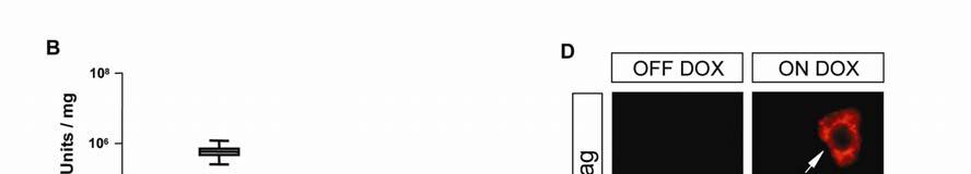
16 Figure S3. Strategy for reversible abrogation of E-cadherin with dn E-cadherin in type II pneumocytes. A: Schematic diagram showing the generation of transgenic mice conditionally expressing dn E-Cadherin in type II pneumocytes. Expression of dn E-cadherin can be induced by DOX treatment and suppressed by DOX withdrawal. B: Luciferase activity in lung lysates of bitransgenic (SP-C rtta/tet-o dn E-cadherin) mice treated with DOX for one week (ON DOX); control littermates without DOX treatment (OFF DOX) showed only background activity. Values are mean ± SD of 15 mice. C: Protein lysates from lungs of bitransgenic mice in the absence or presence (one week pulse) of DOX were immunoblotted for myc-tagged dn E-cadherin. GAPDH was used as loading control. D: Double immunofluorescence staining of paraffin-embedded lung sections from bitransgenic mice before or after a 1 week DOX pulse were stained as indicated for pro SP-C (green) and myc reactivity (red, to visualise dn E-cadherin). Note that coincident staining of myc-tagged dn E-cadherin and pro SPC is only observed after Dox treatment (arrows). Scale bar: 10 µm.


17
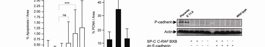
18 Figure S4: Diffuse hyperplasia in lungs after type II cell specific ablation of E cadherin. E-cadherin disruption was accomplished either by Cre-mediated Cdh1 deletion or of dn E cadherin expression in alveolar type II cells. A: Lungs from 6 weeks old mice (SP-C rtta/tet-o-cre /Cdh1 flox/flox or SP-C rtta/tet-o dn E- cadherin) were induced with DOX for two months and stained for H&E as well as proliferation markers. In case of SP-C rtta/tet-o dn E-cadherin mice, reversibility of hyperplasia was examined after DOX withdrawal for two month. Scale bar: 50 µm B: Fraction of apoptotic cells in induced lungs of SP-C rtta/tet-o dn E-cadherin expressing mice before and after DOX withdrawal. M: month; W: week; D: day. C: Reversibility of increased cell proliferation after DOX withdrawal in SP-C rtta/tet-o dn E-cadherin transgenic mice. D: Search for compensatory up-regulation of P-cadherin. Lysates of total lungs from mice in which E-cadherin had been disrupted as indicated were examined with P-cadherin specific antibody by Western blot analysis. Placenta and 11 day old embryo lysates (E 11.0) were used as positive control.


19
20 Figure S5. Ablation of E-cadherin function in SP-C C-RAF BXB lung adenomas causes lymphangiogenesis A: Paraffin embedded sections of lung tumors from four months induced triple transgenic (SP-C C-RAF BXB/SP-C rtta/tet-o dn E-cadherin) and quadruple compound mice (SP-C C- RAF BXB/SP-C rtta/tet-o-cre/cdh1 flox/flox ) were stained with Prox1 antibody. Arrows indicate lymph vessels. Brown nuclear staining identifies lymph endothelial cells. Haematoxylin was used as counterstaining. Scale bar: 50 µm. B: Quantitation of intratumoral lymphatics shows increased number of vessels in lung tumors after four months of treatment (ON DOX). For each group, 5 mice and ten tumors per mouse were counted. All values are mean ± s.d. *** P<


21
22 Figure S6. Lack of evidence for epithelial-mesenchymal transition (EMT) in metastatic lung adenocarcinoma. A: Triple transgenic mice (SP-C C-RAF BXB/SP-C rtta/tet-o dn E-cadherin) were treated with Dox for nine months (ON DOX) and compared with age-matched controls (OFF DOX). Representative pictures of lung tumor sections stained as indicated for vimentin, N-cadherin and cytokeratin. Scale bar: 50 µm B: Double-immunofluoresence for pro SPC/vimentin and pro SPC/N-cadherin. Scale bar: 50 µm. 15 animals were induced for 4-13-months, and 5 tumor sections per animal were stained. C: Western blot analysis of N-cadherin in isolated type-ii cells from 10-months old triple transgenic mice. Lysate from total brain was used as positive control. D: Western blot analysis of N-cadherin in the endothelial fraction. Actin was used as loading control.


23
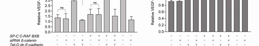
24 Figure S7. ß-catenin dependence of dn E-cadherin-mediated VEGF induction Quantitative RT-PCR from epithelial mouse lung 3041 and human NSCLC A549 cells that had been transfected with sirna and DNA constructs as indicated. Non specific sirna (ns sirna) served as negative control. A constitutively active mutant of ß-catenin (ß-cat 4S), was included. A: Relative VEGF-A and VEGF-C expression in the absence or presence of ß-catenin sirna in 3041 cells. B: Relative VEGF-A and VEGF-C expression in the absence or presence of ß-catenin sirna in A549 cells. Histograms represent the mean of results from triplicate experiments repeated 3 times. Values represent mean ±s.d. (ns=non significant; *P<0.05; **P<0.01; *** P <0.001).

25


26 Figure S8. Distribution of endogenous ß-catenin after of dn E-cadherin transfection Epithelial mouse lung 3041 were transfected with empty plasmid or SP-C rtta/teto dn E- cadherin plasmids and stained for ß-catenin (green). Note relocation of ß-catenin in the cytoplasm.
Supplementary. Table 1: Oligonucleotides and Plasmids. complementary to positions from 77 of the SRα '- GCT CTA GAG AAC TTG AAG TAC AGA CTG C
 Supplementary Table 1: Oligonucleotides and Plasmids 913954 5'- GCT CTA GAG AAC TTG AAG TAC AGA CTG C 913955 5'- CCC AAG CTT ACA GTG TGG CCA TTC TGC TG 223396 5'- CGA CGC GTA CAG TGT GGC CAT TCT GCT G
Electronic Supplementary Information
 Electronic Supplementary Material (ESI) for Molecular BioSystems. This journal is The Royal Society of Chemistry 2017 Electronic Supplementary Information Dissecting binding of a β-barrel outer membrane
Add 5µl of 3N NaOH to DNA sample (final concentration 0.3N NaOH).
.") Bisulfite Treatment of DNA Dilute DNA sample to 2µg DNA in 50µl ddh 2 O. Add 5µl of 3N NaOH to DNA sample (final concentration 0.3N NaOH). Incubate in a 37ºC water bath for 30 minutes. To 55µl samples
Bisulfite Treatment of DNA Dilute DNA sample to 2µg DNA in 50µl ddh 2 O. Add 5µl of 3N NaOH to DNA sample (final concentration 0.3N NaOH). Incubate in a 37ºC water bath for 30 minutes. To 55µl samples
Supplementary Figures
 Supplementary Figures Supplementary Fig. 1 Characterization of GSCs. a. Immunostaining of primary GSC spheres from GSC lines. Nestin (neural progenitor marker, red), TLX (green). Merged images of nestin,
Anti-Pim-1 (Cat#3247), anti-met (Cat#3127), anti-ron (Cat#2654), Anti-EGFR
, anti-met (Cat#3127), anti-ron (Cat#2654), Anti-EGFR") Supplementary Methods Antibodies Anti-Pim-1 (Cat#3247), anti-met (Cat#3127), anti-ron (Cat#2654), Anti-EGFR (Cat#2646), anti-igf1r (Cat#3018), anti-insr (Cat#3020), anti-akt (pan, Cat#4691), anti-phospho-akt
Supplementary Methods Antibodies Anti-Pim-1 (Cat#3247), anti-met (Cat#3127), anti-ron (Cat#2654), Anti-EGFR (Cat#2646), anti-igf1r (Cat#3018), anti-insr (Cat#3020), anti-akt (pan, Cat#4691), anti-phospho-akt
Supplemental Data Supplemental Figure 1.
 Supplemental Data Supplemental Figure 1. Silique arrangement in the wild-type, jhs, and complemented lines. Wild-type (WT) (A), the jhs1 mutant (B,C), and the jhs1 mutant complemented with JHS1 (Com) (D)
Hes6. PPARα. PPARγ HNF4 CD36
 SUPPLEMENTARY INFORMATION Supplementary Table Positions and Sequences of ChIP primers -63 AGGTCACTGCCA -79 AGGTCTGCTGTG Hes6-0067 GGGCAaAGTTCA ACOT -395 GGGGCAgAGTTCA PPARα -309 GGCTCAaAGTTCAaGTTCA CPTa
Supplementary Figure 1A A404 Cells +/- Retinoic Acid
 Supplementary Figure 1A A44 Cells +/- Retinoic Acid 1 1 H3 Lys4 di-methylation SM-actin VEC cfos (-) RA (+) RA 14 1 1 8 6 4 H3 Lys79 di-methylation SM-actin VEC cfos (-) RA (+) RA Supplementary Figure
Supplemental Table 1. Mutant ADAMTS3 alleles detected in HEK293T clone 4C2. WT CCTGTCACTTTGGTTGATAGC MVLLSLWLIAAALVEVR
 Supplemental Dataset Supplemental Table 1. Mutant ADAMTS3 alleles detected in HEK293T clone 4C2. DNA sequence Amino acid sequence WT CCTGTCACTTTGGTTGATAGC MVLLSLWLIAAALVEVR Allele 1 CCTGTC------------------GATAGC
Figure S1. Characterization of the irx9l-1 mutant. (A) Diagram of the Arabidopsis IRX9L gene drawn based on information from TAIR (the Arabidopsis
 Diagram of the Arabidopsis IRX9L gene drawn based on information from TAIR (the Arabidopsis") 1 2 3 4 5 6 7 8 9 10 11 12 Figure S1. Characterization of the irx9l-1 mutant. (A) Diagram of the Arabidopsis IRX9L gene drawn based on information from TAIR (the Arabidopsis Information Research). Exons
1 2 3 4 5 6 7 8 9 10 11 12 Figure S1. Characterization of the irx9l-1 mutant. (A) Diagram of the Arabidopsis IRX9L gene drawn based on information from TAIR (the Arabidopsis Information Research). Exons
Supplementary Figure 1. Localization of MST1 in RPE cells. Proliferating or ciliated HA- MST1 expressing RPE cells (see Fig. 5b for establishment of
 Supplementary Figure 1. Localization of MST1 in RPE cells. Proliferating or ciliated HA- MST1 expressing RPE cells (see Fig. 5b for establishment of the cell line) were immunostained for HA, acetylated
Supplemental Information. Human Senataxin Resolves RNA/DNA Hybrids. Formed at Transcriptional Pause Sites. to Promote Xrn2-Dependent Termination
 Supplemental Information Molecular Cell, Volume 42 Human Senataxin Resolves RNA/DNA Hybrids Formed at Transcriptional Pause Sites to Promote Xrn2-Dependent Termination Konstantina Skourti-Stathaki, Nicholas
Supplement 1: Sequences of Capture Probes. Capture probes were /5AmMC6/CTG TAG GTG CGG GTG GAC GTA GTC
 Supplementary Appendixes Supplement 1: Sequences of Capture Probes. Capture probes were /5AmMC6/CTG TAG GTG CGG GTG GAC GTA GTC ACG TAG CTC CGG CTG GA-3 for vimentin, /5AmMC6/TCC CTC GCG CGT GGC TTC CGC
PCR analysis was performed to show the presence and the integrity of the var1csa and var-
 Supplementary information: Methods: Table S1: Primer Name Nucleotide sequence (5-3 ) DBL3-F tcc ccg cgg agt gaa aca tca tgt gac tg DBL3-R gac tag ttt ctt tca ata aat cac tcg c DBL5-F cgc cct agg tgc ttc
ΔPDD1 x ΔPDD1. ΔPDD1 x wild type. 70 kd Pdd1. Pdd3
 Supplemental Fig. S1 ΔPDD1 x wild type ΔPDD1 x ΔPDD1 70 kd Pdd1 50 kd 37 kd Pdd3 Supplemental Fig. S1. ΔPDD1 strains express no detectable Pdd1 protein. Western blot analysis of whole-protein extracts
Overexpression Normal expression Overexpression Normal expression. 26 (21.1%) N (%) P-value a N (%)
 N (%) P-value a N (%)") SUPPLEMENTARY TABLES Table S1. Alteration of ZNF322A protein expression levels in relation to clinicopathological parameters in 123 Asian and 74 Caucasian lung cancer patients. Asian patients Caucasian
SUPPLEMENTARY TABLES Table S1. Alteration of ZNF322A protein expression levels in relation to clinicopathological parameters in 123 Asian and 74 Caucasian lung cancer patients. Asian patients Caucasian
Arabidopsis actin depolymerizing factor AtADF4 mediates defense signal transduction triggered by the Pseudomonas syringae effector AvrPphB
 Arabidopsis actin depolymerizing factor mediates defense signal transduction triggered by the Pseudomonas syringae effector AvrPphB Files in this Data Supplement: Supplemental Table S1 Supplemental Table
Lecture 10, 20/2/2002: The process of solution development - The CODEHOP strategy for automatic design of consensus-degenerate primers for PCR
 Lecture 10, 20/2/2002: The process of solution development - The CODEHOP strategy for automatic design of consensus-degenerate primers for PCR 1 The problem We wish to clone a yet unknown gene from a known
Supporting Information
 Supporting Information Transfection of DNA Cages into Mammalian Cells Email: a.turberfield@physics.ox.ac.uk Table of Contents Supporting Figure 1 DNA tetrahedra used in transfection experiments 2 Supporting
Supplemental Data. mir156-regulated SPL Transcription. Factors Define an Endogenous Flowering. Pathway in Arabidopsis thaliana
 Cell, Volume 138 Supplemental Data mir156-regulated SPL Transcription Factors Define an Endogenous Flowering Pathway in Arabidopsis thaliana Jia-Wei Wang, Benjamin Czech, and Detlef Weigel Table S1. Interaction
Cat. # Product Size DS130 DynaExpress TA PCR Cloning Kit (ptakn-2) 20 reactions Box 1 (-20 ) ptakn-2 Vector, linearized 20 µl (50 ng/µl) 1
 20 reactions Box 1 (-20 ) ptakn-2 Vector, linearized 20 µl (50 ng/µl) 1") Product Name: Kit Component TA PCR Cloning Kit (ptakn-2) Cat. # Product Size DS130 TA PCR Cloning Kit (ptakn-2) 20 reactions Box 1 (-20 ) ptakn-2 Vector, linearized 20 µl (50 ng/µl) 1 2 Ligation Buffer
Product Name: Kit Component TA PCR Cloning Kit (ptakn-2) Cat. # Product Size DS130 TA PCR Cloning Kit (ptakn-2) 20 reactions Box 1 (-20 ) ptakn-2 Vector, linearized 20 µl (50 ng/µl) 1 2 Ligation Buffer
Converting rabbit hybridoma into recombinant antibodies with effective transient production in an optimized human expression system
 Converting rabbit hybridoma into recombinant antibodies with effective transient production in an optimized human expression system Dr. Tim Welsink Molecular Biology Transient Gene Expression OUTLINE Short
Supplemental material
 Supplemental material Diversity of O-antigen repeat-unit structures can account for the substantial sequence variation of Wzx translocases Yaoqin Hong and Peter R. Reeves School of Molecular Bioscience,
MacBlunt PCR Cloning Kit Manual
 MacBlunt PCR Cloning Kit Manual Shipping and Storage MacBlunt PCR Cloning Kits are shipped on dry ice. Each kit contains a box with cloning reagents and an attached bag with Eco-Blue Competent Cells (optional).
Supporting information for Biochemistry, 1995, 34(34), , DOI: /bi00034a013
, , DOI: /bi00034a013") Supporting information for Biochemistry, 1995, 34(34), 10807 10815, DOI: 10.1021/bi00034a013 LESNIK 10807-1081 Terms & Conditions Electronic Supporting Information files are available without a subscription
Supporting information for Biochemistry, 1995, 34(34), 10807 10815, DOI: 10.1021/bi00034a013 LESNIK 10807-1081 Terms & Conditions Electronic Supporting Information files are available without a subscription
Quantitative reverse-transcription PCR. Transcript levels of flgs, flgr, flia and flha were
 1 Supplemental methods 2 3 4 5 6 7 8 9 1 11 12 13 14 15 16 17 18 19 21 22 23 Quantitative reverse-transcription PCR. Transcript levels of flgs, flgr, flia and flha were monitored by quantitative reverse-transcription
hcd1tg/hj1tg/ ApoE-/- hcd1tg/hj1tg/ ApoE+/+
 ApoE+/+ ApoE-/- ApoE-/- H&E (1x) Supplementary Figure 1. No obvious pathology is observed in the colon of diseased ApoE-/me. Colon samples were fixed in 1% formalin and laid out in Swiss rolls for paraffin
Luo et al. Supplemental Figures and Materials and Methods
 Luo et al. Supplemental Figures and Materials and Methods The supplemental figures demonstrate that nuclear NFAT is situated at PODs, overexpressed PML does not increase NFAT nuclear localization, and
Y-chromosomal haplogroup typing Using SBE reaction
 Schematic of multiplex PCR followed by SBE reaction Multiplex PCR Exo SAP purification SBE reaction 5 A 3 ddatp ddgtp 3 T 5 A G 3 T 5 3 5 G C 5 3 3 C 5 ddttp ddctp 5 T 3 T C 3 A 5 3 A 5 5 C 3 3 G 5 3 G
Dierks Supplementary Fig. S1
 Dierks Supplementary Fig. S1 ITK SYK PH TH K42R wt K42R (kinase deficient) R29C E42K Y323F R29C E42K Y323F (reduced phospholipid binding) (enhanced phospholipid binding) (reduced Cbl binding) E42K Y323F
Supplementary Methods Quantitative RT-PCR. For mrna, total RNA was prepared using TRIzol reagent (Invitrogen) and genomic DNA was eliminated with TURB
 and genomic DNA was eliminated with TURB") Supplementary Methods Quantitative RT-PCR. For mrna, total RNA was prepared using TRIzol reagent (Invitrogen) and genomic DNA was eliminated with TURBO DNA-free Kit (Ambion). One µg of total RNA was reverse
Supplementary Methods Quantitative RT-PCR. For mrna, total RNA was prepared using TRIzol reagent (Invitrogen) and genomic DNA was eliminated with TURBO DNA-free Kit (Ambion). One µg of total RNA was reverse
Materials Protein synthesis kit. This kit consists of 24 amino acids, 24 transfer RNAs, four messenger RNAs and one ribosome (see below).
.") Protein Synthesis Instructions The purpose of today s lab is to: Understand how a cell manufactures proteins from amino acids, using information stored in the genetic code. Assemble models of four very
Protein Synthesis Instructions The purpose of today s lab is to: Understand how a cell manufactures proteins from amino acids, using information stored in the genetic code. Assemble models of four very
PGRP negatively regulates NOD-mediated cytokine production in rainbow trout liver cells
 Supplementary Information for: PGRP negatively regulates NOD-mediated cytokine production in rainbow trout liver cells Ju Hye Jang 1, Hyun Kim 2, Mi Jung Jang 2, Ju Hyun Cho 1,2,* 1 Research Institute
Legends for supplementary figures 1-3
 High throughput resistance profiling of Plasmodium falciparum infections based on custom dual indexing and Illumina next generation sequencing-technology Sidsel Nag 1,2 *, Marlene D. Dalgaard 3, Poul-Erik
Table S1. Bacterial strains (Related to Results and Experimental Procedures)
") Table S1. Bacterial strains (Related to Results and Experimental Procedures) Strain number Relevant genotype Source or reference 1045 AB1157 Graham Walker (Donnelly and Walker, 1989) 2458 3084 (MG1655)
Table S1. Bacterial strains (Related to Results and Experimental Procedures) Strain number Relevant genotype Source or reference 1045 AB1157 Graham Walker (Donnelly and Walker, 1989) 2458 3084 (MG1655)
Supporting Information
 Supporting Information Table S1. Oligonucleotide sequences used in this work Oligo DNA A B C D CpG-A CpG-B CpG-C CpG-D Sequence 5 ACA TTC CTA AGT CTG AAA CAT TAC AGC TTG CTA CAC GAG AAG AGC CGC CAT AGT
SUPPLEMENTARY INFORMATION
 doi: 10.1038/nature07182 SUPPLEMENTAL FIGURES AND TABLES Fig. S1. myf5-expressing cells give rise to brown fat depots and skeletal muscle (a) Perirenal BAT from control (cre negative) and myf5-cre:r26r3-yfp
Supplementary Materials for
 www.sciencesignaling.org/cgi/content/full/10/494/eaan6284/dc1 Supplementary Materials for Activation of master virulence regulator PhoP in acidic ph requires the Salmonella-specific protein UgtL Jeongjoon
Immunocytochemistry was performed on adherent cells grown overnight on
 Supplementary methods Immunocytochemistry was performed on adherent cells grown overnight on coverslips, fixed in 4% (w/v) paraformaldehyde, blocked for 90 min with 2% (v/v) horse serum and incubated with
Disease and selection in the human genome 3
 Disease and selection in the human genome 3 Ka/Ks revisited Please sit in row K or forward RBFD: human populations, adaptation and immunity Neandertal Museum, Mettman Germany Sequence genome Measure expression
Supporting Information. Copyright Wiley-VCH Verlag GmbH & Co. KGaA, Weinheim, 2006
 Supporting Information Copyright Wiley-VCH Verlag GmbH & Co. KGaA, 69451 Weinheim, 2006 Copyright Wiley-VCH Verlag GmbH & Co. KGaA, 69451 Weinheim, 2006 Supporting Information for Expanding the Genetic
RPA-AB RPA-C Supplemental Figure S1: SDS-PAGE stained with Coomassie Blue after protein purification.
 RPA-AB RPA-C (a) (b) (c) (d) (e) (f) Supplemental Figure S: SDS-PAGE stained with Coomassie Blue after protein purification. (a) RPA; (b) RPA-AB; (c) RPA-CDE; (d) RPA-CDE core; (e) RPA-DE; and (f) RPA-C
strain devoid of the aox1 gene [1]. Thus, the identification of AOX1 in the intracellular
![strain devoid of the aox1 gene [1]. Thus, the identification of AOX1 in the intracellular](/thumbs/72/66361212.jpg "strain devoid of the aox1 gene [1]. Thus, the identification of AOX1 in the intracellular") Additional file 2 Identification of AOX1 in P. pastoris GS115 with a Mut s phenotype Results and Discussion The HBsAg producing strain was originally identified as a Mut s (methanol utilization slow) strain
Additional file 2 Identification of AOX1 in P. pastoris GS115 with a Mut s phenotype Results and Discussion The HBsAg producing strain was originally identified as a Mut s (methanol utilization slow) strain
SUPPORTING INFORMATION FILE
 Intrinsic and extrinsic connections of Tet3 dioxygenase with CXXC zinc finger modules Nan Liu, Mengxi Wang, Wen Deng, Christine S. Schmidt, Weihua Qin, Heinrich Leonhardt and Fabio Spada Department of
Isolation, culture, and transfection of primary mammary epithelial organoids
 Supplementary Experimental Procedures Isolation, culture, and transfection of primary mammary epithelial organoids Primary mammary epithelial organoids were prepared from 8-week-old CD1 mice (Charles River)
Supplemental Table 1. Primers used for PCR.
 Supplemental Table 1. Primers used for PCR. Gene Type Primer Sequence Genotyping and semi-quantitative RT-PCR F 5 -TTG CCC GAT CAC CAT CTG TA-3 rwa1-1 R 5 -TGT AGC GAT CAA GGC CTG ATC TAA-3 LB 5 -TAG CAT
Nongenetic Reprogramming of the Ligand Specificity. of Growth Factor Receptors by Bispecific DNA Aptamers
 Supporting Information For Nongenetic Reprogramming of the Ligand Specificity of Growth Factor Receptors by Bispecific DNA Aptamers Ryosuke Ueki,* Saki Atsuta, Ayaka Ueki and Shinsuke Sando* Department
Project 07/111 Final Report October 31, Project Title: Cloning and expression of porcine complement C3d for enhanced vaccines
 Project 07/111 Final Report October 31, 2007. Project Title: Cloning and expression of porcine complement C3d for enhanced vaccines Project Leader: Dr Douglas C. Hodgins (519-824-4120 Ex 54758, fax 519-824-5930)
SUPPLEMENTAL DATA SUPPLEMENTAL FIGURE LEGENDS
 SUPPLEMENTAL DATA SUPPLEMENTAL FIGURE LEGENDS SUPPLEMENTAL FIGURE S1. Identification of BmCREC. (A) Amino acid sequences of BmCREC show the peptides identified in LC-MS/MS analysis (marked by red letters
Supplemental Data. Bennett et al. (2010). Plant Cell /tpc
. Plant Cell /tpc") BRN1 ---------MSSSNGGVPPGFRFHPTDEELLHYYLKKKISYEKFEMEVIKEVDLNKIEPWDLQDRCKIGSTPQNEWYFFSHKDRKYPTGS 81 BRN2 --------MGSSSNGGVPPGFRFHPTDEELLHYYLKKKISYQKFEMEVIREVDLNKLEPWDLQERCKIGSTPQNEWYFFSHKDRKYPTGS 82 SMB
BRN1 ---------MSSSNGGVPPGFRFHPTDEELLHYYLKKKISYEKFEMEVIKEVDLNKIEPWDLQDRCKIGSTPQNEWYFFSHKDRKYPTGS 81 BRN2 --------MGSSSNGGVPPGFRFHPTDEELLHYYLKKKISYQKFEMEVIREVDLNKLEPWDLQERCKIGSTPQNEWYFFSHKDRKYPTGS 82 SMB
SUPPLEMENTARY INFORMATION
 DOI: 10.1038/ncb3240 Supplementary Figure 1 GBM cell lines display similar levels of p100 to p52 processing but respond differentially to TWEAK-induced TERT expression according to TERT promoter mutation
Total RNA was extracted from ESCs or wild-type MEF using an RNeasy Plus mini kit
 Supplementary information, Data S1 Materials and methods RNA-seq Total RNA was extracted from ESCs or wild-type MEF using an RNeasy Plus mini kit (Qiagen). Poly A+ RNA was purified from the total RNA and
-15 diopter negative lenses in wild-type and homozygous CHRM2-deleted mice, and
 Supplementary Materials Supplementary Figure 1: Myopia induction was performed using uniocular -10 and -15 diopter negative lenses in wild-type and homozygous CHRM2-deleted mice, and results at 2, 4 and
Supplementary Fig. 1. Isolation and in vitro expansion of EpCAM + cholangiocytes. For collagenase perfusion, enzyme solution was injected from the
 Supplementary Fig. 1. Isolation and in vitro expansion of EpCAM + cholangiocytes. For collagenase perfusion, enzyme solution was injected from the portal vein for digesting adult livers, whereas it was
Supporting Online Information
 Supporting Online Information Isolation of Human Genomic DNA Sequences with Expanded Nucleobase Selectivity Preeti Rathi, Sara Maurer, Grzegorz Kubik and Daniel Summerer* Department of Chemistry and Chemical
Supporting Information
 Supporting Information Barderas et al. 10.1073/pnas.0801221105 SI Text: Docking of gastrin to Constructed scfv Models Interactive predocking of the 4-WL-5 motif into the central pocket observed in the
An evolutionarily conserved negative feedback mechanism in the hippo pathway reflects functional difference between LATS1 and LATS2
 /, Supplementary Advance Publications Materials 2015 2016 An evolutionarily conserved negative feedback mechanism in the hippo pathway reflects functional difference between LATS1 and LATS2 Supplementary
ORFs and genes. Please sit in row K or forward
 ORFs and genes Please sit in row K or forward https://www.flickr.com/photos/teseum/3231682806/in/photostream/ Question: why do some strains of Vibrio cause cholera and others don t? Methods Mechanisms
The Wnt-5a-derived hexapeptide Foxy-5 inhibits breast cancer. metastasis in vivo by targeting cell motility
 SUPPLEMENTARY MATERIAL: The Wnt-5a-derived hexapeptide Foxy-5 inhibits breast cancer metastasis in vivo by targeting cell motility Annette Säfholm, Johanna Tuomela, Jeanette Rosenkvist, Janna Dejmek, Pirkko
Gene synthesis by circular assembly amplification
 Gene synthesis by circular assembly amplification Duhee Bang & George M Church Supplementary figures and text: Supplementary Figure 1. Dpo4 gene (1.05kb) construction by various methods. Supplementary
Figure S1. Purity of primary cultures of renal proximal tubular epithelial culture ascertained by cytokeratin staining.
 Supplementary information Supplementary figures Figure S1. Purity of primary cultures of renal proximal tubular epithelial culture ascertained by cytokeratin staining. Figure S2. Induction of Nur77 in
NAME:... MODEL ANSWER... STUDENT NUMBER:... Maximum marks: 50. Internal Examiner: Hugh Murrell, Computer Science, UKZN
 COMP710, Bioinformatics with Julia, Test One, Thursday the 20 th of April, 2017, 09h30-11h30 1 NAME:...... MODEL ANSWER... STUDENT NUMBER:...... Maximum marks: 50 Internal Examiner: Hugh Murrell, Computer
Supplemental Information. Target-Mediated Protection of Endogenous. MicroRNAs in C. elegans. Inventory of Supplementary Information
 Developmental Cell, Volume 20 Supplemental Information Target-Mediated Protection of Endogenous MicroRNAs in C. elegans Saibal Chatterjee, Monika Fasler, Ingo Büssing, and Helge Großhans Inventory of Supplementary
An engineered tryptophan zipper-type peptide as a molecular recognition scaffold
 SUPPLEMENTARY MATERIAL An engineered tryptophan zipper-type peptide as a molecular recognition scaffold Zihao Cheng and Robert E. Campbell* Supplementary Methods Library construction for FRET-based screening
Supplementary Figure 1. The level of pri-mir-8 gradually decreases while those of BR-C and E74 increase during 3rd instar larval development.
 qrt-pcr RT-PCR Relative pri-mir-8 level 1.2 1.0 0.8 0.6 0.4 0.2 0.0 Early 3rd (72h) Mid 3rd (96h) Late 3rd (119h) BR-C E74 mtl rrna Early 3rd (72h) Mid 3rd (96h) Late 3rd (119h) Supplementary Figure 1.
Lecture 11: Gene Prediction
 Lecture 11: Gene Prediction Study Chapter 6.11-6.14 1 Gene: A sequence of nucleotides coding for protein Gene Prediction Problem: Determine the beginning and end positions of genes in a genome Where are
SUPPLEMENTARY INFORMATION. Material and methods
 SUPPLEMENTARY INFORMATION Material and methods Cell culture Human hepatocellular carcinoma (HepG) cells and human embryonic kidney (HEK)93 cells were grown in Dulbecco s modified Eagle s medium (Invitrogen
II 0.95 DM2 (RPP1) DM3 (At3g61540) b
 DM3 (At3g61540) b") Table S2. F 2 Segregation Ratios at 16 C, Related to Figure 2 Cross n c Phenotype Model e 2 Locus A Locus B Normal F 1 -like Enhanced d Uk-1/Uk-3 149 64 36 49 DM2 (RPP1) DM1 (SSI4) a Bla-1/Hh-0 F 3 111
Table S2. F 2 Segregation Ratios at 16 C, Related to Figure 2 Cross n c Phenotype Model e 2 Locus A Locus B Normal F 1 -like Enhanced d Uk-1/Uk-3 149 64 36 49 DM2 (RPP1) DM1 (SSI4) a Bla-1/Hh-0 F 3 111
Supplementary Figure 1 Tmod3 expression and phosphorylation. (a) Expression of Tmod3 in 3T3-L1 preadipocytes and differentiated adipocytes.
 Expression of Tmod3 in 3T3-L1 preadipocytes and differentiated adipocytes.") 1 2 3 1 4 5 6 7 8 9 10 11 12 13 14 15 16 17 18 19 20 21 22 23 24 25 26 27 28 29 30 Supplementary Figure 1 Tmod3 expression and phosphorylation. (a) Expression of Tmod3 in 3T3-L1 preadipocytes and differentiated
1 2 3 1 4 5 6 7 8 9 10 11 12 13 14 15 16 17 18 19 20 21 22 23 24 25 26 27 28 29 30 Supplementary Figure 1 Tmod3 expression and phosphorylation. (a) Expression of Tmod3 in 3T3-L1 preadipocytes and differentiated
PCR-based Markers and Cut Flower Longevity in Carnation
 PCRbased Markers and Cut Flower Longevity in Carnation Laura De Benedetti, Luca Braglia, Simona Bruna, Gianluca Burchi *, Antonio Mercuri and Tito Schiva Istituto Sperimentale per la Floricoltura, Corso
Gene Expression Profiling: RNA isolation, crna synthesis and gene expression profiling were
 Gene Expression Profiling: RNA isolation, crna synthesis and gene expression profiling were performed as described by Giordano et al 24. A Unigene cluster was assigned to each of the Affymetrix probe sets
Multiplexing Genome-scale Engineering
 Multiplexing Genome-scale Engineering Harris Wang, Ph.D. Department of Systems Biology Department of Pathology & Cell Biology http://wanglab.c2b2.columbia.edu Rise of Genomics An Expanding Toolbox Esvelt
Supplementary Information. Construction of Lasso Peptide Fusion Proteins
 Supplementary Information Construction of Lasso Peptide Fusion Proteins Chuhan Zong 1, Mikhail O. Maksimov 2, A. James Link 2,3 * Departments of 1 Chemistry, 2 Chemical and Biological Engineering, and
Nature Genetics: doi: /ng Supplementary Figure 1
 Supplementary Figure 1 Coding and noncoding transcription at the vg locus. (a,b) qpcr analysis of developmental and tissue-specific vg mrna (a) and PRE/TRE (b) transcription, shown as percentage of TBP
Supplementary Information
 Supplementary Information Microbead-based biomimetic synthetic neighbors enhance survival and function of rat pancreatic β-cells Wei Li, a Samuel Lee, b Minglin Ma, a, f Soo Min Kim, b Patrick Guye, c
Efficient genome replication of hepatitis B virus using adenovirus vector: a compact pregenomic RNA-expression unit
 Efficient genome replication of hepatitis B virus using adenovirus vector: a compact pregenomic RNA-expression unit Mariko Suzuki 1, Saki Kondo 1, Manabu Yamasaki 2, Norie Matsuda 2, Akio Nomoto 2, Tetsuro
Supplementary Information
 Supplementary Information A general solution for opening double-stranded DNA for isothermal amplification Gangyi Chen, Juan Dong, Yi Yuan, Na Li, Xin Huang, Xin Cui* and Zhuo Tang* Supplementary Materials
Search for and Analysis of Single Nucleotide Polymorphisms (SNPs) in Rice (Oryza sativa, Oryza rufipogon) and Establishment of SNP Markers
 in Rice (Oryza sativa, Oryza rufipogon) and Establishment of SNP Markers") DNA Research 9, 163 171 (2002) Search for and Analysis of Single Nucleotide Polymorphisms (SNPs) in Rice (Oryza sativa, Oryza rufipogon) and Establishment of SNP Markers Shinobu Nasu, Junko Suzuki, Rieko
DNA Research 9, 163 171 (2002) Search for and Analysis of Single Nucleotide Polymorphisms (SNPs) in Rice (Oryza sativa, Oryza rufipogon) and Establishment of SNP Markers Shinobu Nasu, Junko Suzuki, Rieko
SUPPLEMENTARY MATERIALS AND METHODS. E. coli strains, plasmids, and growth conditions. Escherichia coli strain P90C (1)
") SUPPLEMENTARY MATERIALS AND METHODS E. coli strains, plasmids, and growth conditions. Escherichia coli strain P90C (1) dinb::kan (lab stock) derivative was used as wild-type. MG1655 alka tag dinb (2) is
SUPPLEMENTARY MATERIALS AND METHODS E. coli strains, plasmids, and growth conditions. Escherichia coli strain P90C (1) dinb::kan (lab stock) derivative was used as wild-type. MG1655 alka tag dinb (2) is
Marcos-Ramiro et al., http ://www.jcb.org /cgi /content /full /jcb /DC1
 Supplemental material JCB Marcos-Ramiro et al., http ://www.jcb.org /cgi /content /full /jcb.201504038 /DC1 THE JOURNAL OF CELL BIOLOGY Figure S1. Changes in RhoB protein expression in response to inflammatory
Supplementary methods. RNA isolation, cdna synthesis, and quantitative real-time PCR. Total RNAs were
 Supplementary methods RNA isolation, cdna synthesis, and quantitative real-time PCR. Total RNAs were extracted from cells by using an RNeasy Mini Kit (QIAGEN) and then reverse-transcribed using the SuperScript
Lewis x/cd15 expression in human myeloid cell differentiation. is regulated by sialidase activity
 Lewis x/cd15 expression in human myeloid cell differentiation is regulated by sialidase activity Samah Zeineb Gadhoum 1, 2 and Robert Sackstein* 1, 2, 3, 4 From the Departments of Dermatology 1 and Medicine
11th Meeting of the Science Working Group. Lima, Peru, October 2012 SWG-11-JM-11
 11th Meeting of the Science Working Group Lima, Peru, 15-19 October 2012 Russian population genetics studies of jack mackerel in the South Pacific P.K.Afanasiev M.A.Rabchun A.I.Glubokov Introduction. In
RNA was isolated using NucleoSpin RNA II (Macherey-Nagel, Bethlehem, PA) according to the
 according to the") Supplementary Methods RT-PCR and real-time PCR analysis RNA was isolated using NucleoSpin RNA II (Macherey-Nagel, Bethlehem, PA) according to the manufacturer s protocol and quantified by measuring the
Supplementary Methods RT-PCR and real-time PCR analysis RNA was isolated using NucleoSpin RNA II (Macherey-Nagel, Bethlehem, PA) according to the manufacturer s protocol and quantified by measuring the
Targeting the Hedgehog-Gli pathway inhibits bleomycin-induced lung fibrosis in
 Targeting the Hedgehog-Gli pathway inhibits bleomycin-induced lung fibrosis in mice Elika Farrokhi Moshai, Lidwine Wémeau-Stervinou, Natacha Cigna, Stephanie Brayer, Joëlle Marchal Sommé, Bruno Crestani,
S4B fluorescence (AU)
") A S4B fluorescence (AU) S4B fluorescence (AU) dsbb csgba csgd dsbb csgba bcsa 5000 * NS NS 4000 * 3000 2000 1000 0 ΔcsgBAΔbcsA ΔcsgDΔdsbBΔbcsA ΔcsgBA ΔdsbBΔcsgBA ΔcsgDΔdsbB B -1000 4000 * * NS 3500 * 3000
A S4B fluorescence (AU) S4B fluorescence (AU) dsbb csgba csgd dsbb csgba bcsa 5000 * NS NS 4000 * 3000 2000 1000 0 ΔcsgBAΔbcsA ΔcsgDΔdsbBΔbcsA ΔcsgBA ΔdsbBΔcsgBA ΔcsgDΔdsbB B -1000 4000 * * NS 3500 * 3000
SAY IT WITH DNA: Protein Synthesis Activity by Larry Flammer
 TEACHER S GUIDE SAY IT WITH DNA: Protein Synthesis Activity by Larry Flammer SYNOPSIS This activity uses the metaphor of decoding a secret message for the Protein Synthesis process. Students teach themselves
* Correspondence: Ralf Dressel: 1 Supplementary Figures and Tables. - Supplementary Tables 1 to 3. - Supplementary Figures 1 to 6
 s Efficient killing of murine pluripotent stem cells by natural killer (NK) cells requires activation by cytokines and partly depends on the activating NK receptor NKG2D Carina Gröschel, Daniela Hübscher,
evaluated with UAS CLB eliciting UAS CIT -N Libraries increase in the
 Supplementary Figures Supplementary Figure 1: Promoter scaffold library assemblies. Many ensembless of libraries were evaluated in this work. As a legend, the box outline color in top half of the figure
A Genetically Encoded Toolbox for Glycocalyx Engineering: Tunable Control of Cell Adhesion,
 TITLE A Genetically Encoded Toolbox for Glycocalyx Engineering: Tunable Control of Cell Adhesion, Survival, and Cancer Cell Behaviors AUTHORS Carolyn R. Shurer *, Marshall J. Colville *, Vivek K. Gupta,
PILRα Is a Herpes Simplex Virus-1 Entry Coreceptor That Associates with Glycoprotein B
 Satoh et al. Page S1 Cell, Volume 132 PILRα Is a Herpes Simplex Virus-1 Entry Coreceptor That Associates with Glycoprotein B Takeshi Satoh, Jun Arii, Tadahiro Suenaga, Jing Wang, Amane Kogure, Junji Uehori,
SUPPORTING INFORMATION
 SUPPORTING INFORMATION Investigation of the Biosynthesis of the Lasso Peptide Chaxapeptin Using an E. coli-based Production System Helena Martin-Gómez, Uwe Linne, Fernando Albericio, Judit Tulla-Puche,*
Segments of the obstructed intestinal loops were fixed in 4% paraformaldehyde
 Supplementary text Supplementary materials and methods Histopathological examination Segments of the obstructed intestinal loops were fixed in 4% paraformaldehyde (PFA) and embedded in paraffin wax with
Table S1. Alteration of ZNF322A and FBXW7 protein expression levels in relation to clinicopathological parameters in 135 lung cancer patients.
 SUPPLEMENTARY TABLES Table S1. Alteration of ZNF322A and FBXW7 protein expression levels in relation to clinicopathological parameters in 135 lung cancer patients. ZNF322A Normal Overexpression expression
Supplementary Figure 1 - Characterization of rbag3 binding on macrophages cell surface.
 Supplementary Figure 1 - Characterization of rbag3 binding on macrophages cell surface. (a) Human PDAC cell lines were treated as indicated in Figure 1 panel F. Cells were analyzed for FITC-rBAG3 binding
Supplemental Data. Lin28 Mediates the Terminal Uridylation. of let-7 Precursor MicroRNA. Molecular Cell, Volume 32
 Molecular Cell, Volume 32 Supplemental Data Lin28 Mediates the Terminal Uridylation of let-7 Precursor MicroRNA Inha Heo, Chirlmin Joo, Jun Cho, Minju Ha, Jinju Han, and V. Narry Kim Figure S1. Endogenous
Supporting Information
 Supporting Information Sen et al. 10.1073/pnas.1114232109 SI Materials and Methods Cell Culture. Human embryonic lung fibroblasts (HELF) were maintained in Eagle s minimum essential medium (Mediatech)
Gene Expression Profiling Applicable for Cells Cultivated in Channel-Slides
 Gene Expression Profiling Applicable for Cells Cultivated in Channel-Slides Gene expression profiling provides information about the transcriptome of a living cell. The DNA is transcribed into mrna, when
Lecture 19A. DNA computing
 Lecture 19A. DNA computing What exactly is DNA (deoxyribonucleic acid)? DNA is the material that contains codes for the many physical characteristics of every living creature. Your cells use different
Supplementary Material and Methods
 Supplementary Material and Methods Synaptosomes preparation and RT-PCR analysis. Synaptoneurosome fractions were prepared as previously described in 1. Briefly, rat total brain was homogenized in ice-cold