For in vitro killing assays with lysed cells, neutrophils were sonicated using a 550 Sonic
|
|
|
- Polly Mitchell
- 5 years ago
- Views:
Transcription
1 Supplemental Information Cell Host & Microbe, Volume 8 Statins Enhance Formation of Phagocyte Extracellular Traps Ohn A. Chow, Maren von Köckritz-Blickwede, A. Taylor Bright, Mary E. Hensler, Annelies S. Zinkernagel, Anna L. Cogen, Richard L. Gallo, Marc Monestier, Yangming Wang, Christopher K. Glass, and Victor Nizet SUPPLEMENTAL EXPERIMENTAL PROCEDURES In Vitro Bactericidal Killing Assays For in vitro killing assays with lysed cells, neutrophils were sonicated using a 550 Sonic Dismembrator (Fisher Scientific). Neutrophils were cultured in serum-free, antibiotic-free RPMI at 10 6 cells/ml in the presence of 50 µm mevastatin (Sigma) as described above. After 1 hr, cells were stimulated with 156 ng/ml phorbol myristic acetate (PMA, Sigma). For killing assays to determine NET-specificity, micrococcal nuclease was added to a final concentration of 5 U/ml 40 min after addition of PMA. Neutrophils were infected with log-phase S. aureus Newman Strain at an moi of 1 at 1 hr after addition of PMA. After centrifugation for 10 min at 1500 rpm, infected PMNs were incubated for 20 min at 37 C in 5% CO 2. After incubation, surviving bacteria were assessed using a DNase killing assay described by Fuchs et al. with some slight modifications. Briefly, EDTA was added to each well to a final concentration of 0.5 mm. The neutrophils where incubated on ice for 15 min. Following this, cells were detached from plates by scraping and mixing. This mixture was diluted into 0.1% Triton X-100. Lysates were diluted and plated on THA plates for enumeration of surviving bacteria. Percent killing by statin-treated leukocytes was determined by dividing the number of cfu recovered from statin-treated neutrophils by the number of cfu from vehicle-treated neutrophils. 1
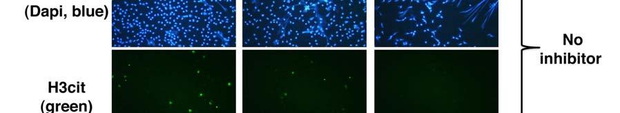
2 Histone Citrullination To block the PAD-4 mediated histone citrullination, PMNs were additionally treated with Clamidine (200 µm) at the same time when treated with mevastatin or simvastatin. After fixation with 4% paraformaldehyde, samples were washed three times with PBS and then blocked for 1 hr with PBST (PBS + 2% BSA + 0.2% Triton X-100). Primary antibody against H3Cit (rabbit anti H3cit) was diluted 1:300 in PBST and cells were stained overnight at 4 C. After washing, cells were stained with the appropriate secondary goat anti rabbit Alexa 488 (1:500 in PBST) for 45 min at room temperature. After washing, cells were mounted in Dapi-Prolongold. Percentage of Net-forming or H3cit-positive cells was determined using a Zeiss Axiolab microscope (Zeiss 40x objective) with an attached Sony Digital Photo Camera DKC-5000 at calibrated magnifications. Induction of Extracellular Traps in Peritoneal Macrophages C57BL/6 mice were intraperitoneally treated with 3 ml 3% thioglycolate solution (BD Biosciences). Peritoneal cells were isolated by peritoneal lavage with PBS 4 days later. Peritoneal lavage was treated with 1x RBC lysis buffer (ebioscience) to lyse erythrocytes and washed with PBS. After centrifuagtion, cells were cultured in RPMI + 10%FBS at 1 x 10 5 cells/500 µl/well on 24-well glass-bottom microtiter plates (MatTek Corporations). Next day, cells were further cultured in serum-free RPMI and stimulated with 50 µm mevastatin (Sigma) or appropriate concentrations of vehicle control (DMSO). After 24 hr cells were additionally treated with 156 ng/ml phorbol myristic acetate (PMA, Sigma) for 2 hr before visualization of extracellular traps (as described in Experimental Procedures). 2
3 LDH Assay LDH release was measured using the CytoTox 96 Non-Radioactive Cytotoxicity Assay (Promega) according to the manufacturer s instructions. 3
4 Figure S1. Mevastatin-Induced Killing Can Be Abrogated by DNase Treatment, related to Figure 3 (A) In vitro killing of S. aureus by intact or sonicated primary human neutrophils treated with mevastatin or vehicle control. (B) In vitro killing of S. aureus by primary human neutrophils treated with mevastatin or vehicle control. Neutrophils were treated with micrococcal nuclease (or water control) prior to infection to disrupt extracellular traps and to confirm NET-specific killing activity. *P < 0.5, ***P < by one-way ANOVA with Tukey s post-test comparing control versus statin-treated grou. 4


5 5
6 Figure S2. Statin-Induced NET Induction Independent of PAD4-Mediated Histone Citrullination, related to Figure 3 (A) Representative fluorescent images of human neutrophils stimulated with mevastatin/simvastatin or vehicle control and PMA to induce NETs. Cl-amidine (200 µm) was used to block the PAD-4 mediated histone citrullination. Net-formation was visualized in blue (DAPI) and histone citrullination (H3Cit) was visualized by Alexa green immunostaining. Bars represent 30 µm. (B) Quantification of results from above experiment by direct visualization and enumeration of % of NET-forming neutrophils (black bars) or H3Cit-positive neutrophils (white bars), average of 4 high-power fields (HPF) counted containing approximately 125 cells. 6
7 Figure S3. Mevastatin Inhibits Macrophage Phagocytosis of Staphylococcus aureus, related to Figure 4 Mean fluorescence intensity was used as parameter for phagocytosis of RAW cells after infection with FITC-labelled S. aureus Wood strain bioparticles measured by flow cytometry. As control, 10 µg/ml cytochalasin D was added to the samples 10 min prior to infection to prevent phagocytosis. ***P < by t-test comparing control versus statin-treated group. 7


8 8
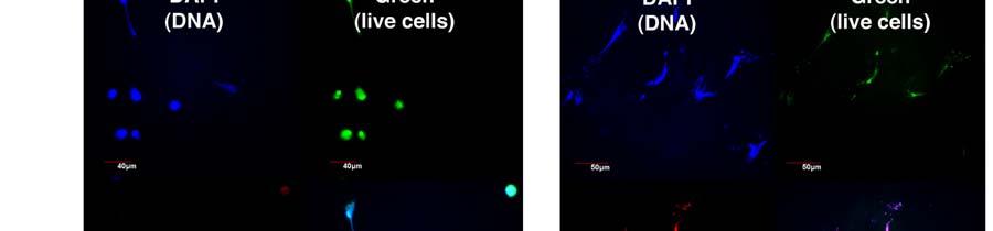
9 Figure S4. Statins Enhance Formation of Extracellular Traps by Different Macrophage Cell Types In Vitro, related to Figure 4 (A) Representative fluorescent images of RAW cells stimulated with simvastatin or vehicle control and PMA to induce METs. MET-formation was visualized in blue (DAPI) and CRAMP-expression (rabbit anti mouse CRAMP, left panel) compared was visualized by Alexa red-immunostaining. (B) Representative fluorescent images of murine peritoneal macrophage extracellular traps, stained with Live/Dead viability/cytotoxicity kit for mammalian cells to determine viability of trap-forming cells after overnight treatment with mevastatin and subsequent stimulation with PMA. Note that all trap-forming macrophages are dead as shown by the red dye. 9


10 10
11 Figure S5. In Vivo Expression of Murine Cathelicidin CRAMP, related to Figure 5 (A) Representative fluorescent images of extracellular trap formation (visualized by Alexa 488 (green)-labeled CRAMP production and counterstained with Dapi) in paraffin-embedded lung sections of mice pre-fed for with standard chow or standard chow supplemented with simvastatin and intranasally infected with 2 x 10 8 cfu of S. aureus strain Newman for 48 hr. Note the white arrows indicating extracellular traps released into alveolar space. The red arrows indicate areas of tissue inflammation with intense intracellular staining of CRAMP-positive cells. (B) Quantification of CRAMP expression compared to total cell amount (DAPI staining) in lung tissue of mice fed with standard chow or standard chow supplemented with simvastatin and infected for 48 hr with S. aureus strain Newman. Mean fluorescence intensity was quantified using Image J 1.41 software. 11
12 Figure S6. Impact of Mevalonate Rescue on RAW Macrophages, related to Figure 6 (A) Killing of S. aureus by RAW cells following treatment with mevastatin or vehicle control ± mevalonate. (B) LDH release as marker for cytotoxicity following treatment of RAW cells with different concentrations of mevalonate. ***P < 0.005, n.s. not significant by two-tailed Student's t-test comparing control versus statin-treated group. Experiments performed 3-4 times with similar results, representative experiment shown ± standard deviation. 12
A Role for Streptococcal Collagen-Like Protein 1 (Scl-1) in M1T1 Group A
 in M1T1 Group A") Supplemental Information for: A Role for Streptococcal Collagen-Like Protein 1 (Scl-1) in M1T1 Group A Streptococcus Resistance to Neutrophil Extracellular Traps Simon Döhrmann 1, Sabina Anik 1, Joshua
Supplemental Information for: A Role for Streptococcal Collagen-Like Protein 1 (Scl-1) in M1T1 Group A Streptococcus Resistance to Neutrophil Extracellular Traps Simon Döhrmann 1, Sabina Anik 1, Joshua
Phagocytosis Assay Kit (IgG PE)
") Phagocytosis Assay Kit (IgG PE) Item No. 600540 www.caymanchem.com Customer Service 800.364.9897 Technical Support 888.526.5351 1180 E. Ellsworth Rd Ann Arbor, MI USA TABLE OF CONTENTS GENERAL INFORMATION
Phagocytosis Assay Kit (IgG PE) Item No. 600540 www.caymanchem.com Customer Service 800.364.9897 Technical Support 888.526.5351 1180 E. Ellsworth Rd Ann Arbor, MI USA TABLE OF CONTENTS GENERAL INFORMATION
Segments of the obstructed intestinal loops were fixed in 4% paraformaldehyde
 Supplementary text Supplementary materials and methods Histopathological examination Segments of the obstructed intestinal loops were fixed in 4% paraformaldehyde (PFA) and embedded in paraffin wax with
Phagocytosis Assay Kit (IgG FITC)
") Phagocytosis Assay Kit (IgG FITC) Item No. 500290 Customer Service 800.364.9897 * Technical Support 888.526.5351 www.caymanchem.com TABLE OF CONTENTS GENERAL INFORMATION 3 Materials Supplied 4 Precautions
Phagocytosis Assay Kit (IgG FITC) Item No. 500290 Customer Service 800.364.9897 * Technical Support 888.526.5351 www.caymanchem.com TABLE OF CONTENTS GENERAL INFORMATION 3 Materials Supplied 4 Precautions
Human neutrophils were isolated from peripheral blood of healthy donors using a dextran-
 Materials and Methods Isolation of neutrophils Human neutrophils were isolated from peripheral blood of healthy donors using a dextran- Ficoll method (S1). Transmission electron microscopy For fine structural
Supplementary Figure. S1
 Supplementary Figure. S1 Supplementary Figure S1. Correlation of phagocytic ability measured with YG and YO beads. Fresh human monocytes (2 10 6 /ml) were labelled with APC conjugated anti CD14 mab alone
E. coli Phagocytosis Assay Kit
 E. coli Phagocytosis Assay Kit Item No. 601370 www.caymanchem.com Customer Service 800.364.9897 Technical Support 888.526.5351 1180 E. Ellsworth Rd Ann Arbor, MI USA TABLE OF CONTENTS GENERAL INFORMATION
This Document Contains:
 This Document Contains: 1. In-Cell Western Protocol II. Cell Seeding and Stimulation Supplemental Protocol III. Complete Assay Example: Detailing the Seeding, Stimulation and Detection of the A431 Cellular
Supporting Information
 Supporting Information Cieslewicz et al. 10.1073/pnas.1312197110 SI Results Human and mouse lesions of atherosclerosis contain both M1 and M2 macrophage phenotypes (1, 2). Previous work has suggested the
Murine in vivo CD8 + T Cell Killing Assay Myoungjoo V. Kim 1*, Weiming Ouyang 2, Will Liao 3, Michael Q. Zhang 4 and Ming O. Li 5
 Murine in vivo CD8 + T Cell Killing Assay Myoungjoo V. Kim 1*, Weiming Ouyang 2, Will Liao 3, Michael Q. Zhang 4 and Ming O. Li 5 1 Department of Immunobiology, Yale University School of Medicine, New
Normalization of Agilent Seahorse XF Data by In-situ Cell Counting Using a BioTek Cytation 5
 Normalization of Agilent Seahorse XF Data by In-situ Cell Counting Using a BioTek Cytation Application Note Authors Yoonseok Kam 1, Ned Jastromb 1, Joe Clayton, Paul Held, and Brian P. Dranka 1 1 Agilent
Neutrophil/Monocyte Respiratory Burst Assay Kit
 Neutrophil/Monocyte Respiratory Burst Assay Kit Item No. 601130 www.caymanchem.com Customer Service 800.364.9897 Technical Support 888.526.5351 1180 E. Ellsworth Rd Ann Arbor, MI USA TABLE OF CONTENTS
Short hairpin RNA (shrna) against MMP14. Lentiviral plasmids containing shrna
 against MMP14. Lentiviral plasmids containing shrna") Supplemental Materials and Methods Short hairpin RNA (shrna) against MMP14. Lentiviral plasmids containing shrna (Mission shrna, Sigma) against mouse MMP14 were transfected into HEK293 cells using FuGene6
Supplemental Materials and Methods Short hairpin RNA (shrna) against MMP14. Lentiviral plasmids containing shrna (Mission shrna, Sigma) against mouse MMP14 were transfected into HEK293 cells using FuGene6
In vivo BrdU Incorporation Assay for Murine Hematopioetic Stem Cells Ningfei An, Yubin Kang *
 In vivo BrdU Incorporation Assay for Murine Hematopioetic Stem Cells Ningfei An, Yubin Kang * Division of Hematology-Oncology, Department of Medicine, Medical University of South Carolina, Charleston,
CytoSelect Leukocyte-Epithelium Adhesion Assay
 Product Manual CytoSelect Leukocyte-Epithelium Adhesion Assay Catalog Number CBA-211 100 assays FOR RESEARCH USE ONLY Not for use in diagnostic procedures Introduction Airway inflammation is a hallmark
In-Cell Western Assay
 In-Cell Western Assay Complete Sample Protocol for Measuring IC 50 of Inhibitor U0126 in NIH3T3 Responding to Acidic Fibroblast Growth Factor (afgf-1) Developed for: Aerius, Odyssey Classic, Odyssey CLx,
SOPVII-7. Panel X: NK-characterization
 Created by judith.eckl Page 1 of 8 09/06/2011 SOPVII-7 Panel X: NK-characterization Date: Author: Petra Prinz, Judith Eckl Experimenter: Date: 08/06/2011 Experiment description: Version: 1.0 Start: End:
Cytotoxicity LDH Assay Kit-WST
 Cytotoxicity LDH Assay Kit-WST Supplementary Information Notice to Users Preparation of Reagent This instruction complements the Technical Manual in the product. Please use this instruction as supplements
No-wash, no-lyse detection of phagocytic cells via a phrodo BioParticles functional assay in human whole blood on the
 APPLICATION NOTE Attune NxT Flow Cytometer No-wash, no-lyse detection of phagocytic cells via a phrodo BioParticles functional assay in human whole blood on the Attune NxT Flow Cytometer Introduction Analysis
Human Pluripotent Stem Cell Functional Identification Kit
 Human Pluripotent Stem Cell Functional Identification Kit Catalog Number SC027B Reagents for the identification of human pluripotent stem cells by in vitro functional differentiation. This package insert
StemXVivo. Mesoderm Kit. Catalog Number SC030B. Reagents for the differentiation of human pluripotent stem cells into mesoderm.
 StemXVivo Mesoderm Kit Catalog Number SC030B Reagents for the differentiation of human pluripotent stem cells into mesoderm. This package insert must be read in its entirety before using this product.
ab Ran Activation Assay Kit
 ab173247 Ran Activation Assay Kit Instructions for Use For the simple and fast measurement of Ran activation. This product is for research use only and is not intended for diagnostic use. Version 1 Last
HMO were isolated from pooled human milk as previously described [1]. Milk was
![HMO were isolated from pooled human milk as previously described [1]. Milk was](/thumbs/76/73774527.jpg "HMO were isolated from pooled human milk as previously described [1]. Milk was") Supplemental Information METHODS Human milk oligosaccharide (HMO) preparation HMO were isolated from pooled human milk as previously described [1]. Milk was obtained and pooled from more than 40 different
Supplemental Information METHODS Human milk oligosaccharide (HMO) preparation HMO were isolated from pooled human milk as previously described [1]. Milk was obtained and pooled from more than 40 different
ab Cell Viability Assay Kit Fluorometric Dual Green/Red
 ab112121 Cell Viability Assay Kit Fluorometric Dual Green/Red Instructions for Use For detecting cell viability in suspension and adherent cells by using dual proprietary green and red fluorescence probes.
Cytotoxicity LDH Assay Kit-WST
 Cytotoxicity LDH Assay Kit-WST Supplementary Information Notice to Users This instruction complements the Technical Manual in the product. Please use this instruction as supplements of the Technical Manual.
SANTA CRUZ BIOTECHNOLOGY, INC.
 TECHNICAL SERVICE GUIDE: Western Blotting 2. What size bands were expected and what size bands were detected? 3. Was the blot blank or was a dark background or non-specific bands seen? 4. Did this same
Supporting Information
 Supporting Information Chakrabarty et al. 10.1073/pnas.1018001108 SI Materials and Methods Cell Lines. All cell lines were purchased from the American Type Culture Collection. Media and FBS were purchased
ApoTrack Cytochrome c Apoptosis ICC Antibody
 ab110417 ApoTrack Cytochrome c Apoptosis ICC Antibody Instructions for Use For the Immunocytochemistry analysis of cytochrome c and a mitochondrial marker (Complex Vα) in apoptotic cells and nonapoptotic
Figure S2. Response of mouse ES cells to GSK3 inhibition. Mentioned in discussion
 Stem Cell Reports, Volume 1 Supplemental Information Robust Self-Renewal of Rat Embryonic Stem Cells Requires Fine-Tuning of Glycogen Synthase Kinase-3 Inhibition Yaoyao Chen, Kathryn Blair, and Austin
Neutrophil Elastase Activity Assay Kit
 Neutrophil Elastase Activity Kit Item No. 600610 www.caymanchem.com Customer Service 800.364.9897 Technical Support 888.526.5351 1180 E. Ellsworth Rd Ann Arbor, MI USA TABLE OF CONTENTS GENERAL INFORMATION
Isolation, culture, and transfection of primary mammary epithelial organoids
 Supplementary Experimental Procedures Isolation, culture, and transfection of primary mammary epithelial organoids Primary mammary epithelial organoids were prepared from 8-week-old CD1 mice (Charles River)
LDH-Cytotoxicity Assay Kit II
 LDH-Cytotoxicity Assay Kit II Catalog Number KA0786 500 assays Version: 08 Intended for research use only www.abnova.com Table of Contents Introduction... 3 Background... 3 General Information... 4 Materials
Direct Cell Counting Assays for Immuno Therapy
 Direct Cell Counting Assays for Immuno Therapy Cytotoxicity assays play a central role in studying the function of immune effector cells such as cytolytic T lymphocytes (CTL) and natural killer (NK) cells.
Calcein AM Cell Viability Kit
 Instructions For Research Use Only. Not For Use In Diagnostic Procedures Calcein AM Cell Viability Kit Catalog# 4892-010-K 1000 Tests* * Calculated based on using 1 μm final concentration of Calcein AM;
Whole Mount IHC Protocol
 Whole Mount IHC Protocol Authors: Ruth Sullivan, Ryan Trevena and Kyle Wegner Creation Date: 03/17/2016 All steps should be conducted with gentle agitation on an orbital shaker, unless otherwise instructed.
Immunofluorescence Staining Protocol for 3 Well Chamber, removable
 Immunofluorescence Staining Protocol for 3 Well Chamber, removable This Application Note presents a simple protocol for the cultivation, fixation, and staining of cells using the 3 Well Chamber, removable.
Developing a real-time fluorescence cell growth monitoring system
 - 65 - Developing a real-time fluorescence cell growth monitoring system Jo-Ting Wang 1, Chun-Han Lu 2, Yao-Nan Wang 2, Ko-Tung Chang 1,* 1 Department of Biological Science and Technology, National Pingtung
TF-1a lymphoblastic leukemia cell line: marking with GFP, phenotyping and sorting
 Supplemental Material Supplemental Methods TF-1a lymphoblastic leukemia cell line: marking with GFP, phenotyping and sorting In order to determine if the multi-parameter FACS approach would be successful
ab Phagocytosis Assay Zymosan Substrate
 Version 1 Last updated 21 November 2016 ab211156 Phagocytosis Assay Zymosan Substrate For the quantitative and accurate measurement of phagocytosis using Zymosan particles as phagocytosis pathogen. This
ab CFSE Fluorescent Cell Labeling Kit
 ab113853 CFSE Fluorescent Cell Labeling Kit Instructions for Use For the durable fluorescent labeling of live cells for fluorescent microscopy and flow cytometry, population growth studies and within sample
64 CuCl 2 in 50 µl 0.1N NaOAc buffer, and 20 µg of each DOTA-antibody conjugate in 40 µl
 Number of DOTA per antibody The average number of DOTA chelators per antibody was measured using a reported procedure with modifications (1,2). Briefly, nonradioactive CuCl 2 (80-fold excess of DOTA antibodies)
Beta3 integrin promotes long-lasting activation and polarization of Vascular Endothelial Growth Factor Receptor 2 by immobilized ligand
 SUPPLEMENTAL FIGURES Beta3 integrin promotes long-lasting activation and polarization of Vascular Endothelial Growth Factor Receptor 2 by immobilized ligand C. Ravelli et al. FIGURE S. I Figure S. I: Gremlin
Nature Immunology: doi: /ni Supplementary Figure 1
 Supplementary Figure 1 BALB/c LYVE1-deficient mice exhibited reduced lymphatic trafficking of all DC subsets after oxazolone-induced sensitization. (a) Schematic overview of the mouse skin oxazolone contact
Total Histone H3 Acetylation Detection Fast Kit (Fluorometric)
") Total Histone H3 Acetylation Detection Fast Kit (Fluorometric) Catalog Number KA1539 48 assays Version: 02 Intended for research use only www.abnova.com Table of Contents Introduction... 3 Intended Use...
Total Histone H3 Acetylation Detection Fast Kit (Fluorometric) Catalog Number KA1539 48 assays Version: 02 Intended for research use only www.abnova.com Table of Contents Introduction... 3 Intended Use...
Flexible Purecell Select System Enables Protocol Modifications to Optimize Enriched MNC Population for Downstream Applications
 Application Note PN3356 Flexible Purecell Select System Enables Protocol Modifications to Optimize Enriched MNC Population for Downstream Applications Introduction Pall s extensive knowledge and experience
PARP-1 (cleaved) Human In-Cell ELISA Kit (IR)
 Human In-Cell ELISA Kit (IR)") ab110215 PARP-1 (cleaved) Human In-Cell ELISA Kit (IR) Instructions for Use For the quantitative measurement of Human PARP-1 (cleaved) concentrations in cultured adherent and suspension cells. This product
ab110215 PARP-1 (cleaved) Human In-Cell ELISA Kit (IR) Instructions for Use For the quantitative measurement of Human PARP-1 (cleaved) concentrations in cultured adherent and suspension cells. This product
*Corresponding author. Tel: ;
 1 SUPPLEMENTARY DATA 2 3 4 5 6 7 8 9 10 11 Integrin 2 1 in nonactivated conformation can induce focal adhesion kinase signaling Maria Salmela 1, Johanna Jokinen 1,2, Silja Tiitta 1, Pekka Rappu 1, Holland
Smooth Muscle-Specific Expression of ipla 2 β Participates in the Initiation and Early Progression of Vascular Inflammation and Neointima Formation
 Smooth Muscle-Specific Expression of ipla 2 β Participates in the Initiation and Early Progression of Vascular Inflammation and Neointima Formation Shu Liu 1, Zhongwen Xie 2, Qingwei Zhao 2, Huan Pang
RayBio LDH-Cytotoxicity Assay Kit
 RayBio LDH-Cytotoxicity Assay Kit User Manual Version 1.0 September 11, 2014 RayBio LDH-Cytotoxicity Assay (Cat#: 68CX-LDH-S400) RayBiotech, Inc. We Provide You With Excellent Support And Service Tel:(Toll
ab Hypoxic Response Human Flow Cytometry Kit
 ab126585 Hypoxic Response Human Flow Cytometry Kit Instructions for Use For measuring protein levels by flow cytometry: hypoxia-inducible factor 1-alpha (HIF1A) and BCL2/adenovirus E1B 19 kda proteininteracting
IgG TrueBlot Protocol for Mouse, Rabbit or Goatderived Antibodies - For Research Use Only
 IgG TrueBlot Protocol for Mouse, Rabbit or Goatderived Antibodies - For Research Use Only Introduction The IgG TrueBlot for mouse, rabbit, or goat-derived antibodies represents unique series of respective
Validation of qpcr Rapid Bacterial Quantification through Viable E. coli cell count in the Saginaw Bay Watershed
 Validation of qpcr Rapid Bacterial Quantification through Viable E. coli cell count in the Saginaw Bay Watershed TYLER LEFEVRE ADVISOR: DR. TAMI SIVY 1 Overview Introduction to fecal coliforms Current
High Throughput Quantitation of Cytokine Biomarkers using LANCE Ultra TR-FRET Assays
 APPLIATION NOTE LANE TR-FRET Authors: Jen arlstrom Stephen Hurt Roger osse PerkinElmer, Inc. Hopkinton, MA High Throughput Quantitation of ytokine iomarkers using LANE Ultra TR-FRET Assays Introduction
Methodology for Immunohistochemistry. Learning Objectives:
 Proteomics Methodology for Immunohistochemistry Methodology for Immunohistochemistry A staining process for identifying the proteins location in cells, tissues by using antigen-antibody property. Immuno
Cell culture and drug treatment. Lineage - Sca-1+ CD31+ EPCs were cultured on
 Supplemental Material Detailed Methods Cell culture and drug treatment. Lineage - Sca-1+ CD31+ EPCs were cultured on 5µg/mL human fibronectin coated plates in DMEM supplemented with 10% FBS and penicillin/streptomycin
T ECHNICAL MANUAL. Culture of Human Mesenchymal Stem Cells Using MesenCult -XF Medium
 T ECHNICAL MANUAL Culture of Human Mesenchymal Stem Cells Using MesenCult -XF Medium i Table of Contents 1.0 Materials... 1 1.1 MesenCult -XF Medium and Required Products... 1 1.2 Additional Required
To isolate single GNS 144 cell clones, cells were plated at a density of 1cell/well
 Supplemental Information: Supplemental Methods: Cell culture To isolate single GNS 144 cell clones, cells were plated at a density of 1cell/well in 96 well Primaria plates in GNS media and incubated at
Nuclear Condensation Assay Kit Green Fluorescence
 ab139479 Nuclear Condensation Assay Kit Green Fluorescence Instructions for Use Designed to assay chromatin condensation in live cells using an intercalating dye which is excitable with a standard 488nm
 Page 1 of 2 ebioscience Fixable Viability Dye efluor 506 Catalog Number: 65-0866 Also known as: FVD efluor 506 For Research Use Only. Not for use in diagnostic procedures. Staining of C57Bl/6 thymocytes
B-27 Plus Neuronal Culture System
 USER GUIDE B-27 Plus Neuronal Culture System Catalog Number A3653401 Pub. No. MAN0017319 Rev. 1.0 WARNING! Read the Safety Data Sheets (SDSs) and follow the handling instructions. Wear appropriate protective
Adoptive Transfer of Isolated Bone Marrow Neutrophils Jimena Tosello Boari 1 and Eva Acosta Rodríguez 2*
 Adoptive Transfer of Isolated Bone Marrow Neutrophils Jimena Tosello Boari 1 and Eva Acosta Rodríguez 2* 1 Bioquímica Clínica, Facultad de Ciencias Quimicas, Universidad Nacional de Cordoba, Cordoba, Argentina
ab JC-10 Mitochondrial Membrane Potential Assay Kit Flow Cytometry
 ab112133 JC-10 Mitochondrial Membrane Potential Assay Kit Flow Instructions for Use For detecting mitochondrial membrane potential changes in cells using our proprietary fluorescence probe. This product
Identification of red and white blood cells from whole blood samples using the Agilent 2100 bioanalyzer. Application Note
 Identification of red and white blood cells from whole blood samples using the Agilent 2100 bioanalyzer Application Note Sylvie Veriac Valérie Perrone Madeleine Avon Abstract Agilent Equipment: 2100 bioanalyzer
QS S Assist KINASE_MSA Kit
 QS S Assist KINASE_MSA Kit Description KINASE MSA Kit is designed for use in pharmacological assays for KINASE based on Off-chip mobility shift assay (MSA). This kit includes Assay Buffer, Termination
Supplemental Materials and Methods
 Supplemental Materials and Methods 125 I-CXCL12 binding assay KG1 cells (2 10 6 ) were preincubated on ice with cold CXCL12 (1.6µg/mL corresponding to 200nM), CXCL11 (1.66µg/mL corresponding to 200nM),
Franzens-Universitaet Graz, Humboldtstrasse 50, 8010 Graz. Phone: ++43 (0) Fax: ++43 (0)
 Fax: ++43 (0)") Extracellular nucleases and extracellular DNA play important roles in Vibrio cholerae biofilm formation Andrea Seper 1, Vera H. I. Fengler 1, Sandro Roier 1, Heimo Wolinski 1, Sepp D. Kohlwein 1, Anne
Extracellular nucleases and extracellular DNA play important roles in Vibrio cholerae biofilm formation Andrea Seper 1, Vera H. I. Fengler 1, Sandro Roier 1, Heimo Wolinski 1, Sepp D. Kohlwein 1, Anne
Multiplex Fluorescence Assays for Adherence Cells without Trypsinization
 Multiplex Fluorescence Assays for Adherence Cells without Trypsinization The combination of a bright field and three fluorescent channels allows the Celigo to perform many multiplexed assays. A gating
CytoPainter Golgi Staining Kit Green Fluorescence
 ab139483 CytoPainter Golgi Staining Kit Green Fluorescence Instructions for Use Designed for the detection of Golgi bodies by microscopy This product is for research use only and is not intended for diagnostic
SUPPLEMENTARY INFORMATION. Small molecule activation of the TRAIL receptor DR5 in human cancer cells
 SUPPLEMENTARY INFORMATION Small molecule activation of the TRAIL receptor DR5 in human cancer cells Gelin Wang 1*, Xiaoming Wang 2, Hong Yu 1, Shuguang Wei 1, Noelle Williams 1, Daniel L. Holmes 1, Randal
1. Cross-linking and cell harvesting
 ChIP is a powerful tool that allows the specific matching of proteins or histone modifications to regions of the genome. Chromatin is isolated and antibodies to the antigen of interest are used to determine
Characterizing Phenotypes of Bacteria by Staining Method
 Experiment 3 Laboratory to Biology III Diversity of Microorganisms / Wintersemester / page 1 Experiment Characterizing Phenotypes of Bacteria by Staining Method Advisor Reading NN Chapters 3.1, 3.7, 3.8,
NETosis Assay Kit. Item No Customer Service Technical Support
 NETosis Kit Item No. 601010 www.caymanchem.com Customer Service 800.364.9897 Technical Support 888.526.5351 1180 E. Ellsworth Rd Ann Arbor, MI USA TABLE OF CONTENTS GENERAL INFORMATION 3 Materials Supplied
Aaron A. Goodarzi, Angela T. Noon, Dorothee Deckbar, Yael Ziv, Yosef Shiloh, Markus Löbrich, and Penny A. Jeggo
 Molecular Cell, Volume 31 Supplemental Data ATM Signaling Facilitates Repair of DNA Double-Strand Breaks Associated with Heterochromatin Aaron A. Goodarzi, Angela T. Noon, Dorothee Deckbar, Yael Ziv, Yosef
Protocol. Micronucleus Assay. with
 with Page 2 of 8 Content 1. Background 3 2. Basic Procedure 3 3. Materials 4 3.1. epics kit components 4 3.2. Additionally needed materials and laboratory equipment 4 4. Method 5 4.1. Tissue and Medium
Corning PureCoat rlaminin-521 (Human) for Expansion and Differentiation of Human Neural Stem Cells
 for Expansion and Differentiation of Human Neural Stem Cells") PureCoat rlaminin-521 (Human) for Expansion and Differentiation of Human Neural Stem Cells Application Note Audrey Bergeron 1, Hilary Sherman 1, Pilar Pardo 1, Hannah Gitschier 1, Himabindu Nandivada 2,
PureCoat rlaminin-521 (Human) for Expansion and Differentiation of Human Neural Stem Cells Application Note Audrey Bergeron 1, Hilary Sherman 1, Pilar Pardo 1, Hannah Gitschier 1, Himabindu Nandivada 2,
Protocol for induction of expression and cell lysate production
 Protocol for induction of expression and cell lysate production AV-04 Doxycyclin induction and cell lysate 1.0 Introduction / Description This method is intended for the treatment of the previously transfected
RayBio LDH-Cytotoxicity Assay Kit II
 RayBio LDH-Cytotoxicity Assay Kit II User Manual Version 1.0 August 1, 2014 RayBio LDH-Cytotoxicity Assay (Cat#: 68CX-LDH-S500) RayBiotech, Inc. We Provide You With Excellent Support And Service Tel:(Toll
Example Protocol for the Culture of the SW620 Cell Line on Alvetex Scaffold in Well Insert and Well Plate Formats
 Introduction: Alvetex Scaffold is currently available in four different cell culture formats: 24-well plate (AVP006), 12-well plate (AVP002), 6-well insert (AVP004), and 12-well insert (AVP005). 24-well
LDH-Cytox Assay Kit. A Colorimetric Cytotoxicity Measuring Kit. Cat. No LDH-Cytox Assay Kit can be used to measure cytotoxicity in vitro
 A Colorimetric Cytotoxicity Measuring Kit Cat. No. 426401 LDH-Cytox Assay Kit can be used to measure cytotoxicity in vitro BioLegend, Inc Biolegend.com It is highly recommended that this manual be read
Cell were phenotyped using FITC-conjugated anti-human CD3 (Pharmingen, UK)
") SUPPLEMENTAL MATERIAL Supplemental Methods Flow cytometry Cell were phenotyped using FITC-conjugated anti-human CD3 (Pharmingen, UK) and anti-human CD68 (Dako, Denmark), anti-human smooth muscle cell α-actin
SUPPLEMENTAL MATERIAL Supplemental Methods Flow cytometry Cell were phenotyped using FITC-conjugated anti-human CD3 (Pharmingen, UK) and anti-human CD68 (Dako, Denmark), anti-human smooth muscle cell α-actin
CytoSelect 96- Well Phagocytosis Assay (Red Blood Cell Substrate)
") Product Manual CytoSelect 96- Well Phagocytosis Assay (Red Blood Cell Substrate) Catalog Number CBA- 220 96 assays FOR RESEARCH USE ONLY Not for use in diagnostic procedures Introduction In mammals, phagocytosis
Product Manual CytoSelect 96- Well Phagocytosis Assay (Red Blood Cell Substrate) Catalog Number CBA- 220 96 assays FOR RESEARCH USE ONLY Not for use in diagnostic procedures Introduction In mammals, phagocytosis
Confocal immunofluorescence microscopy
 Confocal immunofluorescence microscopy HL-6 and cells were cultured and cytospun onto glass slides. The cells were double immunofluorescence stained for Mt NPM1 and fibrillarin (nucleolar marker). Briefly,
Supplementary data Table 1
 Supplementary data Table 1 Amino acid sequences, antimicrobial effects, and hydrophobicity of peptides analysed. For determination of antimicrobial activities, S. aureus ATCC 29213, E. coli ATCC 25922
In-Cell Western Kits I and II
 Odyssey and Aerius Infrared Imaging Systems In-Cell Western Assay Kits I and II Published November, 2006. The most recent version of this protocol is posted at http://biosupport.licor.com/protocols.jsp
M X 500 µl. M X 1000 µl
 GeneGlide TM sirna Transfection Reagent (Catalog # M1081-300, -500, -1000; Store at 4 C) I. Introduction: BioVision s GeneGlide TM sirna Transfection reagent is a cationic proprietary polymer/lipid formulation,
Toll Receptor-Mediated Hippo Signaling Controls Innate Immunity in Drosophila
 Cell Supplemental Information Toll Receptor-Mediated Hippo Signaling Controls Innate Immunity in Drosophila Bo Liu, Yonggang Zheng, Feng Yin, Jianzhong Yu, Neal Silverman, and Duojia Pan Supplemental Experimental
EdU Flow Cytometry Kit. User Manual
 User Manual Ordering information: (for detailed kit content see Table 2) EdU Flow Cytometry Kits for 50 assays: Product number EdU Used fluorescent dye BCK-FC488-50 10 mg 6-FAM Azide BCK-FC555-50 10 mg
Regulatory B Cell Isolation Kit mouse
 For further information refer to our website www.miltenyibiotec.com For technical questions, please contact your local subsidiary or distributor. Technical Support Team, Germany: E-mail: macstec@miltenyibiotec.de
Mayumi Egawa, Kaori Mukai, Soichiro Yoshikawa, Misako Iki, Naofumi Mukaida, Yohei Kawano, Yoshiyuki Minegishi, and Hajime Karasuyama
 Immunity, Volume 38 Supplemental Information Inflammatory Monocytes Recruited to Allergic Skin Acquire an Anti-inflammatory M2 Phenotype via Basophil-Derived Interleukin-4 Mayumi Egawa, Kaori Mukai, Soichiro
sirna Transfection Into Primary Neurons Using Fuse-It-siRNA
 sirna Transfection Into Primary Neurons Using Fuse-It-siRNA This Application Note describes a protocol for sirna transfection into sensitive, primary cortical neurons using Fuse-It-siRNA. This innovative
Strategies for Assessment of Immunotoxicology in Preclinical Drug Development
 Strategies for Assessment of Immunotoxicology in Preclinical Drug Development Rebecca Brunette, PhD Scientist, Analytical Biology SNBL USA Preclinical Immunotoxicology The study of evaluating adverse effects
Biofilm Protocol Optimization For Pseudomonas aeruginosa. Introduction. Materials and Methods. Culture Media, Incubation Time, and Biofilm Measurement
 Biofilm Protocol Optimization For Pseudomonas aeruginosa Culture Media, Incubation Time, and Biofilm Measurement Introduction In addition to the conventional arsenal of antibiotic resistance mechanisms
ab CytoPainter Mitochondrial Staining Kit NIR Fluorescence
 ab176747 CytoPainter Mitochondrial Staining Kit NIR Fluorescence Instructions for Use For staining Mitochondria in live cells with our proprietary NIR probe. This product is for research use only and is
Plasmid DNA transfection of SW480 human colorectal cancer cells with the Biontex K2 Transfection System
 Plasmid DNA transfection of human colorectal cancer cells with the Biontex K2 Transfection System Stephanie Hehlgans and Franz Rödel, Department of Radiotherapy and Oncology, Goethe- University Frankfurt,
Myers Lab ChIP-seq Protocol v Modified January 10, 2014
 Myers Lab ChIP-seq Protocol V011014 1 Contact information: Dr. Florencia Pauli Behn HudsonAlpha Institute for Biotechnology 601 Genome Way Huntsville, AL 35806 Telephone: 256-327-5229 Email: fpauli@hudsonalpha.org
Fluo-8 Medium Removal Calcium Assay Kit
 ab112128 Fluo-8 Medium Removal Calcium Assay Kit Instructions for Use For detecting calcium in cells by using our proprietary fluorescence probe This product is for research use only and is not intended
For labelling sub-cellular organelles in tissue sections, cell cultures and cell free experiments using our proprietary Red fluorescence probe
 ab112127 CytoPainter F-actin Staining Kit - Red Fluorescence Instructions for Use For labelling sub-cellular organelles in tissue sections, cell cultures and cell free experiments using our proprietary
RayBio Apoptotic DNA Ladder Extraction Kit
 RayBio Apoptotic DNA Ladder Extraction Kit User Manual Version 1.1 March 1, 2016 RayBio Apoptotic DNA Ladder Extraction (Cat#: 68SO-DNAL-S50) RayBiotech, Inc. We Provide You With Excellent Support And
Table of Contents. 2.1 NeuroCult NCFC Assay Kit (Rat) Components Additional Required Reagents Required Equipment...
 Components Additional Required Reagents Required Equipment...") i Table of Contents 1.0 Overview of the NeuroCult NCFC Assay 2.0 Materials 2.1 NeuroCult NCFC Assay Kit (Rat) Components... 4 2.2 Additional Required Reagents... 4 2.3 Required Equipment... 4 3.0 Preparation
i Table of Contents 1.0 Overview of the NeuroCult NCFC Assay 2.0 Materials 2.1 NeuroCult NCFC Assay Kit (Rat) Components... 4 2.2 Additional Required Reagents... 4 2.3 Required Equipment... 4 3.0 Preparation