Live Specimen Microscopy
|
|
|
- Neal Washington
- 6 years ago
- Views:
Transcription

1 Jens Jens Rietdorf: Rietdorf: This This presentation presentation is is meant meant give give some some general general hints hints for for live live specimen specimen microscopy microscopy Live Specimen Microscopy
2 Contents Environment Physical integrity Attachment Temperature Gases (CO 2, O 2,H 2 O), ph. Osmolarity Illumination Autluorescence Phodamage Microscopy image processing techniques deconvolution Live specimen microscopes (Time lapse sequence analysis)


3 Jens Jens Rietdorf: Rietdorf: There There different different possibilities possibilities guarantee guarantee physical physical integrity integrity specimen specimen during during imaging. imaging. Physical integrity

4 Jens Jens Rietdorf: Rietdorf: Some Some tricks tricks in in case case sample sample does does not not adhere. adhere. Tissues Tissues or or embryos embryos can can be be trapped trapped under under transpnt, transpnt, gas gas permeable permeable plastic plastic films. films. Bacteria Bacteria or or nonadherent nonadherent cells cells can can be be embedded embedded in in or or overlayed overlayed with with 0.1% 0.1% low low meltingpoint meltingpoint (LMP) (LMP) agarose agarose Attachment Acid cleaning or flaming coverglass Withdraw serum Coating coverglass Poly-L-Lysine Concanavalin A Or 0.1% LMP agarose Transpnt films

5 Jens Jens Rietdorf: Rietdorf: Temperature Temperature is is critical critical both both for for integrity integrity sample sample stability stability microscope. microscope. Several Several solutions solutions discussed. discussed. Temperature

6 Jens Jens Rietdorf: Rietdorf: An An example example movie movie a a TIRF TIRF timelapse timelapse under under bad bad temperature temperature control. control. The The feedback feedback is is o o slow. slow. Temperature Temperature shifts shifts in in order order 0.1degC 0.1degC visible visible with with highna highna lenses. lenses. Example Focusshift
7 Gases (CO 2, O 2,H 2 O), ph, osmolarity Replace carbonate buffer inside medium by HEPES (e.g. 30mM HEPES, 0.5g/l Carbonate instead 2.2g/l Carbonate). Seal sample chamber (no gas exchange) Control CO 2, evaporation Use perfusion chambers Use incubars Jens Jens Rietdorf: Rietdorf: Regunly Regunly culture culture media media contain contain carbonate carbonate buffers buffers which which only only stable stable under under 5% 5% CO2 CO2 atmosphere. atmosphere. Examples Examples open open closed closed incubation incubation chambers discussed.

8 HEPES buffered media Advantages Open system, easy manipulate. Easy hle control. Disadvantages Usable for ca. 1 hour. Toxic conversion HEPES by irradiation. Evaporation.

9 Sealed chambers Advantages Easy hle control. No evaporation. Cheap. Disadvantages Usable for max 3 hours depending on volume. No manipulation.


10 Perfusion chambers Advantages Constant conditions. Manipulation media. Usable for days. Disadvantages Hard assemble control. Expensive.


11 Microscope Incubars Advantages Constant conditions. Manipulation. Usable for days. Expensive. Disadvantages Microscope access impaired.

12 Jens Jens Rietdorf: Rietdorf: Example Example movie movie good good environment environment control. control. 72hour 72hour spanning spanning timelapse timelapse without without focus focus shift, shift, cells cells divide divide express express gfp gfp which which is is good good indication indication y y in in good good shape. shape. Glasgow
13 Contents Environment Physical integrity Attachment Temperature Gases (CO 2, O 2,H 2 O), ph. Osmolarity Illumination Autluorescence Phodamage Microscopy image processing techniques deconvolution Live cell microscopes (Time lapse sequence analysis)
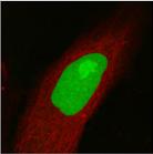
14 Jens Jens Rietdorf: Rietdorf: Autluorescence Autluorescence may may have have different different reasons, reasons, but but is is generally generally stronger, stronger, shorter shorter wavelength wavelength higher higher intensity intensity excitation excitation light light is. is. Stressed Stressed or or damaged damaged specimen specimen show show AF. AF. Autluorescence Specific sources autluorescence (excitation): Aromatic amino acid residues (UV). Reduced pyridine nucleotides (UV). Flavins (UV, blue). Chitin (broad). Chlorophyll (blue, green). General sources autluorescence: Dead cells (broad). Lipuscin (UV, blue). Cures: Long wavelength (also lower energy) light.[except 2-Phon] Avoid stress.
15 Phodamage Illumination energy not converted in emitted light (typ. <1%) or heat can enforce chemical reactions.

16 Recognise damaged cells Cells detach. Blebs form. Michondria swell. Cells do not make it through misis. Necrosis, Apopsis
17 Jens Jens Rietdorf: Rietdorf: Different Different microscopy microscopy techniques techniques will will be be discussed discussed which which very very light light efficient efficient avoid avoid phodamage phodamage discussed discussed in in following following Avoid phodamage Use decent dyes. Optimise illumination detection: Filtersets Detecrs Resolution (xy,z,t,intensity value, channels) Make use image processing ( deconvolution ). Add antioxidants (Trolox, ascorbic acid 2mg/ml) Use appropriate microscope techniques.
18 Contents Environment Physical integrity Attachment Temperature Gases (CO 2, O 2,H 2 O), ph. Osmolarity Illumination Autluorescence Phodamage Microscopy image processing techniques deconvolution Live cell microscopes (Time lapse sequence analysis)

19 Wide-field microscopy + deconvolution -Use a priori knowledge improve image quality -deconvolution is possible for all image dimensions : Along optical axis, time-lapse, color


20 Jens Jens Rietdorf: Rietdorf: Deconvolution Deconvolution can can increase increase signal--noise signal--noise ratio ratio reby reby allows allows reduction reduction excitation excitation light. light. Always Always use use deconvolution deconvolution before before estimating estimating how how much much light light has has be be put put in in sample sample reveal reveal relevant relevant information. information. deconvolution


21 Jens Jens Rietdorf: Rietdorf: Spinning Spinning disc disc confocals confocals a a good good alternative alternative single single beam beam scanning scanning confocals confocals as as y y use use very very low low excitation excitation light light intensities. intensities. Example Yokogawa unit


22 Jens Jens Rietdorf: Rietdorf: TIRF TIRF microscopy microscopy limits limits excitation excitation a a small small a a close close coverglas coverglas provides provides excellent excellent contrast. contrast. Total internal reflection fluorescence microscopy TIRFM prismless system


23 Example membrane fusion in TIRF

24 Jens Jens Rietdorf: Rietdorf: Example Example single single molecule molecule TIRF. TIRF. Example Pascale


25 Jens Jens Rietdorf: Rietdorf: Differently Differently shaped shaped structures structures may may be be labeled labeled with with same same dye dye still still saparable saparable in in different different channels channels by by object object detection detection approaches. approaches. Double Double exposure exposure for for different different fluorophores fluorophores can can be be avoided. avoided. Simultaneous multichannels I
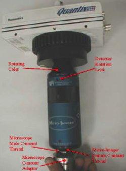
26 scope Simultaneous multichannels II microimager camera unmix
27 Jens Jens Rietdorf: Rietdorf: Different Different microscopy microscopy methods methods more more or or less less suited suited for for different different applications. applications. Mark Mark 1=good. 1=good. A A very very rough rough estimate estimate made made emphasize emphasize pros pros cons cons different different methods methods with with respect respect live live cell cell imaging. imaging. Comparision ols Widefield (+deco) Confocal Light Depth Acquisition Volume Timelapse Flexibility efficiency discrimination speed imaging imaging Multibeam confocal 2-Phon TIRF (n.p) 1 5
28 Conclusions Keep environment constant convenient Use powerful dyes Think about resolution required (xy,z,t,intensity value, channels) minimize phostress Use appropriate microscopy method Use deconvolution

29 People involved ALMF: Rainer Pepperkok Timo Zimmermann Andreas Girod Kota Miura
Widefield Microscopy Bleed-Through
 In widefield microscopy the excitation wavelengths which illuminate the sample, and the emission wavelengths which reach the CCD camera are selected throughout a filter cube. A filter cube consists of
Total Internal Reflection Fluorescence Microscopy
 Total Internal Reflection Microscopy Nicole O Neil Indiana University October 24, 2005 Agenda Why use TIRFM? Theory behind TIR Snell s Law Instrumentation Evanescent Wave Excitation of Fluorophores Advantages/Disadvantages
A Thin Layer Imaging with the Total Internal Reflection Fluorescence Microscopy
 Journal of Optoelectronical Nanostructures Islamic Azad University Summer 2017 / Vol. 2, No. 2 A Thin Layer Imaging with the Total Internal Reflection Fluorescence Microscopy Neda Roostaie 1, Elham Sheykhi
Accessorize Your Imaging. Ian Clements Invitrogen Corp
 Accessorize Your Imaging Ian Clements Invitrogen Corp Imaging Tools and Accessories Antifade Reagents Prolong Gold SlowFade Gold Image-iT FX Signal Enhancer FocalCheck Microscope Test Slides Fluorescent
Microscopy. CS/CME/BioE/Biophys/BMI 279 Nov. 2, 2017 Ron Dror
 Microscopy CS/CME/BioE/Biophys/BMI 279 Nov. 2, 2017 Ron Dror 1 Outline Microscopy: the basics Fluorescence microscopy Resolution limits The diffraction limit Beating the diffraction limit 2 Microscopy:
Confocal Microscopes. Evolution of Imaging
 Confocal Microscopes and Evolution of Imaging Judi Reilly Hans Richter Massachusetts Institute of Technology Environment, Health & Safety Office Radiation Protection What is Confocal? Pinhole diaphragm
Fluorescence Light Microscopy for Cell Biology
 Fluorescence Light Microscopy for Cell Biology Why use light microscopy? Traditional questions that light microscopy has addressed: Structure within a cell Locations of specific molecules within a cell
Microscopy from Carl Zeiss. DirectFRAP. News from the Cell. The New Class of Laser Manipulation for the Analysis of Cell Dynamics
 Microscopy from Carl Zeiss DirectFRAP News from the Cell The New Class of Laser Manipulation for the Analysis of Cell Dynamics DirectFRAP. New Insights into Cell Dynamics. Fluorescence breaks new ground:
Imaging facilities at WUR
 Imaging facilities at WUR Advanced light microscopy facilities at Wageningen UR Programme Thursday 13 June 2013 Lunch meeting organized by Cat-Agro Food 12.00 Welcome and sandwich lunch 12.10 Introduction
Foundations in Microbiology Seventh Edition
 Lecture PowerPoint to accompany Foundations in Microbiology Seventh Edition Talaro Chapter 3 Tools of the Laboratory: The Methods for Studying Microorganisms Copyright The McGraw-Hill Companies, Inc. Permission
BIO 315 Lab Exam I. Section #: Name:
 Section #: Name: Also provide this information on the computer grid sheet given to you. (Section # in special code box) BIO 315 Lab Exam I 1. In labeling the parts of a standard compound light microscope
How to perform-control immunostaining experiment - microscopist subjective point of view. Pawel Pasierbek
 How to perform-control immunostaining experiment - microscopist subjective point of view. Pawel Pasierbek Immunolabeling and fluorescent detection became such a standard procedure in the biomedical research
For outstanding performance in real-time PCR
 Rotor-Gene Q For outstanding performance in real-time PCR Outstanding thermal and optical performance due to rotary format An unmatched optical range spanning UV to infrared wavelengths State-of-the art
BIO 315 Lab Exam I. Section #: Name:
 Section #: Name: Also provide this information on the computer grid sheet given to you. (Section # in special code box) BIO 315 Lab Exam I 1. In labeling the parts of a standard compound light microscope
COPYRIGHTED MATERIAL. Tissue Preparation and Microscopy. General Concepts. Chemical Fixation CHAPTER 1
 CHAPTER 1 Tissue Preparation and Microscopy General Concepts I. Biological tissues must undergo a series of treatments to be observed with light and electron microscopes. The process begins by stabilization
Immunofluorescence Confocal Microscopy of 3D Cultures Grown on Alvetex
 Immunofluorescence Confocal Microscopy of 3D Cultures Grown on Alvetex 1.0. Introduction Immunofluorescence uses the recognition of cellular targets by fluorescent dyes or antigen-specific antibodies coupled
BIOCHEMIST ALL IN ONE ARTICLE
 BIOCHEMIST ALL IN ONE ARTICLE Bringing ease-of-use to microscopy From the Philosopher s Stone to the Researcher s Dream Although naturally occurring luminescence has been observed for many centuries, the
Fluorescence Microscopy. Terms and concepts to know: 10/11/2011. Visible spectrum (of light) and energy
 and energy") Fluorescence Microscopy Louisiana Tech University Ruston, Louisiana Microscopy Workshop Dr. Mark DeCoster Associate Professor Biomedical Engineering 1 Terms and concepts to know: Signal to Noise Excitation
Fluorescence Microscopy Louisiana Tech University Ruston, Louisiana Microscopy Workshop Dr. Mark DeCoster Associate Professor Biomedical Engineering 1 Terms and concepts to know: Signal to Noise Excitation
Live cell microscopy
 Live cell microscopy 1. Why do live cell microscopy? 2. Maintaining living cells on a microscope stage. 3. Considerations for imaging living cells. 4. Fluorescence labeling of living cells. 5. Imaging
D e c N o. 2 8
 D e c. 2 0 0 7 N o. 2 8 CONFOCAL APPLICATION LETTER resolution FRET Acceptor Photobleaching LAS AF Application Wizard FRET with Leica TCS SP5 LAS AF Version 1.7.0 Introduction Fluorescence Resonance Energy
Confocal Microscopy & Imaging Technology. Yan Wu
 Confocal Microscopy & Imaging Technology Yan Wu Dec. 05, 2014 Cells under the microscope What we use to see the details of the cell? Light and Electron Microscopy - Bright light / fluorescence microscopy
A Brief History of Light Microscopy And How It Transformed Biomedical Research
 A Brief History of Light Microscopy And How It Transformed Biomedical Research Suewei Lin Office: Interdisciplinary Research Building 8A08 Email: sueweilin@gate.sinica.edu.tw TEL: 2789-9315 Microscope
Imaging of endocrine organs
 Imaging of endocrine organs Helen Christian Department of Physiology, Anatomy & Genetics St Anne s College, University of Oxford Diabetesforum, Stockholm 2017 Islets of Langerhan Pituitary gland Renin
Simultaneous multi-color, multiphoton fluorophore excitation using dual-color fiber lasers
 Multiphoton Microscopy / Fiber Laser Simultaneous multi-color, multiphoton fluorophore excitation using dual-color fiber lasers Matthias Handloser, Tim Paasch-Colberg, Bernhard Wolfring TOPTICA Photonics
Introduction to histology and its methods of study
 Introduction to histology and its methods of study Li shulei lishulei@tom.com Department of Histology & Embryology 1 What is histology Definition Cell: smallest units functions in the human body Tissue
DNA Microarray Technology
 2 DNA Microarray Technology 2.1 Overview DNA microarrays are assays for quantifying the types and amounts of mrna transcripts present in a collection of cells. The number of mrna molecules derived from
Absorption of an electromagnetic wave
 In vivo optical imaging?? Absorption of an electromagnetic wave Tissue absorption spectrum Extinction = Absorption + Scattering Absorption of an electromagnetic wave Scattering of an electromagnetic wave
Super-resolution imaging: early days w/ Video-enhanced DIC, TIRF, PALM, STORM, etc.
 15/05/2012 Super-resolution imaging: early days w/ Video-enhanced DIC, TIRF, PALM, STORM, etc. Prof. Dr. Rainer Duden duden@bio.uni-luebeck.de 1 Using conventional light microscopy resolution is limited
Selected Techniques Part I
 1 Selected Techniques Part I Gel Electrophoresis Can be both qualitative and quantitative Qualitative About what size is the fragment? How many fragments are present? Is there in insert or not? Quantitative
ab CytoPainter ER Staining Kit Red Fluorescence
 ab139482 CytoPainter ER Staining Kit Red Fluorescence Instructions for Use Designed to detect Human endoplasmic reticulum by microscopy. This product is for research use only and is not intended for diagnostic
PROTOCOL. Live Cell Imaging of 3D Cultures Grown on Alvetex Scaffold Using Confocal Microscopy. Introduction
 Page 1 Introduction Live cell imaging allows real time monitoring of cell shape and form during cell differentiation, proliferation and migration. Alvetex Scaffold offers a unique ability to study cell
Image-iT FX Kits with Alexa Fluor Secondary Detection Conjugates
 Image-iT FX Kits with Alexa Fluor Secondary Detection Conjugates Table 1. Contents and Storage Information. Material Amount Concentration Storage Stability Alexa Fluor IgG conjugates Alexa Fluor streptavidin
Biophotonics?? Biophotonics. technology in biomedical engineering. Advantages of the lightwave
 Biophotonics - Imaging: X-ray, OCT, polarimetry, DOT, TIRF, photon migration, endoscopy, confocal microscopy, multiphoton microscopy, multispectral imaging - Biosensing: IR spectroscopy, fluorescence,
Protocol for. Light sheet microscopy using zebrafish Lightsheet Z.1
 Protocol for Light sheet microscopy using zebrafish Lightsheet Z.1 Zebrafish Facility Department of Organismal Biology Uppsala University / SciLifeLab in collaboration with Biological Visualization (Biovis)
1st Faculty of Medicine, Charles University in Prague Center for Advanced Preclinical Imaging (CAPI)
") ADVANTAGES Optical Imaging OI Optical Imaging is based on the detection of weak light by a highly sensitive and high resolution CCD camera DISADVANTAGES High sensitivity Limited penetration depth Easy
ADVANTAGES Optical Imaging OI Optical Imaging is based on the detection of weak light by a highly sensitive and high resolution CCD camera DISADVANTAGES High sensitivity Limited penetration depth Easy
FLUORESCENCE. Matyas Molnar and Dirk Pacholsky
 FLUORESCENCE Matyas Molnar and Dirk Pacholsky 1 Information This lecture contains images and information from the following internet homepages http://micro.magnet.fsu.edu/primer/index.html http://www.microscopyu.com/
Localization Microscopy
 Localization Microscopy Theory, Sample Prep & Practical Considerations Patrina Pellett & Ann McEvoy Applications Scientist GE Healthcare, Cell Technologies May 27 th, 2015 Localization Microscopy Talk
Live and Dead Cell Assay
 ab115347 Live and Dead Cell Assay Instructions for Use Differential fluorescent labeling of live and dead cells This product is for research use only and is not intended for diagnostic use. Last Updated
In-situ laser-induced contamination monitoring using long-distance microscopy
 In-situ laser-induced contamination monitoring using long-distance microscopy Paul Wagner a, Helmut Schröder* a, Wolfgang Riede a a German Aerospace Center (DLR), Institute of Technical Physics, Pfaffenwaldring
lumox & x-well Technology
 lumox & x-well Technology lumox lumox dish & lumox multiwell lumox cell culture products are characterized by their ultra-thin, gas-permeable film base. Optimum gas exchange is guaranteed due to the gas
Cell Imaging. Cell Imaging 48
 Cell Imaging 48 bio-rad.com/zoe Cell Imaging Bio-Rad s suite of tools for fluorescence microscopy and cell imaging includes the ZOE fluorescent cell imager and nuclear dyes. See Also PureBlu Hoechst 33342
Sample Preparation Manual for the 3D Cell Explorer. November 2016
 Sample Preparation Manual for the 3D Cell Explorer November 2016 Contents 1. IMPORTANT facts FOR THE SAMPLE PREPARATION 3 2. Specifications overview 4 3. Sample preparation 5 3.1 Cell confluency 5 3.2
Characterizing Phenotypes of Bacteria by Staining Method
 Experiment 3 Laboratory to Biology III Diversity of Microorganisms / Wintersemester / page 1 Experiment 3 Characterizing Phenotypes of Bacteria by Staining Method Advisor NN Reading Chapters in BBOM 9
Special Techniques 1. Mark Scott FILM Facility
 Special Techniques 1 Mark Scott FILM Facility SPECIAL TECHNIQUES Multi-photon microscopy Second Harmonic Generation FRAP FRET FLIM In-vivo imaging TWO-PHOTON MICROSCOPY Alternative to confocal and deconvolution
lumox & x-well Technology
 lumox & x-well Technology lumox lumox cell culture products are characterized by their ultra-thin, gas-permeable film base. Optimum gas exchange is guaranteed due to the gas permeability and the short
Characterizing Phenotypes of Bacteria by Staining Method
 Experiment 3 Laboratory to Biology III Diversity of Microorganisms / Wintersemester / page 1 Experiment Characterizing Phenotypes of Bacteria by Staining Method Advisor Reading NN Chapters 3.1, 3.7, 3.8,
Methods of Culturing Microorganisms. Chapter 3. Five Basic Techniques of Culturing Bacteria. Topics
 Chapter 3 Topics Methods of Culturing Microorganisms Microscope (History, Types, Definitions) Staining (Gram s) Methods of Culturing Microorganisms Five basic techniques of culturing Media Microbial growth
Cell Surface-Anchored Fluorescent Aptamer Sensor Enables. Imaging of Chemical Transmitter Dynamics
 Supporting Information for Cell Surface-Anchored Fluorescent Aptamer Sensor Enables Imaging of Chemical Transmitter Dynamics Takeshi Tokunaga, Shigeyuki Namiki, Katsuhiro Yamada, Takahiro Imaishi, Hiroshi
ab CFSE Fluorescent Cell Labeling Kit
 ab113853 CFSE Fluorescent Cell Labeling Kit Instructions for Use For the durable fluorescent labeling of live cells for fluorescent microscopy and flow cytometry, population growth studies and within sample
Concept review: Fluorescence
 16 Concept review: Fluorescence Some definitions: Chromophore. The structural feature of a molecule responsible for the absorption of UV or visible light. Fluorophore. A chromophore that remits an absorbed
The new LSM 700 from Carl Zeiss
 The new LSM 00 from Carl Zeiss Olaf Selchow, Bernhard Goetze To cite this version: Olaf Selchow, Bernhard Goetze. The new LSM 00 from Carl Zeiss. Biotechnology Journal, Wiley- VCH Verlag, 0, (), pp.. .
ab CFSE Fluorescent Cell Labeling Kit
 ab113853 CFSE Fluorescent Cell Labeling Kit Instructions for Use For the durable fluorescent labeling of live cells for fluorescent microscopy and flow cytometry, population growth studies and within sample
ALP (alkaline phosphatase) calibrators were analyzed manually in microtiter plates to find the linearity range by following this protocol:
 calibrators were analyzed manually in microtiter plates to find the linearity range by following this protocol:") Exam Mol 3008 May 2009 Subject 1 (15p) ALP (alkaline phosphatase) calibrators were analyzed manually in microtiter plates to find the linearity range by following this protocol: Reaction solutions: 50
Exam Mol 3008 May 2009 Subject 1 (15p) ALP (alkaline phosphatase) calibrators were analyzed manually in microtiter plates to find the linearity range by following this protocol: Reaction solutions: 50
Caspase-2 BiFC plasmids
 Caspase-2 BiFC plasmids Caspase-2 BiFC - background Bimolecular Fluorescence Complementation (BiFC) describes the use of split fluorescent proteins to measure protein-protein interactions in cells. The
supplementary information
 DOI: 10.1038/ncb1992 Figure S1 (a-b) Verification of actin flow patter using photobleaching of actin:gfp and Lifeact:RFP in migrating dendritic cells (DCs). (a) Actin:GFP and Lifeact:RFP double-trafected
Super Resolution Imaging Solution Provider. Imaging Future
 Super Resolution Imaging Solution Provider Imaging Future Imaging Solution More Than Equipment NanoBioImaging(NBI) is the Industrial Partner of HKUST Super Resolution Imaging Center (SRIC). NBI aims to
Fast, three-dimensional super-resolution imaging of live cells
 Nature Methods Fast, three-dimensional super-resolution imaging of live cells Sara A Jones, Sang-Hee Shim, Jiang He & Xiaowei Zhuang Supplementary Figure 1 Supplementary Figure 2 Supplementary Figure 3
Focal Points. Application Note FP-153. Next Generation Gel Imaging with GelRed and GelGreen Dyes and GelDoc-It Imaging System
 Focal Points Application Note FP-153 MIDSCI 800.227.9997 636.227.9997 Gel Doc Equipment Next Generation Gel Imaging with GelRed and GelGreen Dyes and GelDoc-It Imaging System Introduction By using state-of-the-art
MicroTime 200 STED. Super-resolution add-on for the confocal time-resolved microscopy platform
 MicroTime 200 STED Super-resolution add-on for the confocal time-resolved microscopy platform confocal STED 2 Vision The MicroTime 200... The MicroTime 200 is a high-end confocal fluorescence lifetime
Labware compatibility report. Directly order at:
 Labware compatibility report Directly order at: https://ibidi.com/12-labware Contents 1. Introduction 3 2. Compatible Labware 4 A. 35 mm dishes - high borders 4 µ-dish 35 mm, high 4 µ-dish 35mm, high Glass
Dino-Lite knowledge & education. Fluorescence Microscopes
 Dino-Lite knowledge & education Fluorescence Microscopes Dino-Lite Fluorescence models Smallest fluorescence microscope in the world Revolution to biomedical and educational applications Flexible Easy
Chemotaxis assay using µ-slide Chemotaxis
 Chemotaxis assay using µ-slide Chemotaxis 1. General information The µ-slide Chemotaxis is a tool for observing chemotactical responses of adherent migrating cells over extended periods of time. The linear
Confocal Microscopy Analyzes Cells
 Choosing Filters for Fluorescence A Laurin Publication Photonic Solutions for Biotechnology and Medicine November 2002 Confocal Microscopy Analyzes Cells Reprinted from the November 2002 issue of Biophotonics
Measure of surface protein mobility with u-paint technique
 Measure of surface protein mobility with u-paint technique How dynamic image can solve the situation? Random distribution or cluster? Why live super-resolution microscopy can solve the situation With mobility
Imaging of Cells using fluorescents dyes. By: Josué A. Benjamín Rivera September 27, 2018
 Imaging of Cells using fluorescents dyes By: Josué A. Benjamín Rivera September 27, 2018 1 History Sir William Henry Perkin BRITISH CHEMIST In 1856, at the age of 18, William Henry Perkin set out with
Fluorescence Microscopy: A Biological Perspective
 Fluorescence Microscopy: A Biological Perspective From nanometre to metre: the scale of life Instrumentation and accessible scale limits the questions that can be addressed in biology Why are there limits?
SUMMER SCHOOL LABORATORY ACTIVITIES
 SUMMER SCHOOL LABORATORY ACTIVITIES ACTIVITIES Monday Tuesday Wednesday Thursday 18 th 19 th 20 th 21 st 1 and 2 A B C D 3 and 4 B C D A 5 and 6 C D A B 7 and 8 D A B C The students are divided into 4
Final Exam, 176 points PMB 185: Techniques in Light Microscopy
 Final Exam, 176 points Name PMB 185: Techniques in Light Microscopy Point value is in parentheses at the end of each question. 1) Order the steps in setting up Köhler illumination. It is not necessary
Supplementary Figure 1. Thin layer chromatography of R18 salts with different counterions. The mobility of the R18 salts with TPB counterions is much
 Supplementary Figure 1. Thin layer chromatography of R18 salts with different counterions. The mobility of the R18 salts with TPB counterions is much higher with perchlorate, showing their much higher
Introduction to N-STORM
 Introduction to N-STORM Dan Metcalf Advanced Imaging Manager Outline Introduction Principles of STORM Applications N-STORM overview Biological Scale Mitochondrion Microtubule Amino Acid 1Å Kinesin 1nm
More on fluorescence
 More on fluorescence Last class Fluorescence Absorption emission Jablonski diagrams This class More on fluorescence Common fluorophores Jablonski diagrams to spectra Properties of fluorophores Excitation
Cell Structure and Function
 Cell Structure and Function Dead White Men Who Discovered (and were made of) Cells: Anton Van Leeuwenhoek Robert Hooke Where the Magic Happened Schleiden Cell Theory All plants are made of cells Schwann
MF-ChemiBIS. Today s most comprehensive solution for your bio-imaging needs and applications. Documenting Nature
 MF-ChemiBIS Today s most comprehensive solution for your bio-imaging needs and applications Documenting Nature MF-ChemiBIS Excellence in bio-imaging The DNR Advantage As pioneers in bio-imaging technologies
Microscopy...Seeing the Unseen
 Technical Workshops Series 2013 Three Day Intensive Workshop on Venture Center Microscopy...Seeing the Unseen Organized by Venture Center Learn Organized by For whom When Principles and applications of
Supplementary Materials and Methods
 Supplementary Materials and Methods Reagents Supplementary Material (ESI) for Lab on a Chip RPMI medium, FBS, HEPES buffer solution, sodium pyruvate, penicillin, and streptomycin were obtained from Biological
FRET Sensitized Emission
 Page 1 of 8 FRET Sensitized Emission Function The FRET Sensitized Emission wizard is used for measuring the FRET efficiency. For this purpose, the fluorescence emission of the acceptor that results from
BASICS OF FLOW CYTOMETRY
 BASICS OF FLOW CYTOMETRY AUTHOR: Ana Isabel Vieira APPROVAL: Henrique Veiga Fernandes Ana Sílvia Gonçalves SOP.UCF.002 03-09-2015 Pag. 1/9 Overview Flow: Fluid Cyto: Cell Metry: Measurement Flow cytometry
PRODUCT DATA SHEET. Carboxylated Fluorescent Gold Nanoparticles. Description. Characteristics
 PRODUCT DATA SHEET Carboxylated Fluorescent Gold Nanoparticles Description Cytodiagnostics carboxylated fluorescent gold nanoparticles is a unique product that combines our Cyto fluorescent dyes and gold
SUPPLEMENTARY INFORMATION
 DOI: 1.138/NNANO.211.185 Mixing sub-attolitre volumes in a quantitative and highly parallel manner with soft matter nanofluidics Sune M. Christensen, Pierre-Yves Bolinger,Nikos S. Hatzakis, Michael W.Mortensen
SUMMER SCHOOL LABORATORY ACTIVITIES
 SUMMER SCHOOL LABORATORY ACTIVITIES ACTIVITIES Monday Tuesday Wednesday Thursday 18 th 19 th 20 th 21 st 1 and 2 A B C D 3 and 4 B C D A 5 and 6 C D A B 7 and 8 D A B C The students are divided into 4
2-step or indirect immunofluorescence 1. Substrate on which cells are plated: plastic vs. glass; coating vs. non
 Variables in standard immunostaining protocol 2-step or indirect immunofluorescence 1. Substrate on which cells are plated: plastic vs. glass; coating vs. non 2. Plating density: sparse vs. confluent 3.
Practical light microscopy: an introduction
 Practical light microscopy: an introduction Dr. Mark Leake, Oxford University www.physics.ox.ac.uk/users/leake Aim of today s talk: Explanation of the very (very) basics of how a light microscope works
Obtaining More Accurate Signals: Spatiotemporal Imaging of Cancer Sites Enabled by a Photoactivatable Aptamer-Based Strategy
 Supporting Information Obtaining More Accurate Signals: Spatiotemporal Imaging of Cancer Sites Enabled by a Photoactivatable Aptamer-Based Strategy Heng Xiao,,, Yuqi Chen,, Erfeng Yuan,, Wei Li, Zhuoran
Multiplexed 3D FRET imaging in deep tissue of live embryos Ming Zhao, Xiaoyang Wan, Yu Li, Weibin Zhou and Leilei Peng
 Scientific Reports Multiplexed 3D FRET imaging in deep tissue of live embryos Ming Zhao, Xiaoyang Wan, Yu Li, Weibin Zhou and Leilei Peng 1 Supplementary figures and notes Supplementary Figure S1 Volumetric
Supporting Information
 Supporting Information Koh et al. 10.1073/pnas.1212917110 SI Materials and Methods Protein Purification. N-terminal His 6 -Dicer was purified as previously described with several modifications (1). After
Contact Details. Dr Alexander Galkin. Office: MBC Room 186. Tel: (028) Frequency and wavelength.
 Frequency and wavelength.") Contact Details The electromagnetic spectrum Biological Spectroscopy Dr Alexander Galkin Email: a.galkin@qub.ac.uk Dr Alexander Galkin MSc Biomolecular Function - BBC8045 Office: MBC Room 186 Tel: (028)
Contact Details The electromagnetic spectrum Biological Spectroscopy Dr Alexander Galkin Email: a.galkin@qub.ac.uk Dr Alexander Galkin MSc Biomolecular Function - BBC8045 Office: MBC Room 186 Tel: (028)
SUPPLEMENTARY INFORMATION
 Biosynthesis of Luminescent Quantum Dots in an Earthworm S.R. Stürzenbaum, a# M. Hoeckner, a# A. Panneerselvam, b J. Levitt, b J.-S. Bouillard, b S. Taniguchi, b L.-A. Dailey, d R. Ahmad Khanbeigi, d E.
Live-cell visualization of excitation energy dynamics in chloroplast thylakoid structures
 Supplementary Information Live-cell visualization of excitation energy dynamics in chloroplast thylakoid structures Masakazu Iwai, Makio Yokono, Kazuo Kurokawa, Akira Ichihara & Akihiko Nakano Supplementary
Lab Module 7: Cell Adhesion
 Lab Module 7: Cell Adhesion Tissues are made of cells and materials secreted by cells that occupy the spaces between the individual cells. This material outside of cells is called the Extracellular Matrix
Workflow Spheroids: 3D tissue model in cancer research
 Workflow Spheroids: 3D tissue model in cancer research Irmtraud Steinmetz Updated by: Marco Meijering Application Support Specialist EMEA 2D cell culture and animal models for Cancer research 2D- cell
Living and Dead Cells Staining: -Cellstain- Double Staining Kit
 Introduction -Cellstain - Double Staining Kit combines Calcein-AM (used for fluorescent staining the living cells) and Propidium Iodide (used for a fluorescent staining of the dead cells) for simultaneous
Instructions. Fuse-It-siRNA. Shipping and Storage. Overview. Kit Contents. Specifications. Note: Important Guidelines
 Membrane fusion is a highly efficient method for transfecting various molecules and particles into mammalian cells, even into sensitive and primary cells. The Fuse-It reagents are cargo-specific liposomal
SURFACE ENHANCED RAMAN SCATTERING NANOPARTICLES AS AN ALTERNATIVE TO FLUORESCENT PROBES AN EVALUATION
 APPLICATION NOTE SURFACE ENHANCED RAMAN SCATTERING NANOPARTICLES AS AN ALTERNATIVE TO FLUORESCENT PROBES AN EVALUATION Summary: Interest in using nanoparticles specifically, Surface Enhanced Raman Scattering
In situ semi-quantitative assessment of single cell viability by resonance
 Electronic Supplementary Material (ESI) for Chemical Communications. This journal is The Royal Society of Chemistry 2018 Electronic Supplementary Information (ESI) In situ semi-quantitative assessment
computer controlled cell deforming
 FOUS ON EMERGING TEHNOLOGY computer controlled cell deforming cellstretcher a device for simultaneous live cell imaging with motion compensation for uni-axial mechanical straining or compression RELTIME
Visualizing Cells Molecular Biology of the Cell - Chapter 9
 Visualizing Cells Molecular Biology of the Cell - Chapter 9 Resolution, Detection Magnification Interaction of Light with matter: Absorbtion, Refraction, Reflection, Fluorescence Light Microscopy Absorbtion
ab CytoPainter ER Staining Kit Red Fluorescence
 ab139482 CytoPainter ER Staining Kit Red Fluorescence Instructions for Use Designed to detect Human endoplasmic reticulum by microscopy. This product is for research use only and is not intended for diagnostic
Two-Photon Microscopy for Deep Tissue Imaging of Living Specimens
 for Deep Tissue Imaging of Living Specimens Tilman Franke* and Sebastian Rhode TILL Photonics GmbH, an FEI company, Lochhamer Schlag 21, D-82166 Gräfelfing, Germany *tilman.franke@fei.com Introduction
Super-resolution Microscopy
 Semr oc kwhi t epaperser i es : 1. Introduction Super-resolution Microscopy Fluorescence microscopy has revolutionized the study of biological samples. Ever since the invention of fluorescence microscopy
Immunofluorescence and phalloidin labeling of mammalian cells
 Immunofluorescence and phalloidin labeling of mammalian cells 2 Contents Materials for immunofluorescence and phalloidin labeling of mammalian cells...1 Immunofluorescence-labelling on cultivated adherent