Imaging facilities at WUR
|
|
|
- Kristina Rich
- 5 years ago
- Views:
Transcription
1 Imaging facilities at WUR
2 Advanced light microscopy facilities at Wageningen UR Programme Thursday 13 June 2013 Lunch meeting organized by Cat-Agro Food Welcome and sandwich lunch Introduction to light microscopy Norbert de Ruijter (Cell Biology and WLMC) Interactive presentations about recently purchased state-of-the-art light microscopy equipment and their applications: 12:30 BD Pathway bio-imager: high throughput high-content imaging and analysis of live and fixed cells Jurgen Karczewski (Host Microbe Interactomics) 12:50 Spinning disk confocal microscopy: ideal for imaging of living cells or weak fluorescent signals Norbert de Ruijter (Cell Biology and WLMC) 13:10 Multi-mode confocal laser scanning microscopy: with fluorescence lifetime imaging microscopy (FLIM) option for the detection of protein interactions in living cells Jan Willem Borst (Biochemistry and MSC) 13:30 Single molecule total internal reflection fluorescence microscopy (smtirf): Studying (dynamic) interactions in and between single proteins - Johannes Hohlbein (Biophysics and MSC) End of the programme

3 Introduction to light microscopy Norbert de Ruijter Laboratory of Cell Biology WUR PSG
4 Aims Refresh your basic knowledge on microscopy/ fluorescence/ confocal What equipment best serves my research needs? Where can I get assistance / access to microscopes? What are the main Imaging Facilities within WUR? Explain possibilities with latest CAT AGRO Food investments in LM: 4 small presentations


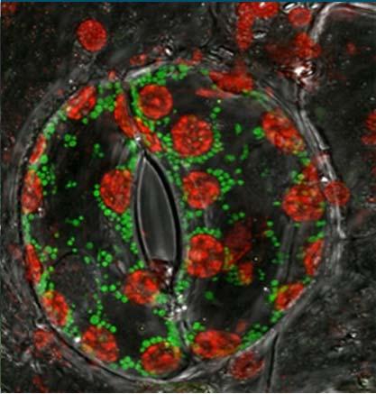
5 LM techniques - Bright field Imaging BF: Contrast by variations in light absorbance DIC/Phase: Contrast by variations in interference/breaking index Polarization microscopy: Contrast by birefringent polymers-crystals - Fluorescence Imaging FL: dark field technique based on FL excitation / emission Many ways for localization-dynamics-quantitative studies
. 3.")
6 Fluorescence: Jablonski energy diagram Discriminate 3 steps: 1. A fluorophore absorbs a high energetic photon (light particle) 2. An electron comes temporarily in an excited vibrational energy state (with short lifetime). 3. A photon of lower energy is emitted, returning the fluorophore back to ground state (S 0 ) = Fluorescence!


7 Fluorescence: photons are emitted with higher wavelength but lower energy Stokes shift Higher energy Lower energy
8 Filters and dichroic mirror allow to select specific emission Excitation light does not reach the detector, since dichroic mirror reflects excitation and transmits emission light. You need much intensity in a specific wavelength band (LED/laser) for optimal excitation.
 on (PMT/CCD) detector.")
9 Out-of-focus blur Focal plane 1 2 d z A cell with stained DNA (blue) and microtubules (green) Cell is illuminated with excitation light from below: Positions 1 and 2 at different heights in the sample receive equal excitation intensity and fluorophores become excited. Emission from the focal plane is detected sharp (in focus) on (PMT/CCD) detector. However, emission light from out-of-focus planes superimposes a blur that prevents us from seeing detail in the sample.
10 Remove out-of-focus blur by optical sectioning Confocal Laser Scanning Microscopy (JW Borst) Spinning Disk Microscopy (N de Ruijter) TIRFM (Johannes Hohlbein) Multi-photon microscopy Light sheet microscopy (SPIM) Mathematical: Image Deconvolution (Image J, Matlab)

11 Confocal Principle Blocking out of focus light


12 Configuration for multiple color detection Pinhole diaphragm Emission filters Secondary dichroic BS HFT: Main dichroic BS Beam path in the scan head of a point scanning LSCM - META Beam combiner xy galvo scanners collimators

13 Prevent signal bleed through by META detector -Diffraction grating splits emission spectrum over set of 32 channels (PMTs) -The spectral signature of each pixel is acquired > Imaging with spectral unmixing : is rapid and increases sensitivity is specific and allows to separate overlapping EM spectra is more reliable than filter sets and band pass acquisition allows high speed multicolor imaging disadvantage: only one pinhole/gain setting for all emissions
14 Confocal laser scanning microscopy (galvo mirrors) With a pinhole the confocal LSM can make optical sections of a sample (xy plane) A z-serie of optical sections is used to reconstruct a sharp 3D image.


15 Optical sectioning with CLSM BY-2 tobacco suspension cell expressing GFP-ER (zs) Orthogonal view; more depth info in (flat) nucleus
16 Confocal Laser Scanning Microscopy a focused laser beam scans (xy) spots on specimen optical slices are collected over time (ts) or (and) in subsequent different focal planes (zs) in various channels (colors) computer reconstructs a 3-dimensional image by compiling optical slices bleaching of out of focus fluorophore however, can still be strong; a pinhole is only present in the emission path 16

17 Photo bleaching - long term imaging Excitation intensity usually decays when imaging a sample for longer time because fluorophores are irreversibly photo bleached. Some fluorophores are more stable than others. Prevent bleaching: low excitation intensity, short exposure... > Find imaging parameters to obtain bright, sharp fluorescent pictures with minimal bleaching.


18 Imaging: balancing sensitivity, speed, resolution Signal / noise ratio relates to scanning speed pixel density pinhole size gain Cygnus cygnus
19 Fluorescent Labels Intrinsic fluorophore e.g. Tryptophan, chlorofyl, lignin amino acids Chemical dye Alexa dyes, Cy dyes Immuno labels Covalent binding Alexa 488 succimidyl ester


20 Fluorescent Label Quantum dots + photo stable excitation emission + high quantum yield + nano particles Green fluorescent protein 2.4 nm + genetic labeling + well protected from environment - size 4.2 nm Ormő et al. Science (1996), 273, 1392
21 Various FL probes allow quantitative functional imaging (seeing molecular dynamics) Fluorescence ratio imaging (internal control) Fluorescence Recovery After Photobleaching (dynamics) Photo activation Photo conversion Uncaging Bimolecular complementation More: Biochemistry FCS study mobility of molecules FRET- follow molecular interactions
22 Imaging centres within WUR NMR: Nuclear Magnetic Resonance Imaging (Transitorium -Dreijen) WEMC: Wageningen Electron Microscopy Centre (Radix West-campus) WLMC: Wageningen Light Microscopy Centre (Radix West-campus) MSC: Micro-Spectroscopy Centre (Transitorium -Dreijen) We share forces and merge nto one virtual centre called WISH: Wageningen Imaging & Spectroscopy Hub Currently in the process of being officially recognized / accepted as a NL-Bio Imaging Centre and Euro Bio Imaging Centre for plant imaging... Aims: more options to further maintain /develop imaging facilities at WUR to facilitate / assist you and foreign visiting scientists for industry/institutes to access up-to-date equipment
23 Where to go? Cell Biology (PSG)- WLMC-Radix Biochemistry (AFSG)- MSC- Dreijen HMI-ASG (Karczewski/Schipper) Macro and FL-stereo X DIC/PhC/BF/HMC (unstained-sterile) X Micro manipulation (Narashigi) and LM X Polarization (quantified) X Fluorescence -wide field - multicolor X X X Confocal - imaging plant structure-morphology X X Confocal - imaging structure-morphology poor signals spinning disk/ MM-CLSM X X Confocal - imaging structure-morphology dynamics/ FRAP spinning disk X Confocal temperature and CO2 control on stage spinning disk X Confocal - ion fluxes dynamics X Confocal combined with Optical tweezers X Confocal - colocalization spectral unmixing X X Confocal - colocalization quantified X X Confocal - protein fast dynamics, quantified MM-CLSM X Confocal - protein interactions localized X X Confocal - protein interactions, quantified, FCS MM-CLSM X Confocal - deep into tissue-/ multi-photon X Structural properties of proteins-distance /FRET MM-CLSM X Structural properties of proteins-folding/dynamics/flim MM-CLSM X TIRF - FL events at the plasma membrane/ in vitro assays sm-tirf X X High througput/content BD pathway X Additional support Embeddings and microtomy X Support for protein purification and labeling X Range of available life probes for testing X X Options for optimal mounting for prolonged life imaging X
24 End intro Questions?
Special Techniques 1. Mark Scott FILM Facility
 Special Techniques 1 Mark Scott FILM Facility SPECIAL TECHNIQUES Multi-photon microscopy Second Harmonic Generation FRAP FRET FLIM In-vivo imaging TWO-PHOTON MICROSCOPY Alternative to confocal and deconvolution
F* techniques: FRAP, FLIP, FRET, FLIM,
 F* techniques: FRAP, FLIP, FRET, FLIM, FCS Antonia Göhler March 2015 Fluorescence explained in the Bohr model Absorption of light (blue) causes an electron to move to a higher energy orbit. After a particular
Workshop advanced light microscopy
 Workshop advanced light microscopy Multi-mode confocal laser scanning microscope Jan Willem Borst Laboratory of Biochemistry Biomolecular Networks www.bic.wur.nl MicroSpectroscopy Centre Wageningen Microspectroscopy
Confocal Microscopy Analyzes Cells
 Choosing Filters for Fluorescence A Laurin Publication Photonic Solutions for Biotechnology and Medicine November 2002 Confocal Microscopy Analyzes Cells Reprinted from the November 2002 issue of Biophotonics
Multiplexed 3D FRET imaging in deep tissue of live embryos Ming Zhao, Xiaoyang Wan, Yu Li, Weibin Zhou and Leilei Peng
 Scientific Reports Multiplexed 3D FRET imaging in deep tissue of live embryos Ming Zhao, Xiaoyang Wan, Yu Li, Weibin Zhou and Leilei Peng 1 Supplementary figures and notes Supplementary Figure S1 Volumetric
Contact Details. Dr Alexander Galkin. Office: MBC Room 186. Tel: (028) Frequency and wavelength.
 Frequency and wavelength.") Contact Details The electromagnetic spectrum Biological Spectroscopy Dr Alexander Galkin Email: a.galkin@qub.ac.uk Dr Alexander Galkin MSc Biomolecular Function - BBC8045 Office: MBC Room 186 Tel: (028)
Contact Details The electromagnetic spectrum Biological Spectroscopy Dr Alexander Galkin Email: a.galkin@qub.ac.uk Dr Alexander Galkin MSc Biomolecular Function - BBC8045 Office: MBC Room 186 Tel: (028)
Resolution of Microscopes Visible light is nm Dry lens(0.5na), green(530nm light)=0.65µm=650nm for oil lens (1.4NA) UV light (300nm) = 0.13µm f
, green(530nm light)=0.65µm=650nm for oil lens (1.4NA) UV light (300nm) = 0.13µm f") Microscopes and Microscopy MCB 380 Good information sources: Alberts-Molecular Biology of the Cell http://micro.magnet.fsu.edu/primer/ http://www.microscopyu.com/ Approaches to Problems in Cell Biology
Microscopes and Microscopy MCB 380 Good information sources: Alberts-Molecular Biology of the Cell http://micro.magnet.fsu.edu/primer/ http://www.microscopyu.com/ Approaches to Problems in Cell Biology
Fluorescence Light Microscopy for Cell Biology
 Fluorescence Light Microscopy for Cell Biology Why use light microscopy? Traditional questions that light microscopy has addressed: Structure within a cell Locations of specific molecules within a cell
Confocal Microscopy & Imaging Technology. Yan Wu
 Confocal Microscopy & Imaging Technology Yan Wu Dec. 05, 2014 Cells under the microscope What we use to see the details of the cell? Light and Electron Microscopy - Bright light / fluorescence microscopy
Widefield Microscopy Bleed-Through
 In widefield microscopy the excitation wavelengths which illuminate the sample, and the emission wavelengths which reach the CCD camera are selected throughout a filter cube. A filter cube consists of
FLIM Fluorescence Lifetime IMaging
 FLIM Fluorescence Lifetime IMaging Fluorescence lifetime t I(t) = F0 exp( ) τ 1 τ = k f + k nr k nr = k IC + k ISC + k bl Batiaens et al, Trends in Cell Biology, 1999 τ τ = fluorescence lifetime (~ns to
Confocal Microscopes. Evolution of Imaging
 Confocal Microscopes and Evolution of Imaging Judi Reilly Hans Richter Massachusetts Institute of Technology Environment, Health & Safety Office Radiation Protection What is Confocal? Pinhole diaphragm
A Brief History of Light Microscopy And How It Transformed Biomedical Research
 A Brief History of Light Microscopy And How It Transformed Biomedical Research Suewei Lin Office: Interdisciplinary Research Building 8A08 Email: sueweilin@gate.sinica.edu.tw TEL: 2789-9315 Microscope
Fluorescence Microscopy. Terms and concepts to know: 10/11/2011. Visible spectrum (of light) and energy
 and energy") Fluorescence Microscopy Louisiana Tech University Ruston, Louisiana Microscopy Workshop Dr. Mark DeCoster Associate Professor Biomedical Engineering 1 Terms and concepts to know: Signal to Noise Excitation
Fluorescence Microscopy Louisiana Tech University Ruston, Louisiana Microscopy Workshop Dr. Mark DeCoster Associate Professor Biomedical Engineering 1 Terms and concepts to know: Signal to Noise Excitation
Concept review: Fluorescence
 16 Concept review: Fluorescence Some definitions: Chromophore. The structural feature of a molecule responsible for the absorption of UV or visible light. Fluorophore. A chromophore that remits an absorbed
More on fluorescence
 More on fluorescence Last class Fluorescence Absorption emission Jablonski diagrams This class More on fluorescence Common fluorophores Jablonski diagrams to spectra Properties of fluorophores Excitation
Dino-Lite knowledge & education. Fluorescence Microscopes
 Dino-Lite knowledge & education Fluorescence Microscopes Dino-Lite Fluorescence models Smallest fluorescence microscope in the world Revolution to biomedical and educational applications Flexible Easy
Lab 5: Optical trapping and single molecule fluorescence
 Lab 5: Optical trapping and single molecule fluorescence PI: Matt Lang Lab Instructor: Jorge Ferrer Summary Optical tweezers are an excellent experimental tool to study the biophysics of single molecule
The new LSM 700 from Carl Zeiss
 The new LSM 00 from Carl Zeiss Olaf Selchow, Bernhard Goetze To cite this version: Olaf Selchow, Bernhard Goetze. The new LSM 00 from Carl Zeiss. Biotechnology Journal, Wiley- VCH Verlag, 0, (), pp.. .
Localization Microscopy
 Localization Microscopy Theory, Sample Prep & Practical Considerations Patrina Pellett & Ann McEvoy Applications Scientist GE Healthcare, Cell Technologies May 27 th, 2015 Localization Microscopy Talk
Imaging & analysis with the LSM780 NLO Discover the secrets beyond the twilight zone
 Imaging & analysis with the LSM780 NLO Discover the secrets beyond the twilight zone Sven Terclavers LSM780 System overview The Scan Module - Core of the LSM 780 1 V/tunable PTC laser ports (405/440, cw/ps;
Final Exam, 176 points PMB 185: Techniques in Light Microscopy
 Final Exam, 176 points Name PMB 185: Techniques in Light Microscopy Point value is in parentheses at the end of each question. 1) Order the steps in setting up Köhler illumination. It is not necessary
FLUORESCENCE. Matyas Molnar and Dirk Pacholsky
 FLUORESCENCE Matyas Molnar and Dirk Pacholsky 1 Information This lecture contains images and information from the following internet homepages http://micro.magnet.fsu.edu/primer/index.html http://www.microscopyu.com/
Fluorescence Microscopy
 Fluorescence Microscopy Dr. Arne Seitz Swiss Institute of Technology (EPFL) Faculty of Life Sciences Head of BIOIMAGING AND OPTICS BIOP arne.seitz@epfl.ch Fluorescence Microscopy Why do we need fluorescence
Visualizing Cells Molecular Biology of the Cell - Chapter 9
 Visualizing Cells Molecular Biology of the Cell - Chapter 9 Resolution, Detection Magnification Interaction of Light with matter: Absorbtion, Refraction, Reflection, Fluorescence Light Microscopy Absorbtion
Welcome! openmicberkeley.wordpress.com. Open Berkeley
 Welcome! openmicberkeley.wordpress.com Agenda Jen Lee: Introduction to FRET Marla Feller: Using FRET sensors to look at time resolved measurements Becky Lamason: Using FRET to determine if a bacterial
Live cell microscopy
 Live cell microscopy 1. Why do live cell microscopy? 2. Maintaining living cells on a microscope stage. 3. Considerations for imaging living cells. 4. Fluorescence labeling of living cells. 5. Imaging
PALM/STORM, BALM, STED
 PALM/STORM, BALM, STED Last class 2-photon Intro to PALM/STORM Cyanine dyes/dronpa This class Finish localization super-res BALM STED Localization microscopy Intensity Bins = pixels xx 2 = ss2 + aa 2 /12
EuBI application for access
 * Title * First name * Last name * Email address * Institution/Company URL of the Institution/Company * Phone number Country code Phone * Street address * Zip Code * City * Country * Position * Not a Principal
Bi177 - Lecture 13 Microscopy Outside the Box. Fluorescence Nanoscopy TIRF 4-pi STED STORM/PALM
 Bi177 - Lecture 13 Microscopy Outside the Box Fluorescence Nanoscopy TIRF 4-pi STED STORM/PALM The diffraction limit: Abbe s law The Problem Diffraction limit 100x larger than molecular scale! Green Fluorescent
Monitoring and Optimizing the Lipopolysaccharides-plasmid DNA interaction by FLIM-FRET
 Transactions on Science and Technology Vol. 4, No. 3-3, 342-347, 2017 Monitoring and Optimizing the Lipopolysaccharides-plasmid DNA interaction by FLIM-FRET Nur Syahadatain Abdul Razak 1#, Clarence M.
Super Resolution Microscopy - Breaking the Diffraction Limit Radiological Research Accelerator Facility
 Super Resolution Microscopy - Breaking the Diffraction Limit Radiological Research Accelerator Facility Sabrina Campelo, Dr. Andrew Harken Outline Motivation Fluorescence Microscopy -Multiphoton Imaging
D e c N o. 2 8
 D e c. 2 0 0 7 N o. 2 8 CONFOCAL APPLICATION LETTER resolution FRET Acceptor Photobleaching LAS AF Application Wizard FRET with Leica TCS SP5 LAS AF Version 1.7.0 Introduction Fluorescence Resonance Energy
11/19/2013. Janine Zankl FACS Core Facility 13. November Cellular Parameters. Cellular Parameters. Monocytes. Granulocytes.
 DEPARTEMENT BIOZENTRUM Janine Zankl FACS Core Facility 13. November 2013 Cellular Parameters Granulocytes Monocytes Basophils Neutrophils Lymphocytes Eosinophils Cellular Parameters 1 What Is Flow Cytometry?
Biochemistry. Biochemical Techniques. 18 Spectrofluorimetry
 Description of Module Subject Name Paper Name 12 Module Name/Title 1. Objectives 1.1 To understand technique of Spectrofluorimetry. 1.2 To explain instrumentation design 1.3 What are applications of Spectrofluorimetry?
Design for Manufacturability (DFM) in the Life Sciences
 in the Life Sciences") T E C H N I C A L N O T E Design for Manufacturability (DFM) in the Life Sciences Fluorescence Spectroscopy Product Platform Realized with TracePro TM Suite of Opto-Mechanical Design Software Tools Authors:
T E C H N I C A L N O T E Design for Manufacturability (DFM) in the Life Sciences Fluorescence Spectroscopy Product Platform Realized with TracePro TM Suite of Opto-Mechanical Design Software Tools Authors:
1. INTRODUCTION 2. EXPERIMENTAL 3. REFERENCES
 1. INTRODUCTION 2. EXPERIMENTAL 3. REFERENCES 1 1. INTRODUCTION Fluorescence spectroscopy is one of the most widely used spectroscopic techniques in the fields of biochemistry and molecular biophysics
Two-Photon Microscopy for Deep Tissue Imaging of Living Specimens
 for Deep Tissue Imaging of Living Specimens Tilman Franke* and Sebastian Rhode TILL Photonics GmbH, an FEI company, Lochhamer Schlag 21, D-82166 Gräfelfing, Germany *tilman.franke@fei.com Introduction
Microscopy from Carl Zeiss
 Microscopy from Carl Zeiss LSM 710 In Tune with Your Application Enjoy new freedom in selecting fluorescent dyes with In Tune, the new laser system for the LSM 710. Whatever the wavelength, you can match
Nodes of regulation in cellular systems
 Nodes of regulation in cellular systems cell membrane signal transduction ligands receptors oligomerization transport signal transduction modified protein Golgi transcription factor transport ER transport
Confocal Microscopy of Electronic Devices. James Saczuk. Consumer Optical Electronics EE594 02/22/2000
 Confocal Microscopy of Electronic Devices James Saczuk Consumer Optical Electronics EE594 02/22/2000 Introduction! Review of confocal principles! Why is CM used to examine electronics?! Several methods
INTRODUCTION TO FLOW CYTOMETRY
 DEPARTEMENT BIOZENTRUM INTRODUCTION TO FLOW CYTOMETRY F ACS C ore F acility Janine Zankl FACS Core Facility 3. Dezember 2015, 4pm Cellular Parameters Granulocytes Monocytes Basophils Lymphocytes Neutrophils
Super Resolution Imaging Solution Provider. Imaging Future
 Super Resolution Imaging Solution Provider Imaging Future Imaging Solution More Than Equipment NanoBioImaging(NBI) is the Industrial Partner of HKUST Super Resolution Imaging Center (SRIC). NBI aims to
Compensation: Fundamental Principles
 Flow Cytometry Seminar Series 2017 : Fundamental Principles Spillover correction in multicolor flow cytometry 28.02.2017 http://www.cytometry.uzh.ch Contents Fluorescence and its detection Absorption and
FRET and FRET based Microscopy Techniques
 Big Question: We can see rafts in Model Membranes (GUVs or Supported Lipid Bilayers, LM), but how to study in cells? Do rafts really exist in cells? Are they static large structures? Are they small transient
Cellular imaging using Nano- Materials. A Case-Study based approach Arun Murali, Srivats V
 Cellular imaging using Nano- Materials A Case-Study based approach Arun Murali, Srivats V Agenda Discuss a few papers Explain a couple of new imaging techniques and their benefits over conventional imaging
Introduction to Fluorescence Jablonski Diagram
 ntroduction to Fluorescence Jablonski Diagram Excited Singlet Manifold S1 internal conversion S2 k -isc k isc Excited riplet Manifold 1 S0 k nr k k' f nr fluorescence k p phosphorescence Singlet round
Fluorescence Microscopy: A Biological Perspective
 Fluorescence Microscopy: A Biological Perspective From nanometre to metre: the scale of life Instrumentation and accessible scale limits the questions that can be addressed in biology Why are there limits?
Rice/TCU REU on Computational Neuroscience. Fundamentals of Molecular Imaging
 Rice/TCU REU on Computational Neuroscience Fundamentals of Molecular Imaging June 2, 2009 Neal Waxham 713-500-5621 m.n.waxham@uth.tmc.edu Objectives Introduction to resolution in light microscopy Brief
Spectral Separation of Multifluorescence Labels with the LSM 510 META
 Microscopy from Carl Zeiss Spectral Separation of Multifluorescence Labels with the LSM 510 META Indians living in the South American rain forest can distinguish between almost 200 hues of green in their
Partha Roy
 Fluorescence microscopy http://micro.magnet.fsu.edu/primer/index.html Partha Roy 1 Lecture Outline Definition of fluorescence Common fluorescent reagents Construction ti of a fluorescence microscope Optical
Macromolecular environments influence proteins
 Research & Development Protein Dynamics Macromolecular environments influence proteins 6 www.q-more.com/en/ q&more 01.16 Studying proteins in the presence of high concentrations of macromolecules ( molecular
The most extensively used technique for tissue analysis is light microscopy.
 Fluorescence Theory Quantum yield Wavelength shift Ligand interactions Membrane interactions Using quenchning effects Fluorescence in-vivo Localization Distance measurements FRET The most extensively used
ALP (alkaline phosphatase) calibrators were analyzed manually in microtiter plates to find the linearity range by following this protocol:
 calibrators were analyzed manually in microtiter plates to find the linearity range by following this protocol:") Exam Mol 3008 May 2009 Subject 1 (15p) ALP (alkaline phosphatase) calibrators were analyzed manually in microtiter plates to find the linearity range by following this protocol: Reaction solutions: 50
Exam Mol 3008 May 2009 Subject 1 (15p) ALP (alkaline phosphatase) calibrators were analyzed manually in microtiter plates to find the linearity range by following this protocol: Reaction solutions: 50
Fluorescence spectroscopy
 Fluorescence spectroscopy The light: electromagnetic wave Tamás Huber Biophysics seminar Dept. of Biophysics, University of Pécs 05-07. February 2013. Luminescence: light emission of an excited system.
Biophotonics?? Biophotonics. technology in biomedical engineering. Advantages of the lightwave
 Biophotonics - Imaging: X-ray, OCT, polarimetry, DOT, TIRF, photon migration, endoscopy, confocal microscopy, multiphoton microscopy, multispectral imaging - Biosensing: IR spectroscopy, fluorescence,
Fluorescence spectroscopy
 Fluorescence spectroscopy The light: electromagnetic wave Zoltán Ujfalusi Biophysics seminar Dept. of Biophysics, University of Pécs 14-16 February 2011 Luminescence: light is not generated by high temperatures!!!
Fluorescence Spectroscopy. Student: Marin Cristina Antonia Coordinator:S.l. Preda Liliana
 Fluorescence Spectroscopy Student: Marin Cristina Antonia Coordinator:S.l. Preda Liliana Fluorescence Electron in the ground state is excited to a higher energy state After loss of some energy in vibrational
Con-focal and Multi-photon Microscope Experiment Fundamental. Qian Hu, Lab of Laser Scanning Confocal & Two-Photon Microscopy, ION, CAS
 Con-focal and Multi-photon Microscope Experiment Fundamental Qian Hu, Lab of Laser Scanning Confocal & Two-Photon Microscopy, ION, CAS 1. Light is Electromagnetic Wave ν = c / λ 2. Image of a Point Source
How to use the SP5 confocal microscope
 How to use the SP5 confocal microscope Mailfert Sébastien (mailfert@ciml.univ-mrs.fr) Tel : 9126 Imaging Immunity (ImagImm) photonic microscopy facility Centre d Immunologie de Marseille-Luminy 2016 /
cell and tissue imaging by fluorescence microscopy
 cell and tissue imaging by fluorescence microscopy Steven NEDELLEC Plateforme Micropicell SFR Santé François Bonamy Nantes 1 A matter of size Limit of resolution 0.15mm aims: building the image of an object
Fluorescence Microscopy
 Fluorescence Microscopy Dr. Arne Seitz Swiss Institute of Technology (EPFL) Faculty of Life Sciences Head of BIOIMAGING AND OPTICS BIOP arne.seitz@epfl.ch Fluorescence Microscopy Why do we need fluorescence
Absorption of an electromagnetic wave
 In vivo optical imaging?? Absorption of an electromagnetic wave Tissue absorption spectrum Extinction = Absorption + Scattering Absorption of an electromagnetic wave Scattering of an electromagnetic wave
Simultaneous multi-color, multiphoton fluorophore excitation using dual-color fiber lasers
 Multiphoton Microscopy / Fiber Laser Simultaneous multi-color, multiphoton fluorophore excitation using dual-color fiber lasers Matthias Handloser, Tim Paasch-Colberg, Bernhard Wolfring TOPTICA Photonics
Single cell molecular profiling using Quantum Dots. Technical Journal Club Rahel Gerosa
 Single cell molecular profiling using Quantum Dots Technical Journal Club 01.10.2013 Rahel Gerosa Molecular Profiling Powerful technique to study complex molecular networks underlying physiological and
Flow Cytometry - The Essentials
 Flow Cytometry - The Essentials Pocket Guide to Flow Cytometry: 1. Know your Cytometer 2. Understanding Fluorescence and Fluorophores 3. Gating Process 4. Controls 5. Optimization 6. Panel Building 7.
Cell analysis and bioimaging technology illustrated
 Cell analysis and bioimaging technology illustrated The Cell Analysis Center Scientific Bulletin Part 1 Sysmex has been studying and exploring principles of automated haematology analysers, making full
ADVANCED LIGHT MICROSCOPY TECHNOLOGY SPECIFIC REVIEW CRITERIA FOR EURO-BIOIMAGING NODE
 ADVANCED LIGHT MICROSCOPY TECHNOLOGY SPECIFIC REVIEW CRITERIA FOR EURO-BIOIMAGING NODE October 15, 2012 TABLE OF CONTENTS Introduction... 3 Common Technology Review Criteria for Advanced Light Microscopy
Each question may have MULTIPLE correct answers. Select all that are correct.
 Knowledge Assessment Flow Cytometry Workshop, Part 1 April 20, 2015 Each question may have MULTIPLE correct answers. Select all that are correct. 1. Tandem dyes are a. highly stable fluorophores after
Imagerie et spectroscopie de fluorescence par excitation non radiative
 Imagerie et spectroscopie de fluorescence par excitation non radiative comment s affranchir de la limite de diffraction Rodolphe Jaffiol, Cyrille Vézy, Marcelina Cardoso Dos Santos LNIO, UTT, Troyes NanoBioPhotonics
Fluorescence microscopy
 Fluorescence microscopy 1 Fluorescence microscopies basic fluorescence, fluorophores Deconvolution Confocal Two-photon/multi-photon 4Pi Light sheet Total internal reflection STED FRAP/FLIP/FCS FRET PALM/STORM/iPALM
 Page 1 of 9 Fundamentals and Applications in Multiphoton Excitation Microscopy Two-photon excitation microscopy (also referred to as non-linear, multiphoton, or two-photon laser scanning microscopy) is
Biophotonics II general remarks
 general remarks BIOPHOTONICS I (WS 2017/18) I. Imaging Systems human vision microscopy II. Light Scattering Mie scattering light propagation in tissue BIOPHOTONICS II (SS 2018) III. Biospectroscopy Fluorescence
Supporting Information for. Electrical control of Förster energy transfer.
 1 Supporting Information for Electrical control of Förster energy transfer. Klaus Becker 1, John M. Lupton 1*, Josef Müller 1, Andrey. L. Rogach 1, Dmitri V. Talapin, Horst Weller & Jochen Feldmann 1 1
Fluorescence spectroscopy
 Fluorescence spectroscopy The light: electromagnetic wave Tamás Huber Biophysics seminar Dept. of Biophysics, University of Pécs 05-06. February 2014. 1 Luminescence: light emission of an excited system.
Practical light microscopy: an introduction
 Practical light microscopy: an introduction Dr. Mark Leake, Oxford University www.physics.ox.ac.uk/users/leake Aim of today s talk: Explanation of the very (very) basics of how a light microscope works
Multiphoton Microscopy: Seeing deeper and clearer
 Multiphoton Microscopy: Seeing deeper and clearer Since the invention of simple microscope by Leuwenhoek and Hooke in the 17th century, different types of light microscopy techniques (such as phase contrast,
Supplementary Table 1. Components of an FCS setup (1PE and 2PE)
") Supplementary Table 1. Components of an FCS setup (1PE and 2PE) Component and function Laser source Excitation of fluorophores Microscope with xy-translation stage mounted on vibration isolated optical
Supplementary Table 1. Components of an FCS setup (1PE and 2PE) Component and function Laser source Excitation of fluorophores Microscope with xy-translation stage mounted on vibration isolated optical
Quantum Dot applications in Fluorescence Imaging for Calibration and Molecular Imaging
 Quantum Dot applications in Fluorescence Imaging for Calibration and Molecular Imaging Introduction In this application note, we will discuss the application of quantum dots in fluorescence imaging, both
Lecture 4: Fluorescence Microscopy
 Lecture 4: Fluorescence Microscopy Basic concepts Fluorescence: a phenomenon that some molecules absorb energy from light and emit another light of longer wavelength. FL-Microscopy: use fluorescence light
Optical Observation - Hyperspectral Characterization of Nano-scale Materials In-situ
 Optical Observation - Hyperspectral Characterization of Nano-scale Materials In-situ Research at the nanoscale is more effective, when research teams can quickly and easily observe and characterize a wide
Innovations To Meet Your Needs
 Innovations To Meet Your Needs Cooled CCD Camera 1340 x 1037 pixel resolution for greatest image quality 12-bit precision provides 3 orders of linear dynamic range Windows and Power Macintosh Software
BASICS OF FLOW CYTOMETRY
 BASICS OF FLOW CYTOMETRY AUTHOR: Ana Isabel Vieira APPROVAL: Henrique Veiga Fernandes Ana Sílvia Gonçalves SOP.UCF.002 03-09-2015 Pag. 1/9 Overview Flow: Fluid Cyto: Cell Metry: Measurement Flow cytometry
Anatomy of a flow cytometer
 Anatomy of a flow cytometer Fluidics Optics Electronics Cells in suspension flow in single-file through an illuminated volume where they scatter light and emit fluorescence that is collected, filtered,
Sapphire. Biomolecular Imager THE NEXT GENERATION OF LASER-BASED IMAGING
 Sapphire Biomolecular Imager THE NEXT GENERATION OF LASER-BASED IMAGING Breakthrough image capture and analysis The Sapphire Biomolecular Imager is a next generation laser scanning system that provides
Fast, three-dimensional super-resolution imaging of live cells
 Nature Methods Fast, three-dimensional super-resolution imaging of live cells Sara A Jones, Sang-Hee Shim, Jiang He & Xiaowei Zhuang Supplementary Figure 1 Supplementary Figure 2 Supplementary Figure 3
A quantitative protocol for intensity-based live cell FRET imaging.
 A quantitative protocol for intensity-based live cell FRET imaging. Kaminski CF, Rees EJ, Schierle GS. Methods Mol Biol. 2014; 1076:445-454. Department of Chemical Engineering and Biotechnology, Pembroke
The New Wave in Spectroscopy FS-2 FluoroMate Fluorescence Spectrometer
 www.scinco.com The New Wave in Spectroscopy FluoroMate FS-2 FluoroMate FS-2 FluoroMate FS-2 The SCINCO FS-2 fluorescence spectrometer delivers exceptional sensitivity for the most accurate measurements.
Sub-micron scale patterning of fluorescent. silver nanoclusters using low-power laser
 Sub-micron scale patterning of fluorescent silver nanoclusters using low-power laser Puskal Kunwar 1,*, Jukka Hassinen 2, Godofredo Bautista 1, Robin H. A. Ras 2, and Juha Toivonen 1 1 Tampere University
How to perform-control immunostaining experiment - microscopist subjective point of view. Pawel Pasierbek
 How to perform-control immunostaining experiment - microscopist subjective point of view. Pawel Pasierbek Immunolabeling and fluorescent detection became such a standard procedure in the biomedical research
Total Internal Reflection Fluorescence Microscopy
 Total Internal Reflection Microscopy Nicole O Neil Indiana University October 24, 2005 Agenda Why use TIRFM? Theory behind TIR Snell s Law Instrumentation Evanescent Wave Excitation of Fluorophores Advantages/Disadvantages
QUICK GUIDE TO STED SAMPLE PREPARATION
 Page 1 13 Author Wernher Fouquet Date 2014-01-23 Quick Guide to STED Sample Preparation This document provides you with information about sample preparation for STED microscopy. It contains some tips and
HYPERSPECTRAL MICROSCOPE PLATFORM FOR HIGHLY MULTIPLEX BIOLOGICAL IMAGING. Marc Verhaegen
 HYPERSPECTRAL MICROSCOPE PLATFORM FOR HIGHLY MULTIPLEX BIOLOGICAL IMAGING Marc Verhaegen CMCS, MONTREAL, MAY 11 th, 2017 OVERVIEW Hyperspectral Imaging Multiplex Biological Imaging Multiplex Single Particle
Principles of flow cytometry: overview of flow cytometry and its uses for cell analysis and sorting. Shoreline Community College BIOL 288
 Principles of flow cytometry: overview of flow cytometry and its uses for cell analysis and sorting Shoreline Community College BIOL 288 Flow Cytometry What is Flow Cytometry? Measurement of cells or particles
The analysis of fluorescence microscopy images for FRET detection
 The analysis of fluorescence microscopy images for FRET detection Ela Claridge, Dale J. Powner and Michael J.O. Wakelam School of Computer Science, The University of Birmingham B5 2TT Institute for Cancer
Lab 1: Ensemble Fluorescence Basics
 Lab 1: Ensemble Fluorescence Basics This laboratory module is divided into two sections. The first one is on organic fluorophores, and the second one is on ensemble measurement of FRET (Fluorescence Resonance
Fluorescence quenching, Fluorescence anisotropy, Fluorescence resonance energy transfer (FRET)
") Fluorescence quenching, Fluorescence anisotropy, Fluorescence resonance energy transfer (FRET) Timescale of fluorescence processes The excited electron decay possibilities k f k ph k q k t k ic Biophysics
Fluorescence quenching, Fluorescence anisotropy, Fluorescence resonance energy transfer (FRET) Timescale of fluorescence processes The excited electron decay possibilities k f k ph k q k t k ic Biophysics
Selected Topics in Electrical Engineering: Flow Cytometry Data Analysis
 Selected Topics in Electrical Engineering: Flow Cytometry Data Analysis Bilge Karaçalı, PhD Department of Electrical and Electronics Engineering Izmir Institute of Technology Outline Experimental design
MicroTime 200 STED. Super-resolution add-on for the confocal time-resolved microscopy platform
 MicroTime 200 STED Super-resolution add-on for the confocal time-resolved microscopy platform confocal STED 2 Vision The MicroTime 200... The MicroTime 200 is a high-end confocal fluorescence lifetime
A Thin Layer Imaging with the Total Internal Reflection Fluorescence Microscopy
 Journal of Optoelectronical Nanostructures Islamic Azad University Summer 2017 / Vol. 2, No. 2 A Thin Layer Imaging with the Total Internal Reflection Fluorescence Microscopy Neda Roostaie 1, Elham Sheykhi